Leukemia 1 Maturation of Myeloid Cells. Definition: Leukemias are a group of disorders characterized...
17
Leukemia r. Rania Alhady 1 Maturation of Myeloid Cells
-
Upload
joshua-harmon -
Category
Documents
-
view
228 -
download
10
Transcript of Leukemia 1 Maturation of Myeloid Cells. Definition: Leukemias are a group of disorders characterized...

Leukemia
Dr. Rania Alhady 1
Maturation of Myeloid Cells
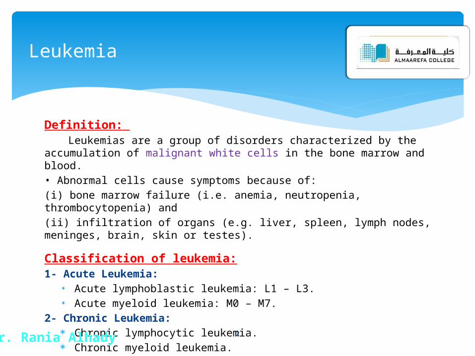
Definition: Leukemias are a group of disorders characterized by the accumulation of
malignant white cells in the bone marrow and blood.
• Abnormal cells cause symptoms because of:
(i) bone marrow failure (i.e. anemia, neutropenia, thrombocytopenia) and
(ii) infiltration of organs (e.g. liver, spleen, lymph nodes, meninges, brain, skin or testes).
Classification of leukemia:1- Acute Leukemia:• Acute lymphoblastic leukemia: L1 – L3.• Acute myeloid leukemia: M0 – M7.
2- Chronic Leukemia: Chronic lymphocytic leukemia. Chronic myeloid leukemia.
Leukemia
Dr. Rania Alhady 2
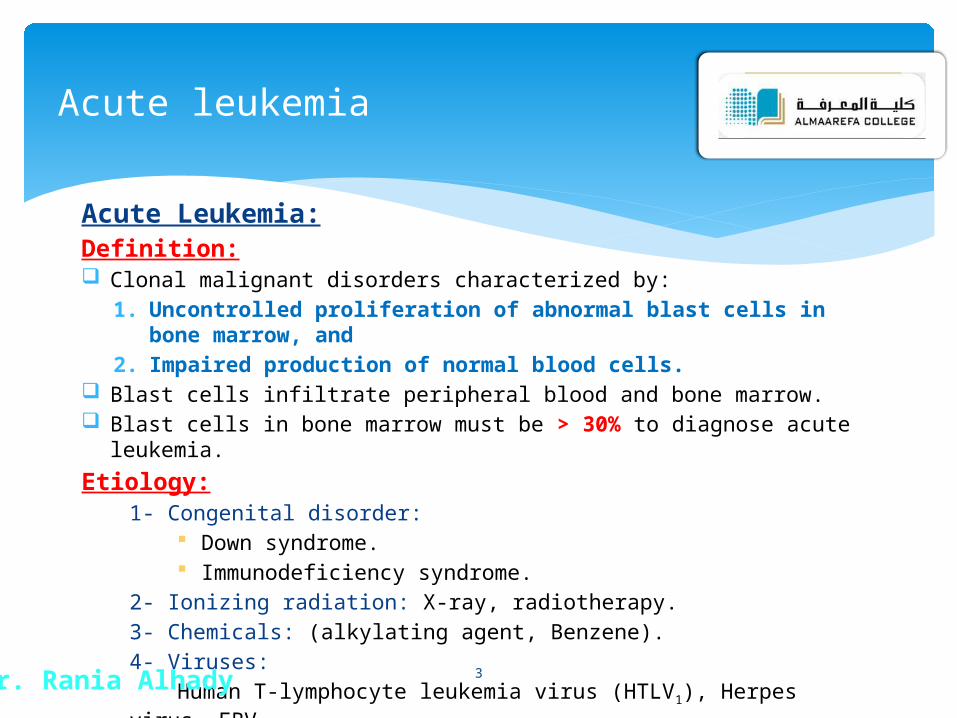
Acute Leukemia:Definition: Clonal malignant disorders characterized by:
1. Uncontrolled proliferation of abnormal blast cells in bone marrow, and
2. Impaired production of normal blood cells. Blast cells infiltrate peripheral blood and bone marrow. Blast cells in bone marrow must be > 30% to diagnose acute leukemia.
Etiology:1- Congenital disorder:
Down syndrome. Immunodeficiency syndrome.
2- Ionizing radiation: X-ray, radiotherapy.
3- Chemicals: (alkylating agent, Benzene).
4- Viruses:
Human T-lymphocyte leukemia virus (HTLV1), Herpes virus, EBV.
Acute leukemia
Dr. Rania Alhady 3

Results:* Chromosomal abnormality:- Imbalance between oncogene and anti-oncogene.
* Blast cells: Uncontrolled proliferation of this clone results in:
1- Arrest of cellular differentiation.
2- Reduced apoptosis.
3- Inhibition of normal marrow elements.
Classification:Acute leukemias are classified according to the type of blast cells into:
1) ALL: Childhood leukemia.
2) AML: Adulthood leukemia.
• Acute leukemias are fatal if not treated.• However, acute leukemias are easier to treat than chronic leukemias.
Acute leukemia
Dr. Rania Alhady 4
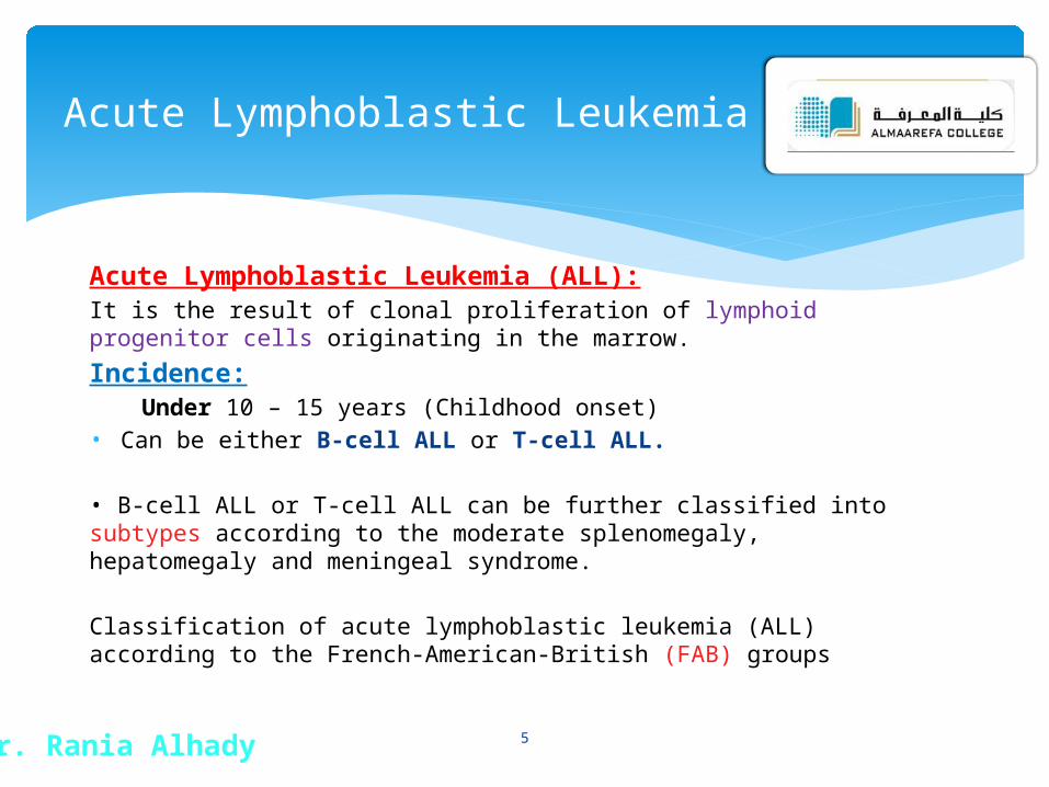
Acute Lymphoblastic Leukemia (ALL):It is the result of clonal proliferation of lymphoid progenitor cells originating in the marrow.
Incidence:Under 10 – 15 years (Childhood onset)
• Can be either B-cell ALL or T-cell ALL.
• B-cell ALL or T-cell ALL can be further classified into subtypes according to the moderate splenomegaly, hepatomegaly and meningeal syndrome.
Classification of acute lymphoblastic leukemia (ALL) according to the French-American-British (FAB) groups
Acute Lymphoblastic Leukemia
Dr. Rania Alhady 5

Clinical features:1- Bone marrow failure: - RBCs → Anemia: pallor, lethargy, malaise and dyspnea.- Leukocytes → neutropenia: fever, malaise, features of mouth, skin, respiratory infections.- Platelets → throbombocytopenia: spontaneous bruises, pupura, bleeding gums.
2- Tissue infiltration:
A. L.N. enlargement.
B. Hepatomegaly. More common with ALL than AML.C. Splenomegaly.
3- Organ infiltration:
A- Bony infiltration.
B- Testicular infiltration.
C- C.N.S. infiltration.
4- Fever.
Acute Lymphoblastic Leukemia
Dr. Rania Alhady 6
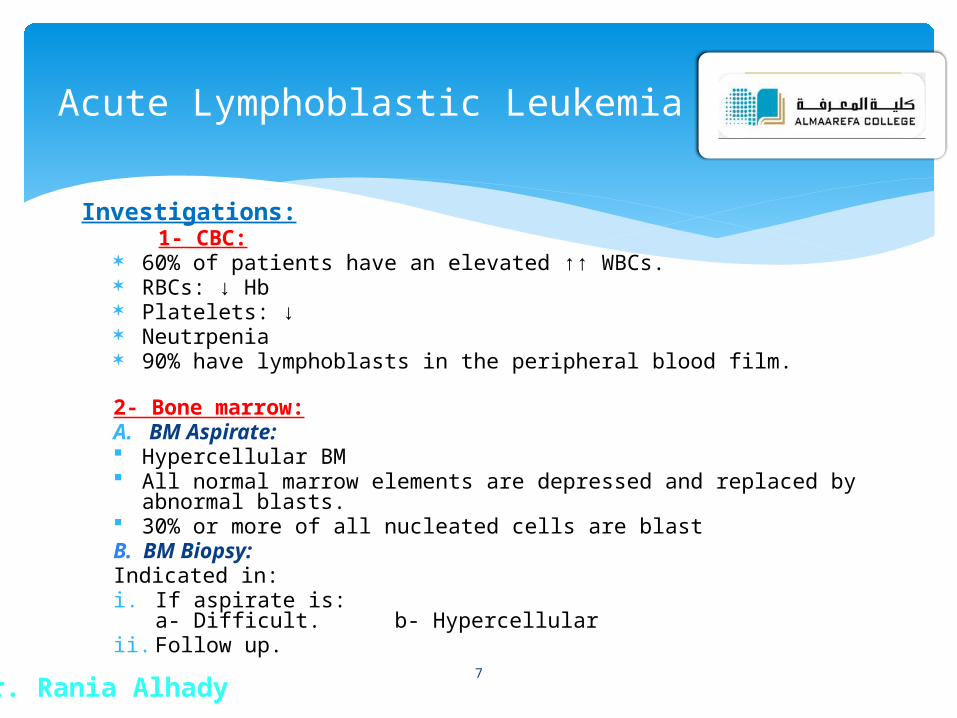
Investigations: 1- CBC:
60% of patients have an elevated ↑↑ WBCs. RBCs: ↓ Hb Platelets: ↓ Neutrpenia 90% have lymphoblasts in the peripheral blood film.
2- Bone marrow:A. BM Aspirate: Hypercellular BM All normal marrow elements are depressed and replaced by abnormal blasts. 30% or more of all nucleated cells are blastB. BM Biopsy:Indicated in:i. If aspirate is:
a- Difficult. b- Hypercellularii. Follow up.
Acute Lymphoblastic Leukemia
Dr. Rania Alhady7

L3 L2 L1
Homogenous Heterogenous in size and shape
Homogenous Cell population
Large Small & Large Small Cell Size
↓ ↓ ↑ N/C ratio
Prominent Prominent (1 - 2) Inconspicuous Nucleoli
Basophilic with vaculations
Larger Scanty Cytoplasm
1% 14% 85% Child Age
9% 60% 31% Adult
Acute Lymphoblastic Leukemia
Dr. Rania Alhady
FAB Classification:
8

Morphological classification of ALL
L1 L2 L3
Acute Lymphoblastic Leukemia
Dr. Rania Alhady 9
(a) L1 subtype-blasts show scanty cytoplasm without granules.(b) L2 subtype-blasts are larger and heterogeneous with more abundant cytoplasm.(c) L3 subtype-blasts are deeply basophilic with cytoplasmic vacuolation.
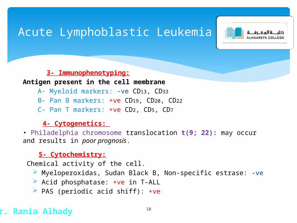
3- Immunophenotyping:
Antigen present in the cell membrane
A- Myeloid markers: -ve CD13, CD33
B- Pan B markers: +ve CD19, CD20, CD22
C- Pan T markers: +ve CD2, CD5, CD7
4- Cytogenetics:
• Philadelphia chromosome translocation t(9; 22): may occur and results in poor prognosis.
5- Cytochemistry:
Chemical activity of the cell. Myeloperoxidas, Sudan Black B, Non-specific estrase: -ve Acid phosphatase: +ve in T-ALL PAS (periodic acid shiff): +ve
Acute Lymphoblastic Leukemia
Dr. Rania Alhady 10
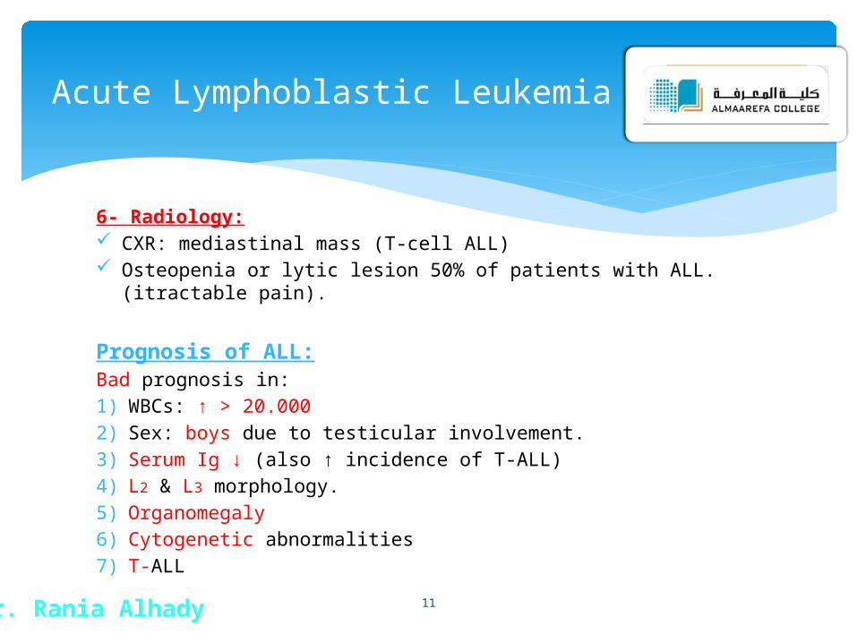
6- Radiology: CXR: mediastinal mass (T-cell ALL) Osteopenia or lytic lesion 50% of patients with ALL.(itractable pain).
Prognosis of ALL:Bad prognosis in:
1) WBCs: ↑ > 20.000
2) Sex: boys due to testicular involvement.
3) Serum Ig ↓ (also ↑ incidence of T-ALL)
4) L2 & L3 morphology.
5) Organomegaly
6) Cytogenetic abnormalities
7) T-ALL
Acute Lymphoblastic Leukemia
Dr. Rania Alhady 11

Acute Myeloid Leukemia (AML):Definition: Clonal proliferation of myeloid precursor cells with reduced capacity
to differentiate into more mature cellular elements.
Results in accumulation of leukemic forms in bone marrow, peripheral blood, and other tissues.
Leads to reduction in RBCs, platelets, PMNs.
It occurs in several morphologic variants; each one has a characteristic clinical & lab. features (M0 – M7)
Acute Myeloid Leukemia
Dr. Rania Alhady 12

Incidence:
AML is the most common leukemia in neonates.
1) 80% of AML is in adults.
2) 20% of AML is in children.
Diagnosis:A. Clinical Picture:• Anemia weakness and easy fatigue• Neutropenia infections• Thrombocytopenia gingival bleeding, ecchymoses, epistaxis, menorrhagia• Anorexia, weight loss, fever.• Organomegaly:
i. HSM in 1/3 of patients.
ii. Lymphadenopathy is uncommon except in monocytic variants.
Acute Myeloid Leukemia
Dr. Rania Alhady 13

B. Laboratory diagnosis:
Bone Marrow Exam: Aspiration and biopsy
* Morphologic classification using cytochemistry and immunophenotyping
Cytochemistry:
A. Peroxidase: +ve
B. Sudan black: +ve
C. Specific esterase: +ve
Immunophenotyping:Pan myeloid markers: +ve CD13, CD33
* Cytogenetic analysis
* Evaluation for presence of significant fibrosis, presence of granulomata
Acute Myeloid Leukemia
Dr. Rania Alhady 14

Diagnosis requires all of the following diagnostic components:
a) Documentation of bone marrow infiltration
b) Myeloid origin of the leukemic cells
c) FAB/WHO classification of the leukemia
FAB ClassificationsM0: minimally differentiated
M1: without maturation
M2: with maturation.
M3: promyelocytic.
M4: myelomonocytic.
M5: monoblastic
M6: erythroleukemia
M7: megakaryoblastic.
Acute Myeloid Leukemia
Dr. Rania Alhady 15

Acute Myeloid Leukemia
Dr. Rania Alhady 16

Acute Myeloid leukemia
Dr. Rania Alhady 17