
LETTER TO THE EDITOR - cambridge.org · abdominal recti muscles: X4-0V: left rectus abdominis...
4
LE JOURNAL CANADIEN DES SCIENCES NEUROLOGIQUES LETTER TO THE EDITOR TO THE EDITOR Epilepsia Partialis Continua of the Abdominal Musculature Caused by Acute Ischemic Stroke Keywords: Abdominal musculature, epilepsia partialis continua, ischemic stroke, status epilepticus Status epilepticus is “a condition resulting either from the fail- ure of the mechanisms responsible for seizure termination or from the initiation of mechanisms that lead to abnormally prolonged seizures”. 1 Epilepsia partialis continua (EPC) is a rare form of focal status epilepticus defined by clonic muscular twitching affecting a limited part of the body occurring for a minimum of 1 hour, and recurring at intervals of no more than 10 seconds. 2 The diagnosis requires the presence of epileptiform electroencephalographic discharges or other neurophysiological abnormalities demonstrat- ing the cortical origin of the muscle jerks. 2 Here we describe the clinical and electrophysiological features of an elderly man with EPC of the abdominal musculature fol- lowing an acute ischemic stroke. A 91-year-old man was admitted to our hospital with an acute right-sided hemiplegia and somnolence, which had appeared the day before. On admission, myoclonic jerks of the right hemibody were noticed, which promptly stopped after levetiracetam (1000 mg intravenous) administration. His medical history was notable for severe Alzheimer's disease (diagnosed 22 years ear- lier), chronic vascular encephalopathy with parkinsonism, mod- erate renal failure, and pancreatic carcinoma. The head CT scan showed ischemic lesions in the left hemisphere, more prominent in the fronto-parietal and temporal lobe, with marked chronic vascular encephalopathy and leukoaraiosis (Figures 1 and 2); the radiological features of the left temporal hypodensity were sug- gestive of a subacute cerebral infarction. The day after the admission, continuous involuntary contractions of the trunk were noted. Neurological examination showed continuous involuntary and rhythmic jerks involving the abdominal muscles, not spreading to other parts of the body (video). The electro- encephalogram (EEG) (Figure 3) revealed rhythmic epileptiform discharges of triphasic/diphasic morphology admixed with poly- spikes confined to the left hemisphere (Supplementary material). The most prominent epileptiform abnormalities were localized, over the left posterior temporo-parieto-occipital area. Equipo- tentiality of epileptiform discharges occurred between electrodes P3 and O1, sometimes with maximal amplitude over O1 > P3 ≥ T5 > T3, C3 (more evident in referential montage). There were also independent epileptiform discharges located anteriorly, close to the anterior Sylvian region in the left fronto-temporal area, sometimes showing phase reversal at F7 or equipotentiality between F7 and T3. A periodicity (frequency of about 1 Hz) was sometimes evident in epileptiform discharges occurring in both posterior and anterior regions (i.e., lateralized periodic dis- charges). The EEG recording also showed brief ictal transitions seen maximally over the left posterior hemispheric structures, especially around T5. Overall, the EEG showed anterior and posterior areas of epileptogenicity, more marked in the left posterior temporo-parieto-occipital region. The myoclonic movements were not synchronous with the rhythmic epileptiform discharges. A concomitant electromyographic recording of the muscles of the abdominal wall showed bursts of quasirhythmic asymmetric polymorphic motor potentials of the right abdominal recti muscles, with a duration lower than 100 ms and a frequency of 0.5-1 Hz. The movements attenuated after intravenous admin- istration of lorazepam (4 mg) and levetiracetam (2000 mg), but then reappeared. The patient died of cardiac arrest the following day. The presence of continuous involuntary movements with electromyography (EMG) correlates of twitches lasting <100 ms (even if not time-locked to the rhythmic epileptiform discharges), the epileptiform discharges in the EEG, and the attenuation of the myoclonic jerks after anti-epileptic therapy confirmed the epi- leptic nature of the symptoms, 3 leading to the diagnosis of EPC. These electroclinical features rule out alternative diagnoses, including a propriospinal myoclonus. The anatomical localization of the epileptogenic zone under- lying EPC of the abdominal muscles or trunk is thought to be in the frontal parasagittal or parietal cortex around the central lobe. 4 Involvement of abdominal musculature in focal motor seizures is rare; this may be explained by the small representation area of the abdomen on the motor cortical homunculus. 5 Only two previous cases of EPC of the abdominal musculature due to ischemic stroke have been published; 4 unlike other etiol- ogies, in both cases post-stroke EPC was preceded by focal myoclonic seizures on the hemibody contralateral to the side of the vascular event. However, unlike our case, they did not occur in the context of acute cerebrovascular lesions. As our case shows, focal-onset motor seizures and even EPC may selectively involve the abdominal musculature. It is noteworthy that in our patient the most prominent epi- leptiform abnormalities were recorded over the left posterior temporo-parieto-occipital area, whereas minor abnormalities were seen more anteriorly, in the left fronto-temporal region. The per- iodicity of epileptiform discharges occurring in both posterior and anterior regions, together with the lack of evolution in discharge amplitude, frequency, morphology, and distribution, is consistent with lateralized periodic discharges in the setting of an acute ischemic stroke. Of note, the myoclonic movements observed in this patient were not synchronous with the rhythmic epileptiform discharges. A strict neurophysiological association between this epileptiform activity and the myoclonic jerks is therefore difficult to prove with certainty. At least 10 cm 2 of synchronous cortical activity is required to record an ictal rhythm on scalp EEG, 6 and this explains why in more than 50% of EPC cases the scalp EEG is completely normal. 2 Furthermore, this dissociation between epi- leptiform discharges over the motor cortex and the motor corre- lates can be also owing to vertical inhibition. 7 In this phenomenon, spiking in lamina V following local penicillin administration leads to contralateral twitching without con- comitant epileptiform activity recorded on the cortical surface. A THE CANADIAN JOURNAL OF NEUROLOGICAL SCIENCES 703 . https://doi.org/10.1017/cjn.2018.330 Downloaded from https://www.cambridge.org/core. IP address: 54.39.129.60, on 22 Jul 2019 at 06:59:49, subject to the Cambridge Core terms of use, available at https://www.cambridge.org/core/terms
Transcript of LETTER TO THE EDITOR - cambridge.org · abdominal recti muscles: X4-0V: left rectus abdominis...
LE JOURNAL CANADIEN DES SCIENCES NEUROLOGIQUES
LETTER TO THE EDITOR
TO THE EDITOR
Epilepsia Partialis Continua of the Abdominal MusculatureCaused by Acute Ischemic Stroke
Keywords: Abdominal musculature, epilepsia partialis continua,ischemic stroke, status epilepticus
Status epilepticus is “a condition resulting either from the fail-ure of the mechanisms responsible for seizure termination or fromthe initiation of mechanisms that lead to abnormally prolongedseizures”.1 Epilepsia partialis continua (EPC) is a rare form of focalstatus epilepticus defined by clonic muscular twitching affecting alimited part of the body occurring for a minimum of 1 hour, andrecurring at intervals of no more than 10 seconds.2 The diagnosisrequires the presence of epileptiform electroencephalographicdischarges or other neurophysiological abnormalities demonstrat-ing the cortical origin of the muscle jerks.2
Here we describe the clinical and electrophysiological featuresof an elderly man with EPC of the abdominal musculature fol-lowing an acute ischemic stroke.
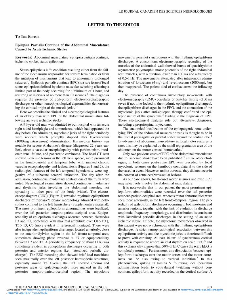
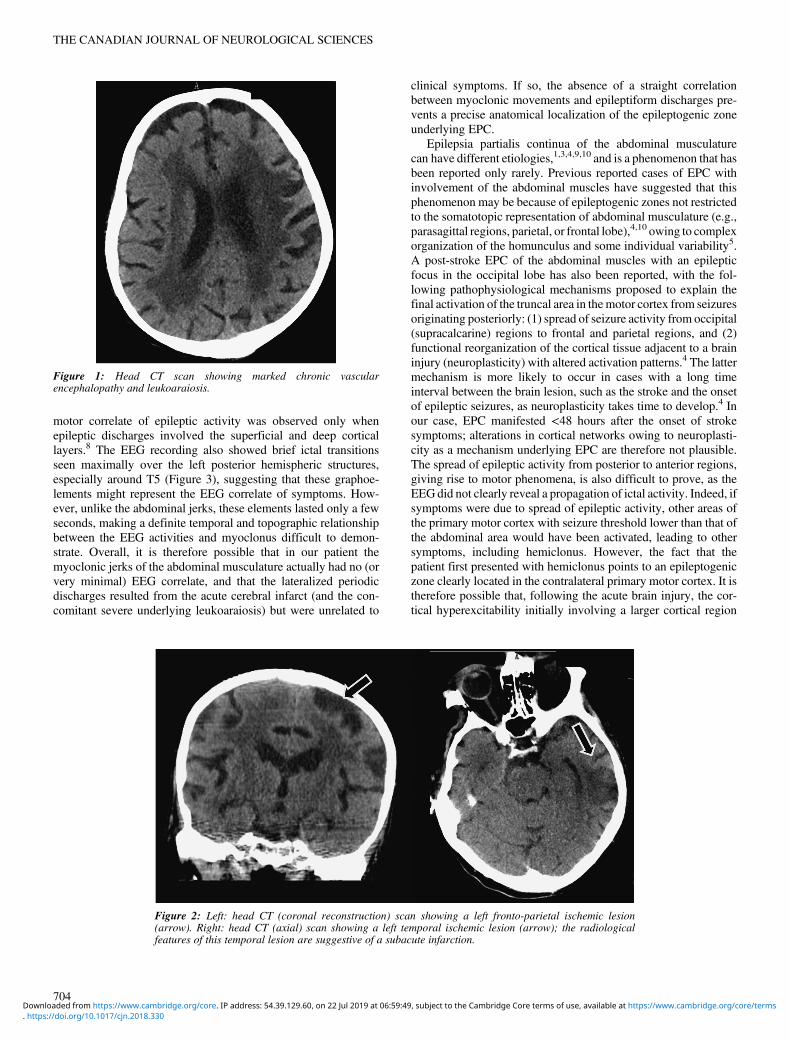
A 91-year-old man was admitted to our hospital with an acuteright-sided hemiplegia and somnolence, which had appeared theday before. On admission, myoclonic jerks of the right hemibodywere noticed, which promptly stopped after levetiracetam(1000mg intravenous) administration. His medical history wasnotable for severe Alzheimer's disease (diagnosed 22 years ear-lier), chronic vascular encephalopathy with parkinsonism, mod-erate renal failure, and pancreatic carcinoma. The head CT scanshowed ischemic lesions in the left hemisphere, more prominentin the fronto-parietal and temporal lobe, with marked chronicvascular encephalopathy and leukoaraiosis (Figures 1 and 2); theradiological features of the left temporal hypodensity were sug-gestive of a subacute cerebral infarction. The day after theadmission, continuous involuntary contractions of the trunk werenoted. Neurological examination showed continuous involuntaryand rhythmic jerks involving the abdominal muscles, notspreading to other parts of the body (video). The electro-encephalogram (EEG) (Figure 3) revealed rhythmic epileptiformdischarges of triphasic/diphasic morphology admixed with poly-spikes confined to the left hemisphere (Supplementary material).The most prominent epileptiform abnormalities were localized,over the left posterior temporo-parieto-occipital area. Equipo-tentiality of epileptiform discharges occurred between electrodesP3 and O1, sometimes with maximal amplitude over O1>P3≥T5>T3, C3 (more evident in referential montage). There werealso independent epileptiform discharges located anteriorly, closeto the anterior Sylvian region in the left fronto-temporal area,sometimes showing phase reversal at F7 or equipotentialitybetween F7 and T3. A periodicity (frequency of about 1Hz) wassometimes evident in epileptiform discharges occurring in bothposterior and anterior regions (i.e., lateralized periodic dis-charges). The EEG recording also showed brief ictal transitionsseen maximally over the left posterior hemispheric structures,especially around T5. Overall, the EEG showed anterior andposterior areas of epileptogenicity, more marked in the leftposterior temporo-parieto-occipital region. The myoclonic
movements were not synchronous with the rhythmic epileptiformdischarges. A concomitant electromyographic recording of themuscles of the abdominal wall showed bursts of quasirhythmicasymmetric polymorphic motor potentials of the right abdominalrecti muscles, with a duration lower than 100ms and a frequencyof 0.5-1Hz. The movements attenuated after intravenous admin-istration of lorazepam (4mg) and levetiracetam (2000mg), butthen reappeared. The patient died of cardiac arrest the followingday.
The presence of continuous involuntary movements withelectromyography (EMG) correlates of twitches lasting <100ms(even if not time-locked to the rhythmic epileptiform discharges),the epileptiform discharges in the EEG, and the attenuation of themyoclonic jerks after anti-epileptic therapy confirmed the epi-leptic nature of the symptoms,3 leading to the diagnosis of EPC.These electroclinical features rule out alternative diagnoses,including a propriospinal myoclonus.
The anatomical localization of the epileptogenic zone under-lying EPC of the abdominal muscles or trunk is thought to be inthe frontal parasagittal or parietal cortex around the central lobe.4
Involvement of abdominal musculature in focal motor seizures israre; this may be explained by the small representation area of theabdomen on the motor cortical homunculus.5
Only two previous cases of EPC of the abdominal musculaturedue to ischemic stroke have been published;4 unlike other etiol-ogies, in both cases post-stroke EPC was preceded by focalmyoclonic seizures on the hemibody contralateral to the side ofthe vascular event. However, unlike our case, they did not occur inthe context of acute cerebrovascular lesions.
As our case shows, focal-onset motor seizures and even EPCmay selectively involve the abdominal musculature.
It is noteworthy that in our patient the most prominent epi-leptiform abnormalities were recorded over the left posteriortemporo-parieto-occipital area, whereas minor abnormalities wereseen more anteriorly, in the left fronto-temporal region. The per-iodicity of epileptiform discharges occurring in both posterior andanterior regions, together with the lack of evolution in dischargeamplitude, frequency, morphology, and distribution, is consistentwith lateralized periodic discharges in the setting of an acuteischemic stroke. Of note, the myoclonic movements observed inthis patient were not synchronous with the rhythmic epileptiformdischarges. A strict neurophysiological association between thisepileptiform activity and the myoclonic jerks is therefore difficultto prove with certainty. At least 10 cm2 of synchronous corticalactivity is required to record an ictal rhythm on scalp EEG,6 andthis explains why in more than 50% of EPC cases the scalp EEG iscompletely normal.2 Furthermore, this dissociation between epi-leptiform discharges over the motor cortex and the motor corre-lates can be also owing to vertical inhibition.7 In thisphenomenon, spiking in lamina V following local penicillinadministration leads to contralateral twitching without con-comitant epileptiform activity recorded on the cortical surface. A
THE CANADIAN JOURNAL OF NEUROLOGICAL SCIENCES 703
. https://doi.org/10.1017/cjn.2018.330Downloaded from https://www.cambridge.org/core. IP address: 54.39.129.60, on 22 Jul 2019 at 06:59:49, subject to the Cambridge Core terms of use, available at https://www.cambridge.org/core/terms
motor correlate of epileptic activity was observed only whenepileptic discharges involved the superficial and deep corticallayers.8 The EEG recording also showed brief ictal transitionsseen maximally over the left posterior hemispheric structures,especially around T5 (Figure 3), suggesting that these graphoe-lements might represent the EEG correlate of symptoms. How-ever, unlike the abdominal jerks, these elements lasted only a fewseconds, making a definite temporal and topographic relationshipbetween the EEG activities and myoclonus difficult to demon-strate. Overall, it is therefore possible that in our patient themyoclonic jerks of the abdominal musculature actually had no (orvery minimal) EEG correlate, and that the lateralized periodicdischarges resulted from the acute cerebral infarct (and the con-comitant severe underlying leukoaraiosis) but were unrelated to
clinical symptoms. If so, the absence of a straight correlationbetween myoclonic movements and epileptiform discharges pre-vents a precise anatomical localization of the epileptogenic zoneunderlying EPC.
Epilepsia partialis continua of the abdominal musculaturecan have different etiologies,1,3,4,9,10 and is a phenomenon that hasbeen reported only rarely. Previous reported cases of EPC withinvolvement of the abdominal muscles have suggested that thisphenomenon may be because of epileptogenic zones not restrictedto the somatotopic representation of abdominal musculature (e.g.,parasagittal regions, parietal, or frontal lobe),4,10 owing to complexorganization of the homunculus and some individual variability5.A post-stroke EPC of the abdominal muscles with an epilepticfocus in the occipital lobe has also been reported, with the fol-lowing pathophysiological mechanisms proposed to explain thefinal activation of the truncal area in themotor cortex from seizuresoriginating posteriorly: (1) spread of seizure activity from occipital(supracalcarine) regions to frontal and parietal regions, and (2)functional reorganization of the cortical tissue adjacent to a braininjury (neuroplasticity) with altered activation patterns.4 The lattermechanism is more likely to occur in cases with a long timeinterval between the brain lesion, such as the stroke and the onsetof epileptic seizures, as neuroplasticity takes time to develop.4 Inour case, EPC manifested <48 hours after the onset of strokesymptoms; alterations in cortical networks owing to neuroplasti-city as a mechanism underlying EPC are therefore not plausible.The spread of epileptic activity from posterior to anterior regions,giving rise to motor phenomena, is also difficult to prove, as theEEGdid not clearly reveal a propagation of ictal activity. Indeed, ifsymptoms were due to spread of epileptic activity, other areas ofthe primary motor cortex with seizure threshold lower than that ofthe abdominal area would have been activated, leading to othersymptoms, including hemiclonus. However, the fact that thepatient first presented with hemiclonus points to an epileptogeniczone clearly located in the contralateral primary motor cortex. It istherefore possible that, following the acute brain injury, the cor-tical hyperexcitability initially involving a larger cortical region
Figure 2: Left: head CT (coronal reconstruction) scan showing a left fronto-parietal ischemic lesion(arrow). Right: head CT (axial) scan showing a left temporal ischemic lesion (arrow); the radiologicalfeatures of this temporal lesion are suggestive of a subacute infarction.
Figure 1: Head CT scan showing marked chronic vascularencephalopathy and leukoaraiosis.
THE CANADIAN JOURNAL OF NEUROLOGICAL SCIENCES
704
. https://doi.org/10.1017/cjn.2018.330Downloaded from https://www.cambridge.org/core. IP address: 54.39.129.60, on 22 Jul 2019 at 06:59:49, subject to the Cambridge Core terms of use, available at https://www.cambridge.org/core/terms
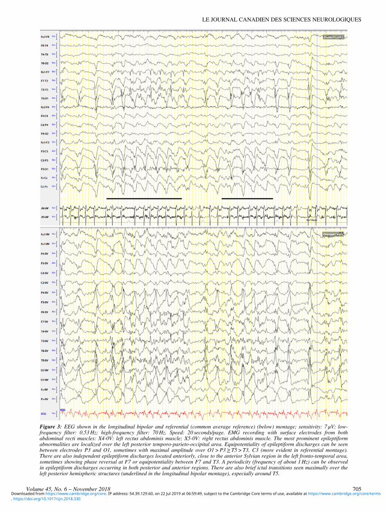
Figure 3: EEG shown in the longitudinal bipolar and referential (common average reference) (below) montage; sensitivity: 7 μV; low-frequency filter: 0.53Hz; high-frequency filter: 70Hz. Speed: 20 seconds/page. EMG recording with surface electrodes from bothabdominal recti muscles: X4-0V: left rectus abdominis muscle; X5-0V: right rectus abdominis muscle. The most prominent epileptiformabnormalities are localized over the left posterior temporo-parieto-occipital area. Equipotentiality of epileptiform discharges can be seenbetween electrodes P3 and O1, sometimes with maximal amplitude over O1>P3≥ T5>T3, C3 (more evident in referential montage).There are also independent epileptiform discharges located anteriorly, close to the anterior Sylvian region in the left fronto-temporal area,sometimes showing phase reversal at F7 or equipotentiality between F7 and T3. A periodicity (frequency of about 1Hz) can be observedin epileptiform discharges occurring in both posterior and anterior regions. There are also brief ictal transitions seen maximally over theleft posterior hemispheric structures (underlined in the longitudinal bipolar montage), especially around T5.
LE JOURNAL CANADIEN DES SCIENCES NEUROLOGIQUES
Volume 45, No. 6 – November 2018 705
. https://doi.org/10.1017/cjn.2018.330Downloaded from https://www.cambridge.org/core. IP address: 54.39.129.60, on 22 Jul 2019 at 06:59:49, subject to the Cambridge Core terms of use, available at https://www.cambridge.org/core/terms
subsequently restricted to a more discrete area of the homuncu-lus.11 In this case, symptoms could have been the result of directictal generation in the truncal area rather than a cortical activationsecondary to propagation from a nearby region.10 This necessarilyimplies a very focal epileptic discharge arising within a smallcortical area, which could have no clear EEG ictal correlates.
Subcortical regions may also have played a role in the patho-genesis of EPC in this patient, considering the entity of the chronicvascular leukoencephalopathy and leukoaraiosis. Similarly, it isalso possible that the coexistent Alzheimer's disease may havecontributed in altering the cortical excitability. However, owing tolack of available data, this remains speculative.
In conclusion, a combined EEG–EMG recording in the event ofinvoluntary continuous muscular jerks in the abdominal muscu-lature should be performed to reach a diagnosis. An ischemic strokein the middle cerebral artery territory can alter the excitability of theprimary motor cortex, lowering the seizure threshold in the trunkmotor area, where threshold is postulated to be high,12 leading toEPC or motor seizures involving the abdominal muscles.
STATEMENT OF AUTHORSHIP
FB, VT, AB and RN contributed to data collection and analysis.FB and RN contributed to writing/editing of this article.
CONFLICT OF INTERESTSThis study was not funded. FB has received speakers' honor-
aria from Eisai and PeerVoice, payment for consultancy fromEisai, and travel support from Eisai, ITALFARMACO, and UCBPharma. Other authors have no conflicts of interest.
SUPPLEMENTARY MATERIAL
To view supplementary material for this article, please visithttps://doi.org/10.1017/cjn.2018.330
Francesco BrigoDepartment of Neurosciences, Biomedicine and Movement
Sciences, University of Verona, Verona, Italy
Department of Neurology, Franz Tappeiner Hospital, Merano,Italy
Arianna BrattiDepartment of Neurology, Franz Tappeiner Hospital, Merano,
Italy
Veronica TavernelliDepartment of Neurology, Franz Tappeiner Hospital, Merano,
Italy
Raffaele NardoneDepartment of Neurology, Franz Tappeiner Hospital, Merano,
Italy
Department of Neurology, Christian Doppler Klinik, ParacelsusMedical University, Salzburg, Austria
Correspondence to: F. Brigo, Department of Neurosciences,Biomedicine and Movement Sciences, University of Verona,Piazzale L.A. Scuro 10, 37134 Verona, Italy. Email: [email protected]
REFERENCES
1. Trinka E, Cock H, Hesdorffer D, et al. A definition and classificationof status epilepticus—report of the ILAE task force on classifi-cation of status epilepticus. Epilepsia. 2015;56:1515-23.
2. Bien CG, Elger CE. Epilepsia partialis continua: semiology anddifferential diagnoses. Epileptic Disord. 2008;10:3-7.
3. Obeso JA, Rothwell JC, Marsden CD. The spectrum of corticalmyoclonus. From focal reflex jerks to spontaneous motorepilepsy. Brain. 1985;108:193-224.
4. Ribeiro JJ, Sousa M, Teotónio R, Bento C, Sales F. Epilepsia par-tialis continua of the abdominal muscles due to cerebrovasculardisease. Epileptic Disord. 2015;17:72-6.
5. Tezer FI, Celebi O, Ozgen B, Saygi S. A patient with two episodes ofepilepsia partialis continua of the abdominal muscles caused bycortical dysplasia. Epileptic Disord. 2008;10:306-11.
6. Tao JX, Ray A, Hawes-Ebersole S, Ebersole JS. Intracranial EEGsubstrates of scalp EEG interictal spikes. Epilepsia. 2005;46(5):669-76.
7. Elger CE, Speckmann EJ. Focal interictal epileptiform discharges(FIED) in the epicortical EEG and their relations to spinal fieldpotentials in the rat. Electroencephalogr Clin Neurophysiol.1980;48:447-60.
8. Elger CE, Speckmann EJ. Penicillin-induced epileptic foci in themotor cortex: vertical inhibition. Electroencephalogr Clin Neu-rophysiol. 1983;56:604-22.
9. Chalk CH, McManis PG, Cascino GD. Cryptococcal meningitismanifesting as epilepsia partialis continua of the abdomen. MayoClin Proc. 1991;66:926-29.
10. Aljaafari D, Nascimento FA, Abraham A, Andrade DM, WennbergRA. Unilateral abdominal clonic seizures of parietal lobe origin:EEG findings. Epileptic Disord. 2018;20(2):158-63.
11. Penfield W, Jasper H. Epilepsy and the functional anatomy ofthe human brain. Boston, MA: Little, Brown & Company;1954:59-72.
12. Oster JM, Aljumairi F, Cosgrove GR. Metabolic imaging correlate oftruncal onset seizures. Arch Neurol. 2011;68:251-53.
THE CANADIAN JOURNAL OF NEUROLOGICAL SCIENCES
706
. https://doi.org/10.1017/cjn.2018.330Downloaded from https://www.cambridge.org/core. IP address: 54.39.129.60, on 22 Jul 2019 at 06:59:49, subject to the Cambridge Core terms of use, available at https://www.cambridge.org/core/terms


