Let’s Talk About Trachs - NCSHLA · Let’s Talk About Trachs Jenna ... relationships to...
22
4/1/2016 1 Let’s Talk About Trachs Jenna Kneepkens MS/CCC-SLP Jaimie Jones, MS/CCC-SLP Disclosures No relevant financial or nonfinancial relationships to disclose. Objectives • Normal respiratory anatomy and physiology • Impact of tracheostomy on respiratory and phonatory anatomy and physiology • Impact of tracheostomy on swallowing • Communication needs of tracheostomy and/or ventilator dependent patients
Transcript of Let’s Talk About Trachs - NCSHLA · Let’s Talk About Trachs Jenna ... relationships to...
4/1/2016
1
Let’s Talk About Trachs
Jenna Kneepkens MS/CCC-SLP
Jaimie Jones, MS/CCC-SLP
Disclosures
No relevant financial or nonfinancial
relationships to disclose.
Objectives
• Normal respiratory anatomy and
physiology
• Impact of tracheostomy on respiratory and
phonatory anatomy and physiology
• Impact of tracheostomy on swallowing
• Communication needs of tracheostomy
and/or ventilator dependent patients

4/1/2016
2
Myth versus Fact
• The cuff cannot be deflated because the patient will aspirate
• A speaking valve should not be placed if the patient has a lot of secretions
• A speaking valve restores taste and smell
• A patient cannot use a speaking valve unless they are off the ventilator
• A speaking valve can facilitate decannulation
• The presence of a tracheostomy will anchor a patients larynx during swallowing
Scope of Practice
• ASHAs Code of Ethics states that clinicians must be competent in any area in which they practice
• ASHAs Scope of Practice in SLP is broad and does not address specific procedures; however, procedures should be related to assessment and treatment of patients with communication and swallowing disorders
• Individual facilities should have specific processes for credentialing staff
• Facilities can provide training and support for teaching SLPs to suction
• SLPs need to consider potential liability issues of related activities such as changing or capping tracheotomy tubes as these may be considered procedure that should be done by medical professionals
• In 2010 Joint Commission released “Advancing Effective Communication, Cultural Competence, and Patient- and Family-Centered Care: A roadmap for hospitals” advocating identification and assessment of communication needs (i.e., AAC)
Respiratory Anatomy and Physiology

4/1/2016
3
What is Normal? (Adults)
• Normal resting respiratory rate
– 12-20
• Normal resting heart rate
– 60-100
• Functional SpO2
– 90-100%
What is Normal? (Pediatrics)
Age Respiratory Rate Heart Rate SpO2
Premature Infant 40-50 140-170 90-100%*
Newborn 30-50 120-160 90-100%*
Infant (1-12 mo) 20-30 80-140 90-100%*
Toddler (1-3 yrs) 20-30 80-130 90-100%*
School Age (6-12yrs) 15-30 70-110 90-100%
Adolescent (13yrs+) 12-20 60-100 90-100%
* This can vary depending on factors including (but not limited to) degree of prematurity, history of need for supplemental oxygen therapy, and cardiopulmonary status. When working with these populations it is imperative to know target ranges as deviations can cause oxygen toxicity, retinal damage, pulmonary overcirculation, right sided heart failure, and other complications.
Respiratory Tract
• Upper Respiratory Tract – Nasal cavity
– Pharynx
– Larynx
• Lower Respiratory Tract – Trachea
– Bronchi
– Lungs • Right lung has 3 lobes; Left lung has 2 lobes
– bronchi>bronchioles>alveoli
• Diaphragm

4/1/2016
4
Respiratory Support
• Supplemental oxygen via nasal cannula
• Continuous Positive Airway Pressure
(CPAP)
– Single pressure
• Bi-level Positive Airway Pressure (BiPAP)
– Delivers an inhale pressure and an exhale
pressure
• Artificial airway with or without ventilator
Artificial Airway
• An artificial airway is indicated when there is a
disruption to the normal respiratory mechanism
• Purposes of an artificial airway include, but are
not limited to:
– Adequate ventilation and oxygenation
– Eliminate airway obstruction/maintain patent airway
– Provide access to the airway for pulmonary toilet
– Reduce the potential for aspiration
Types of Artificial Airway
• Endotracheal intubation – Insertion of a tube through the mouth or nose,
that passes through the pharynx and vocal folds, into the trachea
– Considered temporary
• Tracheostomy
– Surgical or percutaneous placement of a tube through the neck directly into the
trachea (below the vocal folds)
– Extended need for an artificial airway

4/1/2016
5
Indications for a Tracheostomy
• Extended need for artificial airway
• Improved weaning
• Increased options for swallowing and
communication
Common diagnoses/populations for
tracheostomy
• Ventilator dependency
• Cardio-Pulmonary diseases – Bronchopulmonary dysplasia (BPD), congenital
diaphragmatic hernia (CDH), reactive airway disease (RAD), COPD, CHF, diaphragm dysfunction
• Neuromuscular diseases – Guillain-Barre Syndrome, ALS, MS
• Severe trauma to the head, neck or spinal cord
• Airway obstructions – Tumors, edema, infection, vocal cord paralysis,
tracheal stenosis, laryngo/tracheo/bronchomalacia
Trach Placement

4/1/2016
6
Components of a Trach
• Obturator
– Used to insert into trach site and is removed following insertion and replaced by the inner cannula
• Outer Cannula
– Remains in place; maintains
trach site and airway
• Inner Cannula
(*not present in all trachs)
– Collects secretions and needs to be changed or cleaned frequently
Components of a Trach
• Flange
– Rests on the skin of the neck/secures trach
• Cuff
– Used to seal off area between trach tube and trachea to
inhibit air escape to upper airway
• Pilot Balloon
– Indicates how much air is in the
balloon/cuff
• Cap/Button
– Occludes opening prior to decannulation
Types of Trachs
• Manufacturers
– Shiley
– Portex
– Bivona
– Jackson (metal) • PMV adaptor is required
• Sizes/diameters vary depending on the brand

4/1/2016
7
Types of Trachs
• Cuffed
– Foam
– Air
– Sterile water
• Uncuffed
Cuff versus Uncuffed
• Why Cuffed?
– Main purpose of an inflated cuff is to maintain the air delivered from the ventilator to a patient's lungs
– Cuff fills the tracheal space around the tube and prevents breath from escaping through the upper airway
– An inflated cuff prevents leakage of air, thereby creating a closed loop between the ventilator and patient and ensuring a consistent delivery of air
– During periods of cuff inflation, air is not available for phonation and patient is aphonic
Cuff vs Uncuffed
• Why Uncuffed/Cuffless?
– Primarily used in non-ventilated patients
– Since there is no cuff, it allows air to pass
into the upper trachea and larynx

4/1/2016
8
Types of Trachs
• Fenestrated trach – Cuffed or cuffless
– Has singular or multiple holes on the body of the outer cannula
– Allows air to flow from the trachea
through the fenestration to the vocal
folds
• Disadvantages: – With prolonged use granulation tissue can develop
– Secretions can plug fenestration
– Suctioning can be difficult as catheter can pass through fenestration and irritate airway
• “Aerodigestive and Respiratory Changes Post Tracheostomy: A Comprehensive Review” www.passy-muir.com/CEU
How a Trach Changes Physiology
• By-passes the upper airway
• Mouth and nose are unable to function normally with changes in the ability to: – Warm air
– Humidify air
– Filter air, including dust particles and micro-organisms
– Communicate/vocalize
– Swallowing, nutrition & hydration
– Smell
Trach Maintenance
• Care
– Shower shield
• Changes
– Trach*
– Inner cannula
• Downsizing*
– May need to increase space between trachea and trach for improved upper
airway patency
– Be sensitive to secretions
• Capping*
– Air flows through upper respiratory tract
• Decannulation*
– Typically 48 consecutive hours of capping without respiratory issues or need for
deep suctioning (often longer in pediatrics)
*Decision making done in collaboration with treating physician/medical team

4/1/2016
9
Speaking & Swallowing Valves
• How it changes anatomy & physiology
Speaking & Swallowing Valves
• Types – Biased closed vs Biased open valves
• closed at rest vs open at rest
– Different shapes and sizes
– Different resistance levels
• Types of one-way valves – Montgomery Speaking Valve
– Hood Speaking Valve
– Olympic “Trach Talk”
– Kistner One Way Valve
– Shiley Phonate Valve
• Our focus will be on the Passy-Muir Valve (PMV)…
Passy-Muir Valve
• One way tracheostomy speaking valve
that attaches to the tracheostomy hub
• Valve opens when patient inhales and
closes at the end of inhalation to allow
exhaled air to pass through vocal folds
and upper respiratory tract, thus allowing
phonation.

4/1/2016
10
Passy-Muir Speaking Valve
PMV 2001
PMV 007 (inline)
PMV Candidates
• Alert with communicative intent
• Ability to tolerate cuff deflation
• Adequate upper airway patency
• Medical stability
• SpO2 > 90%
• Volitional oral motor movement
• Adequate secretion management
29
Benefits
• Improved communication
• Improved swallow function
• Improved oropharyngeal sensation
• Improved hygiene and infection control
• Improved coughing ability
• Improved pulmonary function
• Improved oxygenation
• Improved mental outlook/higher quality of life
30

4/1/2016
11
Benefits
• Restoration of olfactory
• Decrease in oral/nasal secretions
• Improved communication/speech intelligibility to aid in medical care
• Decreased need for manual occlusion
• Facilitation of decannulation
• Better facilitation of secretions
31
Exclusions and Contraindications
• Inadequate upper airway patency (i.e. significant laryngotracheal stenosis, upper airway obstruction, too large of trach)
• Breath stacking with deflated cuff
• Unconscious or minimally responsive
• Medically unstable
• Severe aspiration risk with inability to tolerate cuff deflation
• Copious secretions
• Severely reduced lung elasticity (Not an absolute contraindication)
32
Evaluation
• Record baseline vitals – HR, RR, Oxygen saturation, and FiO2 requirements
– FiO2 • 21% (room air), 32-36% (3-4 LPM), 40% (5 LPM), 60% (7-8 LPM)
• Ensure oral/tracheal suctioning is completed
• Change or clean inner cannula
• Deflate tracheostomy cuff – Check the amount of ml of air/water taken out of pilot balloon as this
must be the same amount that is placed when re-inflating cuff • An over inflated cuff can lead to fisula, tracheomalacia, tracheal stenosis,
tracheoesophageal fistula, ulceration, necrosis, and scarring
– Deflate cuff at a slow rate
– Patients may experience coughing d/t sensation of upper airway airflow
– Promote cuff deflation it patient is able to tolerate
33

4/1/2016
12
Evaluation
• Check airway patency – Finger occlusion
• Fog on mirror, volitional vocalizations, coughing/throat clearing, finger under nose to feel nasal airflow
– Reasons for reduced/no upper airway patency: • Obstructive: copious secretions, trach size is too large, vocal fold
paralysis/paresis, poor positioning, severe vocal fold edema, upper respiratory tract deformity or edema, significant laryngotracheal stenosis, other upper airway obstruction
• Reduced respiratory drive, diaphragmatic involvement
– “Talking over the trach” • Some patients may be able to speak “around” the trach; however, usually have
breathy vocal quality and short phrase length
• PMV placement remains appropriate given additional benefits it can provide
• Assess for breath stacking/back pressure – Listen/feel for a “whoosh” of air upon removal of finger/trach occlusion
34
Evaluation
• Place PMV
– Stabilize trach via flange
– 1/4 right twist to place; ¼ left twist to remove
• Monitor HR, SpO2, RR and remove PMV with any of the following:
– SpO2 >90%
– RR increase by 8-10 breaths per minute
– HR increase by >20 beats per minute
– Signs/symptoms of respiratory distress,
• Assess patient comfort. Note signs/symptoms of stress caused by PMV, including but not limited to: change in facial color, clavicular breathing, s/s of distress or discomfort, dyspnea, and patient report of difficulty tolerating PMV.
• Assess phonation, speech intelligibility, vocal quality, management of secretions, etc.
35
Evaluation
• SLP concludes trial by determining
success/failure of trial and making
recommendations
– Determine PMV frequency and duration
based on trial
• Educate and ensure patient/family know
guidelines for PMV tolerance and
recommended use
36

4/1/2016
13
Evaluation
• When cleared to wear PMV – Place warning sign on pilot balloon
– Post sign above bed, door and/or chart (see handout)
– Educate patient/family when PMV should be removed
• PMV should be removed when – Sleeping
– Exertion
– Respiratory treatments
– SpO2<90%
– Short of breath
Candidacy for Decannulation
• Decannulation versus chronic trach – Need to know plan of care and patient population
• Secretion management
• Tolerate capping for ~48 consecutive hours without respiratory issues or need for deep suctioning
• Pediatric population typically requires longer trial of capping prior to decannulation (weeks to months)
• SLP may advocate for patient, however, decision making done by ENT/medical team
Intervention
• Trach downsize as medically appropriate
and no contraindications exist
• Promote cuff deflation and/or progress
towards cuffless trach as medically
indicated
• Ongoing trials during therapy
• Initiate Respiratory Muscle Training if
medically appropriate

4/1/2016
14
Speech Pathology Services in Trach
Population
• Communication
• Swallowing
Impact of Trach on Communication
• Air no longer passes through the vocal
folds so the person cannot produce
sounds easily
– Leads to frustration for both patient, family
and medical providers
• Tracheostomies increase risk for long term
disruption in speech-language
development in infants and children
Communication
Effective communication The successful joint establishment of meaning wherein patients and health care providers exchange information, enabling patients to participate actively in their care from admission through discharge, and ensuring that the responsibilities of both patients and providers are understood. To be truly effective, communication requires a two-way process (expressive and receptive) in which messages are negotiated until the information is correctly understood by both parties. Successful communication takes place only when providers understand and integrate the information gleaned from patients, and when patients comprehend accurate, timely, complete, and unambiguous messages from providers in a way that enables them to participate responsibly in their care.
-The Joint Commission, “Advancing Effective Communication, Cultural Competence, and Patient- and Family-Centered Care: A roadmap for hospitals,” 2010

4/1/2016
15
Intervention
• Phonation through speaking valve - Provide education regarding vocal hygiene
- Consider referral to voice specialist if greater intervention needed
• Alternative-Augmentative Communication (AAC) (see handouts)
• Motor assessment
• Non verbal communication strategies assessment
43
AAC
• No Tech
• Gestures (pointing, sign, eye gaze, hand squeezes)
• Mouthing
• Writing
• Communication boards
• Eye Link boards
• Methods of selection
• Direct selection
• Scanning (auditory, row column)
AAC
• Low Tech
– Switches
– Laser pointer
– Simple speech-generating devices: picture
boards that overlay buttons with recorded
voice messages (4-32 icons)
45
4/1/2016
16
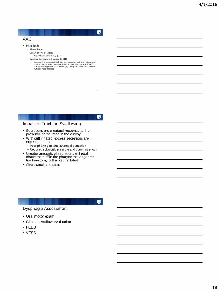
AAC
• High Tech
– Electrolarynx
– Smart phone or tablet
• Passy Muir TrachTools App (new!)
– Speech Generating Devices (SGD)
• A computer or tablet equipped with communication software that provides
digital and/or recorded messages linked to icons that can be activated
directly or through alternative means (e.g., eye gaze, hand, head, or chin
switches, head tracking)
46
Impact of Trach on Swallowing
• Secretions are a natural response to the presence of the trach in the airway
• With cuff inflated, excess secretions are expected due to: – Poor pharyngeal and laryngeal sensation
– Reduced subglottic pressure and cough strength
• Greater amounts of secretions will pool above the cuff in the pharynx the longer the tracheostomy cuff is kept inflated
• Alters smell and taste
Dysphagia Assessment
• Oral motor exam
• Clinical swallow evaluation
• FEES
• VFSS

4/1/2016
17
Dysphagia Assessment –Blue Dye Test
• Aspiration was present in 53% of studies as documented by FEES
– BDT showed an overall 50% false-negative error rate for detection of aspiration as compared with FEES Donzelli et al 2001
• With simultaneous VFSS and BDT overall BDT showed a 50% false-negative error rate
– The BDT identified aspiration in 100% of patients who aspirated more than trace amounts but failed to identify aspiration of trace amounts Brady et al1999
Dysphagia Assessment –Blue Dye Test
• Advantages: conducted at bedside in a
timey fashion, relatively inexpensive, only
suctioning equipment is needed, no
exposure to radiation
• Results suggest using BDT as a screening
tool for presence of gross amounts of
aspiration Donzelli et al 2001; ASHA
Cuff inflation versus deflation with
swallowing
• Cuff provides little protection against penetration or aspiration
• Aspirated material that reaches the cuff will "leak" around the sides of the tube and into the trachea and bronchial airways
• Residue on top of cuff can be aspirated after deflation
• Deflating the cuff makes it possible for the patient to generate airflow to cough via the upper airway

4/1/2016
18
Dysphagia Interventions
• Swallow exercises
• Respiratory Muscle Training
• Increase tolerance of PMV
…all together now!
Case Studies
Ethan
• History: 2y 8m male, Costello Syndrome, trach dependent, bronchomalacia, sialorrhea s/p botox injections 6 months ago, s/p gtube, Robinul for secretion management
• Communication: Crying to communicate; limited gesture use
• Tracheostomy: Did not tolerate speaking valve when trialed 1 year ago – Trach: 3.5 Bivona flextend (cuffless)
– Tracheal suctioning every 20-30 minutes
– Baseline vitals: O2 98% HR 87
4/1/2016
19
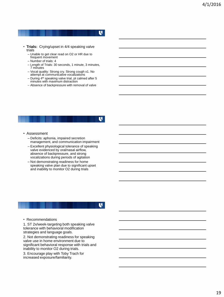
• Trials: Crying/upset in 4/4 speaking valve trials – Unable to get clear read on O2 or HR due to
frequent movement
– Number of trials: 4
– Length of Trials: 30 seconds, 1 minute, 3 minutes, 7 minutes
– Vocal quality: Strong cry. Strong cough x1. No attempt at communicative vocalizations
– During 4th speaking valve trial, pt calmed after 5 minutes with maximum distraction
– Absence of backpressure with removal of valve
• Assessment
– Deficits: aphonia, impaired secretion management, and communication impairment
– Excellent physiological tolerance of speaking valve evidenced by oral/nasal airflow, absence of backpressure, and strong vocalizations during periods of agitation
– Not demonstrating readiness for home speaking valve plan due to significant upset and inability to monitor O2 during trials
• Recommendations
1. ST 2x/week-targeting both speaking valve tolerance with behavioral modification strategies and language goals.
2. Not demonstrating readiness for speaking valve use in home environment due to significant behavioral response with trials and inability to monitor O2 during trials.
3. Encourage play with Toby Trach for increased exposure/familiarity.
4/1/2016
20
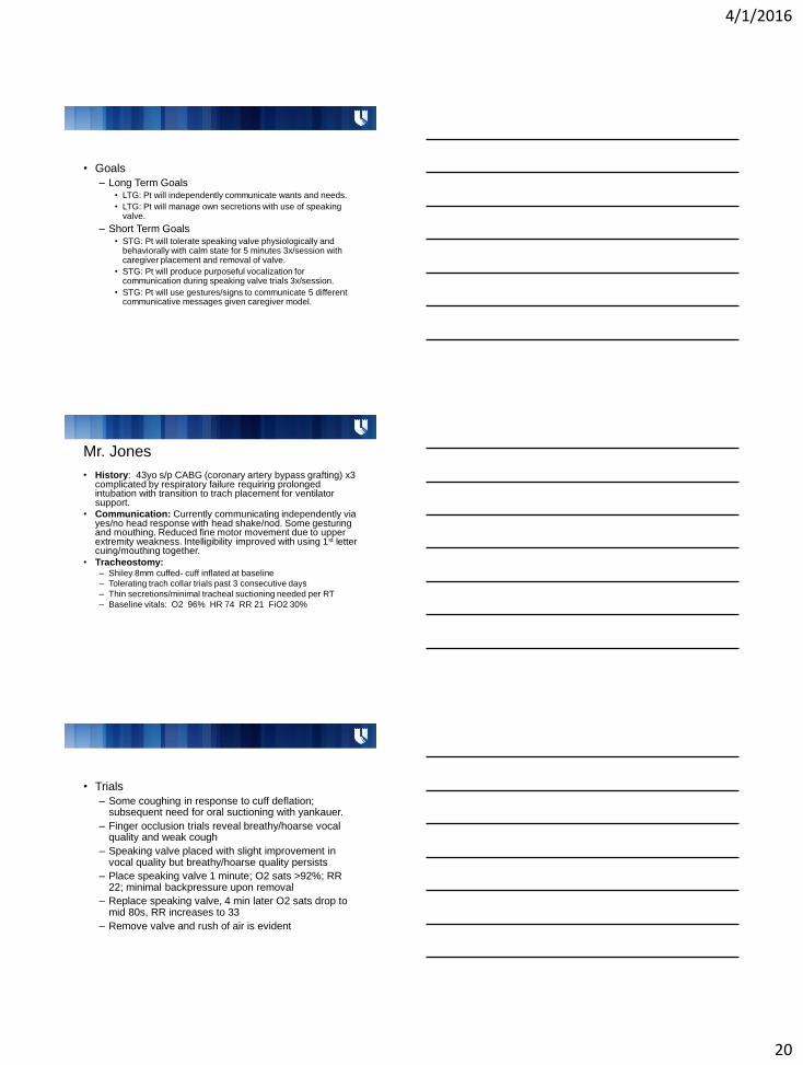
• Goals
– Long Term Goals • LTG: Pt will independently communicate wants and needs.
• LTG: Pt will manage own secretions with use of speaking valve.
– Short Term Goals • STG: Pt will tolerate speaking valve physiologically and
behaviorally with calm state for 5 minutes 3x/session with caregiver placement and removal of valve.
• STG: Pt will produce purposeful vocalization for communication during speaking valve trials 3x/session.
• STG: Pt will use gestures/signs to communicate 5 different communicative messages given caregiver model.
Mr. Jones
• History: 43yo s/p CABG (coronary artery bypass grafting) x3 complicated by respiratory failure requiring prolonged intubation with transition to trach placement for ventilator support.
• Communication: Currently communicating independently via yes/no head response with head shake/nod. Some gesturing and mouthing. Reduced fine motor movement due to upper extremity weakness. Intelligibility improved with using 1st letter cuing/mouthing together.
• Tracheostomy: – Shiley 8mm cuffed- cuff inflated at baseline
– Tolerating trach collar trials past 3 consecutive days
– Thin secretions/minimal tracheal suctioning needed per RT
– Baseline vitals: O2 96% HR 74 RR 21 FiO2 30%
• Trials
– Some coughing in response to cuff deflation; subsequent need for oral suctioning with yankauer.
– Finger occlusion trials reveal breathy/hoarse vocal quality and weak cough
– Speaking valve placed with slight improvement in vocal quality but breathy/hoarse quality persists
– Place speaking valve 1 minute; O2 sats >92%; RR 22; minimal backpressure upon removal
– Replace speaking valve, 4 min later O2 sats drop to mid 80s, RR increases to 33
– Remove valve and rush of air is evident

4/1/2016
21
• Assessment
– Deficits: aphonia secondary to tracheostomy, reduced upper airway patency with reduced tolerance of PMV placement
– Eventual desaturation, increase in RR and notable backpressure upon removal
– Weak cough and dysphonia suggesting reduced respiratory drive and/or reduced glottic closure
– Pt not appropriate for use of PMV at this time and will benefit from trach downsize once medically appropriate
– Consider maintaining cuff deflation if able to tolerate
• Recommendations – ST 4x/week for 1 month
– Prior to using speaking valve recommend consideration of: trach downsize when medically appropriate (may want to consider Shiley 6mm cuffless/cuffed or Shiley 8 cuffless-Shiley 6mm cuffless would be best option for PMV tolerance and moving toward decannulation)
* Be sure to include recommendations for functional means of communication (non verbal, low tech, etc)
• Goals – Long Term Goals
• LTG: Patient will independently communicate wants and needs via verbalization / nonverbal communication
• LTG: Patient will utilize speaking valve to facilitate decannulation.
• LTG: Patient will utilize speaking valve to improve secretion management.
– Short Term Goals • STG: Patient will produce voice over 10/10 trials when phonating at
phrase level with finger occlusion of trach when given moderate verbal cues for deep replenishing breathes.
• STG: Patient will participate in PMV re-assessment without respiratory compromise to determine appropriateness for use
• STG: Patient will communicate 5/5 basic wants/needs when given access to alphabet board for 1st letter cueing in structured situations.
• STG: Patient will activate call bell during 3/3 trials independently

4/1/2016
22
• Goals (cont.) Future goals once pt able to tolerate PMV
• STG: Patient will state (a) how and when to clean the PMV and (b) 4/4 reasons for PMV removal (respiratory treatment, sleeping, discomfort, O2 desaturation) when asked multiple choice questions over two consecutive sessions.
• STG: Patient will place and remove the PMV over 4/4 trials when given mirror, and verbal directions across 2 sessions
• STG: Patient will tolerate PMV throughout waking hours without s/s of respiratory compromise.
Questions?
Resources
• PMV website http://www.passy-muir.com
• Advancing Effective Communication, Cultural Competence, and Patient- and Family-Centered Care: A roadmap for hospitals – http://www.jointcommission.org/assets/1/6/ARoadmapforH
ospitalsfinalversion727.pdf
• ASHA website – www.asha.org
• Hauck, KA. (1999). Communication and Swallowing Issues in Tracheostomized/Ventilator-Dependent Geriatric Patients. Topics in Geriatric Rehabilitation, 15(2), 56-70.