Lenti-X™ iDimerize™ Inducible Homodimer System (with Tet ... · Lenti-X™ iDimerize™...
26
Takara Bio USA, Inc. 1290 Terra Bella Avenue, Mountain View, CA 94043, USA U.S. Technical Support: [email protected] United States/Canada 800.662.2566 Asia Pacific +1.650.919.7300 Europe +33.(0)1.3904.6880 Japan +81.(0)77.565.6999 Page 1 of 26 Takara Bio USA, Inc. Lenti-X™ iDimerize™ Inducible Homodimer System (with Tet-On® 3G) User Manual User Manual Cat. Nos. 635086, 635088, 635090, 635059, 635058, 635069, 632622 (011018)
Transcript of Lenti-X™ iDimerize™ Inducible Homodimer System (with Tet ... · Lenti-X™ iDimerize™...

Takara Bio USA, Inc.
1290 Terra Bella Avenue, Mountain View, CA 94043, USA
U.S. Technical Support: [email protected]
United States/Canada
800.662.2566
Asia Pacific
+1.650.919.7300
Europe
+33.(0)1.3904.6880
Japan
+81.(0)77.565.6999
Page 1 of 26
Takara Bio USA, Inc.
Lenti-X™ iDimerize™ Inducible Homodimer System (with Tet-On® 3G) User Manual User Manual
Cat. Nos. 635086, 635088, 635090, 635059, 635058, 635069, 632622
(011018)

Lenti-X™ iDimerize™ Inducible Homodimer System (with Tet-On® 3G) User Manual
(011018) takarabio.com
Takara Bio USA, Inc.
Page 2 of 26
Table of Contents I. Introduction ................................................................................................................................................................. 4
A. Summary: 4th Generation Lentiviral Packaging System ............................................................................................. 4
B. Summary: Tet-On 3G Inducible Gene Expression ...................................................................................................... 4
C. Summary: iDimerize Inducible Protein Interactions ................................................................................................... 5
D. Lenti-X iDimerize Inducible Homodimer System (with Tet-On 3G) ......................................................................... 6
II. List of Components ..................................................................................................................................................... 7
III. Additional Materials Required .................................................................................................................................... 7
A. In-Fusion® HD Cloning System & Stellar™ Competent Cells .................................................................................. 7
B. Xfect Transfection Reagents ....................................................................................................................................... 7
C. Doxycycline ................................................................................................................................................................ 7
D. Mammalian Cell Culture Supplies .............................................................................................................................. 8
E. Tetracycline-Free Fetal Bovine Serum........................................................................................................................ 8
F. B/B Homodimerizer .................................................................................................................................................... 8
G. B/B Washout Ligand ................................................................................................................................................... 8
H. DmrB Monoclonal Antibody ...................................................................................................................................... 8
I. Tet-On 3G Cell Lines .................................................................................................................................................. 9
J. Lenti-X 293T Cells...................................................................................................................................................... 9
K. High-Titer Packaging System ..................................................................................................................................... 9
L. Lentiviral Titer Determination .................................................................................................................................... 9
M. Lentivirus Concentration ............................................................................................................................................. 9
N. Transduction Enhancers ............................................................................................................................................ 10
O. Antibiotics for Selecting Stable Cell Lines ............................................................................................................... 10
P. Luciferase Assay and Luminometer .......................................................................................................................... 10
IV. Protocol Overview..................................................................................................................................................... 10
A. General Cell Culture.................................................................................................................................................. 10
B. Safety Guidelines for Working with Lentiviruses ..................................................................................................... 10
C. Protocol Summary ..................................................................................................................................................... 12
V. Creating your pLVX-TRE3G-Hom1-GOI Construct ................................................................................................ 13
VI. Pilot Testing Your pLVX-TRE3G-Hom1-GOI Construct for Tet-Based Induction & B/B Homodimerizer-
Mediated Dimerization ......................................................................................................................................................... 13
Materials required ............................................................................................................................................................. 13
A. Confirm Expression of Your pLVX-TRE3G-Hom1-GOI Construct ......................................................................... 14
B. Determine Optimal Doxycycline Concentration ....................................................................................................... 14
C. Determine the Optimal B/B Homodimerizer Concentration ..................................................................................... 15

Lenti-X™ iDimerize™ Inducible Homodimer System (with Tet-On® 3G) User Manual
(011018) takarabio.com
Takara Bio USA, Inc.
Page 3 of 26
VII. Producing Lentivirus from Lenti-X Vectors ............................................................................................................. 15
VIII. Lentivirus Titration ................................................................................................................................................... 15
A. Summary ................................................................................................................................................................... 15
B. Protocol: Determining Viral Titer by Colony Formation .......................................................................................... 16
IX. Transducing Target Cells with the Tet-On 3G Lentiviruses ..................................................................................... 17
A. Summary ................................................................................................................................................................... 17
B. Protocol: Cotransducing Target Cells with Lenti-X Tet-On 3G Lentiviruses........................................................... 17
X. Combining iDimerize Homodimerization with the Tet-On 3G System .................................................................... 18
Materials Required ............................................................................................................................................................ 18
A. Determine the Optimal Doxycycline Concentration ................................................................................................. 18
B. Determine the Optimal B/B Homodimerizer Concentration ..................................................................................... 18
C. Dissociate Dimerized Proteins with the B/B Washout Ligand ................................................................................. 19
XI. References ................................................................................................................................................................. 20
XII. Troubleshooting ........................................................................................................................................................ 21
A. Lenti-X Troubleshooting ........................................................................................................................................... 21
B. Tet-On 3G Troubleshooting ...................................................................................................................................... 22
D. iDimerize Troubleshooting ....................................................................................................................................... 24
Appendix A: Preparing and Handling Tet-On 3G Cell Line Stocks ..................................................................................... 25
A. Protocol: Freezing Tet-On 3G Cell Line Stocks ....................................................................................................... 25
B. Protocol: Thawing Tet-On 3G Cell Line Frozen Stocks ........................................................................................... 25
Table of Figures Figure 1. The Tet-On 3G Systems allow inducible gene expression in the presence of Dox. ................................................ 4
Figure 2. Controlling signal transduction using regulated homodimerization ........................................................................ 5
Figure 3. pLVX-TRE3G-Hom1 and pLVX-TRE3G-Luc Control vector maps ..................................................................... 6
Figure 4. pLVX-Tet3G vector map ......................................................................................................................................... 6
Figure 5. The In-Fusion HD single-tube cloning protocol. ................................................................................................... 13
Figure 6. B/B Washout Ligand can be used to disrupt protein interactions induced by the B/B Homodimerizer................ 19
Figure 7. B/B Washout Ligand has a much more dissociative effect than simple removal of the B/B Homodimerizer ...... 20
Table of Tables Table 1. Recommended antibiotic concentrations for selecting and maintaining stable clones ........................................... 26

Lenti-X™ iDimerize™ Inducible Homodimer System (with Tet-On® 3G) User Manual
(011018) takarabio.com
Takara Bio USA, Inc.
Page 4 of 26
I. Introduction The Lenti-X iDimerize Inducible Homodimer System (with Tet-On 3G) (Cat. No. 635086) is an optimized
system which combines lentiviral gene delivery, Tet-On 3G inducible gene expression, and iDimerize inducible
protein interactions in live cells. One challenge of ligand-dependent dimerization experiments is that non ligand-
induced dimerization events may occur if the protein of interest is expressed at high levels. This is especially
problematic if the target protein is a membrane protein, because the local concentration can increase quickly due
to the limited space on the membrane. For example, overexpression of the Fas receptor can induce apoptosis due
to ligand-independent receptor trimerization when there is a high abundance of the Fas receptor in the cell
membrane.
We have combined the iDimerize Homodimer System with Tet-On 3G technology to reduce potential non-ligand
induced dimerization events that occur due to the protein being dimerized, and lentiviral technology to deliver
your GOI to almost any mammalian cell type, including dividing and nondividing cells, primary cell cultures,
stem cells, and neurons. First, create your lentiviruses and transduce your target cells. Then use doxycycline
(Dox) to optimize the DmrB-tagged protein’s expression to a physiologically relevant level. Finally, induce
dimerization using the B/B Homodimerizer ligand.
A. Summary: 4th Generation Lentiviral Packaging System
Lenti-X Packaging Single Shots (VSV-G), provided with the Lenti-X iDimerize Inducible Homodimer System
(with Tet-On 3G) (Cat. No. 635086), can generate lentiviral titers that are superior to most other commercially
available lentiviral packaging systems. The concerted effects of multiple components in an optimized five-vector
plasmid mix, pre-aliquoted and lyophilized with Xfect™ Transfection Reagent, allow Lenti-X 293T Cells (sold
separately; Cat. No. 632180) to produce the highest amounts of safe, replication-incompetent lentivirus (see
takarabio.com).
B. Summary: Tet-On 3G Inducible Gene Expression
Tet-On 3G systems are inducible gene expression systems for mammalian cells. Target cells that express the Tet-On
3G transactivator protein and contain a gene of interest (GOI) under the control of the PTRE3G will express tightly
controlled levels of your GOI when cultured in the presence of Dox (Figure 1).
Figure 1. The Tet-On 3G Systems allow inducible gene expression in the presence of Dox.
Tet-On 3G Transactivator Protein
Based on the transcriptional regulators described by Gossen & Bujard (1992), Gossen et al. (1995), and Urlinger
et al. (2000), Tet-On 3G is a modified form of the Tet-On Advanced transactivator protein which has been
evolved to display far higher sensitivity to doxycycline (Zhou et al. 2006).
PTRE3G Inducible Promoter
PTRE3G provides for very low basal expression and high maximal expression after induction (Loew et al. 2010). It
consists of 7 repeats of a 19 bp tet operator sequence located upstream of a minimal CMV promoter. In the
presence of Dox, the Tet-On 3G transactivator binds specifically to PTRE3G and activates transcription of the
downstream GOI. PTRE3G lacks binding sites for endogenous mammalian transcription factors, so they are virtually
silent in the absence of induction.
Lenti-X™ iDimerize™ Inducible Homodimer System (with Tet-On® 3G) User Manual
(011018) takarabio.com
Takara Bio USA, Inc.
Page 5 of 26
Doxycycline
Doxycycline is a synthetic tetracycline derivative that is the effector molecule for the Tet-On and Tet-Off® Systems.
When bound by Dox, the Tet-On 3G protein undergoes a conformational change that allows it to bind to tet operator
sequences located in the PTRE3G promoter (Figure 1). The Dox concentrations required for induction of Tet-On
Systems are far below cytotoxic levels for either cell culture or transgenic studies, and Tet-On 3G responds to even
lower concentrations than its predecessors (Zhou et al. 2006).
Note that Tet-On Systems respond well only to doxycycline, and not to tetracycline (Gossen et al. 1995). The half-
life of Dox in cell culture medium is 24 hours. To maintain continuous inducible GOI expression in cell culture, the
medium should be replenished with Dox every 48 hours.
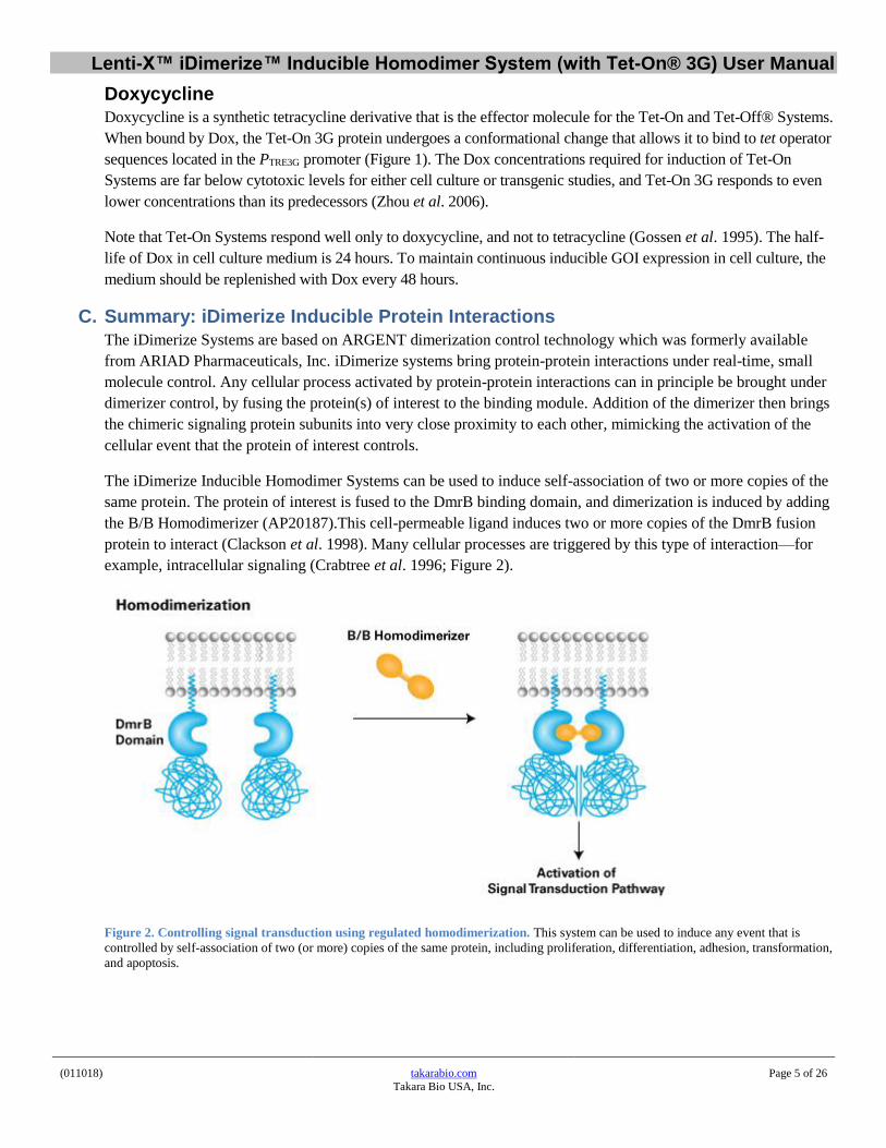
C. Summary: iDimerize Inducible Protein Interactions
The iDimerize Systems are based on ARGENT dimerization control technology which was formerly available
from ARIAD Pharmaceuticals, Inc. iDimerize systems bring protein-protein interactions under real-time, small
molecule control. Any cellular process activated by protein-protein interactions can in principle be brought under
dimerizer control, by fusing the protein(s) of interest to the binding module. Addition of the dimerizer then brings
the chimeric signaling protein subunits into very close proximity to each other, mimicking the activation of the
cellular event that the protein of interest controls.
The iDimerize Inducible Homodimer Systems can be used to induce self-association of two or more copies of the
same protein. The protein of interest is fused to the DmrB binding domain, and dimerization is induced by adding
the B/B Homodimerizer (AP20187).This cell-permeable ligand induces two or more copies of the DmrB fusion
protein to interact (Clackson et al. 1998). Many cellular processes are triggered by this type of interaction—for
example, intracellular signaling (Crabtree et al. 1996; Figure 2).
Figure 2. Controlling signal transduction using regulated homodimerization. This system can be used to induce any event that is
controlled by self-association of two (or more) copies of the same protein, including proliferation, differentiation, adhesion, transformation,
and apoptosis.

Lenti-X™ iDimerize™ Inducible Homodimer System (with Tet-On® 3G) User Manual
(011018) takarabio.com
Takara Bio USA, Inc.
Page 6 of 26
D. Lenti-X iDimerize Inducible Homodimer System (with Tet-On 3G)
The pLVX-TRE3G-Hom1 expression vector included in this system (Figure 3), in combination with the Lenti-X
HTX Packaging System (which allows for packaging and delivery of high-titer lentivirus), enables your DmrB-
tagged protein of interest to be expressed in a wide range of cell types, in a Dox-dependent manner.
Figure 3. pLVX-TRE3G-Hom1 and pLVX-TRE3G-Luc Control vector maps. pLVX-TRE3G-Hom1 allows you to express your
DmrB-tagged protein of interest in a Dox-dependent manner. pLVX-TRE3G-Luc is a control vector that expresses firefly luciferase under
the control of PTRE3GV. When used with standard luciferase detection reagents, this vector can be used as a reporter of induction efficiency.
Figure 4. pLVX-Tet3G vector map. pLVX-Tet3G allows you to express the Tet-On 3G transactivator protein in your cells of
interest.

Lenti-X™ iDimerize™ Inducible Homodimer System (with Tet-On® 3G) User Manual
(011018) takarabio.com
Takara Bio USA, Inc.
Page 7 of 26
II. List of Components Store Lenti-X GoStix™ Plus at room temperature. Store all other components at -20°C.
• 1 each pLVX-TRE3G-Hom1 Vector Set (Cat. No. 635087; not sold separately)
− 20 μl
− 20 μl
pLVX-TRE3G-Hom1 Vector (500 ng/μl)
pLVX-TRE3G-Luc Control Vector (500 ng/μl)
• 20 μl pLVX-Tet3G Vector (500 ng/μl) (Cat. No. 631358; not sold separately)
• 16 rxns Lenti-X Packaging Single shots
• 1 each Lenti-X GoStix Plus (Sample) (Cat. No. 631279; not sold separately)
• 500 µl B/B Homodimerizer (0.5 mM; also sold separately as Cat. Nos. 635059, 635058, 635069 &
632622—see Section III.F)
NOTE: The B/B Homodimerizer is so named because it induces dimerization of two proteins that contain the
DmrB dimerization domain.
III. Additional Materials Required
A. In-Fusion® HD Cloning System & Stellar™ Competent Cells
In-Fusion is a revolutionary technology that greatly simplifies cloning. For more information, visit
takarabio.com/infusion. We recommend using Stellar Competent Cells, which are included in the In-Fusion HD
Cloning Kits listed below. You can also purchase Stellar Competent Cells separately as Cat. No. 636763.
Cat. No. In-Fusion Cloning Kit Size 638909 638910 638911 638920
In-Fusion HD Cloning Plus In-Fusion HD Cloning Plus In-Fusion HD Cloning Plus In-Fusion HD Cloning Plus
10 rxns 50 rxns 100 rxns 96 rxns
638916 638917 638918 638919
In-Fusion HD Cloning Plus CE In-Fusion HD Cloning Plus CE In-Fusion HD Cloning Plus CE In-Fusion HD Cloning Plus CE
10 rxns 50 rxns 100 rxns 96 rxns
B. Xfect Transfection Reagents
Xfect Transfection Reagent provides high transfection efficiency and low cytotoxicity for most commonly used cell
types. Xfect mESC Transfection Reagent is optimized for mouse embryonic stem cells.
Cat. No. Transfection Reagent 631317 631318
Xfect Transfection Reagent (100 rxns) Xfect Transfection Reagent (300 rxns)
631320 631321
Xfect mESC Transfection Reagent (100 rxns) Xfect mESC Transfection Reagent (300 rxns)
C. Doxycycline Cat. No. Transfection Reagent 631311 Doxycycline (5 g)
Dilute to 1 mg/ml in double-distilled H2O. Filter sterilize, aliquot, and store at –20°C in the dark. Use within one year.

Lenti-X™ iDimerize™ Inducible Homodimer System (with Tet-On® 3G) User Manual
(011018) takarabio.com
Takara Bio USA, Inc.
Page 8 of 26
D. Mammalian Cell Culture Supplies
• Medium for Lenti-X 293T Cells:
90% Dulbecco's Modified Eagle's Medium (DMEM) with high glucose (4.5 g/L), 4 mM L-glutamine, and
sodium bicarbonate (Sigma-Aldrich, Cat. No. D5796); 10% Fetal Bovine Serum (FBS); 100 units/ml
penicillin G sodium; and 100 μg/ml streptomycin sulfate.
• Culture medium, supplies, and additives specific for your target cells
• Trypsin/EDTA (e.g., Sigma, Cat. No. T4049)
• Cloning cylinders or discs for isolating colonies of adherent cell lines (Sigma, Cat. No. C1059)
• Cell Freezing Medium, with or without DMSO (Sigma, Cat. Nos. C6164 or C6039)
• 6-well, 12-well, and 24-well cell culture plates, 10 cm cell culture dishes
E. Tetracycline-Free Fetal Bovine Serum
Contaminating tetracyclines, which are often found in serum, significantly elevate basal expression when using
Tet-On 3G. The following functionally tested tetracycline-free sera are available:
Cat. No. Serum Name Size
631106 Tet System Approved FBS 500 ml
631107 50 ml
631101 Tet System Approved FBS, US-Sourced
500 ml
631105 50 ml
F. B/B Homodimerizer
Each Lenti-X iDimerize Inducible Homodimer System (with Tet-On 3G) includes 500 μl B/B Homodimerizer.
Additional B/B Homodimerizer can also be purchased separately in the following sizes:
Cat. No. Product Name Size
635059 B/B Homodimerizer (0.5 mM) 5 x 500 µl
635058 B/B Homodimerizer* 5 mg
635069 25 mg
632622 4 x 25 mg
*Supplied in a dry-down format.
G. B/B Washout Ligand
B/B Washout Ligand is a membrane-permeant ligand that dissociates protein interactions induced by the B/B
Homodimerizer. It dissociates these interactions with a T1/2 of ~10 minutes after adding it to target cells treated
with B/B Homodimerizer.
Cat. No. Product Name Size
635088 B/B Washout Ligand (0.5 mM) 500 µl
H. DmrB Monoclonal Antibody
The DmrB Monoclonal Antibody recognizes the DmrB binding domain expressed using iDimerize Inducible
Homodimer Systems, and is recommended for Western blot analysis.
Cat. No. Product Name Size
635090 DmrB Monoclonal Antibody (0.5 µg/µl) 100 µg
Lenti-X™ iDimerize™ Inducible Homodimer System (with Tet-On® 3G) User Manual
(011018) takarabio.com
Takara Bio USA, Inc.
Page 9 of 26
I. Tet-On 3G Cell Lines
Cat. No. Cell Line
631181 Jurkat Tet-On 3G Cell Line
631182 HEK 293 Tet-On 3G Cell Line
631183 HeLa Tet-On 3G Cell Line
631195 CHO Tet-On 3G Cell Line
631197 NIH/3T3 Tet-On 3G Cell Line
J. Lenti-X 293T Cells
Getting the most from any lentiviral packaging system requires a host 293T cell line that transfects easily and
supports high-level expression of viral proteins. Our Lenti-X 293T Cell Line was clonally selected to meet these
requirements, allowing you to produce the highest possible lentiviral titers when combined with Lenti-X
Packaging Single Shots (VSV-G), an optimized fourth-generation packaging system, pre-mixed and lyophilized
with Xfect Transfection Reagent.
Cat. No. Cell Line 632180 Lenti-X 293T Cell Line
K. High-Titer Packaging System
This 4th generation lentiviral packaging system can generate lentiviral titers that are superior to most other
commercially available lentiviral packaging systems. The concerted effects of multiple components in an
optimized five-vector plasmid mix, pre-aliquoted and lyophilized with Xfect Transfection Reagent, allow Lenti-X
293T Cells to produce the highest amounts of safe, replication-incompetent lentivirus (see takarabio.com).
Cat. No. Packaging System 631275 631276
Lenti-X Packaging Single Shots (VSV-G) (16 rxns) Lenti-X Packaging Single Shots (VSV-G) (96 rxns)
L. Lentiviral Titer Determination
For accurate and consistent transductions, we highly recommend titrating your lentiviral stocks. Various
technologies are available; visit takarabio.com for details.
Cat. No. Lentiviral Titration Technology 632200 Lenti-X p24 Rapid Titer Kit (96 rxns) 631235 Lenti-X qRT-PCR Titration Kit (200 rxns) 631280 Lenti-X GoStix Plus (20 tests)
M. Lentivirus Concentration
Use Lenti-X Concentrator to simply increase your available titer up to 100-fold without ultracentrifugation—visit
takarabio.com for details.
Cat. No. Concentrator 631231 Lenti-X Concentrator (100 ml) 631232 Lenti-X Concentrator (500 ml)

Lenti-X™ iDimerize™ Inducible Homodimer System (with Tet-On® 3G) User Manual
(011018) takarabio.com
Takara Bio USA, Inc.
Page 10 of 26
N. Transduction Enhancers
Use Polybrene (hexadimethrine bromide; Sigma-Aldrich, No. H9268), Lenti-X Accelerator, or RetroNectin®.
• Lenti-X Accelerator is a magnetic bead-based technology designed to accelerate lentiviral and retroviral
transduction experiments; visit takarabio.comfor details.
• RetroNectin is a multivalent molecule that simultaneously binds virus particles and cell surface proteins,
maximizing cell-virus contact. RetroNectin, in particular, is recommended for increasing the transduction
efficiency of suspension cells and stem cells; visit takarabio.comfor details.
Cat. No. Transduction Enhancer 631256 Lenti-X Accelerator (400 µl) 631257 Lenti-X Accelerator (1,000 µl) 631254 Lenti-X Accelerator Starter Kit (1 each) T110A RetroNectin Precoated Dish (10 dishes) T100B RetroNectin Recombinant Human Fibronectin Fragment (2.5 mg) T100A RetroNectin Recombinant Human Fibronectin Fragment (0.5 mg)
O. Antibiotics for Selecting Stable Cell Lines Cat. No. Antibiotic Size 631306 631305
Puromycin Puromycin
100 mg 25 mg
631308 631307
G418 G418
5 g 1 g
P. Luciferase Assay and Luminometer
These items are required when using the pLVX-TRE3G-Luc Vector to screen Tet-On 3G clones. Use any
standard luciferase assay system and luminometer.
IV. Protocol Overview
A. General Cell Culture
This user manual provides only general guidelines for mammalian cell culture techniques. If you require more
information on mammalian cell culture, transfection, and creating stable cell lines, we recommend the following
general reference:
Freshney, R.I. (2005). Culture of Animal Cells: A Manual of Basic Technique, 5th Edition (Wiley-Liss, Hoboken, NJ).
B. Safety Guidelines for Working with Lentiviruses
The protocols in this User Manual require the production, handling, and storage of infectious lentivirus. It is
imperative to fully understand the potential hazards of, and necessary precautions for, the laboratory use of
lentiviruses.
The National Institutes of Health and Centers for Disease Control have designated recombinant lentiviruses as
Level 2 organisms. This requires the maintenance of a Biosafety Level 2 facility for work involving this virus and
others like it. The VSV-G pseudotyped lentiviruses packaged from the HIV-1-based vectors described here are
capable of infecting human cells. The viral supernatants produced by these lentiviral systems could, depending on
your insert, contain potentially hazardous recombinant virus. Similar vectors have been approved for human gene
therapy trials, attesting to their potential ability to express genes in vivo.

Lenti-X™ iDimerize™ Inducible Homodimer System (with Tet-On® 3G) User Manual
(011018) takarabio.com
Takara Bio USA, Inc.
Page 11 of 26
IMPORTANT: For these reasons, due caution must be exercised in the production and handling of any
recombinant lentivirus. The user is strongly advised not to create VSV-G pseudotyped lentiviruses capable of
expressing known oncogenes.
For more information on Biosafety Level 2 agents and practices, download the following reference:
Biosafety in Microbiological and Biomedical Laboratories (BMBL), Fifth Edition (December 2009) HHS
Pub. No. (CDC) 93-8395. U.S. Department of Health and Human Services Centers for Disease Control
and Prevention and NIH.
Available on the web at http://www.cdc.gov/biosafety/publications/bmbl5/
Biosafety Level 2: The following information is a brief description of Biosafety Level 2. It is neither detailed nor
complete. Details of the practices, safety equipment, and facilities that combine to produce a Biosafety Level 2 are
available in the above publication. If possible, observe and learn the practices described below from someone who
has experience working with lentiviruses.
Summary of Biosafety Level 2:
• Practices:
o Standard microbiological practices
o Limited access to work area
o Biohazard warning signs posted
o Minimize production of aerosols
o Decontaminate potentially infectious wastes before disposal
o Use precautions with sharps (e.g., syringes, blades)
o Biosafety manual defining any needed waste decontamination or medical surveillance policies
• Safety equipment:
o Biological Safety Cabinet, preferably a Class II BSC/laminar flow hood (with a HEPA
microfilter) used for all manipulations of agents that cause splashes or aerosols of infectious
materials; exhaust air is unrecirculated
o PPE: protective laboratory coats, gloves, face protection as needed
• Facilities:
o Autoclave available for waste decontamination
o Chemical disinfectants available for spills

Lenti-X™ iDimerize™ Inducible Homodimer System (with Tet-On® 3G) User Manual
(011018) takarabio.com
Takara Bio USA, Inc.
Page 12 of 26
C. Protocol Summary
Please read each protocol completely before starting. Successful results depend on understanding and performing
all the steps correctly.
Section X.C
Use B/B Washout Ligand to dissociate dimerized proteins
Section X.B
Optimize B/B Homodimerizer concentration to control protein dimerization
Section X.A
Optimize Dox concentration to control gene expression
Section IX
Cotransduce LVX-TRE3G-Hom1-GOI & LVX-Tet3G lentiviruses into target cells
Section VIII
Titer LVX-TRE3G-Hom1-GOI & LVX-Tet3G lentiviruses
Section VII
Produce LVX-TRE3G-Hom1-GOI & LVX-Tet3G lentiviruses
Section VI
Pilot test pLVX-TRE3G-Hom1-GOI construct
Section V
Clone GOI in frame with DmrB

Lenti-X™ iDimerize™ Inducible Homodimer System (with Tet-On® 3G) User Manual
(011018) takarabio.com
Takara Bio USA, Inc.
Page 13 of 26
V. Creating your pLVX-TRE3G-Hom1-GOI Construct We recommend using the In-Fusion HD Cloning System (Section III.A) to clone your gene of interest into
pLVX-TRE3G-Hom1. In-Fusion HD cloning is generally recommended over ligation-based cloning because it is
directional, unaffected by internal cut sites, and highly efficient (most clones contain the correct insert).
Follow the protocol outlined in the In-Fusion HD user manual. To find the manual, go to takarabio.com/manuals and
type “In-Fusion HD” in the search box.
Figure 5. The In-Fusion HD single-tube cloning protocol.
VI. Pilot Testing Your pLVX-TRE3G-Hom1-GOI Construct for Tet-Based
Induction & B/B Homodimerizer-Mediated Dimerization Prior to lentivirus production, your pLVX-TRE3G-Hom1-GOI construct can be tested for functionality by plasmid
transfection. Transiently cotransfect your pLVX-TRE3G-Hom1-GOI vector together with pLVX-Tet3G (in a 1:4
ratio for best inducibility) into an easy-to-transfect cell line such as HeLa or HEK 293, or your target cell line.
The optimal concentrations of doxycycline and B/B Homodimerizer that you determine in this pilot test will not
apply directly to your final experiment (Section X), but they may serve as general guidelines for the
concentrations to use in your final experiment.
Materials required
• pLVX-TRE3G-Hom1-GOI (Section V)
• pLVX-Tet3G
• Host cell line: Choose an easy-to-transfect cell line such as HeLa or HEK 293, a premade Tet-On 3G cell
line, or your target cell line
• Xfect transfection reagent (Section III.B)
• Doxycycline (1 mg/ml) (Section III.C)
• Mammalian cell culture supplies (Section III.D)
• Tet Approved FBS (Section III.E)
• B/B Homodimerizer (Section III.F)
Lenti-X™ iDimerize™ Inducible Homodimer System (with Tet-On® 3G) User Manual
(011018) takarabio.com
Takara Bio USA, Inc.
Page 14 of 26
A. Confirm Expression of Your pLVX-TRE3G-Hom1-GOI Construct
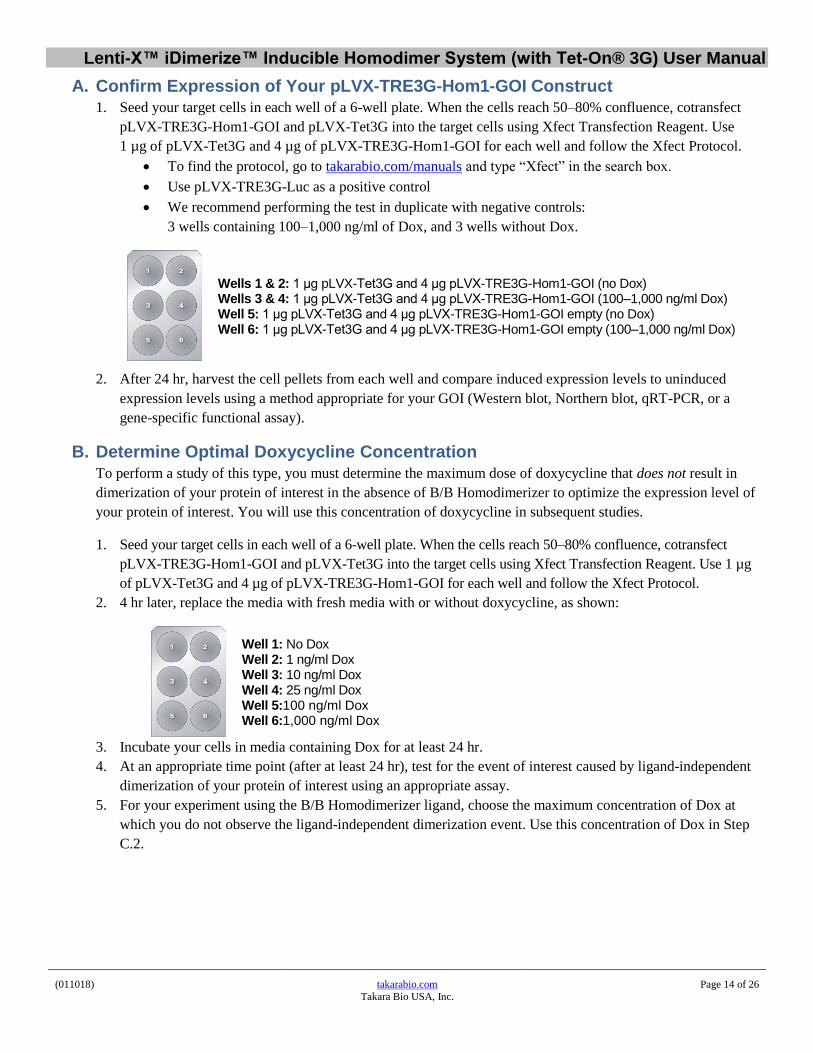
1. Seed your target cells in each well of a 6-well plate. When the cells reach 50–80% confluence, cotransfect
pLVX-TRE3G-Hom1-GOI and pLVX-Tet3G into the target cells using Xfect Transfection Reagent. Use
1 µg of pLVX-Tet3G and 4 µg of pLVX-TRE3G-Hom1-GOI for each well and follow the Xfect Protocol.
• To find the protocol, go to takarabio.com/manuals and type “Xfect” in the search box.
• Use pLVX-TRE3G-Luc as a positive control
• We recommend performing the test in duplicate with negative controls:
3 wells containing 100–1,000 ng/ml of Dox, and 3 wells without Dox.
Wells 1 & 2: 1 μg pLVX-Tet3G and 4 μg pLVX-TRE3G-Hom1-GOI (no Dox) Wells 3 & 4: 1 μg pLVX-Tet3G and 4 μg pLVX-TRE3G-Hom1-GOI (100–1,000 ng/ml Dox) Well 5: 1 μg pLVX-Tet3G and 4 μg pLVX-TRE3G-Hom1-GOI empty (no Dox) Well 6: 1 μg pLVX-Tet3G and 4 μg pLVX-TRE3G-Hom1-GOI empty (100–1,000 ng/ml Dox)
2. After 24 hr, harvest the cell pellets from each well and compare induced expression levels to uninduced
expression levels using a method appropriate for your GOI (Western blot, Northern blot, qRT-PCR, or a
gene-specific functional assay).
B. Determine Optimal Doxycycline Concentration
To perform a study of this type, you must determine the maximum dose of doxycycline that does not result in
dimerization of your protein of interest in the absence of B/B Homodimerizer to optimize the expression level of
your protein of interest. You will use this concentration of doxycycline in subsequent studies.
1. Seed your target cells in each well of a 6-well plate. When the cells reach 50–80% confluence, cotransfect
pLVX-TRE3G-Hom1-GOI and pLVX-Tet3G into the target cells using Xfect Transfection Reagent. Use 1 µg
of pLVX-Tet3G and 4 µg of pLVX-TRE3G-Hom1-GOI for each well and follow the Xfect Protocol.
2. 4 hr later, replace the media with fresh media with or without doxycycline, as shown:
Well 1: No Dox Well 2: 1 ng/ml Dox Well 3: 10 ng/ml Dox Well 4: 25 ng/ml Dox Well 5:100 ng/ml Dox Well 6:1,000 ng/ml Dox
3. Incubate your cells in media containing Dox for at least 24 hr.
4. At an appropriate time point (after at least 24 hr), test for the event of interest caused by ligand-independent
dimerization of your protein of interest using an appropriate assay.
5. For your experiment using the B/B Homodimerizer ligand, choose the maximum concentration of Dox at
which you do not observe the ligand-independent dimerization event. Use this concentration of Dox in Step
C.2.

Lenti-X™ iDimerize™ Inducible Homodimer System (with Tet-On® 3G) User Manual
(011018) takarabio.com
Takara Bio USA, Inc.
Page 15 of 26
C. Determine the Optimal B/B Homodimerizer Concentration
Now that you have determined the optimal level of Dox that induces expression of your protein of interest without
causing ligand-independent dimerization, you can determine the optimal amount of ligand to add in order to
induce dimerization.
1. Seed your target cells in each well of a 6-well plate. When the cells reach 50–80% confluence, cotransfect
pLVX-TRE3G-Hom1-GOI and pLVX-Tet3G into the target cells using Xfect Transfection Reagent. Use
1 μg of pCMV-Tet3G and 4 μg of pTRE3G-Hom1-GOI for each well and follow the Xfect Protocol.
2. 4 hr later, replace the media with fresh media containing the optimal concentration of doxycycline from Step
B.5. Incubate your cells in media containing Dox for at least 12 hr.
3. Add a range of B/B Homodimerizer concentrations to the growth media containing Dox.
Each well should contain the optimal concentration of Dox determined in Step B.5 above, plus:
Well 1: 0 nM B/B Homodimerizer Well 2: 10 nM B/B Homodimerizer Well 3: 50 nM B/B Homodimerizer Well 4: 100 nM B/B Homodimerizer Well 5:250 nM B/B Homodimerizer Well 6:1,000 nM B/B Homodimerizer
4. At an appropriate time point, test for the event caused by ligand-dependent dimerization of your protein of
interest.
Notes:
• Test for the effects of dimerization using any assay that is appropriate for your experiment.
• The amount of time you should wait to perform your analysis after adding B/B Homodimerizer depends
on the nature of the event caused by protein homodimerization. Rapid events such as phosphorylation
may occur within 15–60 min. Slower events, such as differentiation, may require 1–5 days.
VII. Producing Lentivirus from Lenti-X Vectors Follow the Lenti-X Packaging Single Shots (VSV-G) Protocol-At-A-Glance. (Locate this protocol by
searching at takarabio.com/manuals).
VIII. Lentivirus Titration
A. Summary
1. Instant Qualitative Titer Test
You can detect and quantify your lentivirus stock in ten minutes with our Lenti-X GoStix Plus (Cat. Nos.
631280, 631281) and the related smartphone app. The GoStix detect lentiviral p24 in only 20 μl, and can
be used to determine whether virus production is within a usable range or for selecting the best time to
harvest your virus. A 3-prep sample of Lenti-X GoStix Plus is supplied for free with the Lenti-X
iDimerize Inducible Homodimer System (with Tet-On 3G). Visit takarabio.com/gostixhelp for details.

Lenti-X™ iDimerize™ Inducible Homodimer System (with Tet-On® 3G) User Manual
(011018) takarabio.com
Takara Bio USA, Inc.
Page 16 of 26
2. Quantitative Titer Test
a. Determining the viral titer is necessary to obtain the following information:
• Confirmation that viral stocks are viable
• The proper transduction conditions for your particular cell type—obtained by adjusting
the MOI for the desired transduction efficiency
(MOI = No. of infectious virus particles per target cell)
• The maximum number of target cells that can be transduced by a given virus volume
b. To transduce using a known multiplicity of infection (MOI), it is necessary to titrate your
lentiviral stocks. We recommend the Lenti-X qRT-PCR Titration Kit (Cat. No. 631235) or
Lenti-X p24 Rapid Titer Kit (Cat. No. 632200) for very rapid quantitative titrations of virus
stocks (~4 hr), or a standard method that relies on infection.
c. The standard viral titration protocol consists of infecting cells with serial dilutions of the stock,
selecting for stable transductants with antibiotic, and counting the resulting cell colonies (Section
VIII.B).
• Freshly harvested virus can be titered immediately, or frozen in aliquots at –80°C and
then titrated. Note that each freeze-thaw cycle can reduce the functional titers of
infectious virus by up to 2–4 fold.
• Absolute titers will depend heavily on the cell type used for titration, and there may be
significant differences between the titer values determined in cells typically used for
lentiviral titration (i.e. HT-1080) and the number of target cells transduced by the titered
virus. However, titrations serve to determine the relative virus content of different viral
stocks prepared from different vectors.
B. Protocol: Determining Viral Titer by Colony Formation
NOTE: This protocol can be completed in 7–14 days.
1. Plate HT-1080 cells (or other) in 6-well plates the day before performing the titration infections. Plate 2 x 10⁵
cells/well, in 2 ml of medium. Allow at least one well to be used as a “no infection” control.
2. Prepare 20 ml of complete medium and add 60 μl of 4 mg/ml Polybrene. This will be diluted 3-fold for a final
Polybrene concentration of 4 μg/ml.
NOTE: Polybrene is a polycation that reduces charge repulsion between the virus and the cellular membrane.
The optimum final concentration of Polybrene may be determined empirically but generally falls within a range
of 2–12 μg/ml. Excessive exposure to Polybrene (>24 hr) can be toxic to cells.
3. Prepare filtered viral supernatant from packaging cells (Section VII). This is the virus stock.
4. Prepare six 10-fold serial dilutions of the virus stock as follows:
a. Add 1.35 ml of medium containing Polybrene (Step 2) to each of six sterile and numbered 1.5 ml
microfuge tubes.
b. Add 150 μl of the virus stock (Step 3) to Tube 1. Mix.
c. Transfer 150 μl from Tube 1 to Tube 2 and mix. Continue making serial dilutions by transferring 150
μl from each successive dilution into the next prepared tube.
5. Infect the HT-1080 cells by adding 1 ml of each viral dilution (Step 4) to each appropriate well. The final
Polybrene concentration will be 4 μg/ml in ~3 ml. Centrifuge the cultures to improve infection efficiency*.
*NOTE: CULTURE CENTRIFUGATION INCREASES INFECTION EFFICIENCY. Centrifuging the plate
at 1,200 x g for 60–90 min at 32°C can significantly increase infection efficiency. A room temperature
centrifuge is acceptable if a 32°C unit is not available.

Lenti-X™ iDimerize™ Inducible Homodimer System (with Tet-On® 3G) User Manual
(011018) takarabio.com
Takara Bio USA, Inc.
Page 17 of 26
6. After infecting for 8–24 hours, remove supernatants and subject the cells to G418 or puromycin selection
using the selection concentrations that are optimal for your cell line (Section III.O).
7. Allow colonies to form for 7–14 days. Stain the colonies with 1% crystal violet solution (in 10% ethanol) and
count.
8. The titer of virus corresponds to the number of colonies generated by the highest dilution, multiplied by the
dilution factor. For example, the presence of 4 colonies in the 106 dilution would represent a viral titer of 4 x
106 colony forming units.
IX. Transducing Target Cells with the Tet-On 3G Lentiviruses
A. Summary
• Simultaneous Cotransduction: To establish the complete Lenti-X iDimerize Tet-On 3G System, target cells
must be cotransduced with both the LVX-Tet3G and LVX-TRE3G-Hom1-GOI lentiviruses. Using high titers
of each virus ensures that the highest proportion of cells will contain both vectors. Depending on your
application, transduced cells can either be treated immediately with Dox to induce expression of your GOI
and then harvested for analysis, or the cells may be selected with G418 and puromycin to isolate doubly-
transduced clones or to enrich the population for doubly-transduced cells (see Section III.O).
• Virus Ratio Optimization: It is possible to optimize the induction characteristics of your system by infecting
target cells with different ratios of the regulator and response lentiviruses. The optimal ratio can be
determined in pilot studies, but in general we find that a ratio of 1:1 works best.
B. Protocol: Cotransducing Target Cells with Lenti-X Tet-On 3G Lentiviruses
NOTE: This protocol can be completed in 2–3 days.
1. Plate target cells in complete growth medium 12–18 hr before transduction.
2. Thaw aliquots of your LVX-Tet3G and LVX-TRE3G-Hom1-GOI lentiviral stocks, or use filtered virus stocks
freshly prepared from packaging cells (Section VII). Mix gently, but do not vortex.
3. Add Polybrene to the cell cultures to obtain the desired final concentration during the transduction step (e.g.,
4 μg/ml).
NOTE: Lenti-X Accelerator (Cat. Nos. 631256, 631257 & 631254) and RetroNectin (Cat. Nos. T110A, T100B
& T100A) may be used as transduction enhancers instead of Polybrene (see Section III.N).
4. Combine the LVX-Tet3G and LVX-TRE3G-Hom1-GOI lentiviral stocks in the desired ratio and MOI. In
general we find that an MOI ratio of 1:1 works best for most experiments. The optimal ratio should be
determined empirically (for example, compare three different ratios, such as 1:1, 4:1, and 1:4). If titer values
are unknown, use serial dilutions of the viruses mixed at a ratio of 1:1, such that the total volume of
supernatant used makes up no more than 1/3 the final volume of culture medium used in the transduction.
Centrifuge the cultures to improve transduction efficiency (see Section VIII.B).
5. Transduce the cells for 8–24 hr. If you are concerned that exposure to either the Polybrene or to the viral
supernatant (which contains medium conditioned by the packaging cells) may adversely affect your target
cells, limit the transduction to 6–8 hr.
6. Remove and discard the virus-containing medium and replace it with fresh growth medium. Proceed to Part X.

Lenti-X™ iDimerize™ Inducible Homodimer System (with Tet-On® 3G) User Manual
(011018) takarabio.com
Takara Bio USA, Inc.
Page 18 of 26
X. Combining iDimerize Homodimerization with the Tet-On 3G System After cotransducing your target cells with your LVX-Tet3G and LVX-TRE3G-Hom1-GOI lentiviral stocks, you
are ready to study the effects of dimerization in your cells of interest.
Materials Required
• Host cells cotransduced with LVX-Tet3G and LVX-TRE3G-Hom1-GOI lentiviruses(Section IX)
• Doxycycline (1 mg/ml) (Section III.C)
• Mammalian cell culture supplies (Section III.D)
• Tet Approved FBS (Section III.E)
• B/B Homodimerizer (Section III.F)
A. Determine the Optimal Doxycycline Concentration
The first step in a study of this type is to determine the maximum dose of doxycycline that does not result in
dimerization of your protein of interest in the absence of B/B Homodimerizer to optimize the expression level of
your protein of interest. You will use this concentration of doxycycline in subsequent studies.
1. Seed transduced cells expressing Tet-On 3G and TRE3G-Hom1-GOI in each well of a 6-well plate.
2. 6–12 hr later, replace the media with fresh media with or without doxycycline, as shown:
Well 1:No Dox Well 2:1 ng/ml Dox Well 3: 10 ng/ml Dox Well 4: 25 ng/ml Dox Well 5: 100 ng/ml Dox Well 6: 1,000 ng/ml Dox
3. Incubate your cells in media containing Dox for at least 12 hr.
4. At an appropriate time point, test for the event of interest caused by ligand-independent dimerization of your
protein of interest using an appropriate assay.
5. For your experiment using the B/B Homodimerizer ligand, choose the maximum concentration of Dox at which
you do not observe the ligand-independent dimerization event. Use this concentration of Dox in Step X.B.2
below.
B. Determine the Optimal B/B Homodimerizer Concentration
Now that you have determined the optimal level of Dox that induces expression of your protein of interest without
causing ligand-independent dimerization, you can determine the optimal amount of ligand to add in order to
induce dimerization.
1. Seed transduced cells expressing Tet-On 3G and TRE3G-Hom1-GOI in each well of a 6-well plate.
2. 6–12 hr later, add a range of B/B Homodimerizer concentrations to the growth media containing Dox:
Each well should contain the optimal concentration of Dox determined in Step A.5 above, plus:
Well 1:0 nM B/B Homodimerizer Well 2:10 nM B/B Homodimerizer Well 3: 50 nM B/B Homodimerizer Well 4: 100 nM B/B Homodimerizer Well 5: 250 nM B/B Homodimerizer Well 6: 1,000 nM B/B Homodimerizer
Lenti-X™ iDimerize™ Inducible Homodimer System (with Tet-On® 3G) User Manual
(011018) takarabio.com
Takara Bio USA, Inc.
Page 19 of 26
3. At an appropriate time point, test for the event caused by ligand-dependent dimerization of your protein of
interest.
Notes:
• Test for the effects of dimerization using any assay that is appropriate for your experiment.
• The amount of time you should wait to perform your analysis after adding B/B Homodimerizer depends
on the nature of the event caused by protein homodimerization. Rapid events such as phosphorylation
may occur within 15–60 min. Slower events, such as differentiation, may require 1–5 days.
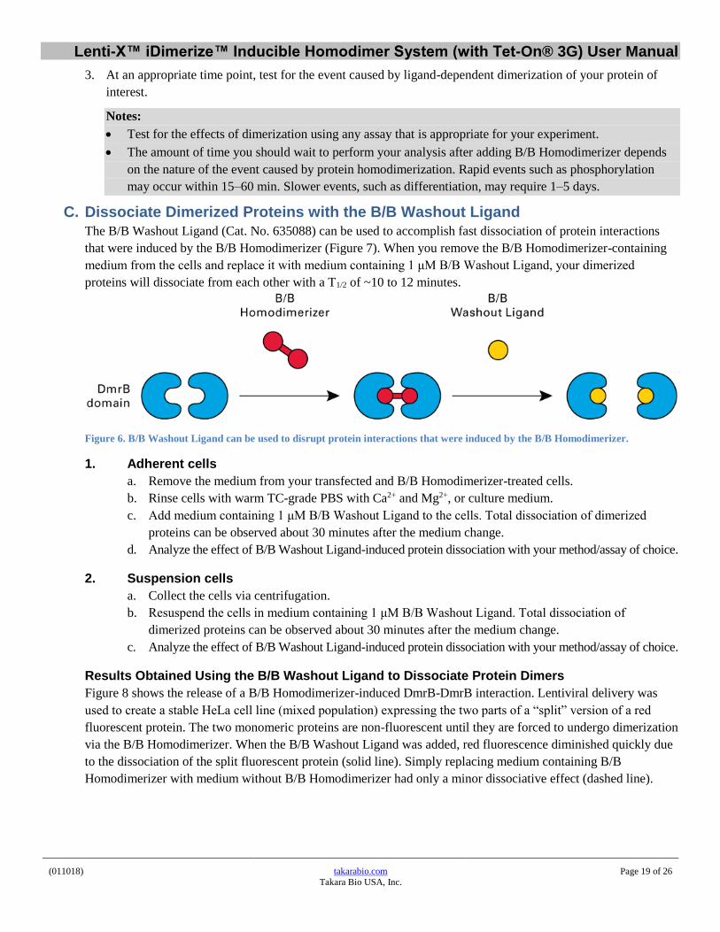
C. Dissociate Dimerized Proteins with the B/B Washout Ligand
The B/B Washout Ligand (Cat. No. 635088) can be used to accomplish fast dissociation of protein interactions
that were induced by the B/B Homodimerizer (Figure 7). When you remove the B/B Homodimerizer-containing
medium from the cells and replace it with medium containing 1 μM B/B Washout Ligand, your dimerized
proteins will dissociate from each other with a T1/2 of ~10 to 12 minutes.
Figure 6. B/B Washout Ligand can be used to disrupt protein interactions that were induced by the B/B Homodimerizer.
1. Adherent cells
a. Remove the medium from your transfected and B/B Homodimerizer-treated cells.
b. Rinse cells with warm TC-grade PBS with Ca2+ and Mg2+, or culture medium.
c. Add medium containing 1 μM B/B Washout Ligand to the cells. Total dissociation of dimerized
proteins can be observed about 30 minutes after the medium change.
d. Analyze the effect of B/B Washout Ligand-induced protein dissociation with your method/assay of choice.
2. Suspension cells
a. Collect the cells via centrifugation.
b. Resuspend the cells in medium containing 1 μM B/B Washout Ligand. Total dissociation of
dimerized proteins can be observed about 30 minutes after the medium change.
c. Analyze the effect of B/B Washout Ligand-induced protein dissociation with your method/assay of choice.
Results Obtained Using the B/B Washout Ligand to Dissociate Protein Dimers
Figure 8 shows the release of a B/B Homodimerizer-induced DmrB-DmrB interaction. Lentiviral delivery was
used to create a stable HeLa cell line (mixed population) expressing the two parts of a “split” version of a red
fluorescent protein. The two monomeric proteins are non-fluorescent until they are forced to undergo dimerization
via the B/B Homodimerizer. When the B/B Washout Ligand was added, red fluorescence diminished quickly due
to the dissociation of the split fluorescent protein (solid line). Simply replacing medium containing B/B
Homodimerizer with medium without B/B Homodimerizer had only a minor dissociative effect (dashed line).

Lenti-X™ iDimerize™ Inducible Homodimer System (with Tet-On® 3G) User Manual
(011018) takarabio.com
Takara Bio USA, Inc.
Page 20 of 26
Figure 7. B/B Washout Ligand has a much more dissociative effect than simple removal of the B/B Homodimerizer. A stable HeLa
cell line expressing DmrB-tagged versions of the two parts of a split red fluorescent protein was treated with 0.1 μM B/B Homodimerizer
for 3 hr. The medium was then removed and replaced with medium ± 1 μM B/B Washout Ligand. After 30 min, the fluorescence level in
the “+ B/B Washout Ligand” sample had dropped virtually to the background level, indicating that the two parts of the red fluorescent
protein were completely dissociated from each other.
XI. References Clackson, T., Yang, W., Rozamus, L. W., Hatada, M., Amara, J. F., Rollins, C. T., Stevenson, L. F., Magari, S.
R., Wood, S. A., Courage, N. L., Lu, X., Cerasoli, F. Jr., Gilman, M. & Holt, D. A. (1998) Redesigning an FKBP-
ligand interface to generate chemical dimerizers with novel specificity. Proc. Natl. Acad. Sci. USA
95(18): 10437–10442.
Crabtree, G. R. & Schreiber, S. L. (1996) Three-part inventions: intracellular signaling and induced proximity.
Trends Biochem. Sci. 21(11): 418–422.
Freshney, R.I. (2005). Culture of Animal Cells: A Manual of Basic Technique, 5th Edition (Wiley-Liss, Hoboken, NJ).
Gossen, M. & Bujard, H. (1992) Tight control of gene expression in mammalian cells by tetracycline responsive
promoters. Proc. Natl. Acad. Sci. USA 89(12):5547–5551.
Gossen, M., Freundlieb, S., Bender, G., Muller, G., Hillen, W. & Bujard, H. (1995) Transcriptional activation by
tetracycline in mammalian cells. Science 268(5218):1766–1769.
Loew, R., Heinz, N., Hampf, M., Bujard, H., & Gossen M. (2010) Improved Tet-responsive promoters with
minimized background expression. BMC Biotechnol. 10:81.
Urlinger, S., Baron, U., Thellmann, M., Hasan, M.T., Bujard, H. & Hillen, W. (2000) Exploring the sequence
space for tetracycline-dependent transcriptional activators: Novel mutations yield expanded range and sensitivity.
Proc. Natl. Acad. Sci. USA 97(14):7963–7968.
Zhou, X., Vink, M., Klave, B., Berkhout, B. & Das, A. T. (2006) Optimization of the Tet-On system for regulated
gene expression through viral evolution. Gene Ther.13(19):1382–1390.

Lenti-X™ iDimerize™ Inducible Homodimer System (with Tet-On® 3G) User Manual
(011018) takarabio.com
Takara Bio USA, Inc.
Page 21 of 26
XII. Troubleshooting
A. Lenti-X Troubleshooting
1. Vector Cloning
Problem Possible Explanation Solution
Plasmid is difficult to grow or clone
Some viral vectors may undergo rearrangement between the 5’ and 3’ LTRs when propagated in less-than-optimal E. coli host strains
Use Stellar Competent Cells (Cat. No. 636763) to produce high DNA yields and to minimize the potential for DNA rearrangements.
2. Lenti-X 293T Packaging Cells
Problem Possible Explanation Solution
Poor viability upon thawing
Improper thawing techniques Use thawing procedure in Appendix A, and/or consult the Lenti-X 293T Cell Line Protocol-at-a-Glance.
Incorrect culture medium Use DMEM with additives listed in Section III.D. Use 10% Tet System Approved FBS (Tc-free).
Improper tissue culture plasticware Use collagen I-coated plates to aid cell adherence during initial seeding.
Slow growth Incorrect culture medium Use DMEM with additives listed in Section III.D. Use 10% Tet System Approved FBS (Tc-free).
Cells do not attach to plate
Improper tissue culture plasticware Use collagen I-coated plates to aid cell adherence during initial seeding.
Cells appear morphologically different
Passage of cell culture is too high (old cells)
Thaw/purchase new aliquot of Lenti-X 293T cells.
3. Virus Production
Problem Possible Explanation Solution
Poor transfection efficiency (as determined by GOI or marker expression in the Lenti-X 293T cell line)
Cells plated too densely Plate 4–5 x 10⁶ cells/100 mm plate, or fewer if the cells divide rapidly. Use at 50–80% confluency. See Section VII.
Transfection is toxic to cells Use the optimized conditions provided in Section VII.
Cells harvested or analyzed too soon after transfection
Wait 48 hr after transfection for maximal expression of GOI or marker to determine efficiency.
Low titers (<105cfu/ml)
Serum in medium contains tetracycline contaminants, which can interfere with the expression of viral proteins, resulting in lower titers
Use Tet System Approved FBS (Cat. Nos. 631101 & 631106) in the Lenti-X 293T cell culture medium.
Virus was harvested too early Harvest virus 48–72 hr after the start of transfection.
Vector is too large The limit for efficient packaging function is 9.7 kb from the end of the 5’-LTR to the end of the 3’-LTR.
Polybrene is missing or at suboptimal concentration
Add Polybrene (4 µg/ml) during transduction or optimize the concentration (2–12 µg/ml).
Low titers (<105cfu/ml)
Virus was exposed to multiple freeze-thaw cycles
Each cycle reduces titer by approximately 2–4 fold. Limit the number of freeze-thaws.
Suboptimal selection procedure during titration
Perform an antibiotic kill curve on the cell line prior to using it for titration.

Lenti-X™ iDimerize™ Inducible Homodimer System (with Tet-On® 3G) User Manual
(011018) takarabio.com
Takara Bio USA, Inc.
Page 22 of 26
4. Transduction of Target Cells
Problem Possible Explanation Solution
Poor transduction efficiency
Low titer See Section C or use the Lenti-X Concentrator (Section III.M) to increase your available titer up to 100-fold without ultracentrifugation.
Poor transfection efficiency Follow the protocol in Section VII.B. Be sure to use 5 µg of transfection-grade plasmid.
Low viability of target cells during transduction
Optimize culture conditions for target cells prior to infection.
Packaging cell line-conditioned media may affect cell growth; dilute viral supernatant or shorten exposure time to viral supernatant. Consider using RetroNectin Reagent and the RetroNectin-Bound Virus transduction protocol or purify your virus prior to transduction using the Lenti-X Maxi Purification Kit (Cat. Nos. 631233 & 631234).
Excessive exposure to Polybrene: optimize amount (titrate) or shorten exposure time to viral supernatant.
Viral supernatant contains transduction inhibitors
Use RetroNectin Reagent or RetroNectin-coated plates in the RetroNectin-Bound Virus transduction protocol, which allows virions to bind the RetroNectin substratum and be washed free of inhibitors prior to target cell infection; or, purify your virus prior to transduction using the Lenti-X Maxi Purification Kit (Cat. Nos. 631233 & 631234).
B. Tet-On 3G Troubleshooting
1. Low Fold Induction of Transient Expression
Description of Problem
Possible Explanation Solution
Decrease in fold induction after several passages or Loss of inducibility after passaging of a (previously frozen) double-stable cell line.
The appropriate antibiotics are missing from the cell culture medium.
Maintain optimal antibiotic concentrations (Section III.O).
Mixed cell population Reselect the current cell line through single colony selection using selective concentrations of puromycin and G418, (Section IX).

Lenti-X™ iDimerize™ Inducible Homodimer System (with Tet-On® 3G) User Manual
(011018) takarabio.com
Takara Bio USA, Inc.
Page 23 of 26
1. Low Fold Induction of Transient Expression (continued)
Description of Problem
Possible Explanation Solution
Low fold induction (ratio of maximal to basal expression of the GOI)
A suboptimal ratio of cotransfected vectors was used.
We recommend a cotransient transfection vector ratio of 1:4 for pCMV-Tet3G:pTRE3G-GOI (Section VI.A). Different vector ratios may result in different maximal to basal gene expression ratios.
Cells were harvested and analyzed too early or too late.
Harvest and analyze cells between 18–48 hr post transfection
Poor transfection efficiency • Optimize transfection protocol
• Optimize density of cell plating; use at 60–90% confluency
Poor target cell viability • Optimize target cell passage number
• Optimize target cell culture conditions
• Optimize tissue culture plasticware
The FBS used in the cell culture medium contains tetracycline derivatives
Use our Tet System Approved FBS (Section III.E). Only Takara Bio performs actual inducibility tests on a sensitive Tet-inducible cell line in order to provide an absolute guarantee that your serum is tetracycline-free.
Transiently transfected cells contain more copies of the TRE-containing plasmid than do stable cell lines.
When testing clones via transient transfection, expect lower fold induction levels than in double-stable clones (sometimes only ~100-fold).
2. Detection and Inhibition of Expression
Description of Problem
Possible Explanation Solution
No detectable GOI expression by Western Blot
Low sensitivity of detection method Check sensitivity of primary and secondary antibodies. Analyze GOI expression by qRT-PCR, using different sets of primers to ensure optimal detection of GOI expression.
Continuous GOI/Fluorescent Protein expression after the removal of doxycycline
Depending on the stability of the protein, it may persist in the cell in the absence of gene induction and de novo synthesis of GOI mRNA. For example, fluorescent proteins tend to have long half-lives.
Upon degradation, GOI/Fluorescent Protein expression will not be detectable in cells in the absence of induction. For faster degradation of an inducible GOI, use pTRE-Cycle Vectors (see takarabio.com).
Doxycycline was not completely removed from the cell culture medium.
• Wash cells three times with PBS, followed by trypsinization and replating in fresh medium supplemented with our Tet System Approved FBS.
• If trypsinization is undesirable, wash cells three times with medium and three times with PBS, then replace with fresh medium supplemented with Tet System Approved FBS.

Lenti-X™ iDimerize™ Inducible Homodimer System (with Tet-On® 3G) User Manual
(011018) takarabio.com
Takara Bio USA, Inc.
Page 24 of 26
D. iDimerize Troubleshooting Description of Problem
Possible Explanation Solution
Dimerization is observed in the absence of the B/B Homodimerizer
The expression level of the protein of interest fused to the DmrB domain is too high, especially in the case of a DmrB-tagged protein of interest localized to the plasma membrane.
Use a lower concentration of Dox to limit the gene expression level:
• Transient cotransfections of pTRE3G-Hom1-GOI and pCMV-Tet3G: See Part VI, Protocol A.1
• Stable Tet3G cell lines transiently transfected with pTRE3G-Hom1-GOI: See Part VI, Protocol B.2
• Double-stable cell lines expressing Tet3G and pTRE3G-Hom1-GOI: See Part VI, Protocol C.3
Addition of B/B Homodimerizer does not result in the expected effect(s)
The B/B Homodimerizer concentration is too low.
Increase the amount of B/B Homodimerizer added.
The monitoring assay is not sensitive enough.
Include a positive control when performing your assay.
The volume of B/B Homodimerizer used causes cells to die, due to high solvent concentration.
Low expression level
Prepare a more concentrated stock solution.
Monitor protein expression in your cells by Western blot using the anti-DmrB antibody (Section III.H).
Lenti-X™ iDimerize™ Inducible Homodimer System (with Tet-On® 3G) User Manual
(011018) takarabio.com
Takara Bio USA, Inc.
Page 25 of 26
Appendix A: Preparing and Handling Tet-On 3G Cell Line Stocks
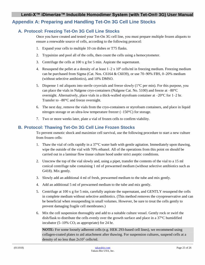
A. Protocol: Freezing Tet-On 3G Cell Line Stocks
Once you have created and tested your Tet-On 3G cell line, you must prepare multiple frozen aliquots to
ensure a renewable source of cells, according to the following protocol:
1. Expand your cells to multiple 10 cm dishes or T75 flasks.
2. Trypsinize and pool all of the cells, then count the cells using a hemocytometer.
3. Centrifuge the cells at 100 x g for 5 min. Aspirate the supernatant.
4. Resuspend the pellet at a density of at least 1–2 x 10⁶ cells/ml in freezing medium. Freezing medium
can be purchased from Sigma (Cat. Nos. C6164 & C6039), or use 70–90% FBS, 0–20% medium
(without selective antibiotics), and 10% DMSO.
5. Dispense 1 ml aliquots into sterile cryovials and freeze slowly (1°C per min). For this purpose, you
can place the vials in Nalgene cryo-containers (Nalgene Cat. No. 5100) and freeze at –80°C
overnight. Alternatively, place vials in a thick-walled styrofoam container at –20°C for 1–2 hr.
Transfer to –80°C and freeze overnight.
6. The next day, remove the vials from the cryo-containers or styrofoam containers, and place in liquid
nitrogen storage or an ultra-low temperature freezer (–150°C) for storage.
7. Two or more weeks later, plate a vial of frozen cells to confirm viability.
B. Protocol: Thawing Tet-On 3G Cell Line Frozen Stocks
To prevent osmotic shock and maximize cell survival, use the following procedure to start a new culture
from frozen cells:
1. Thaw the vial of cells rapidly in a 37°C water bath with gentle agitation. Immediately upon thawing,
wipe the outside of the vial with 70% ethanol. All of the operations from this point on should be
carried out in a laminar flow tissue culture hood under strict aseptic conditions.
2. Unscrew the top of the vial slowly and, using a pipet, transfer the contents of the vial to a 15 ml
conical centrifuge tube containing 1 ml of prewarmed medium (without selective antibiotics such as
G418). Mix gently.
3. Slowly add an additional 4 ml of fresh, prewarmed medium to the tube and mix gently.
4. Add an additional 5 ml of prewarmed medium to the tube and mix gently.
5. Centrifuge at 100 x g for 5 min, carefully aspirate the supernatant, and GENTLY resuspend the cells
in complete medium without selective antibiotics. (This method removes the cryopreservative and can
be beneficial when resuspending in small volumes. However, be sure to treat the cells gently to
prevent damaging fragile cell membranes.)
6. Mix the cell suspension thoroughly and add to a suitable culture vessel. Gently rock or swirl the
dish/flask to distribute the cells evenly over the growth surface and place in a 37°C humidified
incubator (5–10% CO2 as appropriate) for 24 hr.
NOTE: For some loosely adherent cells (e.g. HEK 293-based cell lines), we recommend using
collagen-coated plates to aid attachment after thawing. For suspension cultures, suspend cells at a
density of no less than 2x10⁵ cells/ml.

Lenti-X™ iDimerize™ Inducible Homodimer System (with Tet-On® 3G) User Manual
(011018) takarabio.com
Takara Bio USA, Inc.
Page 26 of 26
7. The next day, examine the cells under a microscope. If the cells are well-attached and confluent, they
can be passaged for use. If the majority of cells are not well-attached, continue culturing for another
24 hr.
NOTE: For some loosely adherent cell lines (e.g., HEK 293-based cell lines), complete attachment of
newly thawed cultures may require up to 48 hr.
8. Expand the culture as needed. The appropriate selective antibiotic(s) should be added to the medium
after 48–72 hr in culture. Maintain stable and double-stable Tet Cell Lines in complete culture
medium containing a maintenance concentration G418 and/or puromycin, as appropriate (Table I).
Table 1. Recommended antibiotic concentrations for selecting and maintaining stable clones.
Cat. No. Antibiotic Recommended Concentration (µg/ml)
Selection1 Maintenance
631308 G418 (5 g) 100–800 200
631307 G418 (1 g)
631306 Puromycin (100 mg) 0.25–10 0.25
631305 Puromycin (25 mg)
1When selecting for single colonies, you must determine the appropriate dose for your specific cell line
empirically. Test a dosage range using dishes of untransfected cells, and choose the dose that kills all
of the cells in 3–5 days. If all the cells die in less than 24 hr, you should use a lower dose.
Contact Us
Customer Service/Ordering Technical Support
tel: 800.662.2566 (toll-free) tel: 800.662.2566 (toll-free)
fax: 800.424.1350 (toll-free) fax: 800.424.1350 (toll-free)
web: takarabio.com web: takarabio.com
e-mail: [email protected] e-mail: [email protected]
Notice to Purchaser
Our products are to be used for research purposes only. They may not be used for any other purpose, including, but not limited to, use in drugs, in vitro diagnostic purposes, therapeutics, or in humans. Our products may not be transferred to third parties, resold, modified for resale, or used to manufacture commercial products or to
provide a service to third parties without prior written approval of Takara Bio USA, Inc.
Your use of this product is also subject to compliance with any applicable licensing requirements described on the product’s web page at takarabio.com. It is your
responsibility to review, understand and adhere to any restrictions imposed by such statements.
©2015 Takara Bio Inc. All Rights Reserved.
All trademarks are the property of Takara Bio Inc. or its affiliate(s) in the U.S. and/or other countries or their respective owners. Certain trademarks may not be
registered in all jurisdictions. Additional product, intellectual property, and restricted use information is available at takarabio.com.
This document has been reviewed and approved by the Quality Department.


![ZZ - fiqhulhadith.com ki kunjyan.pdf69 ----- *****™™*™™ÒÃÃÃÅÅÅÃÅääää™™™™]]]!!!!***hZZZå 71 -----*****™™™*™ÀÀÀÀÂÂÂÂ6666,,,vvvvZZZ{{{Z{ŠŠŠŠcc**c](https://static.fdocuments.net/doc/165x107/5e5e26f29bdb1829b545ee7d/zz-ki-kunjyanpdf-69-aaaaffffaaaahzzz.jpg)
![plasmonic nanoparticles - arXiv · metal-dielectric interfaces [1]. A homodimer of nanoparticles (NPs) with dimensions ˝l o (free space wavelength) provides the basic element of](https://static.fdocuments.net/doc/165x107/5f0a70797e708231d42ba3c8/plasmonic-nanoparticles-arxiv-metal-dielectric-interfaces-1-a-homodimer-of.jpg)