Leica Vibratome Series - 显微镜门户-中国显微图像网 · Leica VT1000 P and VT1000 A...
7
Leica Vibratome™ Series Vibrating Blade Microtomes Living up to Life
Transcript of Leica Vibratome Series - 显微镜门户-中国显微图像网 · Leica VT1000 P and VT1000 A...
Leica Vibratome™ SeriesVibrating Blade Microtomes
Living up to Life

Vibrating blade microtomes are used to produce monolayer or thick sections of fixed or fresh tissue under physiological conditions without freezing or embedding. Sectioning fresh tissue specimens with Leica Microsystems’ VT Series maintain the morphology, enzyme activity and cell viability of the tissue. Their use also minimize artifacts, compression distortion, cell destruction and other inherent deleterious effects of sectioning.
Applications for these instruments include immunohistochemistry, cell culturing of different organs, sections for patchclamping, electrophysiology, free floating sections and many other applications in neuroscience.
In order to maintain physiological conditions while sectioning, chill the buffer and minimize the vertical deflection of the blade holder as well as the blade. During operation, the blade moves laterally and forward and the section thickness is achieved by vertically feeding the specimen stage. Other parameters including section thickness, amplitude, frequency, knife travel speed and blade angle influence section quality. The Leica Vibratome Series of instruments offers a complete product range that control some or all of these parameters.
Cutting Edge Precision
Specifications Leica VT1000 P (formerly Vibratome™ 1000 Plus)
Leica VT1000 A (formerly Vibratome™ 1500) Leica VT1000 S Leica VT1200 Leica VT1200 S
Vibrocheck (measurement device for verticle deflection of the blade) • •
Fully automated cut mode • • •
Specimen retraction • •
Adjustable amplitude • • • • •
Adjustable frequency •
Blade travel speed 0 - 2mm/s 0 - 2mm/s 0.025 - 2.5mm/s 0.01 - 1.5 mm/s 0.01 - 1.5 mm/s
Adjustable cutting window manual manual electronic individually programable front and rear position
Maximum specimen size 60 x 40 x 15 mm 60 x 40 x 15 mm 70 x 40 x 15 mm 33 x 50 x 20 mm 33 x 50 x 20 mm
Total vertical specimen stroke 15 mm 15 mm 15 mm 20 mm 20 mm
Selection of buffer trays • • • •
Cooling options Peltier Vibratome 900 R Peltier Vibratome 900 R Minichiller Minichiller Minichiller
Memory capability for storing section thickness • •
Multiple user settings 8 different user settings
Adjustable return stroke 1- 5 mm/s
Adjustable forward speed in manual mode 1- 5 mm/s
Magnification options 2x magnifier 2x magnifier 2x magnifier 2x magnifier, microscope 2x magnifier, microscope
23
Leica Microsystems offers a wide variety of vibrating blade microtomes that have been developed in collaboration with renowned scientists throughout the world. There is an instrument for every researcher’s application and budget. The features of each instrument vary in the degree of automation, ranging from the Leica VT1000 P to the fully automated Leica VT1200 S with optional Vibrocheck, for measuring and minimizing vertical blade deflection.
Lean principles are revolutionizing the way laboratories operate. Leica Microsystems prides itself on providing high quality, reliable and durable instruments. The Vibratome Series microtomes can greatly improve productivity and reduce costs in the laboratory by producing high quality sections with viable cells without the need of replicating experiments.
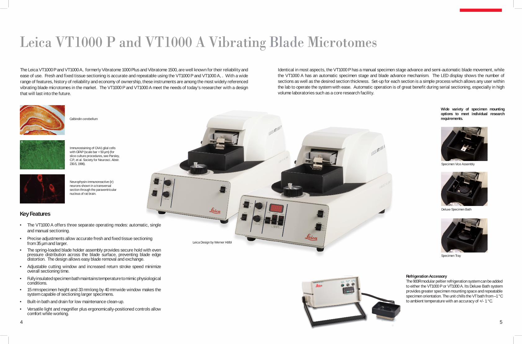
Leica VT1000 P and VT1000 A Vibrating Blade Microtomes
Key Features
The VT1000 A offers three separate operating modes: automatic, single • and manual sectioning.
Precise a• djustments allow accurate fresh and fixed tissue sectioning from 35 µm and larger.The sprin• g-loaded blade holder assembly provides secure hold with even pressure distribution across the blade surface, preventing blade edge distortion. The design allows easy blade removal and exchange.
Adjustable • cutting window and increased return stroke speed minimize overall sectioning time.
F• ully insulated specimen bath maintains temperature to mimic physiological conditions. 15 mm s• pecimen height and 33 mm long by 40 mm wide window makes the system capable of sectioning larger specimens.
Bui• lt-in bath and drain for low maintenance clean-up.
V• ersatile light and magnifier plus ergonomically-positioned controls allow comfort while working.
Identical in most aspects, the VT1000 P has a manual specimen stage advance and semi-automatic blade movement, while the VT1000 A has an automatic specimen stage and blade advance mechanism. The LED display shows the number of sections as well as the desired section thickness. Set-up for each section is a simple process which allows any user within the lab to operate the system with ease. Automatic operation is of great benefit during serial sectioning, especially in high volume laboratories such as a core research facility.
The Leica VT1000 P and VT1000 A, formerly Vibratome 1000 Plus and Vibratome 1500, are well known for their reliability and ease of use. Fresh and fixed tissue sectioning is accurate and repeatable using the VT1000 P and VT1000 A, . With a wide range of features, history of reliability and economy of ownership, these instruments are among the most widely referenced vibrating blade microtomes in the market. The VT1000 P and VT1000 A meet the needs of today’s researcher with a design that will last into the future.
Refrigeration Accessory The 900R modular peltier refrigeration system can be added to either the VT1000 P or VT1000 A. Its Deluxe Bath system provides greater specimen mounting space and repeatable specimen orientation. The unit chills the VT bath from –1 °C to ambient temperature with an accuracy of +/- 1 °C.
Leica Design by Werner Hölbl
5
Immunostaining of CAA1 glial cells with GFAP (scale bar = 50 µm) (for slice culture procedures, see Parsley, C.P., et al. Society for Neurosci. Abstr. 230.5, 1996).
Calbindin cerebellum
Neurophysin-immunoreactive (ir) neurons shown in a transversal section through the paraventricular nucleus of rat brain.
4
Wide variety of specimen mounting options to meet individual research requirements.
Specimen Vice Assembly
Specimen Tray
Deluxe Specimen Bath

Leica VT1000 S Vibrating Blade Microtome
Key Features
Ergonomic design for comfortable working conditions•
5 different amplitude settings from 0.2 - 1 mm•
Linear sectioning speed adjustment from 0.025 mm - 2.5 mm•
Linear sectioning frequency adjustment from 0 - 100Hz•
Programmable specimen retraction•
Freely programmable sectioning window•
Single and continu• ous stroke options for ultimate versatility
Easy • mounting and removing of knife holder and buffer tray for efficient workflow
Dark bu• ffer tray provides excellent contrast to the specimen
The classic design of the Leica VT1000 S make working with the instrument an ease. Ergonomic hand rests and direct access to all functional elements provide exceptional comfort and added safety. Since time is of tremendous importance when dealing with unfixed fresh tissue, every effort has been made to accelerate the sectioning process. The VT1000 S features very fine adjustable knife advance speed, a freely programmable cutting window, and accelerated return knife speed to minimize overall sectioning time of even the smallest specimens. The VT1000 S vibrating blade microtome is designed to consistently produce thin sections of both fresh and fixed tissue specimens, even non-homogeneous specimens that are difficult to section. It is also used for some industrial applications related to structural analysis of foam and other very soft materials and botanical specimens such as plants and roots.
Labeling of cholinergic septal neurons in rat basal forebrain by
using a polyclonal antiserum against choline acetyltransferase (ChAT).
The variable frequency allows the VT1000 S to adapt to a variety of applications. Accurate control of the knife or blade movement is integral in the design of this instrument. The visual clarity provided by the wide large-field magnifier, supplied as standard delivery, can be enhanced with a fiber optic lighting system (optional). Together, these features provide exact, individually adjustable illumination of the entire sectioning range, and prevents surface reflection of the buffer solution for accurate sectioning.
CA3 field of rat hippocampus. Syntaxin positive axon terminals over
pyramidal cells. 40 µm section. 400x.
The Leica VT1000 S sections specimens under physiological conditions, which protects tissue, enzymes and antigens. For that purpose, the specimen is mounted directly onto a specimen plate, using cyanoacrylate adhesive, and placed into a buffer tray filled with cooled physiological buffer solution. The buffer serves two purposes: it maintains the tissue in a live state and also provides a flotation medium for the sections. To maintain stable, cold buffer temperature, the integrated ice bath can be filled with crushed ice or the optional double-walled buffer tray can be connected with the “Minichiller” circulation cooling device. Both the knife holder and buffer tray are easily removed to reduce the risk of reagent carryover or contamination when sectioning.
Transversal section through rat brain cortex at the forebrain level. A
large single neuron was labeled by NADPH-diaphorase histochemistry. The small axon and some branching
dendrites are visible.
Leica Design by Werner Hölbl
6 7
Standard knife holder S and buffer tray S with optional magnetic specimen holder.
The optional double-walled buffer tray is available in two different sizes which allows sectioning of specimens 33 x 40 mm or as large as 70 x 40 mm. Epipremnum pinnatum (ivy).
50 µm section.

Leica VT1200 and VT1200 S Vibrating Blade Microtomes
Key Features
Vertical deflection of the blade can be measured and minimized below 1µm • with the optional Vibrocheck measurement device
Blade• holder can be rotated through 90° to permit accurate insertion of a whole double edged razor blade, sapphire knife or injector blade.
Optimized blade holder designed for minimum buffer spillage. •
Motorized blade holder sectioning speed adjustable between 0.01 to 1.5 mm/sec. •
Palm rests• on the ice bath or double-walled buffer tray allow a relaxed, ergonomic working position.
Bui• lt-in LED illumination provides natural, comfortable lighting during sectioning without adding heat, which could deteriorate the tissue. The Leica VT1200 S features 5-step adjustable light intensity.
Fresh nervous tissues, brain and spinal cord are soft, fragile and extremely susceptible to mechanical damage. The Leica VT1200 and VT1200 S vibrating blade microtomes are designed to meet the highest demands for cutting fresh and fixed tissue in Neuropathology, Neurophysiology (patch-clamping) and Electrophysiology. These robust instruments feature a new blade holder design with the possibility tomeasure vertical deflection using the optional Vibrocheck in order to minimize it. Negative mechanical effects on the tissue are reduced to a minimum. This produces the highest quality sections that retain viable cells on the section surface/s.
Flexible PerformanceThe removable ice bath and buffer tray allow working under physiological conditions and away from the instrument, e.g., under a microscope.
These instruments were designed in collaboration with Prof. Dr. Peter Jonas and his team, Physiology Department, University of Freiburg, Germany.
The semi-automated Leica VT1200 has been designed for users who prefer to manually control sectioning parameters such as section thickness and cutting stroke for each individual section. The VT1200 offers straightforward, intuitive operation, fast sectioning and a full range of accessories at an attractive price.
The fully-automated Leica VT1200 S is recommended for multi user laboratories where users of both semi-automated vibrating blade microtomes and fully automated instruments work together. VT1200 S can be used in both semi- or fully-automated sectioning modes depending on the users’ requirements. The fully automated mode of the VT1200 S offers automatic feeding, specimen retraction, and a cutting window. The mode of operation can be individually selected, and settings can be stored for up to 8 users. Automatic feeding, specimen retraction and use of a cutting window are designed to minimize sectioning time.
Customized ComfortThe separate, foil-protected controlpanel can be placed on either side ofthe instrument depending on the personalpreference of the user.
Modular FunctionalityBoth instrument versions can be enhanced with an optional magnifier (2x) or microscope to improve visual clarity.
Leica Design by Werner Hölbl
9
Optional Measurement Device: Vibrocheck™
The vertical deflection of the blade can be measured by the Vibrocheck measurement device. Both vertical deflection (in µm) and rotation direction of the adjustment screw are displayed on the separate, foil-protected control panel. The adjustment screw on the blade holder allows minimization of the vertical deflection to below 1 µm, which significantly increases the number of viable cells.
8

Leica VT1200 Cutting frequency . . . . . . . . . . . . . . . . . . . . . . . . . . . . . . . . . . . . .Rated voltage range . . . . . . . . . . . . . . . . . . . . . . . . . . . . . . . . . . .Rated frequency . . . . . . . . . . . . . . . . . . . . . . . . . . . . . . . . . . . . . .Power consumption . . . . . . . . . . . . . . . . . . . . . . . . . . . . . . . . . . .Amplitude . . . . . . . . . . . . . . . . . . . . . . . . . . . . . . . . . . . . . . . . . . . .Sectioning speed . . . . . . . . . . . . . . . . . . . . . . . . . . . . . . . . . . . . . Maximum specimen size: With standard blade holder (LxW) . . . . . . . . . . . . . Specimen orientation, rotating. . . . . . . . . . . . . . . . Specimen plate, swiveling . . . . . . . . . . . . . . . . . . . .Section thickness adjustment . . . . . . . . . . . . . . . . . . . . . . . . . . .Total vertical specimen stroke . . . . . . . . . . . . . . . . . . . . . . . . . . Return speed . . . . . . . . . . . . . . . . . . . . . . . . . . . . . . . . . . . . . . . . .Specimen retraction. . . . . . . . . . . . . . . . . . . . . . . . . . . . . . . . . . . Sectioning range . . . . . . . . . . . . . . . . . . . . . . . . . . . . . . . . . . . . . .Size L x W x H (basic instrument without control unit) . . . . . .Weight (basic instrument without control unit) . . . . . . . . . . . .
Leica VT1200 S – same as Leica VT1200 above, plus:
Return speed . . . . . . . . . . . . . . . . . . . . . . . . . . . . . . . . . . . . . . . . . Cutting window . . . . . . . . . . . . . . . . . . . . . . . . . . . . . . . . . . . . . . . Specimen retraction . . . . . . . . . . . . . . . . . . . . . . . . . . . . . . . . . . .Section thickness adjustment . . . . . . . . . . . . . . . . . . . . . . . . . . .
Leica VT1000 PRated voltage range. . . . . . . . . . . . . . . . . . . . . . . . . . . . . . . . . . . . Rated frequency. . . . . . . . . . . . . . . . . . . . . . . . . . . . . . . . . . . . . . . Amplitude. . . . . . . . . . . . . . . . . . . . . . . . . . . . . . . . . . . . . . . . . . . . . Sectioning speed. . . . . . . . . . . . . . . . . . . . . . . . . . .. . . . . . . . . . . . Maximum specimen size (LxW). . . . . . . . . . . . . . . . . . . . . . . . . . Specimen orientation, rotating. . . . . . . . . . . . . . . . . . . . . . Specimen plate, swiveling. . . . . . . . . . . . . . . . . . . . . . . . . .Section thickness adjustment . . . . . . . . . . . . . . . . . . . . . . . . . . .Total vertical specimen stroke. . . . . . . . . . . . . . . . . . . . . . . . . . . Return speed . . . . . . . . . . . . . . . . . . . . . . . . . . . . . . . . . . . . . . . . .Size LxWxH (basic instrument). . . . . . . . . . . . . . . . . . . . . . . . . . .Weight (basic instrument). . . . . . . . . . . . . . . . . . . . . . . . . . . . . . .
Leica VT1000 A – Same as Leica VT1000 P above, plus:
Section thickness adjustment . . . . . . . . . . . . . . . . . . . . . . . . . . .
Leica VT1000 SCutting frequency . . . . . . . . . . . . . . . . . . . . . . . . . . . . . . . . . . . . . .Rated voltage range. . . . . . . . . . . . . . . . . . . . . . . . . . . . . . . . . . . .Rated frequency . . . . . . . . . . . . . . . . . . . . . . . . . . . . . . . . . . . . . .Power consumption. . . . . . . . . . . . . . . . . . . . . . . . . . . . . . . . . . . .Amplitude. . . . . . . . . . . . . . . . . . . . . . . . . . . . . . . . . . . . . . . . . . . . .Sectioning speed. . . . . . . . . . . . . . . . . . . . . . . . . . . . . . . . . . . . . . .Maximum specimen size: With standard blade holder (LxW) . . . . . . . . . . . . . With knife holder L (LxW). . . . . . . . . . . . . . . . . . .. . . Section thickness adjustment . . . . . . . . . . . . . . . . . . . . . . . . . . .Total vertical specimen stroke. . . . . . . . . . . . . . . . . . . . . . . . . . . Return speed. . . . . . . . . . . . . . . . . . . . . . . . . . . . . . . . . . . . . . . . . .Specimen retraction . . . . . . . . . . . . . . . . . . . . . . . . . . . . . . . . . . .Sectioning range. . . . . . . . . . . . . . . . . . . . . . . . . . . . . . . . . . . . . . .
85 Hz (± 10%)100 V to 240 V (± 10%)50/60 Hz (± 10%)35 VAfrom 0 to 3 mm, in 0.05 mm increments0.01 to 1.5 mm/s (± 10%)
33 mm x 50 mm360°0 to 10°manual in 1 µm increments or automatic max. 1000 µm 20 mm (motorized) 2.5 mm/s (± 10%)0 to 100 µm (adjustable, can be deactivated)45 mm (adjustable)600 mm x 250 mm x 230 mm56 kg
1 to 5 mm/s, in 0.5 mm increments (± 10%)0.5 mm to 45 mm0 to 100 µm (adjustable, can be deactivated)manual in 1 µm increments or automatic max. 1000 µm
100 V or 220 V (specified at time of order)50/60 Hz from 0 to 3 mm (± 1.5 mm)0 to 5 mm/s33 mm x 40 mm360 ° (full rotation with specimen tray)0 to 15 ° (range of specimen vice assembly)Manual, 5 µm increments 15 mm (± 10%)5 to 7 mm/s(± 10%)534 mm x 305 mm x 305 mm23 kg
manual in 5 µm increments, automated in 1 µm increments
Linear adjustment from 0 to100 Hz100 V to 240 V (± 10 %)50/60 Hz (± 10 %)35 VA5 different settings selectable: 0.2; 0.4; 0.6; 0.8; 1.0 mm0.025 to 2.5 mm/s
33 mm x 40 mm70 mm x 40 mm1 to 999 µm (in 1 µm increments)15 mm (motorized) standard or 20 mm (optional)5 mm/s0 to 999 µm (motorized)1 to 40 mm
Complete the Package
Leica LMD7000The Leica LMD7000 Laser Microdissection system gives researchers the power to conduct cell-specific molecular analysis. LMD makes it possible to obtain homogeneous, ultra-pure samples from heterogeneous start-ing material. Designed for selective cell isolation leading to subsequent molecular analysis, this tool improves the specificity of downstream tech-niques in Mass Spectrometry, PCR, Microarray, or Microfluidics. Since the Leica LMD7000 is the only system that collects specimens by gravity, this cell selection method is both contact-free and contamination-free.
Leica DM6000B Designed to allow researchers to spend more time on their science and less time adjusting equipment or training new users, the Leica DM6000 B research upright microscope is extremely easy to use. Both expert and novice users can quickly learn to capture high-resolution morphological images of biological specimens. Switching between contrast techniques happens with the simple push of a button on this fully automated system, plus all imaging parameters can be instantly recalled to reproduce brilliant images with the next specimen.
Perfusion Two™ Automating the perfusion system gives much greater consistency and more reproducible perfusions, as well as greater convenience and ease of operation. The Perfusion Two system features an automated air compressor that quietly pumps to 300 mm Hg and stops. As each animal is being prepared, the pressure is set up to begin at the turn of a switch. An aperture between the air tank and the fluids provides the correct rate of pressure rise after the perfusion is started. There is no need to pump up the pressure, and less need for the hands to be involved.
Technical Specifications
10 11
Leica CM3050SThe Leica CM3050S cryostat meets the demanding cryosectioning needs of neuroscience. When working with delicate specimens such as brain, the precise specimen orientation and the specimen feed system via step motor offer reproducible, high-quality thin serial sections. The motorized Leica CM3050S is available with or without active specimen cooling. The optional ergonomic height adjustment can be retrofitted, which enables the user to operate the cryostat in a sitting or standing position.
Leica Microsystems expresses its thanks to Prof. Dr. Peter Jonas, Francisco Javier García Ladona, Ph.D., Shawn Hochman, Ph.D., and Dr. Andreas Schober for granting their permission to use the application photographs contained in this brochure.
Leica Microsystems operates internationally in four divi-sions, where we rank with the market leaders.
• Life Science DivisionThe Leica Microsystems Life Science Division supports the imaging needs of the scientific community with advanced innovation and technical expertise for the visualization, measurement, and analysis of microstructures. Our strong focus on understanding scientific applications puts Leica Microsystems’ customers at the leading edge of science.
• Industry DivisionThe Leica Microsystems Industry Division’s focus is to support customers’ pursuit of the highest quality end result. Leica Microsystems provide the best and most innovative imaging systems to see, measure, and analyze the micro-structures in routine and research industrial applications, materials science, quality control, forensic science investigation, and educational applications.
• Biosystems DivisionThe Leica Microsystems Biosystems Division brings histopathology labs and researchers the highest-quality, most comprehensive product range. From patient to pathologist, the range includes the ideal product for each histology step and high-productivity workflow solutions for the entire lab. With complete histology systems featuring innovative automation and Novocastra™ reagents, Leica Microsystems creates better patient care through rapid turnaround, diagnostic confidence, and close customer collaboration.
• Surgical DivisionThe Leica Microsystems Surgical Division’s focus is to partner with and support surgeons and their care of patients with the highest-quality, most innovative surgi cal microscope technology today and into the future.
“With the user, for the user”Leica Microsystems
The statement by Ernst Leitz in 1907, “with the user, for the user,” describes the fruitful collaboration with end users and driving force of innovation at Leica Microsystems. We have developed five brand values to live up to this tradition: Pioneering, High-end Quality, Team Spirit, Dedication to Science, and Continuous Improvement. For us, living up to these values means: Living up to Life.
Active worldwideAustralia: North Ryde Tel. +61 2 8870 3500 Fax +61 2 9878 1055
Austria: Vienna Tel. +43 1 486 80 50 0 Fax +43 1 486 80 50 30
Belgium: Groot Bijgaarden Tel. +32 2 790 98 50 Fax +32 2 790 98 68
Canada: Richmond Hill/Ontario Tel. +1 905 762 2000 Fax +1 905 762 8937
Denmark: Herlev Tel. +45 4454 0101 Fax +45 4454 0111
France: Nanterre Cedex Tel. +33 811 000 664 Fax +33 1 56 05 23 23
Germany: Wetzlar Tel. +49 64 41 29 40 00 Fax +49 64 41 29 41 55
Italy: Milan Tel. +39 02 574 861 Fax +39 02 574 03392
Japan: Tokyo Tel. +81 3 5421 2800 Fax +81 3 5421 2896
Korea: Seoul Tel. +82 2 514 65 43 Fax +82 2 514 65 48
Netherlands: Rijswijk Tel. +31 70 4132 100 Fax +31 70 4132 109
People’s Rep. of China: Hong Kong Tel. +852 2564 6699 Fax +852 2564 4163
Portugal: Lisbon Tel. +351 21 388 9112 Fax +351 21 385 4668
Singapore Tel. +65 6779 7823 Fax +65 6773 0628
Spain: Barcelona Tel. +34 93 494 95 30 Fax +34 93 494 95 32
Sweden: Kista Tel. +46 8 625 45 45 Fax +46 8 625 45 10
Switzerland: Heerbrugg Tel. +41 71 726 34 34 Fax +41 71 726 34 44
United Kingdom: Milton Keynes Tel. +44 1908 246 246 Fax +44 1908 609 992
USA: Bannockburn/lllinois Tel. +1 847 405 0123 Fax +1 847 405 0164 and representatives in more than 100 countries
© L
eica
Mic
rosy
stem
s Gm
bH •
HRB
518
7 •
06/2
009
• 95
.853
9 Re
v A
www.leica-microsystems.com