LECTURER: WILLIAM GARIBA AKANWARIWIAK Senior … · WILLIAM GARIBA AKANWARIWIAK Senior Lecturer,...
331
BIOL 151 CELL STRUCTURE LECTURER: WILLIAM GARIBA AKANWARIWIAK Senior Lecturer, Biological Sciences 7/1/2017 1
Transcript of LECTURER: WILLIAM GARIBA AKANWARIWIAK Senior … · WILLIAM GARIBA AKANWARIWIAK Senior Lecturer,...

BIOL 151 CELL STRUCTURE
LECTURER:
WILLIAM GARIBA
AKANWARIWIAK
Senior Lecturer, Biological Sciences
7/1/2017 1

7/1/2017 2
ATTENDANCE AT LECTURES
Attendance at lectures is an
integral part of the requirements of
assessment for the course.
10% of the marks will be awarded
to students at the end of the
semester for 100% attendance at
lectures.

7/1/2017 3
ATTENDANCE AT LECTURES
CONT’D.
However, if any student absents
herself/himself for a cumulative total of three
(3) lecture periods before mid-semester,
she/he will not be eligible to write both the
mid-semester and end of semester
examinations or will not be eligible to write
the end of semester examination if this occurs
after the mid-semester examination.

ATTENDANCE AT LECTURES CONT’D.
Any student(s) affected by this rule will however,
be scored deferred (Df) and not zero (0%) for the
course.
Any student who absents herself or himself from
lectures with my express permission or for any
reason acceptable to me will not be adversely
affected.
Pass mark for the course is 40%. Mid-Semester is
30% and end of Semester is 70%.
7/1/2017 4

READING OR REFERENCE MATERIAL
Principles and techniques of practical
Biochemistry. Fourth Edition and edited by
Keith Wilson and John Walker.
Techniques used in Bioproduct Analysis.
Published by Butterworth-Heinemann Ltd.
Structure and Function of cells, by Colin R.
Hopkins.
7/1/2017 5

7/1/2017 6
READING OR REFERENCE
MATERIAL CONT’D.
Molecular and Cell Biochemistry:
Cell Biology, 1st Edition by C. A.
Smith and E. J. Wood.
Molecules of Life, Structure and
Function at a glance, 2nd Edition,
by J. P. Adjimani.

7/1/2017 7
READING OR REFERENCE
MATERIAL CONT’D.
Biology, Concepts and Connections,
2nd Edition, by Neil A. Campbell,
Lawrence G. Mitchell and Jane B.
Reece.
Microbiology: An Introduction (8th
Edition) by Gerard J. Tortora, Berdell
R. Funke and Christine L. Case.

7/1/2017 8
READING OR REFERENCE
MATERIAL CONT’D.
Cell Biology, Structure, Biochemistry
and function (2nd edition), by Philip
Sheeler and Donald E. Bianchi etc.
Plant Cell Structure and Metabolism
by J. L. Hall, T. J. Flowers and R. M.
Roberts.
8
7/1/2017 9
READING OR REFERENCE MATERIAL
CONT’D.
Cell and Molecular Biology, Concepts and
Experiments, 3rd Edition, by Gerald Karp.
Biology, 8th Edition, Pearson International,
by N. A. Campbell, J. B. Reece, L. A.
Urry, M. L. Cain, S. A. Wasserman, P. V.
Minorsky and R. B. Jackson.
9

13
BROAD COURSE OBJECTIVES
At the end of the course, you will berequired to:
Know the theory and uses of a
variety of types of lightmicroscopes.
7/1/2017
BROAD COURSE OBJECTIVES CONT’D.
Describe the advances made over the last
three centuries in studying cell structure
and function.
Know the theory and uses of the electron
microscope in Biology.
Evaluate the advantages and
disadvantages of electron microscopy in
Biology.7/1/2017 11

1212
BROAD COURSE OBJECTIVES CONT’D.
Discriminate between organisms on the
basis of their cellular structure.
Describe the structure and functions of
organelles of eukaryotic cells.
Summarize the major methods of disrupting
and fractionating cells and tissues.
7/1/2017

SPECIFIC COURSE OBJECTIVES ON
MICROSCOPY
At the end of this chapter on microscopy,
you should be able to:
Know why there is the need to invent new
microscopes that continue to be more
powerful than the earlier ones.
Know what a simple microscope is.
Know the defects of simple microscopes.
7/1/2017 13

SPECIFIC COURSE OBJECTIVES ON
MICROSCOPY CONT’D.
Know how to overcome the defects of the simple
microscopes.
Classify modern/conventional light microscopes.
Know the uses of compound and dissecting
microscopes.
Define magnification.
Distinguish between ‘useful’ and ‘empty’
magnification.7/1/2017 14

SPECIFIC COURSE OBJECTIVES ON
MICROSCOPY CONT’D.
Contrast:
Identify a use for darkfield, phase-
contrast, fluorescence, utra-violet,
ultra violet – fluorescence
microscopy and compare each with
brightfield microscopy.
7/1/2017 15

SPECIFIC COURSE OBJECTIVES ON
MICROSCOPY CONT’D.
Know what parfocal is.
Define resolution.
Distinguish between limit of resolution and
the resolving power of the microscope.
Know the factors that determine the
resolving power of a microscope.
7/1/2017 16

16
UNITS OF MEASUREMENTS OF
CELLS IN MICROSCOPY
1000 microns or micrometers (µm)
= 1mm
1000 mµ or nanometers (nm) = 1µm
10 Angstrom (Ǻ) = 1mµ
106 mµ = 1mm
7/1/2017
UNITS OF MEASUREMENT CON’T.
Because microorganisms and their
components are very small, they are measured
in small units such as micrometers and
nanometers. A micrometer (µm) is equal to
0.000001 m (10-6 m). A nanometer (nm) is
equal to 0.000000001 m (10-9 m). Angstrom
(Ǻ) was previously used for 10-10 m or 0.1
nm.
7/1/2017 18

MICROSCOPY
The unaided human eye can distinguishobjects as small as 0.1 mm (100µm), butnot smaller than this.
To be able to view cell structure down tothe molecular level while counteracting thetransparency of the cell by increasingcontrast requires the invention ofmicroscopes with high resolving power.
7/1/2017 19

TYPES OF LIGHT MICROSCOPES
There are two types of light
microscopes:
Simple microscopes and
Compound microscopes
7/1/2017 20
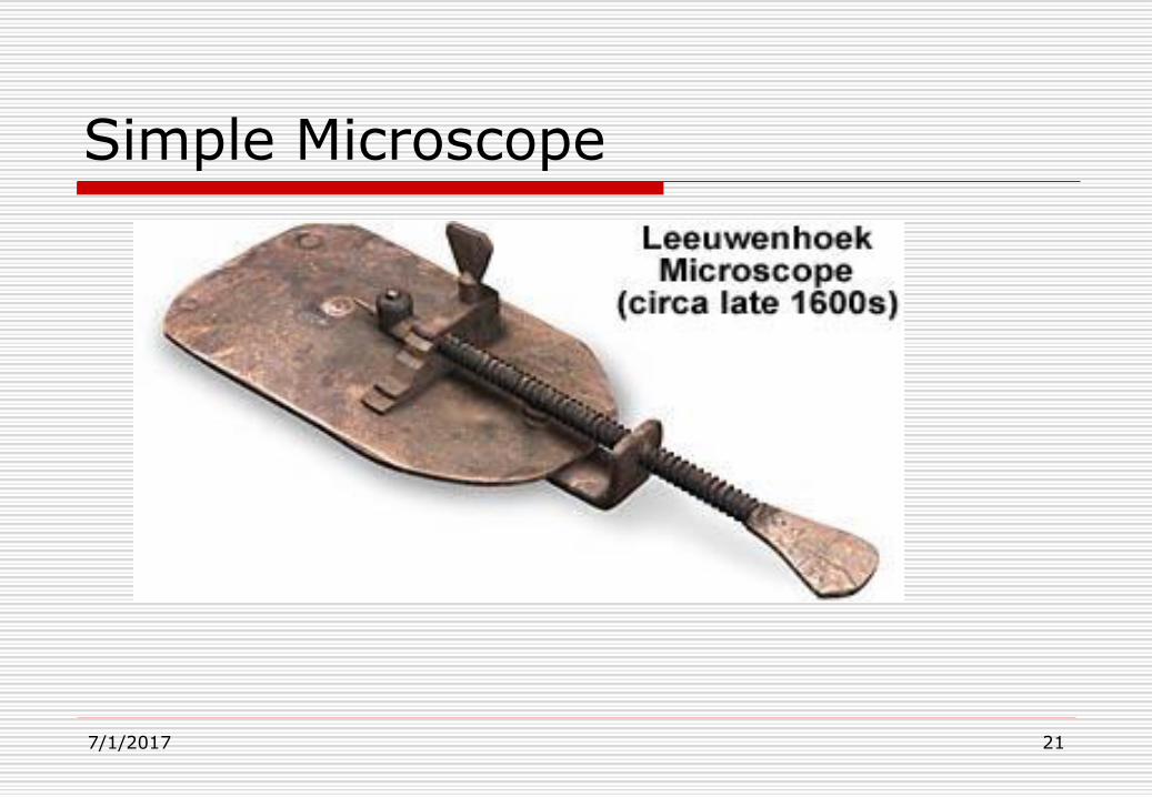
Simple Microscope
7/1/2017 21

Simple Microscope Cont’d
7/1/2017 22

Simple magnifier – forming an image
7/1/2017 23

SIMPLE MICROSCOPES
A simple microscope is a microscope
which only has one lens, as opposed to
the compound lenses used in more
complex microscope designs.
Magnifying glasses and loupes are two
well-known examples of the simple
microscope.7/1/2017 24

SIMPLE MICROSCOPES CONT’D.
Modern simple microscopes are usually
hand held, designed for field work or quick
viewing of objects which require
magnification. Historical designs included
mounts which resemble those used with
modern microscopes, although instead of
viewing the object through a series of
lenses, the user had only one lens to use.7/1/2017 25

18
DEFECTS OF SIMPLE MICROSCOPES
There are two inherent optical defects
associated with simple microscopes:
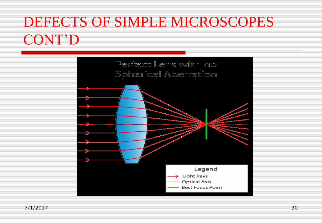
a)Spherical aberration: Blurred
images are formed because light
rays from a specimen are not
brought to simultaneous focus.
7/1/2017

DEFECTS OF SIMPLE MICROSCOPES
CONT’D
7/1/2017 27

DEFECTS OF SIMPLE MICROSCOPES
CONT’D
Spherical Aberration is an optical problem
that occurs when all incoming light rays
end up focusing at different points after
passing through a spherical surface. Light
rays passing through a lens near its
horizontal axis are refracted less than rays
closer to the edge or “periphery” of the lens
and as a result, end up in different spots
across the optical axis.7/1/2017 28

DEFECTS OF SIMPLE MICROSCOPES
CONT’D
In other words, the parallel light rays
of incoming light do not converge at
the same point after passing through
the lens. Because of this, Spherical
Aberration can affect resolution and
clarity, making it hard to obtain sharp
images.7/1/2017 29
DEFECTS OF SIMPLE MICROSCOPES
CONT’D
7/1/2017 30

3131
DEFECTS OF SIMPLE MICROSCOPES
CONT’D
Spherical aberration also occurs when
the specimen and the objective lens are
separated by several materials of
different refractive indices e.g. air,
glass etc. This results in the repeated
bending of the light rays at different
angles.7/1/2017

Ways to Reduce Spherical Aberration
Modern lenses employ different techniques
to dramatically reduce spherical aberration.
One of the methods employs using a
specialized aspherical (meaning non-
spherical) lens surface, which is curved
outwards on one side for the sole purpose
of converging light rays into a single focal
point.7/1/2017 32

Ways to Reduce Spherical Aberration
Cont’d
7/1/2017 33
3434
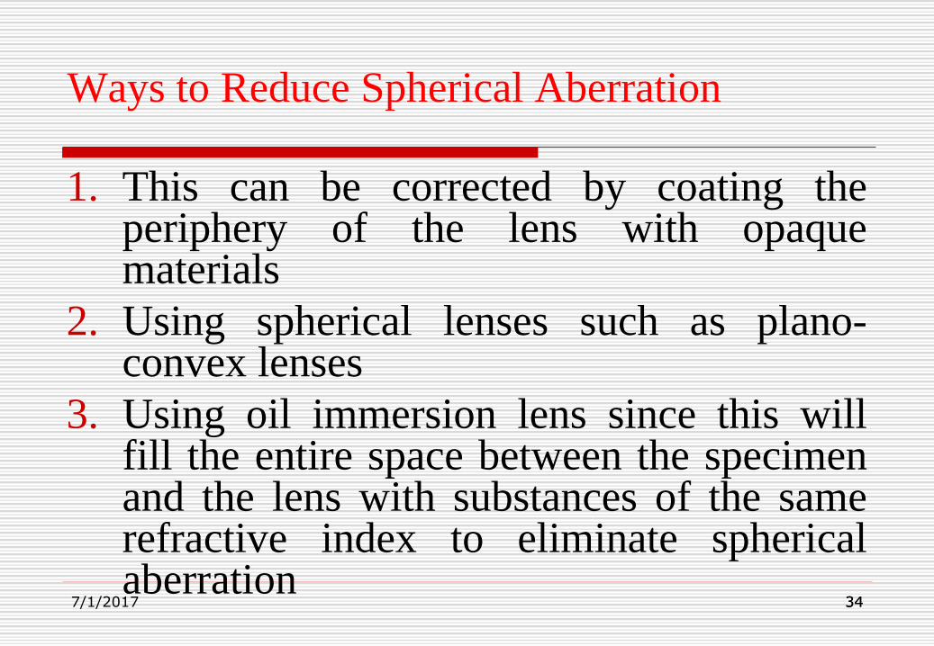
Ways to Reduce Spherical Aberration
1. This can be corrected by coating theperiphery of the lens with opaquematerials
2. Using spherical lenses such as plano-convex lenses
3. Using oil immersion lens since this willfill the entire space between the specimenand the lens with substances of the samerefractive index to eliminate sphericalaberration
7/1/2017

3535
b) Chromatic Aberration
Chromatic Aberration, also known as
“colour fringing” or “purple fringing”,
is a common optical problem that
occurs when a lens is either unable to
bring all wavelengths of color to the
same focal plane, and/or when
7/1/2017

Chromatic Aberration Cont’d
wavelengths of color are focused at different
positions in the focal plane. Chromatic
aberration is caused by lens dispersion, with
different colors of light travelling at different
speeds while passing through a lens. As a result,
the image can look blurred or noticeable colored
edges (red, green, blue, yellow, purple, magenta)
can appear around objects, especially in high-
contrast situations.7/1/2017 36

Chromatic Aberration Cont’d
7/1/2017 37

Chromatic Aberration Cont’d
7/1/2017 38
3939
Ways of correcting chromatic aberration
1. Using flint glass and crown which have
equal but opposite focal lengths. This
combination of crown and flint is known as
Acromatic Doublet.
2. Using more complex systems of lenses.
7/1/2017
4040
LIGHT MICROSCOPY.
Light microscopy refers to the use of anykind of microscope that uses visible light toobserve specimens. Here we examineseveral types of light microscopy.
The curvatures of the lens materials andtheir composition are designed to minimizedistortion of image shapes and colours.
Light microscopes are of two different basictypes:
7/1/2017
4141
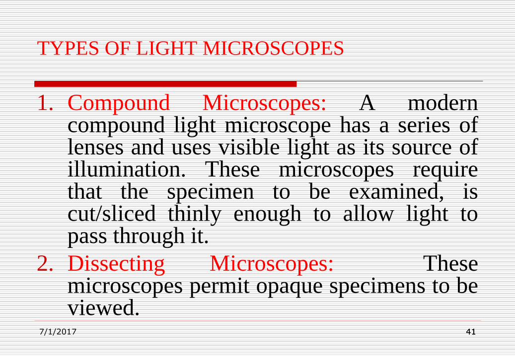
TYPES OF LIGHT MICROSCOPES
1. Compound Microscopes: A moderncompound light microscope has a series oflenses and uses visible light as its source ofillumination. These microscopes requirethat the specimen to be examined, iscut/sliced thinly enough to allow light topass through it.
2. Dissecting Microscopes: Thesemicroscopes permit opaque specimens to beviewed.
7/1/2017
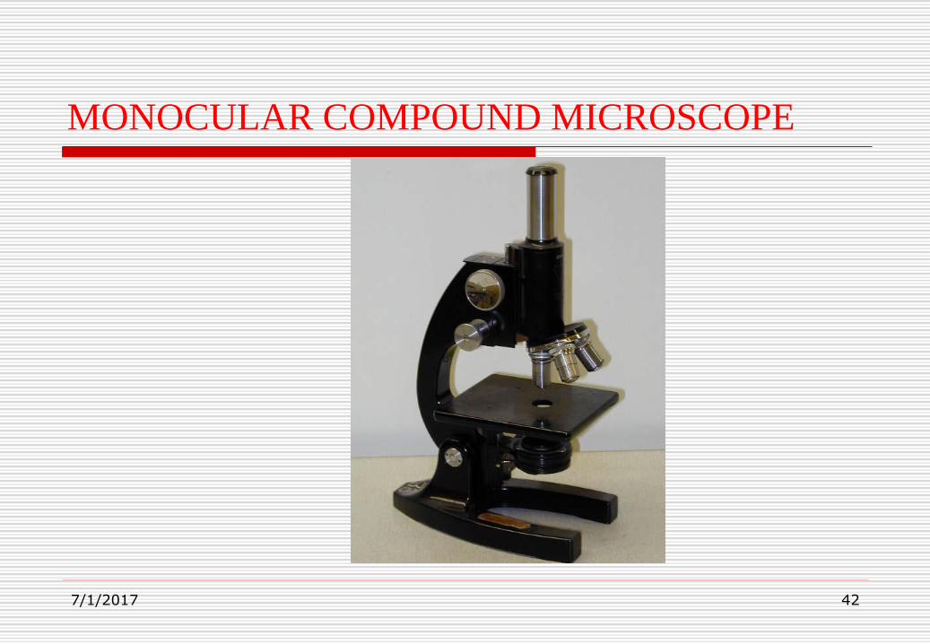
MONOCULAR COMPOUND MICROSCOPE
7/1/2017 42

BINOCULAR COMPOUND MICROSCOPE
7/1/2017 43

COMPOUND MICROSCOPE
7/1/2017 44
FUNCTIONS OF MICROSCOPES
Microscopy serves two
independent functions of
enlargement (magnification) and
improved resolution (the rendering
of two objects as separate entities).
7/1/2017 45
4646
46
MAGNIFICATION OF LIGHT MICROSCOPES
In their most basic essentials, all microscopes
aim at:
a) Magnifying the specimen and
b) Displaying the specimen in greater detail.
These aims are interdependent and it is
important to realise that to increase
magnification without a commensurate
improvement in the degree of discernible detail
7/1/2017

4747
47
Magnification of light microscopes Cont’d
is of little advantage.
a) Magnification: The magnification ofany optical system is dependent uponthe focal length of the lenses in thesystem and their mutual arrangement.
7/1/2017
4848
48
Magnification of light microscopes Cont’d
It is usually expressed as the ratio of the length
of the final image to that of the specimen, and
for the ordinary class microscope it is between
25x and 1500x. Some compound microscopes
can achieve a useful magnification of 2000x
with the oil immersion lens.
7/1/2017
4949
49
Magnification of light microscopes Cont’d
Most dissecting microscopes magnify up to
30x.
Magnifications of more than 2000x for
compound microscopes however, is considered
“empty” because resolution does not improve
with magnification beyond a certain point.
7/1/2017

PARFOCAL
Parfocal microscope objectives stay in
focus when magnification is changed; i.e.,
if the microscope is switched from a lower
power objective (e.g., 10×) to a higher
power objective (e.g., 40×), the object stays
in focus.
7/1/2017 50

Objective working and Parfocal distance
7/1/2017 51
Objective working and Parfocal distance
Cont’d.
Working distance is the distance
between the objective lens and the
specimen. At low magnification the
working distance is relatively long. As
you increase the magnification the
working distance decreases
considerably.7/1/2017 52

Objective working and Parfocal distance
Cont’d.
Oil immersion lenses practically touch
the specimen. Be aware of this change
in working distance with increasing
magnification so as to prevent damage
to your specimens and the lens in
particular.
7/1/2017 53

Relationship between working distance and
Magnification
Generally speaking, the working distance
of any given microscope is reduced as
magnification increases. As in most
compound microscopes, the lens is moved
physically closer to the specimen to
increase magnification; the working
distance available between the lens and the
specimen is reduced considerably as
magnification increases.7/1/2017 54

5555
55
Resolution of light microscopes
b) Resolution: The resolving power of a lens indicates the fineness of detail that it allows to be seen. Thus, if one examines two small specimens with a microscope lens, provided the specimens are well separated, they will be resolved as separate entities. If, however, they are then gradually moved closer together, a situation will eventually arise in
7/1/2017

5656
56
Resolution of light microscopes Cont’d
which the two specimens, though still separate,
can no longer be seen to be distinct from each
other. In this situation, only by improving the
resolution i.e. by using a lens with better
resolving power, will it again be possible to
render the two specimens as separate entities.
7/1/2017

5757
57
Factors that determine resolution
1. Numerical aperture: When light rays pass
through a specimen containing fine detail
they interfere with each other and they are
variously diffracted; increasingly fine detail
increases their angles of diffraction. Since
the resolving power of a lens depends upon
its ability to collect these diffracted rays, the
7/1/2017

5858
58
Factors that determine resolution Cont’d
wider the angle of rays collected, the better is
the resolution.
The capacity of a lens to collect rays emerging
from a specimen is defined by its numerical
aperture (NA), and this depends upon both its
angular aperture (U) and the refractive index (η) of
the medium through which the rays pass.
7/1/2017

5959
59
Factors that determine resolution Cont’d
The relationship is expressed as:
NA = η sin U
In any given lens, the NA and thus the resolution, is
at its best when the cone of rays emerging from the
specimen just fills the angular aperture. When setting
up a microscope this optimum requirement is only
obtained by careful focusing of the illumination system.
7/1/2017
6060
60
Factors that determine resolution Cont’d
In the conventional light microscope, the
medium between the low power (less than x100)
objective lenses and the specimen is air (i.e. the
refractive index, η = 1). However, for lenses of
higher power, where maximum resolution is
required, the refractive index may be increased by
filling this space with a special “immersion” oil.
The refractive index of the immersion oils used
with7/1/2017

6161
61
Factors that determine resolution Cont’d
glass-covered microscope slides is optimally
about 1.55. This arrangement increases the
NA and results in fewer light rays being lost
due to refraction. Resolution is thus
improved.
7/1/2017

6262
627/1/2017
6363
63
Factors that determine resolution Cont’d
2. Wavelength:
Resolution also depends upon the wavelength
of the transmitted wave form; the smaller the
wavelength the better is the resolution.
7/1/2017

6464
64
Factors that determine resolution Cont’d
3. Limit of resolution (r):
Resolution is the ability of a lens
to separate or distinguish between
small objects that are close
together. With reference to the
numerical aperture (NA) and
7/1/2017

6565
65
Factors that determine resolution Cont’d
wavelength (λ), it is expressed as:
r = 0.61 λ /NA
In practice, the maximum NA available for light microscope objective lenses is about 1.4. The component of white light to which the human
eye is most sensitive is green light (λ = 560nm);
7/1/2017

Factors that determine resolution Cont’d
Because sin(U) is always smaller than 1 and n
cannot rise above 1.7, the maximal resolving
power of a microscope is about
r=λ/2
and thus only depends on the wavelength of the
light used, which normally will be about 600nm.
7/1/2017 66

6767
67
Factors that determine resolution
therefore, this equation indicates that the
best resolution limit obtainable when using
white light is about 240 nm (0.24µm). A
red blood cell has a diameter of about 7µm.
7/1/2017

BRIGHT-FIELD MICROSCOPY
A standard brightfield microscope relies upon
light from the lamp source being gathered by the
substage condenser and shaped into a cone
whose apex is focused at the plane of the
specimen. Specimens are seen because of their
ability to change the speed and the path of the
light passing through them. This ability is
dependent upon the refractive index and the
opacity of the specimen.7/1/2017 68

BRIGHT-FIELD MICROSCOPY CONT’D.
To see a specimen in a brightfield microscope,
the light rays passing through it must be changed
sufficiently to be able to interfere with each
other which produces contrast (differences in
light intensities) and, thereby, build an image. If
the specimen has a refractive index too similar to
the surrounding medium between the
microscope stage and the objective lens, it will
not be seen.7/1/2017 69

BRIGHT-FIELD MICROSCOPY CONT’D.
To visualize biological materials well, the materials
must have this inherent contrast caused by the
proper refractive indices or be artificially stained.
These limitations require instructors to find
naturally high contrast materials or to enhance
contrast by staining them which often requires
killing them. Adequately visualizing transparent
living materials or thin unstained specimens is not
possible with a bright-field microscope.
7/1/2017 70

DARKFIELD MICROSCOPY
The structure of many biological specimens
are of low contrast that cannot be revealed
by the brightfield compound microscopes.
Microscopes that improve the contrast of
these specimens through special optics are
prohibitively expensive. An inexpensive
darkfield microscope is one that is
modified from a brightfield microscope.7/1/2017 71

DARKFIELD MICROSCOPY CONT’D.
Dark field microscopy is a very simple yet
effective technique and well suited for uses
involving live and unstained biological
samples, such as a smear from a tissue
culture or individual water-borne single-
celled organisms. Considering the
simplicity of the setup, the quality of
images obtained from this technique is
impressive.7/1/2017 72

DARKFIELD MICROSCOPY CONT’D.
A darkfield microscope is used for
examining live microorganisms that either
are invisible in the ordinary light
microscope, cannot be stained by standard
methods, or are so distorted by staining that
their characteristics then cannot be
identified. Instead of the normal
condenser, a darkfield microscope uses a7/1/2017 73

DARKFIELD MICROSCOPY CONT’D.
darkfield condenser that contains an opaque
disc. The disc blocks light that would enter
the objective lens directly. Only light that is
reflected off the specimen enters the objective
lens. Because there is no direct background
light, the specimen appears light against a
dark background. This technique is frequently
used to examine unstained microoganisms7/1/2017 74

DARKFIELD MICROSCOPY CONT’D.
suspended in liquid such as yeast cells.
7/1/2017 75

Bright-field and Dark-field compared
7/1/2017 76

7777
777/1/2017

7878
787/1/2017

PHASE-CONTRAST MICROSCOPY
Differences in light absorption are often
negligible between living cells and their
surrounding nutrient medium, as well as
between the various intracellular
components and plasma membranes,
rendering these entities barely visible when
observed by brightfield illumination.
7/1/2017 79

PHASE-CONTRAST MICROSCOPY
CONT’D.
When living cells are examined by normal
light microscopy it is frequently difficult to
see cell contents. This is because the human
eye normally detects contrast by differences
in colour or in intensity of illumination, and
living cells are normally colourless and more
or less transparent. One common method of
overcoming this difficulty is to stain cells7/1/2017 80

PHASE-CONTRAST MICROSCOPY
CONT’D.
with various dyes but, as this usually results
in death or disruption, it cannot be used to
examine living cells. One technique which is
now widely used to solve this problem is
known as phase contrast microscopy.
The wavelength of light is a measure of
colour whilst amplitude is a measure of
brightness. When a specimen is examined7/1/2017 81

PHASE-CONTRAST MICROSCOPY
CONT’D.
with a light microscope, the light passing
through the specimen will be affected in two
ways in relation to the light passing through
the surroundings, and will produce the
contrast in the image. Firstly, the light will be
diffracted or scattered, and may be lost to the
image; the greater the diffraction, the darker
the image. Secondly, the diffracted rays that7/1/2017 82

PHASE-CONTRAST MICROSCOPY
CONT’D.
do pass through the microscope will be
retarded in relation to the light that did not
pass through the specimen, the degree of
retardation depending on the thickness of the
specimen. Thus two sets of rays will arrive at
the eye, the diffracted and undiffracted. The
slight phase change produced in the diffracted
rays will interfere with the other rays7/1/2017 83
PHASE-CONTRAST MICROSCOPY
CONT’D.
Resulting in a net reduction in amplitude, and
so a lowering of brightness. The image seen
by the eye is, in fact a complex interference
pattern of diffracted and undiffracted rays.
Normally the change in phase, and amplitude,
is slight, and produces little contrast. The
phase contrast microscope functions by
further retarding the deffracted rays7/1/2017 84

PHASE-CONTRAST MICROSCOPY
CONT’D.
producing a greater change in amplitude on
interference, and so a greater reduction in
brightness. In the phase contrast microscope
the light rays from the specimen background
pass through the thinner portion of the
positive phase plate and those that pass
through the specimen will pass through the
thicker portion of the positive phase plate7/1/2017 85

PHASE-CONTRAST MICROSCOPY
CONT’D.
and so suffer a greater diffraction
(retardation), enhancing the phase difference
produced by passage through the specimen. In
addition, further light will be lost by the
greater scatter produced by the thicker part of
the positive phase plate, and so contrast will
be enhanced by both greater amplitude
changes and loss of light.7/1/2017 86

PHASE-CONTRAST MICROSCOPY
CONT’D.
Phase contrast microscopy is now widely used to
study living cells and tissues. Considerable
cytological detail of cell movement, changes in
nuclei and other organelles, and cytoplasmic flow
have been observed although resolution is still
limited by the wavelength of visible light. Phase
contrast microscopy has been particularly useful
when used in conjunction with time-lapse motion
photography of living cells.7/1/2017 87

PHASE-CONTRAST MICROSCOPE
7/1/2017 88

PHASE-CONTRAST MICROSCOPY
CONT’D.
Figure 2 in the slide below is a comparison
of living cells in culture imaged in both
brightfield and phase contrast illumination.
In brightfield illumination (Figure 2(a)),
the cells appear semi-transparent with only
highly refractive regions, such as the
membrane, nucleus, and unattached cells
(rounded or spherical), being visible.7/1/2017 89

PHASE-CONTRAST MICROSCOPY
CONT’D.
7/1/2017 90
9191
91
ULTRA VIOLET MICROSCOPY
This type of microscope uses U/V light that tends
to increase resolution two-fold compared with a
microscope that uses white light.
This type of microscope makes use of specimens
that either have the natural ability to absorb U/V
light and re-emit it as light of longer wavelength or
stained with specific dyes to aid them absorb U/V
light and re-emit it as light of longer wavelength
visible to the human eye.7/1/2017

ULTRA VIOLET MICROSCOPY CONT’D
The main disadvantages of using this
microscope are:
U/V cannot pass through glass lenses and
hence the lenses have to be replaced with
quartz lenses which are expensive.
The U/V light carrying the image of the
specimen is not visible to the human eye.
7/1/2017 92
ULTRA VIOLET MICROSCOPY CONT’D
U/V light has the tendency to damage the eye
and requires that the microscope be fitted with a
camera or the image of the specimen projected
onto a fluorescent screen.
7/1/2017 93

What is a Fluorescence Microscope?
A fluorescence microscope is basically a
conventional light microscope with added
features and components that extend its
capabilities.
Basic Requirements of Fluorescence Microscope
Optics
Nearly all fluorescence microscopes use the
objective lens to perform two functions:
7/1/2017 94

Basic Requirements of Fluorescence
Microscope Optics Cont’d
1. Focus the illumination (excitation) light on the
sample. In order to excite fluorescent species in a
sample, the optics of a fluorescent microscope must
focus the illumination (excitation) light on the
sample to a greater extent than is achieved using the
simple condenser lens system found in the
illumination light path of a conventional
microscope.
7/1/2017 95

Basic Requirements of Fluorescence
Microscope Optics Cont’d
2. Collect the emitted fluorescence. This type of
excitation-emission configuration, in which both
the excitation and emission light travel through
the objective, is called epifluorescence. The key
to the optics in an epifluorescence microscope is
the separation of the illumination (excitation)
light from the fluorescence emission emanating
from the sample.
7/1/2017 96

Basic Requirements of Fluorescence
Microscope Optics Cont’d
In order to obtain either an image of the
emission without excessive background
illumination, or a measurement of the
fluorescence emission without background
"noise", the optical elements used to separate
these two light components must be very
efficient.
7/1/2017 97

The Dichroic Mirror
In a fluorescence microscope, a dichroic
mirror is used to separate the excitation and
emission light paths. Within the objective, the
excitation/emission share the same optics.
7/1/2017 98

FLUORESCENCE MICROSCOPY
Certain chemical substances emit visible light
when they are illuminated with ultra violet light.
The effect is termed fluorescence and is put to
use in the fluorescence microscopy in which
ultraviolet light rays are focused on the
specimen. Some cellular components possess a
natural fluorescence and appear in various
colours such as the chromosomes, nuclei etc.
7/1/2017 99

FLUORESCENCE MICROSCOPY CONT’D
Other, nonfluorescing structures can be made to
fluoresce by staining them with fluorescent dyes
(fluorochromes). One of the most popular
contemporary uses of fluorescence microscopy
involves the preparation of antibodies that will
bind to specific cellular proteins. The antibodies
are first complexed with fluorecein (a
fluorescent dye), and the fluorescein-labelled
antibody is then applied to the cells.7/1/2017 100

FLUORESCENCE MICROSCOPY CONT’D
Cell structures containing the specific proteins
capable of binding the fluorescein-labelled
antibody are caused to fluoresce when examined
with fluorescence microscope. This technique in
microbiology is called immunofluorescence.
7/1/2017 101
102102
102
U/V – FLUORESCENCE MICROSCOPY
Fluorescence microscopy is used to detect
structures, molecules or proteins within a cell.
Fluorescent molecules absorb light at one
wavelength and emit at another, longer wavelength.
When fluorescent molecules absorb a specific
absorption wavelength for an electron in a given
orbital, the electron rises to a higher energy level.
Electrons in this state are unstable and will return to
the ground state, releasing energy in the form of7/1/2017

U/V – FLUORESCENCE MICROSCOPY
CONT’D
light and heat. This emission of energy is
fluorescence. In fluorescence microscopy, a cell is
stained with a dye and the dye is illuminated with
filtered light at the absorbing wavelength; the light
emitted from the dye is viewed through a filter that
allows only the emitted wavelength to be seen. The
dye glows brightly against a dark background
because only the emitted wavelength is allowed to
reach the eye pieces or camera port of the
microscope.7/1/2017 103

104104
1047/1/2017

105105
ELECTRON MICROSCOPY
All matter has a dual nature, both particulateand wave-like. Thus, electrons have wave-likeproperties and their wave-length depends uponthe speed at which they are moving. Theapproximate λ of electrons, over a potentialdifference of V volts, can be calculated from:
7/1/2017

ELECTRON MICROSCOPY CONT’D
λ =√1.5/V nm
Thus, electrons at 60 KV have an apparent λ of
0.005 nm, which should give a huge increase in
resolution over the light microscope. Thus,
phenomenally high resolving power
theoretically available with electron microscopy
unfortunately is not realizable. The resolving
power is limited by specimen contrast and the
highly imperfect electromagnetic lenses.7/1/2017 106

ELECTRON MICROSCOPY CONT’D
There are two types of electron microscopes:
The Transmission electron microscope (TEM)
and the Scanning electron microscope (SEM).
Transmission electron microscope (TEM)
In transmission electron microscope, a finely
focussed beam of electrons from an electron gun
passes through a specially prepared, ultrathin
section of the specimen. The ray of
7/1/2017 107

ELECTRON MICROSCOPY CONT’D
electrons is produced by a pin-shaped cathode
heated up by current. The electrons are
vacuumed up by a high voltage at the anode.
The acceleration voltage is between 50 and 150
kV. The higher it is, the shorter are the electron
waves and the higher is the power of resolution.
But this factor is hardly ever limiting. The
power of resolution of electron microscopy is
7/1/2017 108

ELECTRON MICROSCOPY CONT’D
usually restrained by the quality of the lens-
systems and especially by the technique with
which the preparation has been achieved.
Modern gadgets have powers of resolution that
range from 0.2 – 0.3 nm. The useful resolution is
therefore around 300,000 x. The accelerated ray
of electrons passes a drill-hole at the bottom of
the anode.
7/1/2017 109

ELECTRON MICROSCOPY CONT’D
It follows a way analogous to that of a ray of light
in a light microscope. The lens-systems consist of
electronic coils generating an electromagnetic field.
The ray is first focused by a condenser. It then
passes through the object, where it is partially
deflected. The degree of deflection depends on the
electron density of the object. The greater the mass
of the atoms, the greater is the degree of deflection.
Biological objects have only weak contrasts since7/1/2017 110

ELECTRON MICROSCOPY CONT’D
they consist mainly of elements with low atomic
numbers (C, H, N, O). After passing through the
object the scattered electrons are collected by an
objective. Thereby an image is formed, that is
subsequently enlarged by an additional lens-system
(called projective with electron microscopes). The
thus formed image is made visible on a fluorescent
screen or it is documented on photographic
material.7/1/2017 111

112112
TRANSMISSION ELECTRON MICROSCOPY
7/1/2017

1131137/1/2017

SCANNING ELECTRON MICROSCOPY
The Scanning Electron Microscope (SEM) is
a microscope that uses electrons rather than
light to form an image. There are many
advantages to using the SEM instead of a
light microscope. The SEM has a large depth
of field, which allows a large amount of the
sample to be in focus at one time.
1147/1/2017

SCANNING MICROSCOPY CONT’D
The SEM also produces images of high
resolution, which means that closely spaced
features can be examined at a high
magnification. The combination of higher
magnification, larger depth of focus, greater
resolution, and ease of sample observation
makes the SEM one of the most heavily used
instruments in research areas today.
1157/1/2017
SCANNING MICROSCOPY CONT’D
A scanning electron microscope (SEM) is a
powerful microscope that uses electrons
rather than light to form an image of objects
such as fractured metal components, foreign
particles and residues, polymers, electronic
components, biological samples, and
countless others.
1167/1/2017

SCANNING MICROSCOPY CONT’D
The shorter wavelength of electrons permits
image magnifications of up to 100,000X, as
compared to about 2,000X for conventional light
microscopy. An SEM also provides a greater
depth of field than a light microscope, allowing
complex, three-dimensional objects to remain
sharp and in focus. This capability reveals
details that cannot be resolved by light
microscopy.1177/1/2017

118118
SCANNING MICROSCOPY CONT’D
SEM is used to look at the surface of a solid
specimen. The resolution is usually only 10
nm (unless FESEM is used), but with 20 000
× magnification. We get very attractive 3D-
looking images because of the large depth of
field.
7/1/2017

119119Scanning electron microscope image of pollen.
7/1/2017

1201207/1/2017

121121
PREPARATION OF SPECIMENS FOR
LIGHT AND ELECTRON MICROSCOPY
There are problems in analysing cell structure and consequently complex preparative procedures must be used. The include the ff:
1. FIXATION:
It is the rapid killing and preservation of a tissue. Correct fixation is the fundamental basis of all histological work.
7/1/2017

Fixation Cont’d
The tissue must be fixed to prevent autolysis,
bacterial or fungal attack and also to make the
tissue resistant to any damage that might caused by
later procedures. Fixation may be by either physical
and chemical means. Physical methods involve
immersing the specimen in liquid nitrogen, thereby,
freezing it so rapidly that ice crystals which would
disrupt and distort the tissue, do not form.
7/1/2017 122

Fixation Cont’d
This method of fixation is often essential if it is
necessary to preserve the tissue structure and to
prevent any damage occurring to the enzymatic
components of cells. Frozen tissue is only fixed
while frozen and if brought to room temp. would
rapidly undergo autolysis. Thus, if it is
necessary to keep permanent preparations of
frozen sections, they must be chemically fixed.
7/1/2017 123

Fixation Cont’d
Chemical methods of fixation involve chemicals
that would stabilise proteins and lipids, which
are the major structural components of cells. If
the specimen is to be examined with the electron
microscope, it is essential to use fixatives whose
reactions lead to the formation of precipitates
that do not obscure the structure of the cell
organelles.
7/1/2017 124
Fixation Cont’d
The most widely used fixatives for light
microscopy include acetic acid, alcohol,
acetone and mercuric chloride, chromic acid,
formaldehyde, while formaldehyde and
glutaraldehyde (which fix proteins) and
osmium tetroxide and potassium
permanganate, (which fix lipids) are used for
electron microscopy.7/1/2017 125

PREPARATION OF SPECIMENS FOR LIGHT
AND ELECTRON MICROSCOPY CONT’D
2. EMBEDDING:
An electron beam has a low penetrating power and
so, the column of the microscope is kept under high
vacuum. This means that water must be removed
from the specimens otherwise the vacuum will be
destroyed. Therefore, specimens are dehydrated
after fixation, usually by immersion in a series of
ethanol – water mixtures of increasing alcohol
concentration up to 100% alcohol which7/1/2017 126
PREPARATION OF SPECIMENS FOR LIGHT
AND ELECTRON MICROSCOPY CONT’D
replaces the water in the specimen. A wax solvent
such as xylene is used to replace the alcohol in the
specimen in another series of baths and finally
replacing the xylene with molten paraffin wax. A
similar dehydration procedure is used to embed
material in epoxy resin, but instead of xylene, a
resin solvent such as propylene oxide is used. The
resin-impregnated specimen is then baked in an
oven to polymerise it.7/1/2017 127

PREPARATION OF SPECIMENS FOR LIGHT
AND ELECTRON MICROSCOPY CONT’D
Associated with the low penetration property of
the electron beam is the question of specimen
thickness. The formation of the image in an
electron microscope depends upon the electrons
passing through the specimen. If the specimen is
too thick, all of the electrons will be scattered to
some extent, resulting in an image with poor
contrast and considerable loss of detail.
7/1/2017 128

PREPARATION OF SPECIMENS FOR LIGHT
AND ELECTRON MICROSCOPY CONT’D
Therefore, it is necessary to cut thin
slices/sections of the material to be examined,
and these must be of the order of 50 nm thick.
To cut these ultra-thin sections, the tissue must
first be embedded in a hard support medium
since biological material has little mechanical
strength. The supporting medium used depends
on the thickness of the section required.
7/1/2017 129

PREPARATION OF SPECIMENS FOR LIGHT
AND ELECTRON MICROSCOPY CONT’D
For relatively thick sections of plant material,
specimens may be supported by sandwiching
them between easily cut material such as raw
carrot or raw unripe pawpaw fruit. Thinner
sections can be cut if the specimen is embedded
in paraffin wax and for the ultra sections
required for electron microscopy, a more rigid
support such as the epoxy resin, araldite etc can
be used.7/1/2017 130

PREPARATION OF SPECIMENS FOR LIGHT
AND ELECTRON MICROSCOPY CONT’D
3. SECTIONING:
To produce thin sections (1000 – 20000 nm for
light microscopy, but 50 – 100 nm for electron
microscopy), an instrument called a microtome
is used. All microtomes of whatever pattern
consist of a specimen holder, a sharp cutting
edge and a means of regulating the thickness of
the section being cut. The sharp cutting edge
7/1/2017 131
PREPARATION OF SPECIMENS FOR LIGHT
AND ELECTRON MICROSCOPY CONT’D
may be that of a steel razor for wax - embedded
specimens or of a glass or diamond knife for
araldite – embedded specimens. Sections of frozen
specimens are cut with a microtome mounted in a
freezer maintained at – 20oC. When the sections
have been cut, they are supported by being mounted
either on glass microscope slides for examination
with light microscopy or on a grid of fine copper
strands for electron microscopy.7/1/2017 132

133133
TYPES OF MICROTOMES
7/1/2017

1341347/1/2017

1351357/1/2017

136136
4. STAINING:
Thin sections of cells or tissues are usually
transparent or nearly so and it is therefore, usually
necessary to stain the sections before they are
examined.
For light microscopy, most of the commonly used
stains are organic aromatic dyes originally
produced for use in the textile industry. Some, like
toluidin blue colour all tissues; others colour only
particular components of cells or parts of tissues.7/1/2017

137137
Of these more specific stains, there are essentially two
groups; basic stains and acidic stains. The specificity
of these stains depends on the difference in the charge
of the different cell components.
A commonly used basic stain is haematoxylin – the
colour imparting (chromogenic) group is cationic
(+vely charged) and reacts primarily with negatively
charged molecules, such as the nucleic acids in the
nucleus, to produce colour.
STAINING contd
7/1/2017

138138
STAINING contd
7/1/2017

139139
STAINING contd
7/1/2017
140140
STAINING contd
Haematoxylin is most frequently used incombination with a second dye, eosin. In anacidic solution, the chromogenic group ofeosin is anionic (-vely charged) and it willreact with basic groups in the cell, which arelargely present in the cytoplasm, stainingthem red. Haematoxylin and eosin are used asroutine stains for most animal tissues.
7/1/2017

141141
STAINING contd
In electron microscopy, different stains areused to emphasise different cell componentsas they are for light microscopy, but in thiscase, the stains must impart contrast to thesections instead of colour. This is because inelectron microscopy, the formation of animage depends on a beam of electrons, whichpasses through the section and hits afluorescent screen. The image is formed by7/1/2017

142142
STAINING contd
the removal of electrons from this beam bythe specimen, but because the electrons passeasily through the resin and the unstainedtissue, the image is faint and with lowcontrast. By staining cell components withelements of high atomic number like Pb andUranium, their contrast is increased becausetheir electron density is increased.
7/1/2017

143143
STAINING contd
Uranium binds preferentially to nucleic acidsand proteins, whereas Pb binds to lipids andmembranes. Hence, cell components rich inlipids or nucleic acids appear dark orelectron-dense and areas of cytoplasm or theresin surrounding the tissue appear light.Osmium will impart electron density tomembranes as well as to lipids and nucleicacids.7/1/2017

144144
STAINING contd
This is known as positive staining technique
which is frequently used in the examination
of fragments of isolated organelles and
membranes and of small particles such as
viruses and ribosomes. The double staining
technique of Uranium and Pb is to electron
microscopy as H and E is to light microscopy.
7/1/2017

145145
STAINING contd
Negative staining produces an electron –densebackground against which the less dense specimensare observed. Negative stains consist of the salts ofheavy metals and include potassiumphosphotungstate, Uranyl acetate and ammoniummolybdate. The electron-dense stain appears to drymore rapidly on the support film than on thespecimen and forms a sharp boundary; thespecimen appearing bright on a dark background.
7/1/2017

Cellular level of organisation:
Specific Objectives
• Appreciate the cell as the basic functional unit of
life
• Basic differences between prokaryotes and
eukaryotes
• Describe the structure of the plasma membrane and
explain its functional significance.
• Describe the structure of the cell nucleus and explain its significance.
• Describe the structure & function of the cellular
organelles in the cytoplasm.7/1/2017 146
Fundamentals of the cell
Wide range of cell types but generally two
kinds of cell types:
• Cells without a nucleus = Prokaryotes
• Cells with a nucleus = eukaryotes
7/1/2017 147
What is the cell
Like ourselves, the individual cells that form our bodies can
grow, reproduce, process information, respond to stimuli and
carry out an amazing array of chemical reactions. These
abilities define life.
7/1/2017 148

What is the cell Cont’d
We and other multi-cellular organisms contain billions and
trillions of cells organized into complex structures, but many
organisms consist of a single cell.
• Even simple unicellular organisms exhibit all the major
properties of life,
7/1/2017 149

What is the cell Cont’d
This indicates that the cell is the fundamental unit of life.
Definition of Cell
A cell is the smallest unit of life that is
capable of independently performing
life functions.
7/1/2017 150

Cellular Level of Organization
The cell:
• Basic, living, structural and functional unit of
life, capable of independent reproduction
• Organismal activity depends on individual and
collective activity of cells
• Biochemical activities dictated by subcellular
structure.
7/1/2017 151

General functions of the cell
• Compartmentalization of chemical reactions
within specialized structures
• Regulate inflow & outflow of materials
• Use genetic material to direct cell activities
• Continuity of life originates from the cell
7/1/2017 152

Cellular Diversity
7/1/2017 153

Cellular Diversity Cont’d.
• 100 trillion cells in the body -- 200
different types
• Vary in size and shape related to their
function
7/1/2017 154

A typical cell
7/1/2017 155

156156
Cell Structure Cont’d
7/1/2017

157157
Cell Structure Cont’d
7/1/2017
158158
Factors that determine cell size and shape
The shape of the cell depends partly on the
surface tension and viscosity of the cytoplasm,
the mechanical action which the adjoining cells
exert, the rigidity of the membrane and the
functional adaptation.
7/1/2017

159159
Cell Structure Cont’d
ASSIGNMENTS:
Cell Theory.
Factors that determine cell size and cell
shape.
Differences between Prokaryotic and
Eukaryotic cells.
7/1/2017

160160
Use of Geometric relationships to explain why most cells are microscopic
7/1/2017

161161
Detailed Structure of some of the organelles
MICROBODIES:
Microbodies are a heterogeneous group of smallvesicle-like organelles, concerned largely withoxidation. They are usually oval or spherical andbounded by a single membrane. Microbodies arefound in the liver and kidneys of vertebrates, in theleaves and seeds of plants as well as in protozoa,yeasts and other fungi. Microbodies consist ofperoxisomes, glyoxysomes, hydrogenosomes andglycosomes.7/1/2017

162162
PEROXISOMES
These contain the enzymes urate oxidase, D-aminoacid oxidase and catalase.
Urate oxidase is a purine – catabolising enzyme,converting urate into allantoin, CO2 and H2O2.Thus, peroxisomes not only play a role in the breakdown of nitrogenous bases derived from nucleicacids but they are also involved in the degradationof L-α-hydroxy acids to oxoacids and H2O2 and ofoxoacids to smaller products including acetyl CoA.
7/1/2017

163163
PEROXISOMES CONT’D
D-Amino acid oxidase and catalase have
protective functions. D-amino acids may be
absorbed from the gut following their release
by the break down of cell walls of gut
bacteria. These “unnatural” amino acids are
degraded to give oxoacids and H2O2 .
H2O2 is a powerful oxidising agent and is
potentially toxic. Catalase catalyses the rapid
degradation of H2O2 to water and O2.7/1/2017

Peroxisomes Cont’d.
• Membranous vesicles
– smaller than lysosomes
– form by division of pre-existing peroxisomes
– contain enzymes that oxidize organic material
• Function
– part of normal metabolic breakdown of amino acids
and fatty acids
– oxidizes toxic substances such as alcohol and
formaldehyde
– contains catalase which decomposes H2O27/1/2017 164

165165
MICROBODIES CONT’D
GLYOXYSOMES:
These are microbodies of plant cells in which theenzymes of the glyoxylate cycle are functionallymore important than those involved in oxidativemechanisms. The glyoxylate cycle allows therelatively immobile fatty reserves of e.g. seeds tobe converted to sugars and therefore, more easilytransported to growing tissues.
The fats are degraded to acetyl CoA which feedsinto the glyoxylate cycle to give succinate as a netproduct.7/1/2017

166166
MICROBODIES CONT’D
Succinate is then transported to the
mitochondria where it becomes a substrate for
gluconeogenesis.
7/1/2017

167167
PEROXISOME
7/1/2017

Lysosomes
7/1/2017 168
•Membranous vesicles
–formed in Golgi complex
–filled with digestive enzymes
–pumps in H+ ions until internal pH
reaches 5.0
•Functions
–digest foreign substances
•recycles own organelles
–autolysis
•lysosomal damage after death

169169
Lysosomes Cont’d.
Lysosomes are vesicular structures, limited by asingle smooth membrane, and containinghydrolases active at acid pH values. Lysosomesarise initially as primary lysosomes, which appearto be derived from coated vesicles released fromtrans cisternae of the Golgi apparatus. Lysosomescontain about 60 hydrolytic enzymes whoseconcerted action will degrade most biologicalmaterials. Coated vesicles arising from receptor-mediated endocytosis deliver their contents to a7/1/2017

LYSOSOMES CONT’D:
vesicle called an endosome. Fusion of an endosome
with a primary lysosome forms a secondary
lysosome. The activity of a H+ - ATPase in the
lysosome membrane pumps protons into the
intralysosomal space producing a pH of about 5.0
which activates the lysosomal enzymes.
Phagocytic vesicles (phagosomes) also fuse with
primary lysosomes to give secondary lysosomes.
7/1/2017 170
LYSOSOMES CONT’D:
This allows digestion of the ingested material, and
subsequently absorption of the products into the
cytosol. Following digestive activity, a residual
body containing non-degradable material may
remain. Residual bodies are retained within the cell
and may accumulate. The digestion of extracellular
material is called heterophagy. However, lysosomes
can also degrade material of intracellular origin,
such as mitochondria and ribosomes. This process7/1/2017 171

Lysosomes Cont’d.
is called autophagy.
It is apparent that the lysosomal membrane is
unusual. Not only is it resistant to digestion by the
hydrolases of the lysosome, but also, under normal
circumstances, it is impermeable to both the
enzymes and their substrates. Despite this, the
lysosomal membrane is freely permeable to the low
molecular weight products of hydrolysis.
7/1/2017 172

173173
Lysosomes Cont’d.
Lysosomes are important in many clinical andmedical aspects of biochemistry. For example,phagocytic cells in tissues of lungs and livercontain large lysosomes which are important indigesting foreign materials. Silicosis is a conditionresulting from the inhalation of silica particles intothe lungs which are taken up by phagocytes.Reactions between the silica and the lysosomalmembranes lead to the rupture of the membrane,the release of the lysosomal enzyme and eventually7/1/2017

174174
Lysosomes Cont’d.
the death of the phagocyte. The death of largenumbers of phagocytes stimulates fibroblasts todeposit collagen fibres which decrease lungelasticity, impair breathing and contribute to thepathology of disease. Silicosis is similar toasbestosis and black lung disease, conditionscaused by breathing in asbestos fibres and coal dustrespectively.
The absence of specific hydrolase enzymes fromlysosomes leads to the accumulation of substrate7/1/2017
175175
Lysosome Cont’d.
for that enzyme within the lysosomes. This usuallyhas severe medical consequences.
7/1/2017

176176
Lysosome Cont’d.
7/1/2017

Tay-Sachs Disorder
• Affects children of Eastern European descent
– seizures, muscle rigidity, blindness, demented and die before the age of 5
• Genetic disorder caused by absence of a single lysosomal enzyme
– enzyme normally breaks down glycolipid commonly found in nerve cells
– as glycolipid accumulates, nerve cells lose functionality
– chromosome testing now available7/1/2017 177

Nucleus
• Large organelle with double membrane nuclear envelope
– outer membrane continuous with rough ER
– perforated by water-filled nuclear pores
• Nucleolus
– spherical, dark bodies within the nucleus (no membrane)
– site of ribosome assembly
7/1/2017 178

Function of Nucleus
• 46 human DNA chromosomes
– genes found on chromosomes
– gene directs synthesis for a specific protein
• Non-dividing cells contain nuclear chromatin
– loosely packed DNA
• Dividing cells contain chromosomes
– tightly packed DNA
– it doublescopies itself before condensing7/1/2017 179

Protein synthesis
• Instructions for making
specific
proteins is found in the DNA
(your genes)
– transcribe that information
onto a messenger RNA
molecule
– translate the “message” into
a sequence of amino acids
in order to build a protein
molecule7/1/2017 180

181181
THE NUCLEUS:
In eukaryotic organisms, the DNA is protected in
the nucleus which has a surrounding envelope. The
nuclear envelope consists of a double membrane
separated by a perinuclear space.
The nuclear envelope is interrupted by pores, found
at regions where the outer and inner membranes
fuse. The pores are surrounded by highly organised
annulus consisting of eight protein granules
symmetrically arranged around the pore. The term7/1/2017

182182
THE NUCLEUS CONT’D:
porosome has been applied to the nuclear pore andits associated annulus. There are molecules that aremuch bigger than the nuclear pores but are freelyable to pass through yet there are molecules farsmaller than the nuclear pores but are preventedfrom entering the nucleus. Nuclear proteins, like allproteins, are produced in the cytosol but mustaccumulate in the nucleus for functional reasons.Such proteins contain sequences of amino acidresidues which act as signals, enabling them to beconcentrated selectively in the nucleus.7/1/2017

183183
THE NUCLEUS CONT’D:
Glycoproteins present in the porosome appear toregulate the entry of material into the nucleus.
Contained in the nucleus is the nucleolus and thenucleoplasm. Found embedded in the nucleoplasmare chromatin materials which are precursors ofchromosomes. Eukaryotic chromosomes consist ofabout one-third DNA and two-thirds protein. Thecomplex of chromosomal DNA and protein (anucleoprotein) is called chromatin. The protein ofchromatin consists of small, basic proteins knownas histones and additional proteins generally called7/1/2017

THE NUCLEUS CONT’D:
chromatin is made up of repeating structural units
called nucleosomes.
The nuclear envelope is perforated with holes
called nuclear pores. These pores regulate the
passage of molecules between the nucleus and
cytoplasm, permitting some to pass through the
membrane, but not others. Building blocks for
building DNA and RNA are allowed into the
nucleus as well as molecules that provide the
energy for constructing genetic material.7/1/2017 184

THE NUCLEOLUS
The nucleolus is a membrane-less organelle within
the nucleus that manufactures ribosomes, the cell's
protein-producing structures. Through the
microscope, the nucleolus looks like a large dark
spot within the nucleus. A nucleus may contain up
to four nucleoli, but within each species the number
of nucleoli is fixed. After a cell divides, a nucleolus
is formed when chromosomes are brought together
into nucleolar organizing regions. During cell
division, the nucleolus disappears.7/1/2017 185

THE NUCLEOLUS CONT’D:
Nucleoli are prominently staining regions of the
nucleoplasm. They are composed of groups of
ribosomal genes surrounded by their rRNA
transcripts, together with many proteins.
Nucleoli are sites of synthesis of rRNA
molecules and for the assembly of ribosomal
subunits using rRNA molecules, and the
ribosomal proteins produced in the cytoplasm.
7/1/2017 186

THE NUCLEOLUS CONT’D:
Some studies suggest that the nucleolus may be
involved with cellular aging and, therefore, may
affect the senescence of an organism.
7/1/2017 187

188188
Chromatin and Chromosomes
7/1/2017

189189
Chromatin and Chromosomes Cont’d
7/1/2017

Centrosome
• Found near nucleus
• Pericentriolar area
– formation site for mitotic spindle
and microtubules
• Centrosome
– 2 centrioles (90 degrees to each
other)
– 9 clusters of 3 microtubules
– role in formation of cilia & flagella
7/1/2017 190

Centrosome
Centrosomes often lie close to the cell nucleus and microtubules radiate from here in all directions towards the edge of the cell (plasma membrane).
The ‘plus’ end of the microtubule is furthest away from the centrosome. This is where microtubules rapidly lengthen or shorten in response to signals.

• In animal cells, the centrosome has a pair of centrioles, each with nine triplets of microtubules arranged in a ring.
• During cell division the centrioles replicate.

Centrosome
194194
Cytoskeleton
Eukaryotic cells have a wide variety of distinctshapes and internal organizations. Cells are capableof changing their shape, moving organelles, and inmany cases, move from place to place. Thisrequires a network a protein filaments placed in thecytoplasm known as the cytoskeleton.
The two most important protein filaments are calledthe actin filaments and the microtubules. The actinis responsible for contraction (like in muscles) andthe microtubules are for structural strength.
7/1/2017

In this lecture, you will learn
1.What is cytoskeleton?
2.Functions of the cytoskeleton
3.Composition and associated proteins
4.How cytoskeleton brings about movement
5.Dynamic instability of microtubules
6.Centrosome, Cilia and flagella
7.Microtubules and medicine/disease
8.Actin filaments and associated binding proteins
9.Actin filament and RBC cytoskeletal architecture
10.Actin filaments and muscle contraction

Red Blood Cells
Why aren't they spherical?
They have an internal cytoskeleton

197
Cytoskeleton Cont’d.
7/1/2017


What is the CytoskeletonThe cytoskeleton acts much like our own skeletons
in supporting the general shape of a cell.
– Unlike our skeletons though, the cytoskeleton is highly dynamic and internally motile, shifting and rearranging in response to the needs of the cell.
It also has a variety of purposes beyond simply providing the shape of the cell.
Generally, these can be categorized as
– Structural
– Transport.
All 3 major components performs each of these functions, but not equally, because their biophysical characteristics are quite different

With respect to structure, at some point in the life of every cell, it must change shape,
– whether simply increasing or decreasing in size,
May be a drastic alteration like the super-elongated form of neurons with axons,
– the cytoskeleton must be able to respond by dynamically increasing and decreasing the size of the internal structures as needed.
The Cytoskeleton

Structure
– also applies to the relative position of internal cellular elements, such as organelles or proteins, to one another.
– In many highly specialized cells, the segregation of particular structures within certain parts of the cell is crucial for it to function.
Transport
Refers to the movement of molecules and organelles within the cell
Movement of the cell as a whole.
Structure and Function

Structural Support
Mechanical support
– Maintains shape
Fibers act like a geodesic dome to stabilize and balance opposing forces
Provides anchorage for organelles
Dynamic
– Dismantles in one spot and reassembles in another to change cell shape

Introduction
The cytoskeleton is a network of fibers extending throughout the cytoplasm.
The cytoskeleton organizes the structures and activities of the cell.

Cytoskeleton This is the overall name given to protein filaments and
motor proteins in the cell.
These protein filaments form an enormous three dimensional (3D) meshwork.
Filaments can be cross linked to other similar filaments, and to membranes, by means of accessory proteins.
This inter-linking greatly increases rigidity and provides mechanical strength
Some filaments are used as trackways for motor proteins to transport cargoes

205
The eukaryotic cytoskeleton. Actin filaments are shown in red, microtubules are in green, and the nuclei are in
blue.
The eukaryotic cytoskeleton

Functions of the cytoskeletonThey help the cell remain rigid but also help it move and
change its shape when instructed to do so.
Components of the cytoskeleton also enable cilia, flagella and sperm to swim, cell organelles to be moved and positioned, and muscles contraction.
During cell division these components also assist by pulling the daughter chromosomes to opposite ‘poles’ in the dividing process.
Throughout the life of the cell various molecules and cargo containing vesicles are transported around the cell by motor proteins.
These move along the protein filaments using them as trackways rather like a railway locomotive runs on rail tracks.

Cytoskeleton forms the architecture and transport system of the cell
During the evolution of eukaryotic cells, the compartmentalization of cell organelles into membrane bounded structures, was accompanied by the evolution of a system that positioned and anchored them.
This system therefore contributes to the architecture of the cell, its rigidity and in some cases to its ability to move.
It also contributes by providing a physical transport system that enables cargo filled vesicles, some individual molecules, and even some cell organelles to be moved within the cell.

Cytoskeleton: Movers and shapers
Three main groups of shapers, the protein filaments: microtubules, intermediate filaments and actin filaments.
There are three groups of movers, the motor proteins: kinesin, dynein and myosin,

Shapers (protein filaments) come in three sizes
The variable shape and rigidity of the cell and its ability to move is largely dependent on three groups of cytoskeletal protein filaments:
Microtubules - size: about 25nm external diameter
Intermediate filaments – size: about 10nm external diameter
Actin filaments – size: about 8nm external diameter
All three groups of protein filaments are polymers made up of protein sub-units.

There are three kinds of intracellular fibers
Microtubules - long tubes made from the
protein, tubulin
Microfilaments - thinner than microtubules;
they are made from actin
Intermediate filaments - intermediate in
diameter; they are made from a family of
proteins that are all similar to keratin, the
protein of hair

The Cell Cytoskeleton 211
The Cell Cytoskeleton
What are the 3 primary types
of cytoskeletal proteins?
Actin filaments
-- membrane contraction
-- muscle cells
-- cytokinesis
Intermediate filaments
-- resist mechanical stress
Microtubules
-- cytoplasmic transport
-- axoneme movement
-- chomosome movement

Cytoskeleton – mammalian cell

actinCytoskeleton – mammalian cell
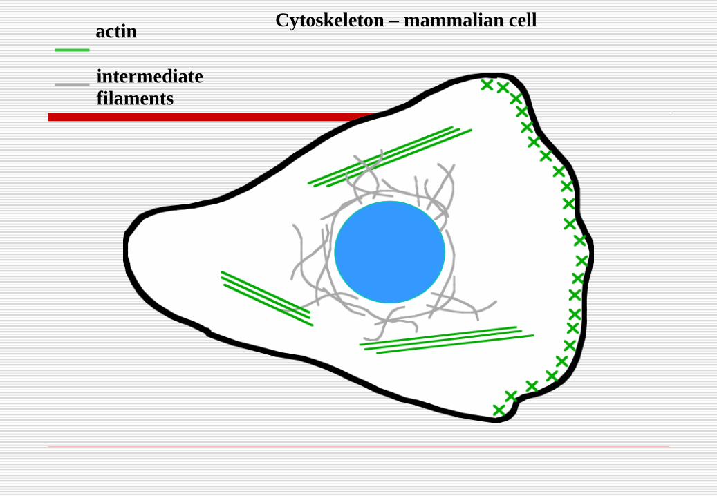
actin
intermediate
filaments
Cytoskeleton – mammalian cell
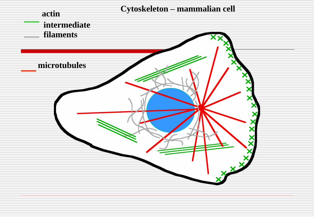
microtubules
intermediate
filaments
actinCytoskeleton – mammalian cell

Actin


Microtubules

Intermediate filaments

Compared!

MicrotubulesMicrotubules are hollow tubes of variable length and
about 25nm diameter.
They are stiff but flexible.
Microtubules carry cargoes along the length of nerve axons; in humans axons these can be more than a meter in length.
Microtubules are assembled linearly from building blocks of tubulin molecules grouped into pairs called a dimer.
Dimers are joined end-to-end by the process of polymerization to form a linear polymer called a protofilament.

MicrotubulesThe microtubule consist of thirteen (13)
protofilaments lying in parallel are formed into a circular tube with the duct running down the middle.
For microtubule assembly to take place the concentration of tubulin molecules in solution must exceed a critical level.
Each tubulin building block molecule is said to be ‘polar’ (has polarity) since it has a different molecular configuration at each end.
One end is called the ‘plus’ end; the other the ‘minus’ end. The ‘plus’ end of one molecule can only link to the ‘minus’ end of another to form a linear polymer.

D & E: Electron
micrographs of
microtubules
The Cell Cytoskeleton 224
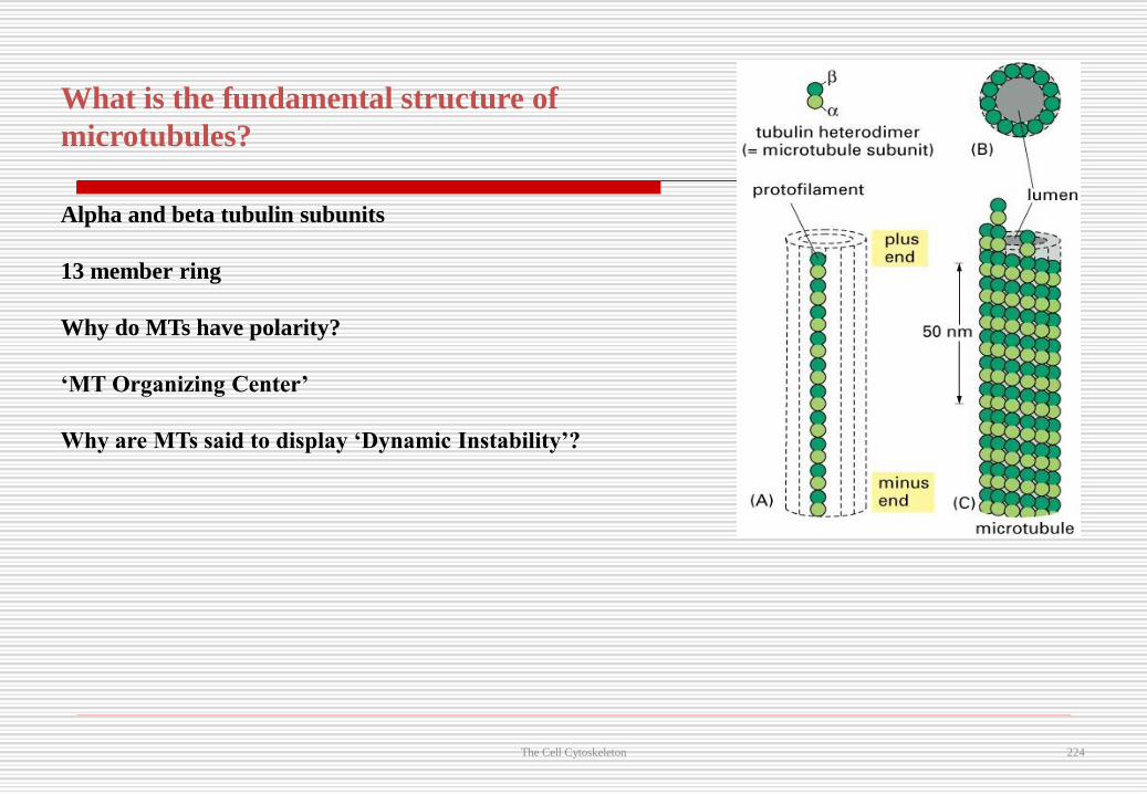
What is the fundamental structure of
microtubules?
Alpha and beta tubulin subunits
13 member ring
Why do MTs have polarity?
‘MT Organizing Center’
Why are MTs said to display ‘Dynamic Instability’?

Cytosol
• 55% of cell volume
• 75-90% water with other components
– large organic molecules (proteins,
carbohydrates & lipids) suspended by
electrical charges
– small organic molecules (simple sugars) &
ions dissolved
7/1/2017 225

Cytosol Cont’d.
– inclusions (large aggregates of one
material)
• lipid droplets
• glycogen granules
• Site of many important chemical reactions
– production of ATP, synthesis of building
blocks (amino Acids)
7/1/2017 226

Plasma Membrane
The plasma membrane is the membrane surrounding a cell that separates the cell from its external environment, consisting of a phospholipid bilayer and associated membrane lipids and proteins.
Each organelle is also surrounded by a Biomembrane and each type of organelle contains a unique complement of proteins – some embedded in its membranes other in its aqueous interior space or lumen
These proteins enable each organelle to carry out its unique cellular functions.

Biomembranes
The cytoplasm is the part of the cell outside the largest organelle, the nucleus.
The Cytosol is the aqueous part of the cytoplasm outside all of the organelles; it also contains its own distinctive proteins.
All Biomembranes form closed structures, separating the lumen on the inside from the outside and are based on a similar bilayer structure
They control the movement of molecules between the inside and outside of all cells and into and out of the organelles of eukaryotic cells.

Cellular membranes are fluid mosaics of lipids and proteins
Phospholipids are the most abundant lipid in the plasma membrane
Phospholipids are amphipathic molecules, containing hydrophobic and hydrophilic regions
The fluid mosaic model states that a membrane is a fluid structure with a “mosaic” of various proteins embedded in it

Membrane is a collage of proteins & other molecules embedded in the fluid matrix of the lipid bilayer
Extracellular fluid
Cholesterol
Cytoplasm
Glycolipid
Transmembraneproteins
Filaments ofcytoskeleton
Peripheralprotein
Glycoprotein
Phospholipids

Phospholipid bilayer
polarhydrophilicheads
nonpolarhydrophobictails
polarhydrophilicheads

Phospholipids
Fatty acid tails
hydrophobic
Phosphate group head
hydrophilic
Arranged as a bilayer
Fatty acid
Phosphate

Hydrophilic
head
Hydrophobic
tail
WATER
WATER

Phospholipids Are the Main Lipid Constituents of Most Biomembranes
•The most abundant lipid components in most membranes are phospholipids,
which are amphipathic molecules (i.e., they have a hydrophilic and
a hydrophobic part).
• In phosphoglycerides, a principal class of phospholipids, fatty acyl side chains
are esterified to two of the three hydroxyl groups in glycerol, and the third
hydroxyl group is esterified to phosphate.


Cholesterol
Cholesterol within the animal cell membrane


The Fluidity of Membranes
Phospholipids in the plasma membrane can move within the bilayer
Most of the lipids, and some proteins, drift laterally
Rarely does a molecule flip-flop transversely across the membrane

ViscousFluid
Unsaturated hydrocarbon
tails with kinks
Membrane fluidity
Saturated hydro-
carbon tails
The Fluidity of Membranes
The Fluidity of Membranes
As temperatures cool, membranes switch from a fluid state to a solid state
The temperature at which a membrane solidifies depends on the types of lipids
Membranes rich in unsaturated fatty acids are more fluid than those rich in saturated fatty acids
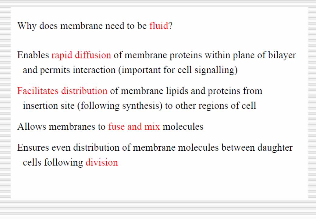
Membranes must be fluid to work properly; they are usually about as fluid as salad oil

The Fluidity of Membranes
The steroid cholesterol has different effects on membrane fluidity at different temperatures
At warm temperatures (such as 37°C), cholesterol restrains movement of phospholipids
At cool temperatures, it maintains fluidity by preventing tight packing

Lateral movement
(~107 times per second)
Flip-flop
(~ once per month)
Movement of phospholipids

Plasma Membrane Structural Components
7/1/2017 247
Plasma Membrane Components Cont’d.
• Phospholipids
• Comprises 75% of lipids
• Phospholipid bilayer = 2 parallel layers of molecules
• Each molecule is amphipathic (has both a polar & non-polar region)
– polar parts (heads) are hydrophilic and face on both surfaces of a watery environment
– nonpolar parts (tails) are hydrophobic and line up next to each other in the interior7/1/2017 248

249249
Membrane Structure Components Cont’d.
7/1/2017

250
Membrane Structure Components Cont’d.
7/1/2017

Glycolipids within the Cell Membrane
• Comprises 5% of the lipids of the cell membrane
• Carbohydrate groups form a polar head only on the side of the membrane facing the extracellular fluid
7/1/2017 251

Types of Membrane Proteins
• Integral proteins
• Extend into or completely across cell membrane
-if it extends completely across =
transmembrane protein
• All are amphipathic with hydrophobic portions
hiding among the phospholipid tails
7/1/2017 252

Types of Membrane Proteins Cont’d.
• Glycoproteins have the sugar portion facing the
extracellular fluid to form a glycocalyx
– Gives cell “uniqueness”; creates a stickiness
to hold it to other cells and also it can hold a
fluid layer creating a slippery surface
• Peripheral proteins
– Attached to either inner or outer surface of cell membrane and are easily removed from it
7/1/2017 253

7/1/2017 254
Membrane ProteinsMembrane Proteins

7/1/2017 255
Functions of Membrane Proteins (1)
255

Summary of functions of membrane proteins
(I)
• Formation of Channel
-passageway to allow specific substance to pass
through
• Transporter Proteins
-bind a specific substance, change their shape &
move it across membrane
• Receptor Proteins
-cellular recognition site -- bind to substance
7/1/2017 256

7/1/2017 257
Functions of Membrane Proteins (2)
257

Selective Permeability of Membrane
• Lipid bilayer
– permeable to non-polar, uncharged molecules --oxygen, CO2, steroids
– permeable to water which flows through gaps that form in hydrophobic core of membrane as phospholipids move about
• Transmembrane proteins act as specific channels
– for small and medium polar & charged particles
• Macromolecules unable to pass through the membrane– vesicular transport7/1/2017 258

Gradients across the Plasma Membrane
• Membrane can maintain difference in concentration of a
substance inside and outside of the membrane (concentration
gradient)
– more O2 & Na+ outside of cell membrane
– more CO2 and K+ inside of cell membrane
• Membrane can maintain a difference in charged ions between
inside & outside of membrane (electrical gradient or membrane
potential)
• Thus, substances move down their concentration gradientand towards the oppositely charged area
– E.g. ions have electrochemical gradients
7/1/2017 259

Gradients across membrane
7/1/2017 260

261
MEMBRANE STRUCTURE CONT’D
7/1/2017

262262
Cell Fractionation
The functions of different parts of the cell may beinvestigated by disrupting the cell and separating it intoits individual compartments. The process is called cellfractionation and consists of:
Disruption or homogenisation of cells, usually in asupportive iso-osmotic medium, to give a homogenate
Fractionation or separation of the various cellfragments and organelles in the homogenate forindividual study and
Analysis of chemical activity of the separatedfragments.7/1/2017

263263
CELL HOMOGENISATION
Cell lysis is the first step in cell fractionation andprotein purification and as such opens the door to amyriad of biological studies. Many techniques areavailable for the disruption of cells, including physicaland detergent-based methods.
Cell Lysis Using Traditional (Non-detergent)Methods:
Several methods are commonly used to physically lysecells, including mechanical disruption, liquidhomogenization, high frequency sound waves,freeze/thaw cycles and manual grinding.7/1/2017

264264
Mechanical Disruption
These methods have been reviewed extensively.
Mechanical methods rely on the use of rotating blades
to grind and disperse large amounts of complex tissue,
such as liver or muscle. The Waring blender and the
Polytron are commonly used for this purpose. Unlike
the Waring blender, which is similar to a standard
household blender, the Polytron draws tissue into a
long shaft containing rotating blades. The shafts vary
in size to accommodate a wide range of volumes, and
can be used with samples as small as 1 ml.7/1/2017

265265
Liquid Homogenization
Liquid-based homogenization is the most widely
used cell disruption technique for small volumesand cultured cells. Cells are lysed by forcing thecell or tissue suspension through a narrow space,thereby shearing the cell membranes. Threedifferent types of homogenizers are in commonuse. A Dounce homogenizer consists of a roundglass pestle that is manually driven into a glasstube. A Potter-Elvehjem homogenizer consistsof a manually or mechanically driven Teflonpestle shaped to fit a rounded or conical vessel.
7/1/2017

266266
Liquid Homogenization Cont’d
The number of strokes and the speed at which thestrokes are administered influences the effectiveness ofDounce and Potter homogenization methods. Bothhomogenizers can be obtained in a variety of sizes toaccommodate a range of volumes. A French pressconsists of a piston that is used to apply high pressureto a sample volume of 40 to 250 ml, forcing it througha tiny hole in the press. Only two passes are requiredfor efficient lysis due to the high pressures used withthis process. The equipment is expensive, but theFrench press is often the method of choice for breakingbacterial cells mechanically.7/1/2017

267267
Sonication
Sonication is the third class of physical disruptioncommonly used to break open cells. The methoduses pulsed, high frequency sound waves to agitateand lyse cells, bacteria, spores and finely dicedtissue. The sound waves are delivered using anapparatus with a vibrating probe that is immersed inthe liquid cell suspension. Mechanical energy fromthe probe initiates the formation of microscopicvapour bubbles that form momentarily andimplode, causing shock waves to7/1/2017

Sonication Cont’d
radiate through a sample. To prevent
excessive heating, ultrasonic treatment is
applied in multiple short bursts to a sample
immersed in an ice bath. Sonication is best
suited for volumes <100 ml.
7/1/2017 268
269269
Freeze and thaw
The freeze and thaw method is commonly used tolyse bacterial and mammalian cells. The techniqueinvolves freezing a cell suspension in a dryice/ethanol bath or freezer and then thawing thematerial at room temperature or 37°C. This methodof lysis causes cells to swell and ultimately break asice crystals form during the freezing process andthen contract during thawing. Multiple cycles arenecessary for efficient lysis, and the process can be
7/1/2017

Freeze and thaw Cont’d
quite lengthy. However, freeze and thaw has
been shown to effectively release
recombinant proteins located in the cytoplasm
of bacteria and is recommended for the lysis
of mammalian cells in some protocols.
7/1/2017 270
271271
Mortar and Pestle
Manual grinding is the most common method
used to disrupt plant cells. Tissue is frozen in
liquid nitrogen and then crushed using a
mortar and pestle. Because of the tensile
strength of the cellulose and other
polysaccharides comprising the cell wall, this
method is the fastest and most efficient way
to access plant proteins and DNA.7/1/2017
272272
Additives/Facilitators
Cells can be treated with various agents to aid thedisruption process. Lysis can be promoted bysuspending cells in a hypotonic buffer, which causethem to swell and burst more readily under physicalshearing. 200 µg/ml can be used to digest thepolysaccharide component of yeast and bacterialcell walls. Alternatively, processing can beexpedited by treating cells with glass beads in orderto facilitate the crushing of cell walls. Thistreatment is commonly used with yeast cells.7/1/2017

273273
Additives/Facilitators Cont’d
Viscosity of a sample typically increases during lysis
due to the release of nucleic acid material. DNase can
be added to samples (25-50 µg/ml) along with RNase
(50 µg/ml) to reduce this problem.
Nuclease treatment is not required for sonicated
material since sonication shears chromosomes. Finally,
proteolysis can be a problem whenever cells are
manipulated; therefore, protease inhibitors should be
added to all samples undergoing lysis.
7/1/2017

274274
Fractionation or Centrifugation
Centrifugation is the process of isolating
components of a cell. There are two common
centrifugation techniques for separating
bacteria components. The two methods are
differential centrifugation and bouyant
density centrifugation.
7/1/2017

Fractionation or Centrifugation Cont’d
Once the homogenate has been prepared it is then
necessary to fractionate or separate the various
intracellular components from each other. The most
widely used method for doing this is centrifugation.
When a centrifugal force is applied to a suspension
of cellular particles the rate of sedimentation of any
given particle will depend on its size, density and
shape.
7/1/2017 275

276276
Differential Centrifugation
Differential centrifugation is the process
where a homogenate undergoes repeated
centrifugations at increasing centrifugal force.
Centrifugation is the use of increased gravity
to quicken the sedimentation of substances to
the bottom. The tool used here is the
centrifuge that spins at various speeds.
7/1/2017

277277
Differential Centrifugation Cont’d
The centrifuge separates the cell's parts into pellets
and supernatant. The pellets are the large cell
structures that are settled at the test tube's bottom.
The supernatant are smaller parts of the cell
suspending in liquid, the supernatant is decanted
and undergoes another centrifugation. The process
is repeated at increased speed with each trial to
collect successively smaller parts of a cell in
pellets.7/1/2017
Differential Centrifugation Cont’d
Separation is achieved primarily based on the
size of the particles in differential
centrifugation. This type of separation is
commonly used in simple pelleting and in
obtaining partially-pure preparation of
subcellular organelles and macromolecules.
7/1/2017 278

279279
Differential centrifugation Cont’d.
For the study of subcellular organelles, tissuesor cells are first disrupted to release theirinternal contents. This crude disrupted cellmixture is referred to as a homogenate.During centrifugation of a cell homogenate,larger particles sediment faster than smallerones and this provides the basis for obtainingcrude organelle fractions by differentialcentrifugation.
7/1/2017

280280
Differential centrifugation Cont’d.
A cell homogenate can be centrifuged at a series ofprogressively higher g-forces and times to generatepellets of partially-purified organelles.
When a cell homogenate is centrifuged at 1000 x gfor 10 minutes, unbroken cells and heavy nucleipellets move to the bottom of the tube. Thesupernatant can be further centrifuged at 10,000 x gfor 20 minutes to pellet subcellular organelles ofintermediate velocities such as mitochondria,lysosomes, and microbodies.
7/1/2017

281281
Differential centrifugation Cont’d.
Some of these sedimenting organelles can beobtained in partial purity and are typicallycontaminated with other particles. Repeatedwashing of the pellets by re-suspending inisotonic solvents and re-pelleting may resultin removal of contaminants that are smaller insize (Figure 1 on slide # 203). Obtainingpartially-purified organelles by differentialcentrifugation serves as the preliminary step forfurther purification using other types of centrifugalseparation (density gradient separation).7/1/2017

282282
Differential centrifugation Cont’d.
7/1/2017

283283
Differential Centrifugation
7/1/2017

284284
Density gradient centrifugation or Buoyant
density centrifugation
Density gradient centrifugation is thepreferred method to purify subcellularorganelles and macromolecules. Densitygradients can be generated by placing layerafter layer of gradient media (Table 2 on slide# 214) such as sucrose in a tube with theheaviest layer at the bottom and the lightest atthe top in either a discontinuous or continuousmode.7/1/2017

285285
Density gradient centrifugation or Buoyant
density centrifugation Cont’d
The buoyant density centrifugation involves viruseswith densities of 1.1-1.2 g/cm and a sucrosegradient. The cell suspension is added to the top ofthe sucrose gradient. In this centrifugation thedensest components move fastest down the tubeand stops at the sucrose density equal to its own.The sucrose gradient bands at the bottom containcell components with high buoyant densities andthe components at the top have low buoyantdensities.7/1/2017

286286
Buoyant density centrifugation
7/1/2017
287287
Density gradient centrifugation Cont’d.
The cell fraction to be separated is placed on topof the layer and centrifuged. Density gradientcentrifugation can be classified into twocategories. (a) Rate-zonal (size) separation. (b)Isopycnic (density) separation.
(a) Rate zonal (size) separation
Rate-zonal separation takes advantage ofparticle size and mass instead of particle densityfor sedimentation.
7/1/2017

288288
Rate zonal (size) separation Cont’d.
Examples of common applications includeseparation of cellular organelles such asendosomes or separation of proteins, such asantibodies. For instance, antibody classes allhave very similar densities, but different masses.Thus, separation based on mass will separate thedifferent classes, whereas separation based ondensity will not be able to resolve these antibodyclasses (Figure 2, slide # 210).
7/1/2017

289289
Rate zonal (size) separation
Cont’d.
7/1/2017

290290
(b) Isopycnic separation
In this type of separation, a particle of a
particular density will sink during
centrifugation until a position is reached
where the density of the surrounding
solution is exactly the same as the density
of the particle. Once this quasi-equilibrium
is reached, the length of centrifugation does
7/1/2017
291291
Isopycnic separation Cont’d
not have any influence on the migration of the
particle. A common example for this method
is separation of nucleic acids in a CsCl
gradient (Figure 3, slide # 213).
7/1/2017

292292
Isopycnic separation Cont’d.
7/1/2017
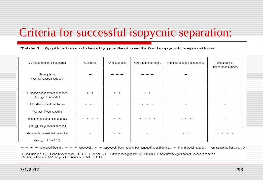
293293
Criteria for successful isopycnic separation:
7/1/2017
294294
Analytical Ultra-Centrifuge
Analytical ultracentrifugation is a classicalmethod of biochemistry and molecularbiology. Because analyticalultracentrifugation relies on the principalproperty of mass and the fundamental laws ofgravitation, it has broad applications. It is aprimary technique requiring no standards forcomparison.
7/1/2017

295295
Analytical Ultra-Centrifuge Cont’d.
Sedimentation can be used to analyze thesolution behavior of nearly any molecule overa wide range of solute concentrations and in awide variety of solvents.
It is a further refinement of the preparativecentrifuges. It can be used to separate andcharacterize very small molecules andmacromolecules such as ribosomes andproteins.
7/1/2017

296296
Analytical Ultra-Centrifuge Cont’d.
The specimen is mixed with a sucrose or caesium
chloride medium in a quartz centrifuge cell and
centrifuged at very high speeds to generate forces
of up to 500,000g. Such extreme forces causes
small particles and macromolecules to sediment
from the solution leaving pure solvent at the top
of the cell and forming a sharp boundary between
the pure solvent and solvent containing specimen.
7/1/2017
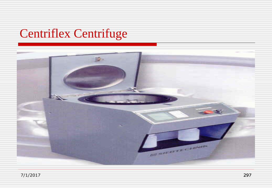
297297
Centriflex Centrifuge
7/1/2017

298298
Gilson GmCLab Mini Centrifuge
7/1/2017

299299
Analysis
Once cell fractions have been isolated, they
can then be examined by standard
biochemical procedures such as
spectrophotometry, autoradiography,
chromatography etc. in an attempt to
characterize their biochemical activities.
7/1/2017

300300
Spectrophotometry
Principles of Spectrophotometry:
A spectrophotometer consists of twoinstruments, namely a spectrometer forproducing light of any selected colour(wavelength), and a photometer for measuringthe intensity of light. The instruments are soarranged that liquid in a cuvette can be placedbetween the spectrometer beam and thephotometer.7/1/2017

301301
Spectrophotometry Cont’d
The amount of light passing through the tubeis measured by the photometer. Thephotometer delivers a voltage signal to adisplay device, normally a galvanometer. Thesignal changes as the amount of lightabsorbed by the liquid changes.
If development of colour is linked to theconcentration of a substance in solution
7/1/2017

302302
Spectrophotometry Cont’d
then the concentration can be measured by
determining the extent of absorption of light at the
appropriate wavelength.
For example haemoglobin appears red because the
hemoglobin absorbs blue and green light rays much
more effectively than red. The degree of
absorbance of blue or green light is proportional to
the concentration of haemoglobin in solution.
7/1/2017

303303
Spectrophotometer
7/1/2017
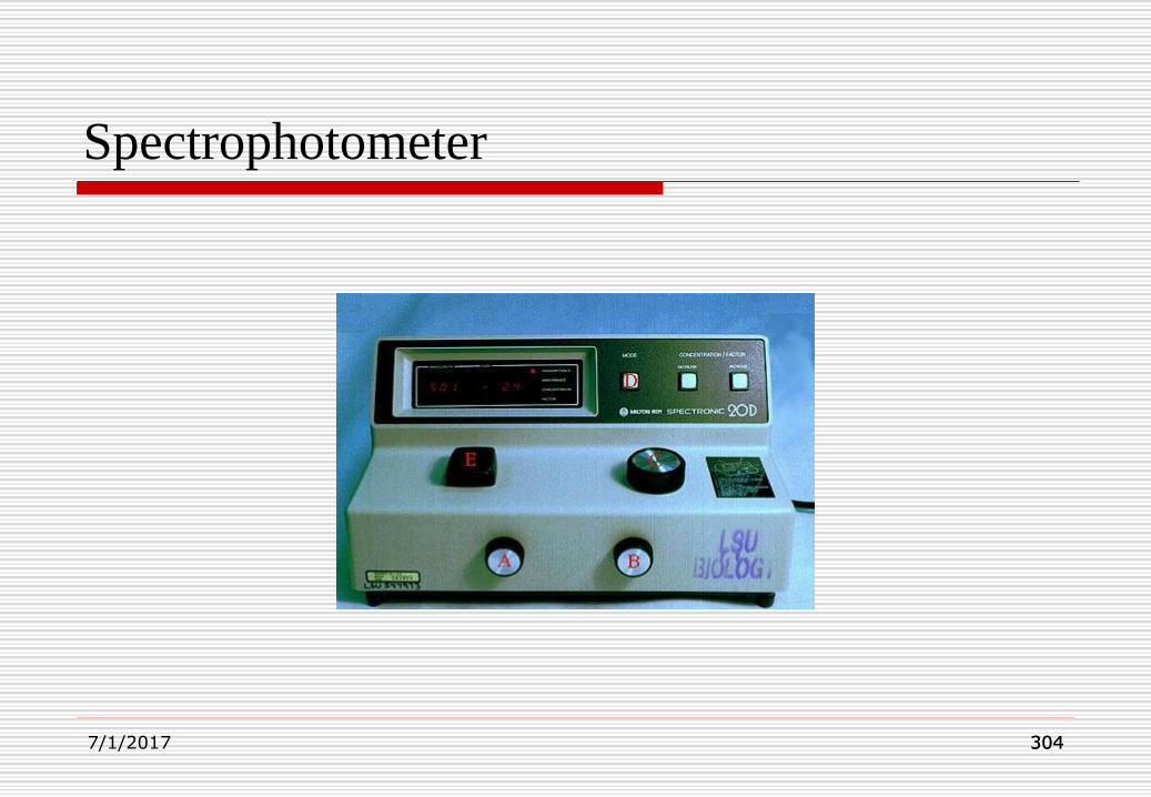
304304
Spectrophotometer
7/1/2017
305305
Spectrophotometry Cont’d
Water quality photometers and colorimeters
are ion-specific computer-interfaced probes
designed to determine the concentration of a
solution from its colour intensity.
7/1/2017
306306
Autoradiography
This technique, which is now widely used incell biology, involves the visual localizationof radioactive substances in cells and tissues.Tissues are incubated in solutions containingradioactive isotopes which becomeincorporated into the cells. The tissue issectioned and the sites of incorporation aretraced by placing a photographic emulsionover the section.7/1/2017

307307
Autoradiography Cont’d.
The radioactive particles emitted from theisotope sensitizer react with silver halidecrystals in the emulsion and these areconverted to metallic silver by normalphotographic development. Theunsensitized crystals are washed awayduring the subsequent fixation and the site
7/1/2017

308308
Autoradiography Cont’d.
of radioactive incorporation is then seen as
a deposit of silver grains on the sections.
The most widely used isotopes in
biological research are tritium (3H), 14C,32P, 45Ca, 35S etc.
These can be used to label a wide range of
molecules which, when incubated with the
7/1/2017
309309
Autoradiography Cont’d.
appropriate tissues, are incorporated into
various cellular constituents.
Sites of protein synthesis in tissues can be
detected by immersing the tissue in a
radioactive precursor to proteins i.e. amino
acids.
In medicine, barium meal is used to test for
ulcer in the GIT, which is an isotope of barium.
7/1/2017

310310
Chromatography
Chromatography is a technique used toseparate components of mixtures. It is basedon the partitioning of compounds between astationary phase and a moving phase. Inpartition chromatography, the stationaryphase is a liquid whilst in adsorptionchromatography the stationary phase is asolid.
7/1/2017

311311
Chromatography Cont’d
Partition chromatography depends primarily
on the relative solubility of substances in two
or more immiscible solvents. When a solute
is added to a mixture of equal volumes of two
immiscible solvents, it may dissolve entirely
in one or other of the solvents. If the solute
dissolves in both solvents, it may not dissolve
to the same extent in each solvent.7/1/2017

312312
Chromatography Cont’d
7/1/2017
313313
Chromatography Cont’d
partition coefficient to separate components
of mixtures.
In contrast, adsorption chromatography
relies mainly on electrostatic interaction
between solutes and the stationary phase.
Some adsorption also occurs in partition
chromatography.
7/1/2017
314314
Paper chromatography
Paper chromatography is an analytical
technique for separating and identifying
mixtures that are coloured or can be made
coloured, especially pigments. This method
has been largely replaced by thin layer
chromatography, however it is still a powerful
research tool.
7/1/2017

315315
Paper chromatography Cont’d.
Technique:
A small concentrated spot of solution thatcontains the sample is applied to a strip ofchromatography paper about 2 cm away fromthe base of the plate, usually using a capillarytube for maximum precision. This sample isabsorbed onto the paper and may forminteractions with it.
7/1/2017

316316
Paper chromatography Cont’d.
Any substance that reacts or bonds with
the paper cannot be measured using this
technique. The paper is then dipped in to a
suitable solvent, such as ethanol or water,
or even acetone taking care that the spot is
above the surface of the solvent, and
placed in a sealed container.
7/1/2017

317317
Paper chromatography Cont’d.
The solvent moves up the paper by capillary
action, which occurs as a result of the attraction
of the solvent molecules to the paper and to one
another. As the solvent rises through the paper it
meets and dissolves the sample mixture, which
will then travel up the paper with the solvent.
Different compounds in the sample mixture
travel at different rates due to differences in
7/1/2017

318318
Paper chromatography Cont’d.
solubility in the solvent, and due to
differences in their attraction to the fibre or
cellulose in the paper. Paper
chromatography takes anywhere from
several minutes to several hours to
complete. In some cases, paper
chromatography does not separate
pigments completely; this occurs7/1/2017

319319
Paper chromatography Cont’d.
when two substances appear to have thesame values in a particular solvent. In thesecases, two-way chromatography is used toseparate the multiple-pigment spots. Thechromatogram is turned by ninety degrees90o, and placed in a different solvent in thesame way as before; some spots separate inthe presence of more than one pigment.
7/1/2017

320320
Paper chromatography Cont’d.
As before, the value is calculated, and the two
pigments are identified. The Rf value
(retention factor) is the distance travelled by a
particular component from the origin (where
the sample was originally spotted) as a ratio
to the distance travelled by the solvent front
from the origin. Rf values for each substance
will be unique, and can be7/1/2017

321321
Paper chromatography Cont’d.
used to identify components. A particular
component will have the same Rf value if it
is separated under identical conditions.
7/1/2017

322322
Paper chromatography
7/1/2017

323323
CHROMATOGRAM
The figure on the next slide shows a simple separationby chromatography. A continuous flow of solventcarries a solution of solutes A and B down a column.(a) As the solvent carries the two solutes down thecolumn, we begin to see some separation of thesolution. (b) At some later point in time, it can be seenthat solute B is moving at a much faster rate than A. (c)In (d), solute B emerges first, while solute A finallyemerges in (e). Thus, solute A has a greater affinity forthe stationary phase than solute B. By varying the pHof the solvent or temperature of the column, the outputof the column can be significantly altered, such as thetiming of when individual species emerge.7/1/2017

324324
CHROMATOGRAM
7/1/2017

325
Interpreting the Data
The Rf value for each spot should be
calculated. Rf stands for "ratio of fronts" and
is characteristic for any given compound on
the same stationary phase using the same
mobile phase for development of the plates.
7/1/2017

Interpreting the Data Cont’d
7/1/2017 326

327
Interpreting the Data Cont’d.
7/1/2017

328
Calculate the Rf values for the spots on the
TLC slide below.
7/1/2017

329
Solution:
Use the equation:
Measure the distances from the starting
point to the solvent front and from the
starting point to the center of each spot, and
calculate the Rf values:
7/1/2017
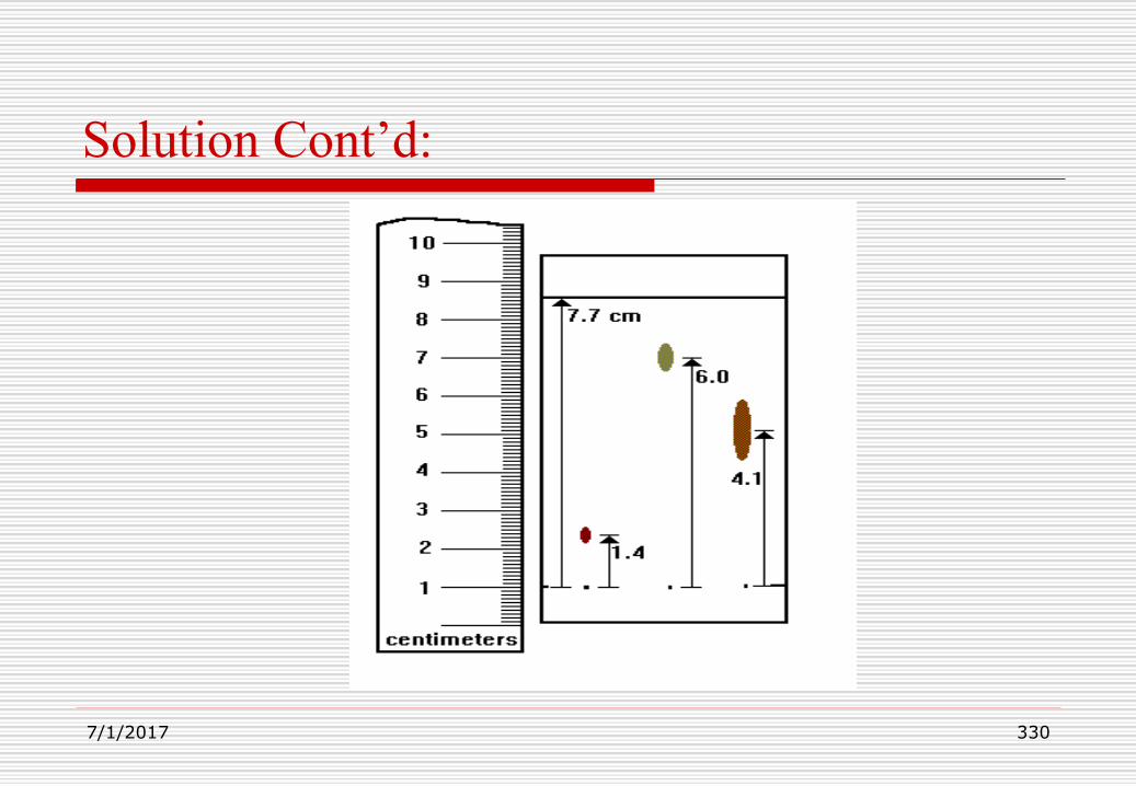
330
Solution Cont’d:
7/1/2017
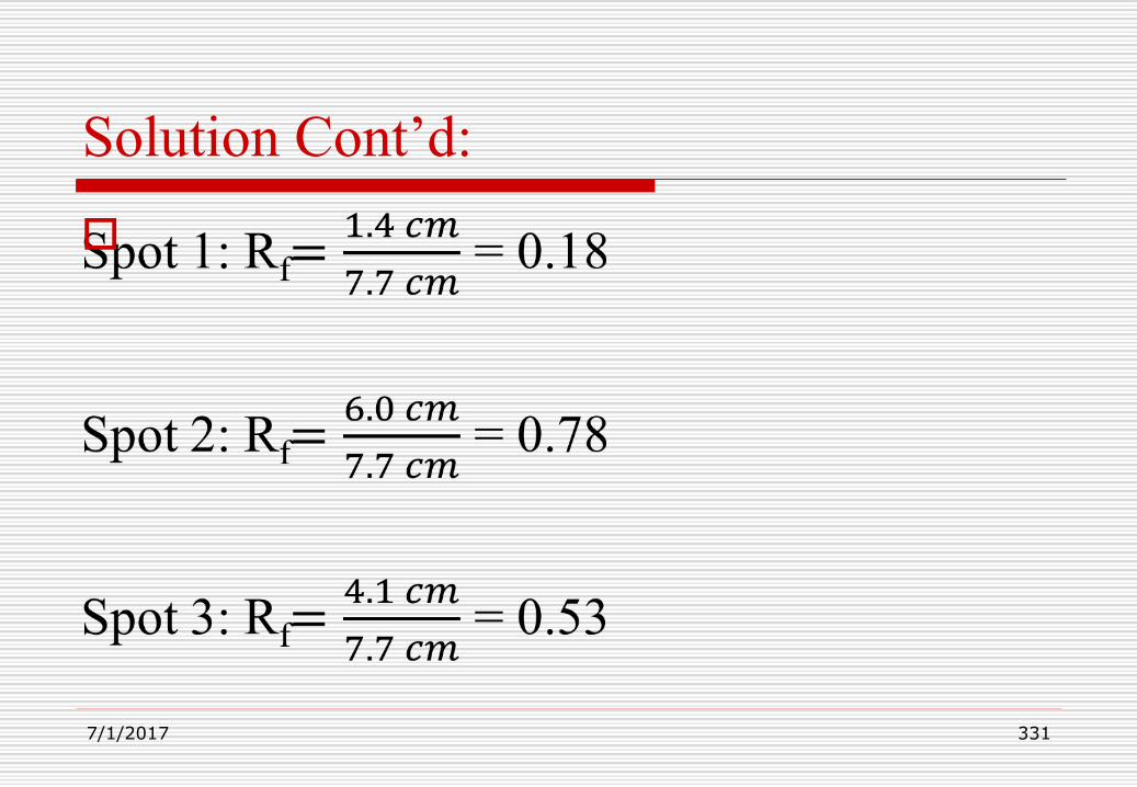
Solution Cont’d:
7/1/2017 331