Lecture Topics Protein Synthesis Mitosis Epithelial Tissue.
200
Lecture Topics Lecture Topics • Protein Synthesis • Mitosis • Epithelial Tissue
Transcript of Lecture Topics Protein Synthesis Mitosis Epithelial Tissue.

Lecture TopicsLecture Topics
• Protein Synthesis
• Mitosis
• Epithelial Tissue

NucleusNucleus
• Most cells have one nucleus.

NucleusNucleus
• Exceptions:
• Skeletal muscle cells are multinucleated.
• Some cardiac muscle cells are binucleated.
• Mature rbc lack a nucleus.

NucleusNucleus
• Nuclear envelope – a double membrane that surrounds the nucleus
NucleusNucleus
• Both layers of the membrane are lipid bilayers

NucleusNucleus
• Contains a dark spherical body called a nucleoli where rRNA are made.
NucleusNucleus
• The nucleoli assembles rRNA and proteins into ribosomes.

NucleusNucleus
• Ribosomes are exported into the cytosol and play a major role in protein synthesis (translation).

NucleusNucleus
• Contains chromsomes. Humans have 46.

NucleusNucleus
• 23 pairs of chromosomes
• 23 from mother
• 23 from father

NucleusNucleus
• All chromsomes are referred to as autosomes except one pair. In other words 22 of the pairs are autosomes.

NucleusNucleus
• The last or 23rd pair are referred to as the sex chromsomes.

NucleusNucleus
• The two chromosomes of each pair are called homologous chromosomes

NucleusNucleus
• Each Chromosome is a long molecule of DNA.
NucleusNucleus
• Each Chromosomes contain thousands of genes arranged in a single file.

NucleusNucleus
• Each gene is a segment of DNA

NucleusNucleus
• Each gene represents a protein

NucleusNucleus
• The DNA molecule resembles a spiral ladder called a double Helix.


NucleusNucleus
• Monomers of DNA are called nucleotides.

NucleusNucleus
• Each monomer or unit of DNA contains a 1. pentose sugar
2. phosphate group,
3. nitrogenous base.


NucleusNucleus
• There are four different nitrogenous bases;
1. Adenine
2. Thymine
3. Cytosine
4. Guanine

NucleusNucleus
• Cytosine always pairs with Guanine

NucleusNucleus
• Thymine always pairs with Adenine

NucleusNucleus
• These bases are held together by hydrogen bonds.


NucleusNucleus
DNA Template DNA Complementary
A T
T A
G C
C G
A T
T A

Protein SynthesisProtein Synthesis
• Two major Parts
• 1. Transcription (takes place in nucleus)
• 2. Translation ( takes place in ribosomes in the cytosol)
Protein SynthesisProtein Synthesis
• Basic order:
DNA → mRNA → Protein

Protein Synthesis: TranscriptionProtein Synthesis: Transcription
• DNA molecules have a template strand and a complementary strand.


TranscriptionTranscription
• In transcription an RNA strand is made from the DNA template strand.

TranscriptionTranscription
• There are three different types of RNA that are transcribed; mRNA, rRNA, tRNA

TranscriptionTranscription
• RNA molecules are single stranded unlike DNA molecules

Protein Synthesis: TranscriptionProtein Synthesis: Transcription
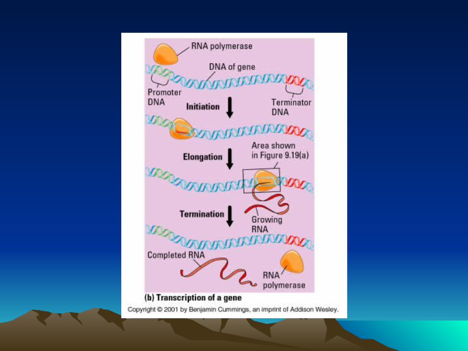
• At the beginning of a gene there is a DNA sequence called a promoter.

TranscriptionTranscription
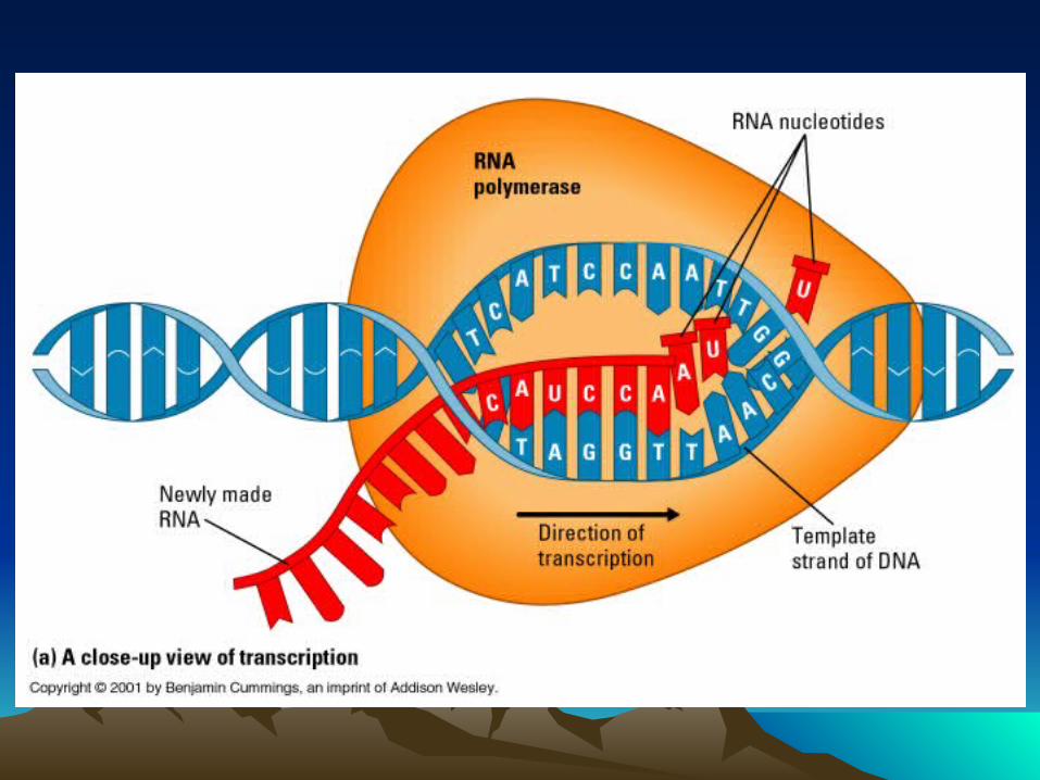
• This promoter tells RNA polymerase where to start transcription. RNA polymerase catalyzes transcription

TranscriptionTranscription
• As the DNA molecule unzips, bases pair with the template strand of the DNA molecule and a complementary RNA strand is formed.


Protein Synthesis: TranscriptionProtein Synthesis: Transcription
• RNA have adenine, guanine, and cytosine bases, but do not have thymine. Instead they have uracil.

TranscriptionTranscription
• Cytosine, Guanine, and Thymine in the DNA template pair with Guanine, Cytosine, and Adenine in the RNA strand.

TranscriptionTranscription
• Adenine in the DNA template pairs with uracil not thymine in RNA

Protein Synthesis: TranscriptionProtein Synthesis: Transcription
DNA Template RNA Strand
A U
T A
G C
C G
A U
T A


Protein Synthesis: NucleusProtein Synthesis: Nucleus
DNA Template DNA Complementary
A T
T A
G C
C G
A T
T A

Protein Synthesis: TranscriptionProtein Synthesis: Transcription
• The terminator is a nucleotide sequence that specifies the end of the gene.

TranscriptionTranscription
• RNA polymerase detaches itself from the transcribed RNA molecule and DNA strand.

TranscriptionTranscription
• The transcribed mRNA molecule is referred to as pre mRNA.

TranscriptionTranscription
• A DNA segment is a gene

TranscriptionTranscription
• Genes codes for proteins
• DNA segment or Gene → RNA → Protein
TranscriptionTranscription
• Not all parts of a gene code for a protein.

TranscriptionTranscription
• A gene can be divided into introns and exons.

TranscriptionTranscription
• Introns are the parts that don’t code for a protein.

TranscriptionTranscription
• Exons are the parts that do code for a protein.

TranscriptionTranscription
• Pre mRNA contains exons and introns


Protein Synthesis: TranscriptionProtein Synthesis: Transcription
• Introns are removed from the pre mRNA and the exons are spliced together by small nuclear ribonucleoproteins (snRNPs).


TranscriptionTranscription
• The end product is a mRNA molecule that exits the nucleus through a nuclear pore.


TranscriptionTranscription
• The mRNA travels through the cytosol until it reaches a ribosome where translation takes place.

QuestionQuestion
• Why do introns exist if it is useless informaiton?

QuestionQuestion
• If there are only 35,000 to 45,000 genes, why are there actually 500,000 to 1 million genes?

Protein Synthesis: TranslationProtein Synthesis: Translation
• RNA stores genetic information in sets of three nucleotides called codons.

Protein Synthesis: TranslationProtein Synthesis: Translation
• Each codon specifies a particular amino acid.

TranslationTranslation
• There are 64 codons and only 20 amino acids.

TranslationTranslation
• This means there are more than one codon for each amino acid. In other words, several codons specify for the same amino acid.

QuestionQuestion
Why this redundancy?


Six Steps in TranslationSix Steps in Translation
1. The mRNA molecule binds to the small ribosomal subunit at the mRNA binding site.

TranslationTranslation
1. Then the initiator tRNA that contains the anticodon attaches to the mRNA codon.

TranslationTranslation
1. The tRNA contains the amino acid that corresponds to the codon.


TranslationTranslation
1. The first codon of an mRNA strand is always AUG, therefore methionine is always the first amino acid in a protein.


Translation Steps Cont.Translation Steps Cont.
2. The large ribosomal subunit attaches to the small ribosomal subunit-mRNA complex, creating a functional ribosome.

TranslationTranslation
2. The initiator tRNA, with the amino acid methionine, are now in the P site of the ribosome.


TranslationTranslation
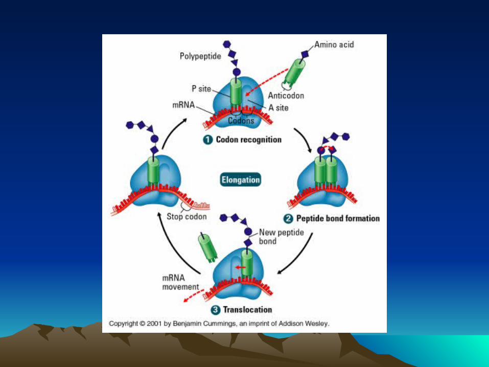
3. Now another tRNA with another amino acid attach to the second mRNA codon at the A site of the ribosome.


Translation Steps Cont.Translation Steps Cont.
4. A component of the large ribosomal subunit catalyzes the formation of a peptide bond between methionine in the P site and the amino acid at the A site.

TranslationTranslation
4. Then methionine detaches itself from the tRNA at the P site.


TranslationTranslation
5. The tRNA at the P site leaves.


TranslationTranslation
5. The ribosome shifts the mRNA strand by one codon. Now the tRNA that was in the A site is now in the P site.

TranslationTranslation
5. This allows another tRNA with an amino acid to attach to the codon at the A site. Steps 3 through 5 occur repeatedly.


Translation Steps Cont.Translation Steps Cont.
6. Protein synthesis stops when the ribosome reaches a stop codon at the A site.


TranslationTranslation
6. When a ribosome reaches a termination codon on the mRNA, the A site of the ribosome accepts a protein called a release factor instead of tRNA


TranslationTranslation
6. Release factor hydrolyzes the bond between the tRNA in the P site and last a.a. of the protein.


TranslationTranslation
6. Then the completed protein detaches from the final tRNA.


TranslationTranslation
6. After the tRNA leaves the P site the ribosome disassociates into small and large subunits.


DNA VirusesDNA Viruses
• In a DNA virus the, the virus uses the host cells machinery to replicate itself.

DNA VirusesDNA Viruses
• The virus is made up of a protein capsid with viral DNA inside.

DNA VirusesDNA Viruses
• It uses the host cells machinery and duplicates the DNA and makes new protein capsids via protein synthesis.

RNA VirusesRNA Viruses
• In RNA retroviruses like HIV, it is a little different from DNA viruses but same concept. It uses the host cell’s machinery to replicate itself.

RNA VirusesRNA Viruses
• The virus is made up of viral RNA surrounded by a protein capsid.

RNA VirusesRNA Viruses
• It forms a complementary DNA strand via reverse transcriptase.

RNA VirusesRNA Viruses
• After the DNA forms double strands, it then replicates more viral RNA via transcription

RNA VirusesRNA Viruses
• It also makes more capsid proteins via translation.

10 minute Break10 minute Break

Cell DivisionCell Division
• Interphase
• Mitosis
• Cytokinesis

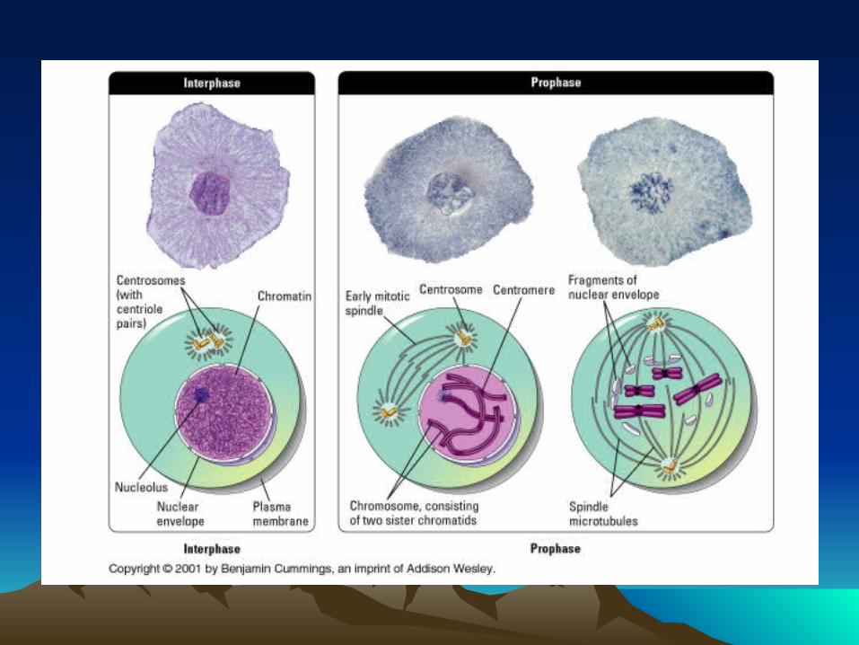
InterphaseInterphase
• Our cells are in interphase 90% of the time.

InterphaseInterphase
• During this time the DNA, protein, and RNA are referred to as chromatin.

InterphaseInterphase
• The chromatin looks like a diffuse granular mass.

InterphaseInterphase
• There are three phases of interphase.

3 Stages of Interphase3 Stages of Interphase
1. G1 phase
During this phase it duplicates most of its organelles.

3 Stages of Interphase3 Stages of Interphase
2. S phase
Chromosomes duplicate during this stage. The duplicated chromosomes are attached at the centromere are referred to as chromatids.
3 Stages Cont.3 Stages Cont.
3. G2
Cell growth continues and enzymes and other proteins are synthesized.
* Some cells for example remain in the G1 stage forever for example nerve cells. They are said to be in the G0 stage

4 Stages of Mitosis4 Stages of Mitosis
• Prophase
• Metaphase
• Anaphase
• Telophase

ProphaseProphase
• Chromatin fibers condense and are now visible underneath the microscope as individual chromosomes.

ProphaseProphase
• The chromosomes have been replicated and are attached to its double or sister chromatid by the centromere.

ProphaseProphase
• Later in prophase mitotic spindle radiating from the centrioles attach to the kinetochore ( a protein complex outside the centromere).


Prophase Cont.Prophase Cont.
• Nucleoli disappears
• Nuclear envelope disappears as well


MetaphaseMetaphase
• The mitotic spindle aligns the centromeres of the chromatid pairs at the metaphase plate.


AnaphaseAnaphase
• The centromeres split separting the two members of each chromatid pair, which move toward opposite poles of the cell.

AnaphaseAnaphase
• Once separated, the chromatids are termed chromosomes.

AnaphaseAnaphase
• The chromosomes appear V shaped because the centromeres lead the way as they are being pulled by the mitotic spindle.


TelophaseTelophase
• Most events are opposite of prophase

TelophaseTelophase
• Chromosome revert back to a chromatin like appearance.
TelophaseTelophase
• Nuclear envelope develops around each set of chromosomes.

TelophaseTelophase
• Nucleoli reappears

TelophaseTelophase
• Mitotic spindle disappears

TelophaseTelophase
• Cleavage furrow appears


CyokinesisCyokinesis
• The cytoplasm, organelles and the two nuclei are divided into two daughter cells.
TissuesTissues
• Epithelial Tissue
• Connective Tissue
• Muscle Tissue
• Nervous Tissue

Epithelial TissueEpithelial Tissue
• Covers the external body surface (epidermis), lines cavities and tubules, and generally marks off our insides from our outsides

Epithelial TissueEpithelial Tissue
• Contain cell junctions

Epithelial TissueEpithelial Tissue
• Avascular

Epithelial TissueEpithelial Tissue
• Contains nerve supply

Epithelial TissueEpithelial Tissue
• High rate of mitotic division
Cell junctionsCell junctions
• They are contact points between the cell membranes of tissue cells.
• Five types:Tight JunctionsAdherens JunctionsDesmosomesHemidesmosomesGap Junctions

Tight JunctionsTight Junctions
• This prevents the passage of substances between cells.

Adherens JunctionsAdherens Junctions
• Helps epithelial surfaces resist separation

DesmosomesDesmosomes
• Contribute to stability
• Prevent epidermal cells from separating under tension and cardiac muscle cells from pulling apart during contraction.

HemidesmosomesHemidesmosomes
• Anchor cells

Gap JunctionsGap Junctions
• Allows cells in tissues to communicate

Epithelial Cell SurfacesEpithelial Cell Surfaces
1. Apical Surface – Faces the body surface, a body cavity, the lumen, or a tubular duct
Epithelial Cell SurfacesEpithelial Cell Surfaces
2. Lateral surfaces - Face adjacent cells. Contain cell junctions except hemidesmosomes

Epithelial Cell SurfacesEpithelial Cell Surfaces
3. Basal surface - Opposite of apical surface. Attaches to the basal lamina of the basement membrane, an extracellular layer

Types of Epithelial TissueTypes of Epithelial Tissue
1. Simple Squamous
2. Simple Cuboidal
3. Simple Columnar
4. Ciliated Simple Columnar
5. Stratified Squamous
6. Stratified Cuboidal
7. Stratified Columnar

Types Cont.Types Cont.
8. Transitional
9. Pseudostratified columnar

Simple SquamousSimple Squamous
• Single layer of cells
• Scale like

Simple SquamousSimple Squamous
• Functions in filtration, diffusion, osmosis, and secretion

Simple SquamousSimple Squamous
• Lines heart, blood vessels, air sacs, glomerular capsule of kidneys, serous membranes

Simple CuboidalSimple Cuboidal
• Single layer
• Cube Shaped
Simple CuboidalSimple Cuboidal
• Function in secretion and absorption
Simple CuboidalSimple Cuboidal
• Covers surface of ovary, lines kidney tubules and small ducts of glands (thyroid and pancreas)


Simple Columnar epitheliumSimple Columnar epithelium
• Single layer
• Rectangular shaped
• Some contain goblet cells


Simple Columnar epitheliumSimple Columnar epithelium
• Function in secretion and absorption

Simple Columnar epitheliumSimple Columnar epithelium
• Lines G.I. tract from stomach to the anus, gallbladder


Ciliated Simple ColumnarCiliated Simple Columnar
• Single layer
• Columnar shaped
• Some contains goblet cells
• Ciliated

Ciliated Simple ColumnarCiliated Simple Columnar
• Function in moving mucus and other substances

Ciliated Simple ColumnarCiliated Simple Columnar
• Uterine tubes, uterus, central canal of spinal cord
Stratified SquamousStratified Squamous
• Several Layers
• Scale like shaped
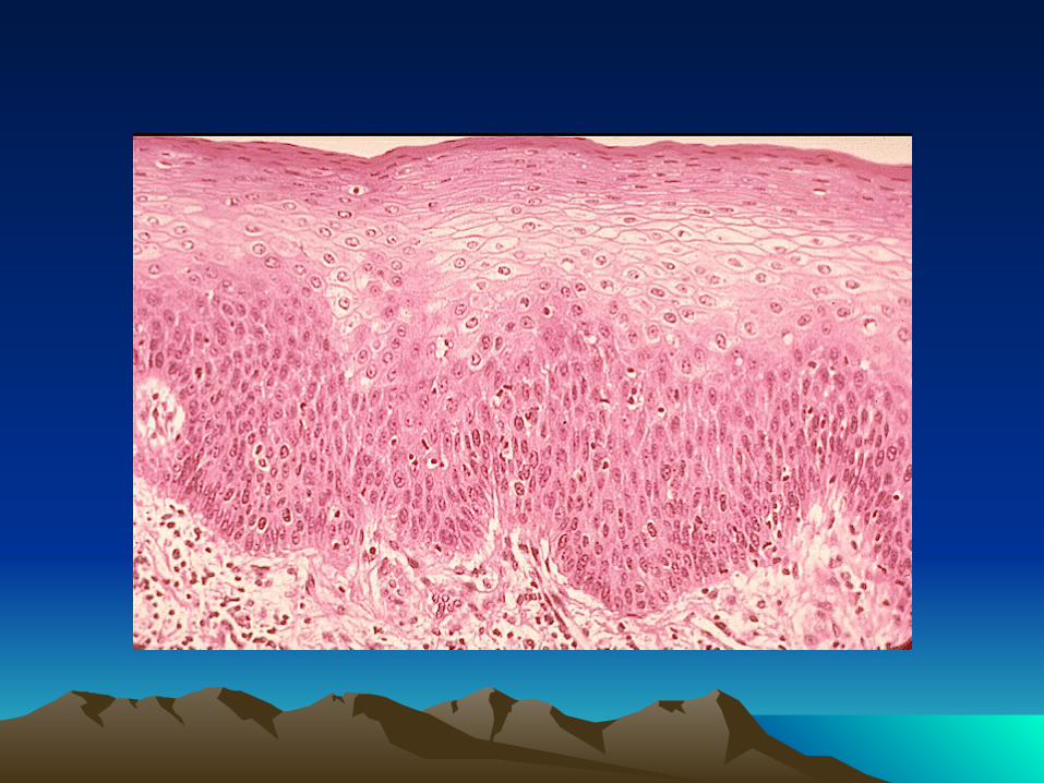


Stratified SquamousStratified Squamous
• Function in protection

Stratified SquamousStratified Squamous
• Superficial layer of the skin, lining of the mouth, esophagus, epiglottis, vagina, and tongue

Stratified CuboidalStratified Cuboidal
• Several layers
• Square or cube shaped in apical layer

Stratified CuboidalStratified Cuboidal
• Function in protection and some secretion and absorption

Stratified CuboidalStratified Cuboidal
• Ducts of sweat glands, esophageal glands, and male urethra


Stratified ColumnarStratified Columnar
• Several Layers
• Rectangular shaped in apical layer

Stratified ColumnarStratified Columnar
• Function in protection and secretion
Stratified ColumnarStratified Columnar
• Part of urethra, large excretory ducts of some glands (esophageal)

Transitional EpitheliumTransitional Epithelium
• Several layer
• Scale to cube shaped

Transitional EpitheliumTransitional Epithelium
• Function in permitting distention

Transitional EpitheliumTransitional Epithelium
• Lines urinary bladders and portions of ureters and urethra

Pseudostratified ColumnarPseudostratified Columnar
• Not stratified

Pseudostratified ColumnarPseudostratified Columnar
• ciliated
Pseudostratified ColumnarPseudostratified Columnar
• Nucleus of cells are at different levels, all cells are attached to a basement
membrane, but not all reach the surface

Pseudostratified ColumnarPseudostratified Columnar
• Function in secretion and movement of mucus

Pseudostratified ColumnarPseudostratified Columnar
• Trachea, epididymis, and part of male urethra


ENDEND