Lecture Content - American Academy of Osteopathy
31
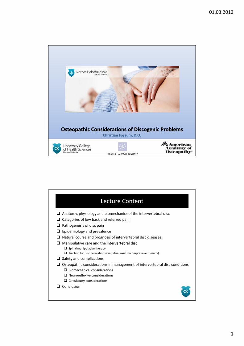
01.03.2012 1 Osteopathic Considerations of Discogenic Problems Osteopathic Considerations of Discogenic Problems Christian Fossum, D.O. Lecture Content Anatomy, physiology and biomechanics of the intervertebral disc Categories of low back and referred pain Pathogenesis of disc pain Pathogenesis of disc pain Epidemiology and prevalence Natural course and prognosis of intervertebral disc diseases Manipulative care and the intervertebral disc Spinal manipulative therapy Traction for disc herniations (vertebral axial decompressive therapy) Safety and complications Ot thi id ti i t fi t tb l di diti Osteopathic considerations in management ofintervertebral disc conditions Biomechanical considerations Neuroreflexive considerations Circulatory considerations Conclusion
Transcript of Lecture Content - American Academy of Osteopathy
01.03.2012
1
Osteopathic Considerations of Discogenic ProblemsOsteopathic Considerations of Discogenic ProblemsChristian Fossum, D.O.
Lecture Content
Anatomy, physiology and biomechanics of the intervertebral disc
Categories of low back and referred pain
Pathogenesis of disc pain Pathogenesis of disc pain
Epidemiology and prevalence
Natural course and prognosis of intervertebral disc diseases
Manipulative care and the intervertebral disc Spinal manipulative therapy
Traction for disc herniations (vertebral axial decompressive therapy)
Safety and complications
O t thi id ti i t f i t t b l di diti Osteopathic considerations in management of intervertebral disc conditions
Biomechanical considerations
Neuroreflexive considerations
Circulatory considerations
Conclusion
01.03.2012
2
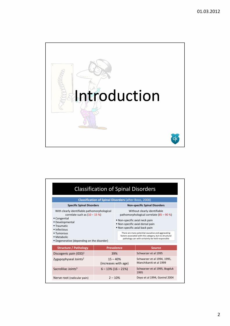
IntroductionIntroduction
Classification of Spinal Disorders
Classification of Spinal Disorders (after Boos, 2008)
Specific Spinal Disorders Non‐specific Spinal Disorders
With clearly identifiable pathomorphological Without clearly identifiable correlate such as (10 – 15 %)
Congenital Developmental Traumatic Infectious TumorousMetabolic Degenerative (depending on the disorder)
pathomorphological correlate (85 – 90 %)
Non‐specific axial neck pain Non‐specific axial dorsal pain Non‐specific axial back pain
There are many potential causative and aggravating factors associated with this category, but no structural pathology can with certainity be held responsible
Structure / Pathology Prevalence SourceStructure / Pathology Prevalence Source
Discogenic pain (IDD)1 39% Schwarzer et al 1995
Zygapophyseal Joints2 15 – 40% (increases with age)
Schwarzer et al 1994, 1995, Manchikaniti et al 1999
Sacroliliac Joints3 6 – 13% (16 – 21%) Schwarzer et al 1995, Bogduk 1995
Nerve root (radicular pain) 2 – 10% Deyo et al 1994, Govind 2004

01.03.2012
3
Back Pain and Red Flags
Patients with low back pain can present with a variety of signs, symptoms and clinical findings of variable significance relative to degree of severity og the problem, injury,
pathoanatomical chances and frank pathology. Low back pain where sinister pathology ( l f f d ) h l l(spinal infections, fracture, metastases and tumors) as the cause is relatively rare in
primary care with an estimated prevalence of less than 1 %.
Since patients with low back pain often presents with a clinical picture that may be multifactorial, it is important for the clinician to be able to recognize and identify
conditions and possible comorbidities that may have serious consequences for the patient, or may hinder full or optimal recovery from the presenting problem. Numerous Red Flags indicating varying degrees of possible pathology or factors influencing the treatment of the
patient is well described in the medical litteraturepatient is well described in the medical litterature.
Jensen S. Back pain‐clinical assessment. Aust Fam Physician. 2004;33:393–395, 397–401.
Roach KE et al. The use of patient symptoms to screen for serious back problems. J Orthop Sports Phys Ther. 1995;21:2–6
Sizer PS et al. Medical screening for red flags in the diagnosis and management of musculoskeletal spina pain. Pain Practice 1(7): 53 – 71, 2007
Wilk V. Acute low back pain: assessment and management. Aust Fam Physician. 2004;33:403–407.
Bogduk N. On the definitions and physiology of back pain, referred pain and radicular pain. Pain 147(2009) 17 ‐ 19
1. Nociceptive back pain
Ligaments in the lumbosacral spine
Lumbar zygapophyseal joints
S ili j i Sacroiliac joints
Posterior surface of the lumbar intervertebral disc
2. Somatic referred pain
Lumbar zygapophyseal joints
Lumbar intervertebral disc
Myofascial triggerpoints (MTrP)
3. Radicular pain
Pain evoked by ectopic discharges emanating from a dorsal root or its ganglion → disc hernia ons and inflamma on of affected nerve seems to be the critical pathophysiological process
4. Radiculopathy
Neurological state in which conduction is blocked along a spinal nerve or its roots. It is not defined by pain but by its objective neurological signs

01.03.2012
4
Prevalens Prolapsed DIV Lumbar Region A symptomatic lumbar disc herniation occurs during the lifetime of 2% of the
general population
90 – 95 % off all lumbar discogenic problems are confined to L4 – L5 or L5 – S1
Risk factors for development of discogenic problems are Risk factors for development of discogenic problems are
• Male gender aged 30 – 50 years
• Physical labour with heavy lifting and torsional movement
• Low job satisfaction and low socioeconomical status / income
• Smoking
• Long‐term exposure to vibration
Not all disc pathology has clinical importance
Abnormal MRI findings in lumbar IVD in asymptomatic subjects (Vacarro 2005)
Age Prolaps Protrusion Disc degeneration
20 – 39 21 % 56 % 34 %
40 – 59 22 % 50 % 59 %
60 ‐ 80 36 % 79 % 53 %
Vaccaro AR. Spine: core knowledge in orthopaedics. Philadelphia: Mosby Elsevier 2005, page 74 ‐ 75
Discogenic PainDiscogenic Pain

01.03.2012
5
Anatomy of the Lumbar IVD
Each IVD consists of three componentsI. Central gelatinous nucleus pulposus
II. Surrounding anulus fibrosus
III P i f t b l d l t th t d i h th NPIII. Pair of vertebral endplates that sandwich the NP
Nucleus Pulposus Consists of a matrix of proteoglycans that bind a
considerable amount of water
Anulus Fibrosus Concentric laminae of Collagen fibers
Inner fibers of the AF envelop the NP and are attached to the vertebral endplate 15‐25 concentric lamina in adults
Outer fibers of the AF are attached to the margins of the vertebral bodies and constitute the ligamentous portion of the AF
Vertebral endplates Cartilaginous structures that are encircled by the
ring apophysis. Covers the NP and attaches to AF
Only weakly attached to the vertebral bodies
15 25 concentric lamina in adults Cells in ECM <1% of total volume Different cell types in NP and AF
Fibroblasts (AF) Chondrocytes (NP
Bogduk N. Anatomy and biomechanics. In. Cole A, Herring S. Low back pain handbook: a guide for the practicing clinician. Philadelphia: Stanley & Belfus, 2nd edition, 2003, p. 9 – 26
Roberts S et al. Histology and pathology of the human intervertebral disc. JBJS Vol 88A, Supplement 2, 2006
Disc Degeneration Lumbar Spine
Disc degeneration may be a sequel to injuries and/or incompetence of the disc tissue to bear normal load
Pathological disc degeneration or deterioration of the disc is characterized by dysfunctional cells and a decrease in extracellular components, leading to a gradual loss of intradiscal fluid
The initiating events are not fully understood, but it may involve poor nutrition as well as mechanical injuries to the disc. In addition, biochemical, immunologic, genetic and nociceptive factors that predispose the IVD to dysfunction has been proposed
Depending on grade of degeneration, several changes can be seen with use of epe d g o g ade o dege e at o , se e a c a ges ca be see t use oimaging modalities
Decrease of disc height, irregular disc contour with bulging due to deterioration of the anulus fibrosus, centrally abundant presence of gas, osteophyte formation and endplate erosion
Borenstein DG, Weisel SW, Boden SD. Low back and neck pain: comprehensive diagnosis and management. Philadelphia: Saunders, 3rd ed 2004Brisby H. Pathology and possible mechanisms of nervous system response to disc degeneration. JBJS, Vol 88A, Supp. 2, 2006

01.03.2012
6
General Classification of Disc Lesions
Normal
Congenital / developmental variant
Degenerative / traumatic lesion Anular tear
Herniation
Protrusion / Extrusion
Intravertebral
Degeneration
Spondylosis deformans
Intervertebral osteochondrosis
Inflammation / Infection Inflammation / Infection
Neoplasia
Morphologic variant of unknown significance
Milette PC. Classification, diagnostic imaging, and imaging characterization of a lumbar herniated disc.
Radiol Clin North Am 2000; 38: 1267 ‐ 1292
Disc Disruption (IDD)
Internal
ses
Disc Prolaps
Mage DJ et al. Orthopaedic physical assessment. St. Louis: Mosby, 5th edition, 2008Vernon‐Roberts B et al. The natural history of age‐related disc degeneration: the pathology and sequela of tears. Spine 32(25): 2007

01.03.2012
7
Innervation: normal and degenerated IVD
In human and animal models of degenerating IVDs, especially painful IVDs, it has been observed that innervation is increased and that theinnervation is increased and that the nociceptive nerve fibers grow into what are usually aneural inner parts of the AF and even into the NP, sometimes together with blood vessels
It has also been observed in degenerated IVDs an increase in the number of Golgi‐tendon organ‐like structures such as Ruffini’s and Pacinian corpsules
In addition to sensory nerve fibers, there is growing evidence that sympathetic afferents are also increased in degenerated IVDs and that they play a significant role in LBP
Garcia‐Cosamalon et al (2010)
01.03.2012
8
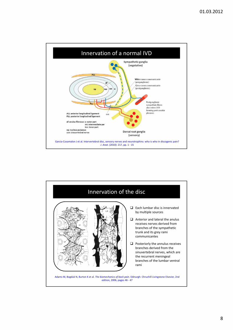
Innervation of a normal IVD
Garcia‐Cosamalon J et al. Intervertebral disc, sensory nerves and neurotrophins: who is who in discogenic pain? J. Anat. (2010): 217, pp. 1 ‐ 15
Innervation of the disc
Each lumbar disc is innervated by multiple sources
Anterior and lateral the anulus receives nerves derived from branches of the sympathetic trunk and its grey rami communicantes
Posteriorly the annulus receives branches derived from the i t b l hi hsinuvertebral nerves, which are the recurrent meningeal branches of the lumbar ventral rami
Adams M, Bogduk N, Burton K et al. The biomechanics of back pain. Edinurgh: Chruchill Livingstone Elsevier, 2nd edition, 2006, pages 46 ‐ 47

01.03.2012
9
Innervation of the disc
Sympathetic trunk and itsgrey rami communicantes (gr)
Anterior
Posterior
Sinusvertebral nerve
Innervation Lumbar ZAJs: Dorsal Rami
Giles (1989, 2009)

01.03.2012
10
IVD Innervated by Multiple Segments
It has been widely believed that the lumbar IVD is segmentally innervated by the dorsal root ganglion (DRG) neurons through the sinuvertebral nerve
However, data from animal studies in the 1990s demonstrated that the L5 –L6 disc in r was innervated by upper (L2) DRG neurons in rats and otherL6 disc in r was innervated by upper (L2) DRG neurons in rats and other studies confirmed the L1 and L2 innervation (Takahashi et al, 1993, Moringa et al, 1996)
It was also shown that the posterior potion of the lumbar disc was innervated by sympathetic nerves multisegmentally and bilaterally (Nakamura et al, 1996)
Discogenic groin pain (L2 segment) has been confirmed in human subjects (Oikawa et al 2012) adding credibility to data from animal studies(Oikawa et al, 2012) adding credibility to data from animal studies
Core Message: The Lumbar IVD receives multisegmental innervation through the sinuvertebral
nerve and sympathetic trunks via the dorsal root ganglion (DRG) Treating discogenic pain from the osteopathic perspective should include evaluation
and treatment of the thoracolumbar region
The Role of Neurotrophins in Discogenic Pain
The increased innervation of the injured lumbar IVD is not fully understood
Neurotrophins (NTs), known to have both neurotrophic and neurotropic popertiesneurotrophic and neurotropic poperties and regulate the density and distribution of nerve fibers in peripheral tissue, seems to play an important role
Indeed, biological evidence suggests that NTs play a role in the genesis and maintenance of painful stimuli from degenerating IVDs
NTs together with extracellular matrix NTs, together with extracellular matrix modifications and some cytokines, regulate the nerve ingrowth into the IVD, the synthesis of pain‐related peptides in the IVD itself but especially in the DRGs
Garcia‐Cosamalon J et al. Intervertebral disc, sensory nerves and neurotrophins: who is who in discogenic pain? J. Anat. (2010): 217, pp. 1 ‐ 15

01.03.2012
11
Schematic hypothesis for a reflex system for motion segment stabilization
Holm S, Indahl A, Solomonow M. Sensorimotor control of the spine. Journal of Electromyography and Kinesiology 12 (2002) 219 ‐ 234
The Degenerative Cascade (after The Degenerative Cascade (after KirkaldyKirkaldy‐‐Willis 1983)Willis 1983)
Asymmetric Disc Injury at Asymmetric Disc Injury at One Motion SegmentOne Motion Segment
Before changes in the physiology of the disc:Before changes in the physiology of the disc:physiological movements of the spine as described physiological movements of the spine as described
by Fryette (1918, 1954) may applyby Fryette (1918, 1954) may apply
Disturbed Kinematics of Disturbed Kinematics of the Motion Segmentthe Motion Segment
Unequal sharing of facet Unequal sharing of facet loadsloads
Cartilage Cartilage Facet hypertrophy and Facet hypertrophy and narrowing of spinal canalnarrowing of spinal canal
Coupled motion pattern of the motion segment Coupled motion pattern of the motion segment influenced by changes in weight distributioninfluenced by changes in weight distribution
Lumbar facet joints Load Transfer:Lumbar facet joints Load Transfer:
Normal: ca. 18% Now: ca. 70%Normal: ca. 18% Now: ca. 70%
Degeneration Degeneration narrowing of spinal canalnarrowing of spinal canal
The progress of degeneration can be divided into three phases:
► Stage I (Dysfunction): changes in biochemical composition, physiology and biomechanics of the
motion segment may result in the clinical symptoms
► Stage II (Instability): increased mobility at the affected level
► Stage III (Stabilization): spinal osteophyte formation (symptoms dye to facet joint hypertrophy and spinal stenopsis)

01.03.2012
12
Manipulative Care and theManipulative Care and theManipulative Care and the Manipulative Care and the Intervertebral DiscIntervertebral Disc
Pictures from: Cyriax JH, Cyriax PJ. Cyriax’s illustrated manual of orthopaedic medicine. London: Butterworth & Heinman 1983, 1993
James Cyriax, M.D.:Major advocate of manipulative therapy for treatment of discs
Used Mixter and Barr’s seminal 1934 NEJM on intervertebral disc problems to discredit the theories and methods of osteopathic physicians osteopathsand methods of osteopathic physicians, osteopaths and chiropractors in treating back pain
The advocacy of his approach was not based so much on existing pathology but on the clinical model he proposed
His manipulative techniques in the spine can best be described as long‐lever, low specificity, high‐velocity thrusts with excessive traction and multiple assistants
James Cyriax (1904 – 1985), British orthopaedic physician
thrusts with excessive traction and multiple assistants
Based on the premise that tightening up the anterior and posterior longitudinal ligaments would “reposition” the disc pathology (protrusion, prolaps, extrusion, or sequestration)
Little or no experimental or clinical data to support this claim and practice
who advocated the use of spinal manipulative therapy for the treatment of disc
herniations. Erroneously held the belief that ZALs and SIJs could be the cause of back pain, nor manipulated. Published numerous
textbooks and advocated use of manipulation by PTs

01.03.2012
13
Stoddard A. Manipulative procedures in the treatment of intervertebral disc lesions. The British Journal of Physical Medicine, May 1951
“Since manipulation, as practiced by orthopedic surgeons, has proved unsuccessful in most cases, the general attitude to disc lesions now is to prescribe restgeneral attitude to disc lesions now is to prescribe rest and, if this fails, to operate and remove part of the disc”
“This is unfortunate, because manipulation has a most important place in the treatment of intervertebral disc conditions”
“There is a great deal of difference between accurate skilled manipulation and the haphazard snapping of
Alan Stoddard, MB, DO (1915 – 2002) graduated from the
skilled manipulation and the haphazard snapping of spines under anesthesia”
“By correct and appropriate manipulation it is often possible to reduce prolapsed discs, to re‐align joints and to remove mechanical stresses and strains”
“The essential point about the manipulation is that it should never aggravate the condition”
British School of Osteopathy in 1935 and earned his medical degree from King’s College in London in 1942. He wrote two
seminal textbooks on osteopathic technique and
practice (1959, 1965) and was a strong advocate of the use of osteopathic manipulation in
PM&R
Risk Management: Considerations
The prevalence of symptomatic disc herniation has been estimated at 1 and 3 %, with 95 % occurring in the lower lumbar spine (Andersson, 1997)
Sciatica accompanies about 10 % of low back pain episodes, and nerve root compression by disc herniation is regarded as the most frequent cause of sciatica (Vroomen et al, 2000)
Symptomatic disc herniation is therefore probably a common presentation in osteopathic clinics (Snelling, 2006)
With regard to manipulation, a great deal of controversy exists
Some authors state that it is absolutely contraindicated in cases of disc herniations (Corrigan and Maitland, 1983)e at o s (Co ga a d a t a d, 983)
Others advocates its use and clinical efficacy (Cyriax, 1985, Cox, 1993)
Attitudes of UK osteopaths in a survey indicated that 54 % of respondents would sometimes employ manipulation in the presence of disc herniation, and most others described this practice as “dangerous” (Rebain et al, 2003)
In the United States it is reported that disc herniation is the leading cause of claims against chiropractors (Jagbandhangsingh et al, 1997)

01.03.2012
14
Snelling N. Spinal manipulation in patients with disc herniation: a critical review of risk and benefit. International Journal of Osteopathic Medicine 9(2006) 77 ‐ 84
Osteopathic Manipulative Osteopathic Manipulative Considerations of Discogenic ProblemsConsiderations of Discogenic Problems
01.03.2012
15
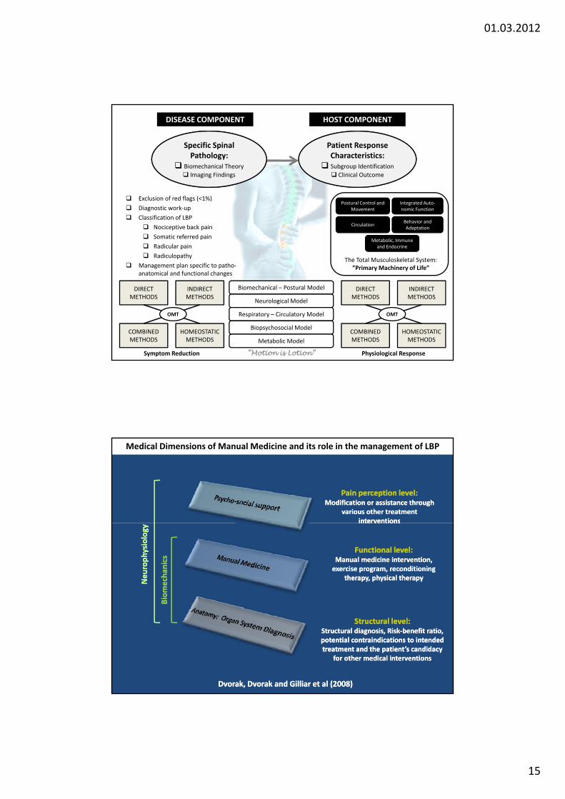
Specific Spinal Pathology:
Biomechanical Theory
Imaging Findings
Patient Response Characteristics:
Subgroup Identification
Clinical Outcome
HOST COMPONENTDISEASE COMPONENT
Exclusion of red flags (<1%)
Diagnostic work‐up
Classification of LBP
Nociceptive back pain
Somatic referred pain
Radicular pain
Radiculopathy
Management plan specific to patho‐anatomical and functional changes
Postural Control and Movement
Circulation
Metabolic, Immune and Endocrine
Integrated Auto‐nomic Function
Behavior and Adaptation
The Total Musculoskeletal System: ”Primary Machinery of Life”
Biomechanical – Postural Model
Neurological Model
Respiratory – Circulatory Model
Biopsychosocial Model
Metabolic Model
anatomical and functional changes
DIRECT METHODS
INDIRECT METHODS
COMBINED METHODS
HOMEOSTATIC METHODS
OMT
DIRECT METHODS
INDIRECT METHODS
COMBINED METHODS
HOMEOSTATIC METHODS
OMT
Symptom Reduction Physiological Response”Motion is Lotion”
Pain perception level:Pain perception level:Modification or assistance through Modification or assistance through
various other treatment various other treatment interventionsinterventions
Medical Dimensions of Manual Medicine and its role in the management of LBP
interventionsinterventions
Functional level:Functional level:Manual medicine intervention, Manual medicine intervention, exercise program, reconditioning exercise program, reconditioning
therapy, physical therapytherapy, physical therapy
Biomechan
ics
Biomechan
ics
Neurophysiology
Neurophysiology
Structural level:Structural level:Structural diagnosis, RiskStructural diagnosis, Risk‐‐benefit ratio, benefit ratio, potential contraindications to intended potential contraindications to intended treatment and the patient’s candidacy treatment and the patient’s candidacy
for other medical interventionsfor other medical interventions
Dvorak, Dvorak and Gilliar et al (2008)Dvorak, Dvorak and Gilliar et al (2008)
01.03.2012
16
Perspectives
LBP: one of the leading causes for why patients seek medical care
Studies and Guidelines support that Osteopathic Manipulative
Prevalens Effectiveness of Care
why patients seek medical care
97% of all LBP are of a mechanical nature (Deyo et al, 2001)
“Tissue causing symptoms” prevalence lists three major progenitors of LBP:
Lumbar IVD 39%
L b ZAJ 15 40 %
that Osteopathic Manipulative Treatment (OMT) is effective in management of LBP Licciardone JC et al. Osteopathic
manipulative treatment for low back pain: a systematic review and meta‐analysis of randomized controlled trials. BMC Musculoskeletal Disorders 2005; 6: 43
NICE Guidelines. Low back pain: the acute management of patients with chronic Lumbar ZAJ 15 – 40 %
Sacroiliac Joints 16 – 21 %
Patients with CLBP have greater severity of diagnostic findings characterizing the somatic dysfunction (TART)
management of patients with chronic (longer than 6 weeks) non‐specific low back pain. Issued May 2009, (The National Collaborating Center for Primary Care and the Royal College of General Practitioners, United Kingdom)
Seffinger M et al. American Osteopathic Association Guidelines for OMT for patients with low back pain. J Am Osteopath Assoc. 11(110): 653‐666, 2010
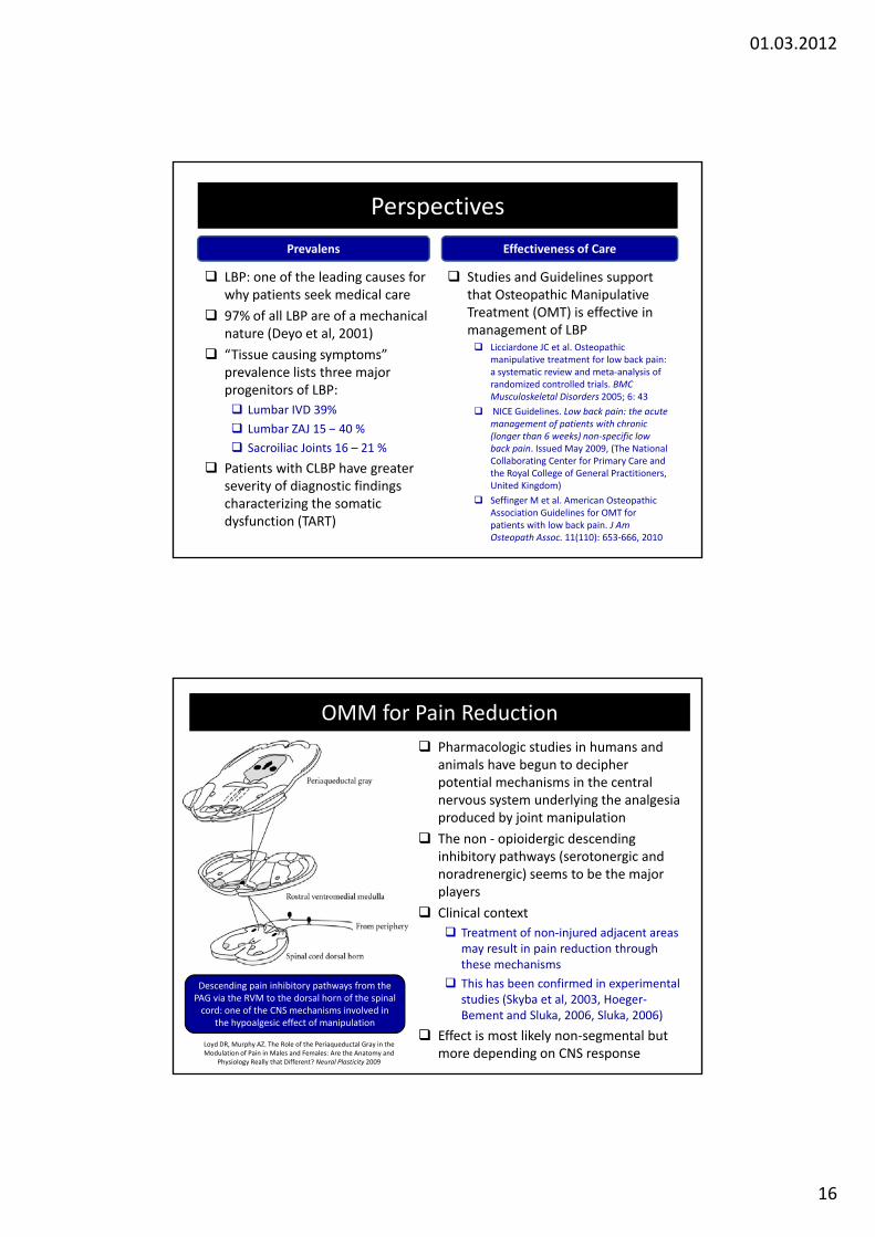
Pharmacologic studies in humans and animals have begun to decipher potential mechanisms in the central nervous system underlying the analgesia
OMM for Pain Reduction
produced by joint manipulation
The non ‐ opioidergic descending inhibitory pathways (serotonergic and noradrenergic) seems to be the major players
Clinical context
Treatment of non‐injured adjacent areas may result in pain reduction through these mechanisms
This has been confirmed in experimental studies (Skyba et al, 2003, Hoeger‐Bement and Sluka, 2006, Sluka, 2006)
Effect is most likely non‐segmental but more depending on CNS response
Descending pain inhibitory pathways from the PAG via the RVM to the dorsal horn of the spinal cord: one of the CNS mechanisms involved in
the hypoalgesic effect of manipulation
Loyd DR, Murphy AZ. The Role of the Periaqueductal Gray in the Modulation of Pain in Males and Females: Are the Anatomy and
Physiology Really that Different? Neural Plasticity 2009

01.03.2012
17
Snider KT, Johnson JC, Snider EJ, Degenhardt BF
Increased Incidence and Severity of Somatic Dysfunction in Subjects With Chronic Low Back PainJ Am Osteopath Assoc. 108(8): 372 ‐ 378, August 2008
Results: Resistance to anterior springing (P<.001) and tenderness (P=.002) were found at significantly greater incidence in the chronic LBP group than in the non‐LBP group, but there were no significant differences between groups for incidence of tissue texture changes or static rotational asymmetry. Significantly greater severity of tissue texture changes (P=.006), static rotational asymmetry (P=.008), resistance to anterior springing (P<.001), and tenderness (P=.001) were observed in the chronic LBP group than in the non‐LBP group.Conclusion: When compared with non‐LBP subjects, chronic LBP subjects had overall greater severity for each of the four elements of somatic dysfunction evaluated, as well as greater incidence of resistance to anterior springing and tenderness. Somatic dysfunction is more severe in individuals with chronic LBP than in individuals without chronic LBP.
Natural history Favorable indications for non‐operative treatment*
sequestrated disc herniation young ageminor neural compromise
small herniationmild disc degenerationmild to moderate sciatica
*Although based more on anecdotal experience than scientific evidence several factors have*Although based more on anecdotal experience than scientific evidence, several factors have been associated with a favorable outcome of non‐operative treatment
Boos N, Aebi M. Spinal disorders: fundamentals of diagnosis and treatment. Berlin: Springer Verlag 2008, page 494
• The natural history of discogenic problems and radiculopathy is usually benign
• Acute episodes of sciatica takes a brief course: this phase is normally followed by a subacute or chronic period of residual symptoms but most patients recover within 1 month, but the recurrence rate is approximately 10 – 15 %
Prognostic factors of positive outcome with conservative intervention for lumbar disc herniation*
Favorable Absence of crossed SLR; spinal motion in extension does not reproduce leg pain; relief or > 50% reduction in leg pain within 6 first weeks after onset; limited psychosocial issues; self‐employed; educational level > 12 y; absence of spinal stenosis; progressive return of neurological deficit within first 12 weeks
Unfavorable Positive crossed SLR, leg pain produced with extension; lack of >50% pain reduction in first 6 w; overbearing psychosocial issues, Worker’s compensation; Educational level >12 y; concomitantspinal stenosis; cauda equina syndrome
*Saal JA. Natural history and nonoperative treatment of lumbar disc herniation. Spine 21: 2S – 9S, 1996

01.03.2012
18
Lumbar Zygapophyseal or Facet Joint Pain (FJP)Discogenic Pain:
Internal disc disruption
Effective office management of LBP
Sacroiliac Joint Pain (SJP)
Internal disc disruption HNP
Protrusion, Extrusion orSequestration
Pain intensity and pain distribution Degree of neurological involvement Contributing factors: what is amenable to osteopathic manipulative treatment ?
Predicted probabilities and 95 % CI for internal disc disruptions (IDD), facet joint pain (FJP), sacroiliac joint pain (SIJP) and other sources of LBP
DePalma MJ et al. What is the source of chronic low back pain and does age play a role? Pain Medicine 2011; 12: 224 ‐ 233

01.03.2012
19
Diagnostic Considerations: Radicular Pain and Radiculopathy
The affected nerve root and its degree of involvement can usually be determined through sensory, motor and reflex testing
Sensitivity for sciatic distribution of pain in the
The differential diagnosis can be aided by adescription of the
distribution of the patient’s Sensitivity for sciatic distribution of pain in the diagnosis of lumbar disc herniation of 90 % and calculated likelihood ratio of disc herniation being present in the absence of sciatic pain as 0.1%
Radiculopathy is such a sensitive finding (95%) that its absence almost rules out a clinically important disc herniation
The SLR and its variations are good markers for
symptoms and the results of the physical examination
Consider:Map pain pattern → radicular pain is dermatomal in distribution
Physical examination including orthopedic and neurological assessment
The SLR and its variations are good markers for radicular pain / radiculopathy involving the 4th and 5th lumbar as well as 1st to 3rd sacral nerve roots
SLR may be positive with 3rd lumbar nerve root involvement, but the likelihood is greater that the femoral nerve stretch test will be positive
Deyo et al (1992), Dutton (2010)
Use dural signs or nerve stretch tests for radicular pain Determine if the herniated NP is of “shoulder” or “axilla” type Peripheralization vs. centralizationof pain Determine degree of involvementof radiculopathy through sensory, motor and reflex changes
Radicular Pain and Radiculopathy
• L3 – L4 Disc → L4 Nerve Root Pain in L4 dermatome (radicular pain)
Sensory loss in L4 dermatome (radiculopathy)
Loss of muscle force: inversion of foot (radiculopathy)
↓ DTR: Patellareflex
• L4 – L5 Disc → L5 Nerve Root Pain in L5 dermatome (radicular pain)
Sensory loss in L5 dermatome(radiculopathy)
Loss of muscle force: Extensor Hallucis Longus Muscle
↓ DTR: None
• L5 S1 Disk→ S1 Nerve Root• L5 – S1 Disk → S1 Nerve Root Pain in S1 dermatome(radicular pain)
Sensory loss in S1 dermatome (radiculopathy)
Loss of muscle force: eversion of foot (radiculopathy)
↓DTR: Achillesreflex
• Central Prolaps → Symptoms may be as in CES

01.03.2012
20
ative to Nerve Root
HNP rela
cteristics
”Shoulder” ”Axillae”
Rad
icular Pain Charac
Magee et al (2008)
Pain Patterns: IDD, ZAJ and SIJ This study looked at the predictive value of pain
localization relative to structures causing it
N = 170, Average age 54.4 yr, LBP average 12 months
Provocative discography, ZA‐ and SI jointblocks
Discogic (IDD), Lumbar ZAJ og SI joint pain
Calculated sensitivity, spesificity, positive and negative predictive value, diagnostic accuracy and LR +/‐
Pain median (over the SPs) → IDD and reduces the likelihood for ZAJ and SIJ as pain generators
Isolated paramedian pain→ increased likelihood for
DePalma MJ et al. Does the location of low back pain predict its source? PM R 2011; 3: 33 – 39, 2011DePalma MJ et al. Does the location of low back pain predict its source? PM R 2011; 3: 33 – 39, 2011
Isolated paramedian pain → increased likelihood for ZAJ and SIJ as pain generators
Distribution of pain in internal disc disruption (IDD):
Axial midline pain (over SPs) with possibility for pain in buttock and groin region (Skogsbergh et al, 2001)

01.03.2012
21
Use of Translatory Motion for Pain Provocation with IDD
If tolerated by the patient, translatory motion in seated position may be used to
induce mechanical stress oninduce mechanical stress on the IVD with IDD, and may
be used for pain provocation. In addition, resistance to motion at
segment with pain is noted
Traditional motion testing using flexion, extension, SB and rotation, may, despite potential displacement of the nucleus, be insufficient to provoke pain from a
lumbar IVD with IDD. With a HNP these tests, especially flexion and extension may
produce pain
Summary: Findings with IDD
A provocative discogram is considered the ”gold standard” for determining pain of discogenic origin (IASP). However, there are numerous clues from the physical
examination that may help indicate the condition:
Age: <40 years Age: <40 years
Translatory motion testing to stress the lumbar IVD Motion stress to the IVD to provoke pain with IDD
Compression and traction test Positional loading testing to reproduce the pain: midline pain, well‐localized and well‐defined
Provocative AP pressure in prone position Overpressure at vertebra with well‐localized, well‐defined midline pain corresponds well with
IDD, especially in the lower lumbar spine and lumbosacral junction, p y p j
Vibratory pain provocation on spinous processes In lateral recumbent position : symptomatic side down→ vibratory s mula on applied to SP
Can elicit pain associated wit internal disc disruption: agrees with provocative discography in 70.9% to 85.9% of discs. Comparatively, MRI specificity was 55.7
Vanharanta H et al. Vibration pain provocation can improve the specificity of MRI in the diagnosis of symptomatic lumbar disc rupture. Clin J Pain 1998; 3(4): 239 – 247
Yjma M et al. Bony vibration stimulation test combined with magnetic resonance imaging. Can discography be replaced? Spine 1997; 22(7): 808 ‐ 803

01.03.2012
22
Somatic Dysfunction
From a biomechanical perspective the spinal somatic dysfunction is to be considered a tripod system where dysfunction involves all active and passive structures of the functional spinal unit (FSU)
Three legs of support
The sensorimotor control of the spine depends on normal interaction between the active and passive components of the FSU
Somatic dysfunction alters the sensorimotor control of the spine through the FSU
The three legs of support in the FSU is the Intervertebral disc and the two zygapophyseal joint
They are all three involved in the mechanics of somatic They are all three involved in the mechanics of somatic dysfunction
Although the osteopathic literature describes Type I and Type II mechanics in the lumbar spine, natural aging and degeneration of the structures of the FSU will alter the coupling behavior of the motion segment making it less predictable
Sensorimotor Control
Instantaneous Axes of Rotation: Lumbar Spine
Flexion and extension Caudal portion of intervertebral disc close to the intervertebral endplate
Sidebending I th t l ti f th i t t b l di li ht di l t t th l ft th In the central portion of the intervertebral disc: slight displacement to the left or the
right during sidebending
Rotation Central (Panjabi, 1978) or posterior (Farfan, 1986) portion of the intervertebral disc
Key Message: The lumbar IAR all seem to be located in the IVD
Penning L. Hals‐ under Lendenwirbelsäule in Biomechanick und Pathologie. München: Pflaum Verlag 1996

01.03.2012
23
80 – 85% of Imposed Weight through Body and
IVD
15 – 20% of Imposed Weight through Facet
Joints
The IVD is weight bearing element and controls movement of the Functional Spinal Unit
The facet joints are only in spatial apposition whose function is to guide movement
Coupled motions are less influenced by contractile elements
Type I (NSXRY) and Type II (E or F RXSX) mechanics would apply here
The rest of Imposed Weight through Body and
IVD
Up to 70% of Imposed Weight through Facet
Joints
With disc degradation or degenerative changes, the weight bearing of the facet joints
can be up to 70% of imposed weight (Adams et al 2001)
The facet joints are in apposition through the arc of movement
Type I (NSXRY) and Type II (E or F RXSX) mechanics would not necessarily apply here

01.03.2012
24
Osteopathic Considerations: Biomechanical
Muscle Energy Techniques (MET)
If tolerated by the patient → seated posi on
Use translatory motions
A P l i f fl i d i A – P translation for flexion and extension: may prevent unwanted movement of the nucleus pulposus as with pure F or E
Lateral translation: with the sidebending component the coupled rotation will follow automatically. Less concerns about altered coupling behavior at FSU
PIR variation: restore motion at FSU
Ruddy variation: reduce venolymphatic congestion
MET can also effectively be used to improve sensorimotor control of the spinal motion segment
Ensure to keep to contraction effort from the patient light in order to prevent recruitment from the polyarticular muscles
Kimberly PE, Dickey J, Halma KD. Outline of osteopathic
manipulative procedures : the Kimberly manual. Kirksville: A.T.
Still University 2009
Combined Leverage and Thrust in the Lumbar Spine in Patients with Discogenic Pain
Advantages:
Using combined leverages allows the operator to manufacture a
The spine is kept in a neutral position and the technique utilizes physiological locking of the spine from above and below (e.g. myofascial locking as opposed to approximation of joint surfaces)
the operator to manufacture a pre‐manipulative barrier within the physiological range that is
usually not at the end‐point. This minimizes stress on tissues and is done short of pain for patient Force applied perpendicular to
joint surface = Gapping of ZAJ
approximation of joint surfaces)
Why neutral position?
Mathematical analyses of the lumbar IVD shows that with bending moments (flexion) the stress on the AF is 450 times greater than with spinal rotation and they conclude that there is no radial expansion or extrusion (bulging) during twisting*
Extension may aggravate the patients symptoms
In neutral there is less stress or strain on inflamed or injured tissue (pain ↓)*Chaudhry H et al. Viscoelastic stresses on anisotropic annulus fibrosus of lumbar disc under compression, rotatoon and flexion in manual treatment. Journal
of Bodywork and Movement Therapies (2009) 13, 182 ‐ 191

01.03.2012
25
Thoracolumbar Junction:Thoracolumbar Junction:A major player in LBP and Discogenic PainA major player in LBP and Discogenic Pain
LBP of Thoracolumbar Origin
LBP of thoracolumbar origin is common in clinical practice, it may be acute or chronic, and its character is similar to that of pain of lumbosacral origin
Distribution of spinal T12 d L1 similar to that of pain of lumbosacral origin
This may also include pseudovisceral abdominal pain, pubic and trochanteric tenderness associated with this
Note the anterior ramus (1): groin pain of discogenic origin has recently been demonstrated in L2 distribution
nerves T12 and L1:
1. Anterior ramus2. Posterior ramus3. Perforating lateral
cutaneous branch
Maigne R. Diagnosis and treatment of pain of vertebral origin. Florida: Taylor and Francis Group, 2nd edition, 2006Oikawa Y et al. Lumbar disc degeneration iduces persistent groin pain. Spine 37(2): 114 – 118, 2012

01.03.2012
26
TL Junction: Biomechanical Stressinducer
The lumbar zygapophyseal joints (ZAJ) are designed to limit or block axial rotation
The protects the intervertebral disc from excessive torsion
Although approximating the sagittal plane, the surface of the lumbar ZAJ are either flat, C – shaped or J – shaped
On average there is 2 – 3 O of axial rotation possible per segment
There is a sudden change of joint structure occurring in the region of a single vertebra T12: orientation of ZAJ changes abruptly
Lack of axial rotation in the lumbar spine is compensated at this junction
Motion restriction at T12 may increase the biomechanical stress in the lumbar spine
Maigne (2001), Adams et al (2006), Lewit (2010)
Stabilizing Role of the TL Junction
An early signal to the stabilizing role of the human thoracolumbar junction (TLJ) appears in the sequence of ossification of the vertebral centra
a number of reports have indicated that the first sites of ossification are consistently located in the lower two thoracic and first lumbar vertebral bodiesconsistently located in the lower two thoracic and first lumbar vertebral bodies before a progressive cranial and caudal pattern of ossification commences in the adjacent vertebrae
The reciprocal change in curvature of the thoracic kyphosis and lumbar lordosis produces an inflexion point commonly located between T11 and L1
The morphology of the TLJ is highly variable, but the morphology may aptly be described as an “anti‐torsion” device limiting torsional stress in the region
Data from Donish and Basmajian (1972) indicates that the multifidi muscles Data from Donish and Basmajian (1972) indicates that the multifidi muscles in the TLJ are antagonistic to axial rotation and thereby prevents torsion
In addition to resisting axial rotation, in the TLJ extension is “closed‐packed position” → mechanics of spinal HVLA in the region may needs revisi ng
Singer KP. Anatomy and biomechanics of the thoracolumbar junction. In: Boyling JD, Palastanga N. Grieve’s modern manual therapy. Edinburgh: Churchill Livingstone, 2nd edition, 1994, pages 85 ‐ 97
Giles L, Singer KP. Clinical anatomy and management of low back pain. London: Butterworth & Heineman 1997
01.03.2012
27
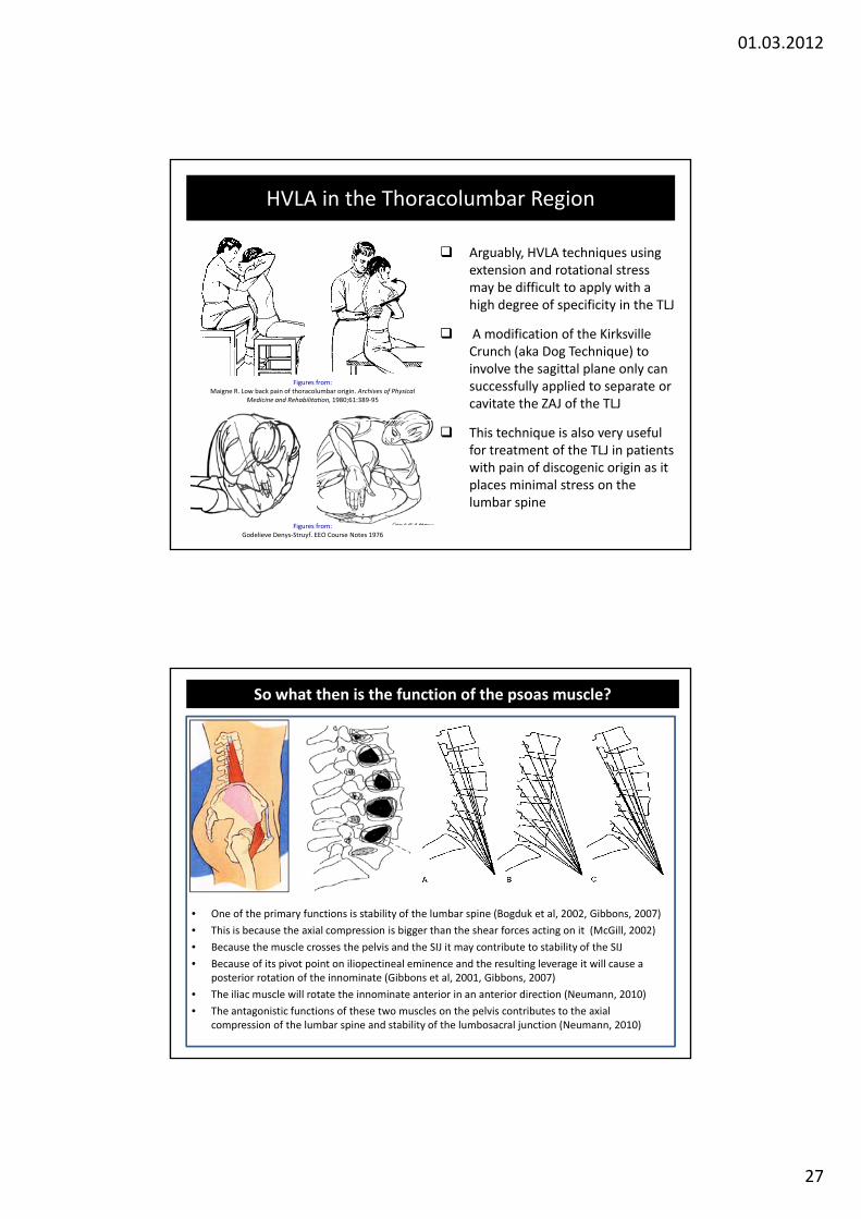
HVLA in the Thoracolumbar Region
Arguably, HVLA techniques using extension and rotational stress may be difficult to apply with amay be difficult to apply with a high degree of specificity in the TLJ
A modification of the Kirksville Crunch (aka Dog Technique) to involve the sagittal plane only can successfully applied to separate or cavitate the ZAJ of the TLJ
Figures from:Maigne R. Low back pain of thoracolumbar origin. Archives of Physical
Medicine and Rehabilitation, 1980;61:389‐95
This technique is also very useful for treatment of the TLJ in patients with pain of discogenic origin as it places minimal stress on the lumbar spine
Figures from:Godelieve Denys‐Struyf. EEO Course Notes 1976
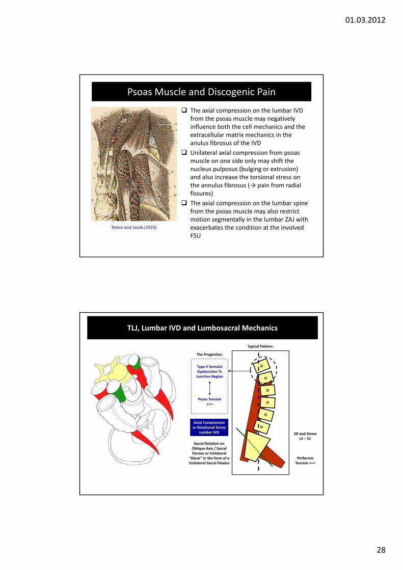
So what then is the function of the psoas muscle?
• One of the primary functions is stability of the lumbar spine (Bogduk et al, 2002, Gibbons, 2007)
Thi i b th i l i i bi th th h f ti it (M Gill 2002)• This is because the axial compression is bigger than the shear forces acting on it (McGill, 2002)
• Because the muscle crosses the pelvis and the SIJ it may contribute to stability of the SIJ
• Because of its pivot point on iliopectineal eminence and the resulting leverage it will cause a posterior rotation of the innominate (Gibbons et al, 2001, Gibbons, 2007)
• The iliac muscle will rotate the innominate anterior in an anterior direction (Neumann, 2010)
• The antagonistic functions of these two muscles on the pelvis contributes to the axial compression of the lumbar spine and stability of the lumbosacral junction (Neumann, 2010)
01.03.2012
28
Psoas Muscle and Discogenic Pain
The axial compression on the lumbar IVD from the psoas muscle may negatively influence both the cell mechanics and the extracellular matrix mechanics in theextracellular matrix mechanics in the anulus fibrosus of the IVD
Unilateral axial compression from psoas muscle on one side only may shift the nucleus pulposus (bulging or extrusion) and also increase the torsional stress on the annulus fibrosus (→ pain from radial fissures) )
The axial compression on the lumbar spine from the psoas muscle may also restrict motion segmentally in the lumbar ZAJ with exacerbates the condition at the involved FSU
Testut and Jacob (1923)
TLJ, Lumbar IVD and Lumbosacral Mechanics
Typical Pattern:
Type II Somatic Dysfunction TL
The Progenitor:
yJunction Region
SD and Stress
Psoas Tension +++
Axial Compression or Rotational Stress
Lumbar IVD
Sacral Rotation on Oblique Axis / Sacral Torsion or Unilateral
“Shear” in the form of a Unilateral Sacral Flexion
SD and Stress L5 – S1
Piriformis Tension +++

01.03.2012
29
Suggestions on treating the Psoas Muscle
Treatment of the thoracolumbar junction and upper lumbar spine prior to treating the psoas muscle in patients with discogenic pain
Counterstrain: this technique is helpful in addressing the psoas muscle specifically. Although the position requires a great deal of flexion with SB and rot, patients with discogenic pain usually tolerates it well
If condition is very acute, consider holding the position of comfort for longer than 90 seconds
J C i M D d h “D lli T h i ” James Cyriax, M.D. used the “Dallison Technique” (see picture) on patients with acute discs
This technique bears resemblance to how osteopathic physicians treat the psoas muscle using counterstrain
Cyriax and Cyriax (1983), Myers (2006)
Thoraco‐Abdomino‐Pelvic Pump(Mitchell FL Jr. The respiratory‐circulatory model: concepts and application. In: Greenman PE. Concepts and mechanisms of
neuromuscular function. New York: Springer 1984)
Respiration is an activity where numerous systems and all body tissues are involved (Cathie, 1965, 1974)
THORAX
o‐Pelvic
Thoracoabdominal junction: vertical and transverse plane where we have integration of somatic, respiratory, vascular, neural and visceral functions (Cathie, 1974)
< 23,000 respiratory cycles per day
The diaphragm is the extrinsic pump of the
Abdomino The diaphragm is the extrinsic pump of the
venous and lymphatic system: asymmetries with reduce the respiratory efficacy through distortion of the cylinder (Zink, 1970, 1973, 1977, Mitchell, 1984)
Important in LBP and discogenic conditions
01.03.2012
30
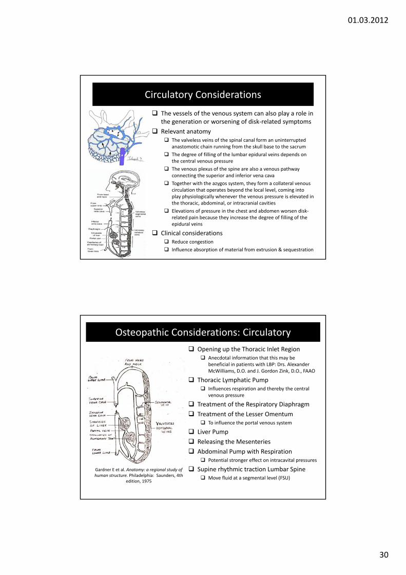
Circulatory Considerations
The vessels of the venous system can also play a role in the generation or worsening of disk‐related symptoms
Relevant anatomy The valveless veins of the spinal canal form an uninterrupted
anastomotic chain running from the skull base to the sacrum
The degree of filling of the lumbar epidural veins depends on the central venous pressure
The venous plexus of the spine are also a venous pathway connecting the superior and inferior vena cava
Together with the azygos system, they form a collateral venous circulation that operates beyond the local level, coming into play physiologically whenever the venous pressure is elevated in play physiologically whenever the venous pressure is elevated inthe thoracic, abdominal, or intracranial cavities
Elevations of pressure in the chest and abdomen worsen disk‐related pain because they increase the degree of filling of the epidural veins
Clinical considerations Reduce congestion
Influence absorption of material from extrusion & sequestration
Osteopathic Considerations: Circulatory
Opening up the Thoracic Inlet Region Anecdotal information that this may be
beneficial in patients with LBP: Drs. Alexander McWilliams, D.O. and J. Gordon Zink, D.O., FAAO
Thoracic Lymphatic Pump Influences respiration and thereby the central
venous pressure
Treatment of the Respiratory Diaphragm
Treatment of the Lesser Omentum To influence the portal venous system
Liver Pump
Gardner E et al. Anatomy: a regional study of human structure. Philadelphia: Saunders, 4th
edition, 1975
Releasing the Mesenteries
Abdominal Pump with Respiration Potential stronger effect on intracavital pressures
Supine rhythmic traction Lumbar Spine Move fluid at a segmental level (FSU)
01.03.2012
31
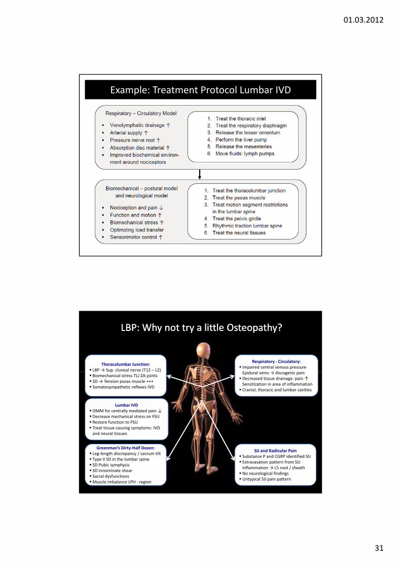
Example: Treatment Protocol Lumbar IVD
Respiratory ‐ Circulatory: Impaired central venous pressureE id l i → di i i
Thoracolumbar Junction: LBP → Sup. cluneal nerve (T12 – L2)
LBP: Why LBP: Why not try a little Osteopathy?not try a little Osteopathy?
Epidural veins → discogenic pain Decreased tissue drainage: pain ↑Sensitization in area of inflammation Cranial, thoracic and lumbar cavities
p ( ) Biomechanical stress TLJ ZA‐joints SD → Tension psoas muscle +++ Somatosympathetic reflexes IVD
Lumbar IVD OMM for centrally mediated pain ↓ Decrease mechanical stress on FSU Restore function to FSU Treat tissue causing symptoms: IVD
d l ti
Greenman’s Dirty‐Half Dozen: Leg‐length discrepancy / sacrum tilt Type II SD in the lumbar spine SD Pubic symphysis SD Innominate shear Sacral dysfunctionsMuscle imbalance LPH ‐ region
SIJ and Radicular Pain Substance P and CGRP identified SIJ Extravasation pattern from SIJ: Inflammation → L5 root / sheath No neurological findings Untypical SIJ pain pattern
and neural tissues