Cozen O’Connor, Wilmington, Delaware. O’Connor, New York ...
Upload
maliyah-harborCategory
view
220download
1
Components of BoneExtracellular Matrix• 90% mineral
• Hydroxyapatite• Ca10(PO4)6(OH)2
• 10% organic• Type I collagen• Osteocalcin• other collagens• other proteins• growth factors
Cellular• Osteoblasts
• Osteocytes• Periosteal• Endosteal
• Osteoclasts• Hematopoietic Cells• Mesenchymal Stem Cells• Chondrocytes
• growing or healing bones• Articular surfaces

Bone Development: Endochondral Ossification

Bone Development: Intramembraneous Ossification
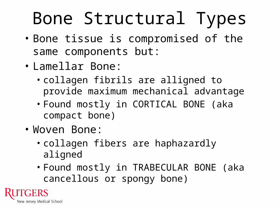
Bone Structural Types• Bone tissue is compromised of the same
components but:• Lamellar Bone:• collagen fibrils are alligned to provide maximum
mechanical advantage• Found mostly in CORTICAL BONE (aka compact
bone)• Woven Bone:• collagen fibers are haphazardly aligned• Found mostly in TRABECULAR BONE (aka
cancellous or spongy bone)

Function Form►• Mechanical Functions• cortical bone• lamellar tissue
• Physiological Functions• trabecular bone• woven tissue• surface area
• 7 m2 (lungs 70 m2)
• Bone is Anisotropic

Osteoblasts Make Bone1. Collagen and alkaline
phosphatase secretion2. Forms poorly
mineralized OSTEOID3. Calcium precipitates
quickly in osteoid (days to weeks)
4. Calcium salt converted in hydroxapatite (weeks to months)
5. Bone remodeling6. Induced by many stimuli

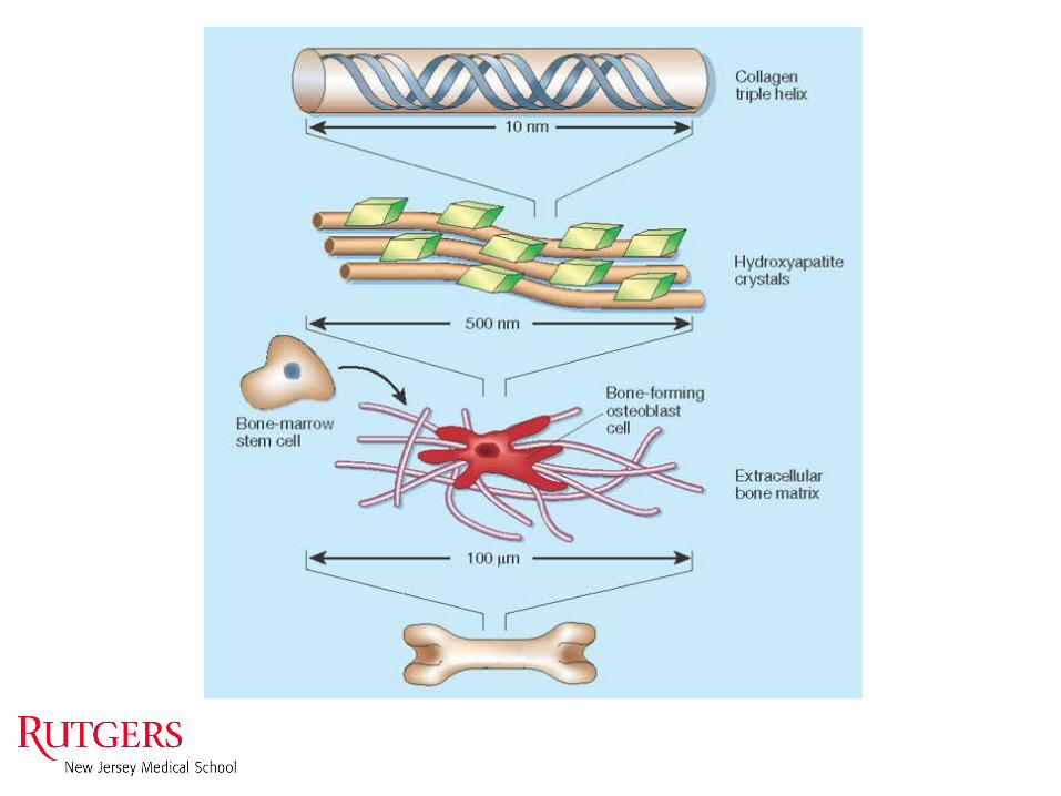
Bone Microstructure: collagen fibrils


Bone Functions:Resorption & Formation Follow Function
• Protection: heart and brain• Breathing: rib cage• Hematopoiesis• Mechanical strength: sustain body weight
and movements• Locomotion: sites for muscle attachment
• Calcium Regulation

Response to Mechanical Loading: Wolff’s Law
• “The Law of Transformation of Bone” By Julius Wolff (1892)
• Bone structure and shape adapt to mechanical loading conditions
• Microdamage Repair

Calcium Homeostasis• Calcium content of adult human: 1.1 kg• Total plasma calcium: 2.5 mM• Functions:• Blood coagulation• Cardiac and muscle contraction• Nerve function
• Serum calcium concentration principally regulated by 3 hormones• 1,25-dihydroxycholecalciferol (Vitamin D)
• ↑ osteoclast function;• ↑ intestinal Ca2+ adsorption
• Parathyroid hormone (PTH)• ↑ osteoclast formation;• ↑kidney Ca2+ re-absorption;• ↑ Vit D
• Calcitonin• ↓ osteoclast activity

Bone Homeostasis
Normally, bone resorption and formation arebalanced allowing for continual renewal of skeletaltissue and repair of micro-damage.

• Howship’s Lacunae• Carbonic Anhydrase II• Tartate Resistant Acid Phosphatase• Cathepsin K
Osteoclasts Resorb Bone

Osteoclast DifferentiationRequires M-CSF and RANKL
M-CSF RANKL OPG

Osteoblasts Regulate Osteoclasts: Coupling and RANKL & OPG
Enhances osteoclastdifferentiationRANKL
OPGReducing bone
resorbtion
OR “Coupling”

When things go wrong:
• Osteoporosis: reduced amount of bone tissue• Osteomalacia: poorly mineralized bone tissue• Osteopetrosis: excessive bone tissue with
enhanced mineralization• Osteosclerosis: localized areas of increased
bone density

Osteopetrosis
• Abnormally high bone mineralization
• Increased bone tissue• Affects tooth eruption
and formation• Generally caused by
defects in osteoclast development

Osteosclerosis: Padget’s Disease• Localized areas of
sclerotic bone• Sclerotic bone is
abnormally dense.• Approx. 50% of
Padget’s patients have complications involving mandible or maxilla
• Cause unknown

Osteomalacia
• Poorly mineralized bone tissue
• Generally caused by Vitamin D defiency
• Rickets in children– Bowed legs

Osteoporosis• Low bone mass• Microarchitectural
deterioration of bone tissue• Enhanced bone fragility• ~25% of women over 65
years old have osteoporosis-related vertebral fractures
• Age, gender, genetic background, environment, endocrinology, and bone structure

Localized Bone Loss: Periodontal Disease
• Usually caused by local acute or chronic inflammation
• Bone loss exposes roots of teeth making them susceptible to:– Cavities– Loosening

Treating Osteoporosis• Estrogen therapy
– Reduces RANKL expression• Bisphosphonates
– Induce osteoclast apoptosis• Behavior (diet and exercise)• Estrogen receptor drugs
– raloxifene• PTH like drugs• Calcitonin• Anti-RANKL
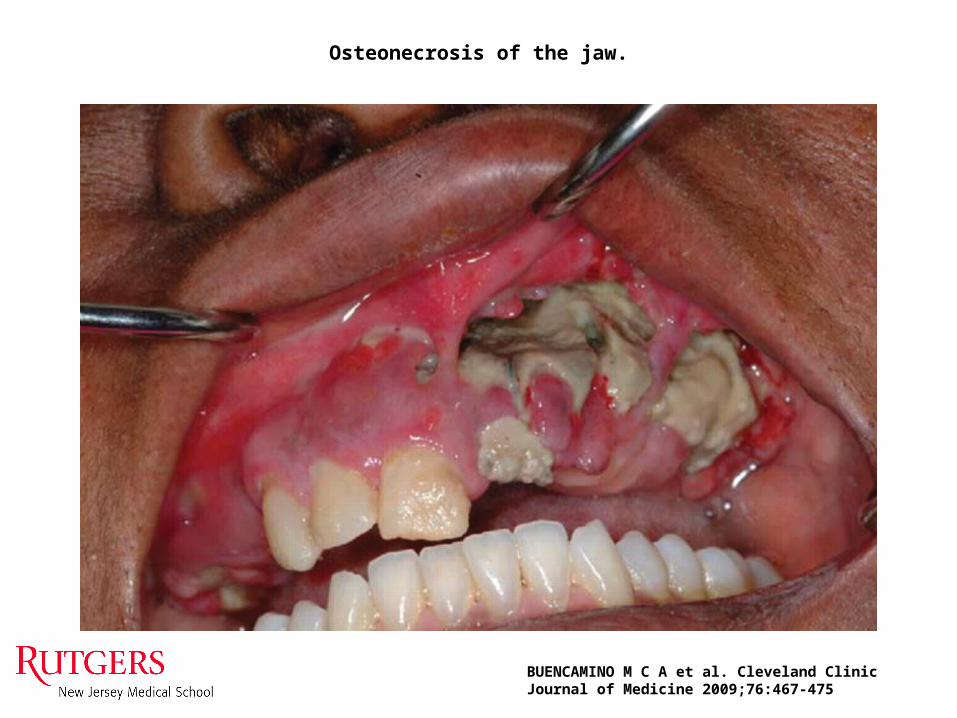
Osteonecrosis of the jaw.
BUENCAMINO M C A et al. Cleveland Clinic Journal of Medicine 2009;76:467-475

Alternative Osteoporosis TherapiesDensumab: anti-RANKL antibody
• Anti-CATABOLIC– Densumab: anti-RANKL
antibody
• Pro-ANABOLIC– Teriparatide: PTH1-34 (Forteo)

Treating Peridontal Bone Loss
• Prevention• Stopping loss• Bone graft• Tissue graft• Guided Tissue
Regeneration• Growth Factors• Dentures

Differential Protein Composition in Mammalian Calcified Tissue
Component Cartilage Bone Dentin EnamelType I Collagen + ++ + -Type II Collagen ++ - - -GLA Proteins + + + -Osteocalcin - + + -Glycoproteins + + + +Proteoglycans + + + ND

Questions