Lecture # 21: The Brain and Cranial Nerves (Chapter 14) Objectives: 2- Name the three coverings of...
15
Lecture # 21: The Brain and Cranial Nerves (Chapter 14) Objective s: 2- Name the three coverings of the central nervous system and give the characteristics, location, and function of each. 1- Name the major regions of the brain and give the functions of each. 3- Give the functions of cerebrospinal fluid and explain its formation, circulation, and resorption.
-
Upload
alexis-garrett -
Category
Documents
-
view
214 -
download
1
Transcript of Lecture # 21: The Brain and Cranial Nerves (Chapter 14) Objectives: 2- Name the three coverings of...
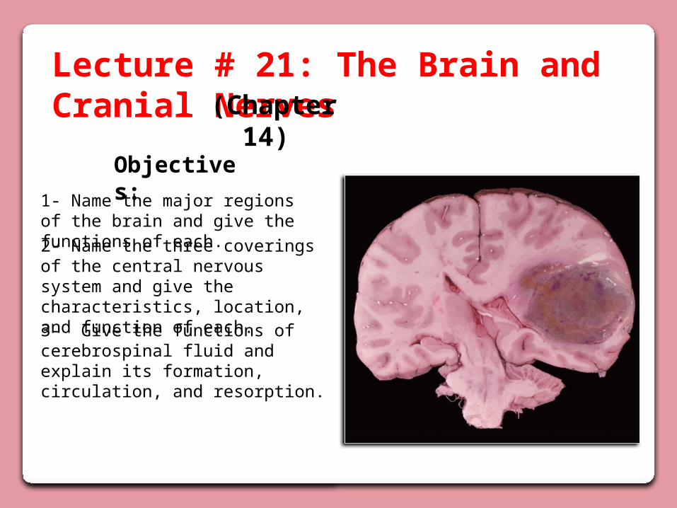
Lecture # 21: The Brain and Cranial Nerves (Chapter 14)
Objectives:
2- Name the three coverings of the central nervous system and give the characteristics, location, and function of each.
1- Name the major regions of the brain and give the functions of each.
3- Give the functions of cerebrospinal fluid and explain its formation, circulation, and resorption.
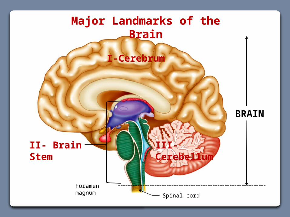
Major Landmarks of the Brain
I-Cerebrum
III- Cerebellum
Spinal cord
Foramen magnum
BRAIN
II- Brain Stem
I- Cerebrum
II- Brain Stem
III- Cerebellum
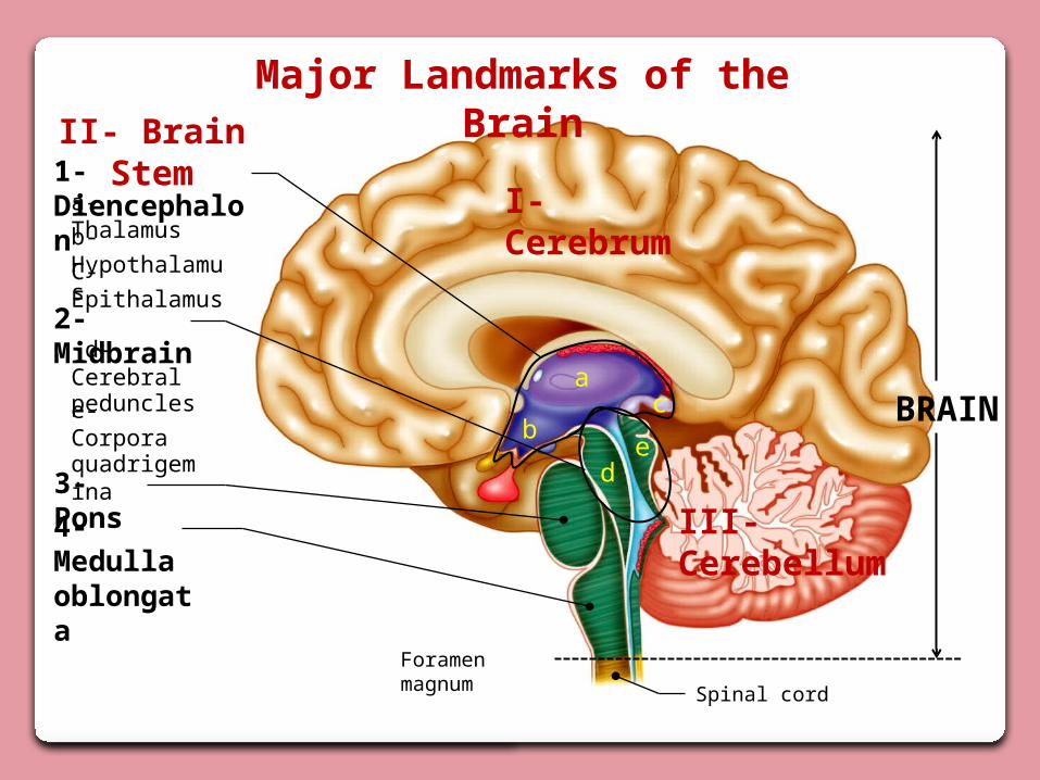
a-Thalamusb- HypothalamusC- Epithalamus
4- Medulla oblongata
2- Midbrain d- Cerebral pedunclese- Corpora quadrigemina
3- Pons
1- Diencephalon
a
bc
de
Major Landmarks of the Brain
BRAIN
Spinal cord
Foramen magnum
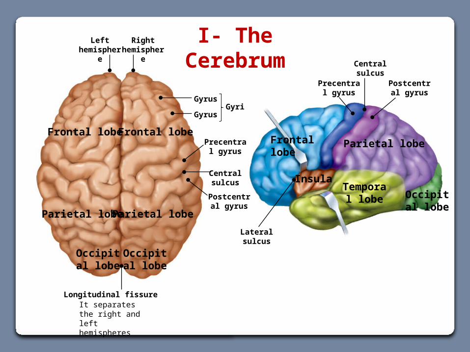
Longitudinal fissureIt separates the right and left hemispheres
Right hemisphere
Left hemisphere
Central sulcus
Frontal lobe Frontal lobe
Occipital lobe
Occipital lobe
Parietal lobe Parietal lobe
Gyrus
Gyrus
Precentral gyrus
Postcentral gyrus
Gyri
Frontal lobe Parietal lobe
Occipital lobe
Temporal lobe
Precentral gyrus
Central sulcus
Postcentral gyrus
I- The Cerebrum
Lateral sulcus
Insula
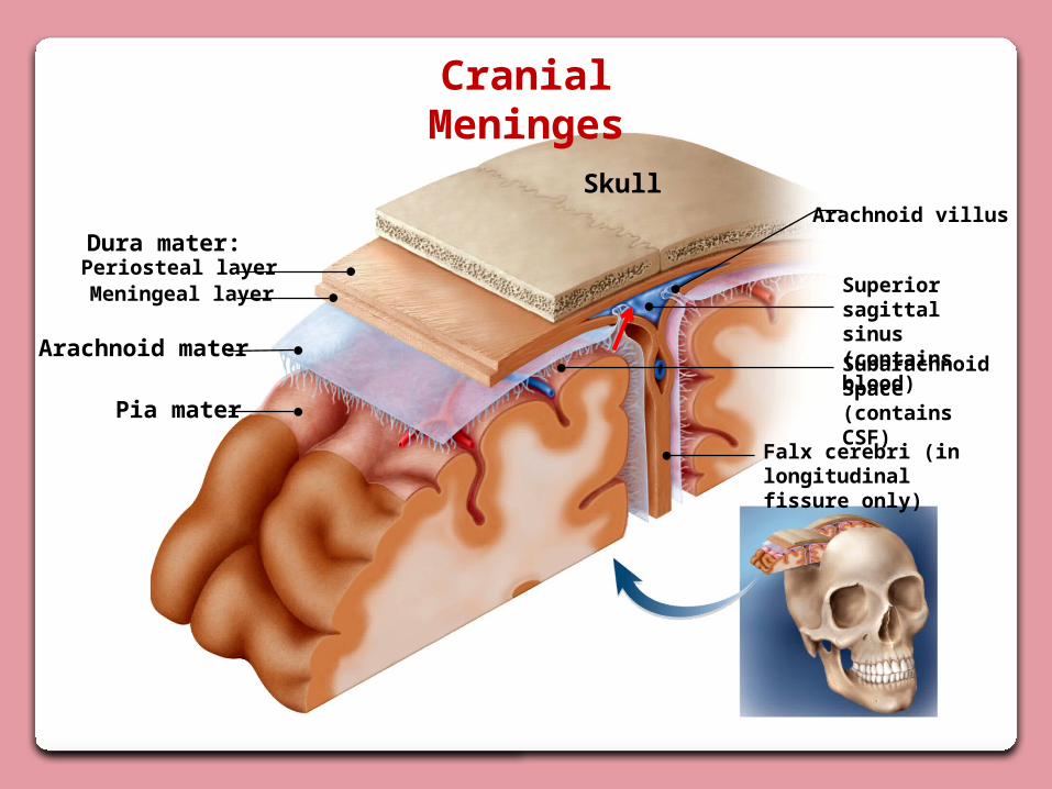
Cranial Meninges
Skull
Dura mater:
Superior sagittalsinus (contains blood)
Pia mater
Arachnoid mater
Meningeal layer Periosteal layer
SubarachnoidSpace (contains CSF)
Arachnoid villus
Falx cerebri (in longitudinalfissure only)
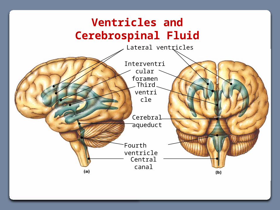
Lateral ventricles
Third ventricle
Fourth ventricle
Cerebral aqueduct
Central canal
Interventricular foramen
Ventricles and Cerebrospinal Fluid
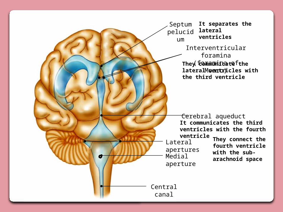
Septum pelucidum
It separates the lateral ventricles
They communicate the lateral ventricles with the third ventricle
It communicates the third ventricles with the fourth ventricle
Cerebral aqueduct
They connect the fourth ventricle with the sub- arachnoid space
Central canal
Interventricular foramina (foramina of Monro)
Medial aperture
Lateral apertures
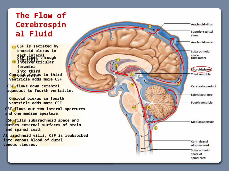
The Flow of Cerebrospinal Fluid
Choroid plexus in fourthventricle adds more CSF.
CSF flows out two lateral aperturesand one median aperture.
CSF fills subarachnoid space andbathes external surfaces of brainand spinal cord.
At arachnoid villi, CSF is reabsorbedinto venous blood of duralvenous sinuses.
CSF is secreted by choroid plexus in each lateral ventricle.
CSF flows throughInterventricular foraminainto third ventricle.
Choroid plexus in thirdventricle adds more CSF.
CSF flows down cerebralaqueduct to fourth ventricle.
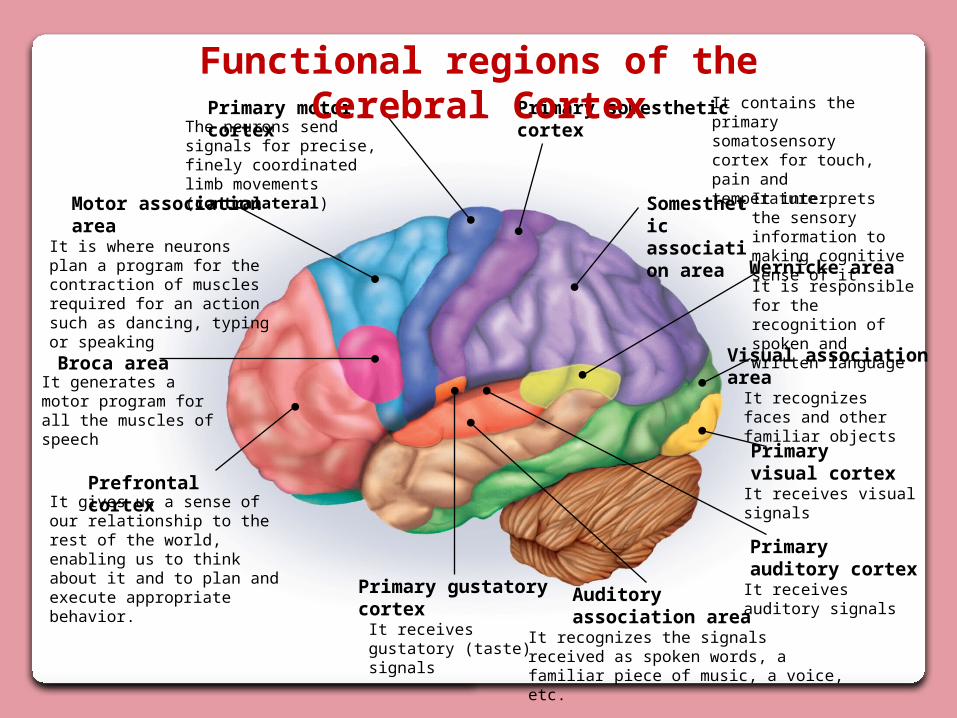
Broca area
Primary motor cortex
Motor associationarea
Prefrontal cortex
Primary somestheticcortex
Somestheticassociation area
Primary gustatorycortex
Visual associationarea
Primaryvisual cortex
Primaryauditory cortex
Auditoryassociation area
It is where neurons plan a program for the contraction of muscles required for an action such as dancing, typing or speaking
It contains the primary somatosensory cortex for touch, pain and temperature.
The neurons send signals for precise, finely coordinated limb movements (contralateral)
It interprets the sensory information to making cognitive sense of it
Wernicke areaIt is responsible for the recognition of spoken and written language
It generates a motor program for all the muscles of speech
It receives visual signals
It receives auditory signals
It recognizes the signals received as spoken words, a familiar piece of music, a voice, etc.
It recognizes faces and other familiar objects
It receives gustatory (taste) signals
It gives us a sense of our relationship to the rest of the world, enabling us to think about it and to plan and execute appropriate behavior.
Functional regions of the Cerebral Cortex
Median section
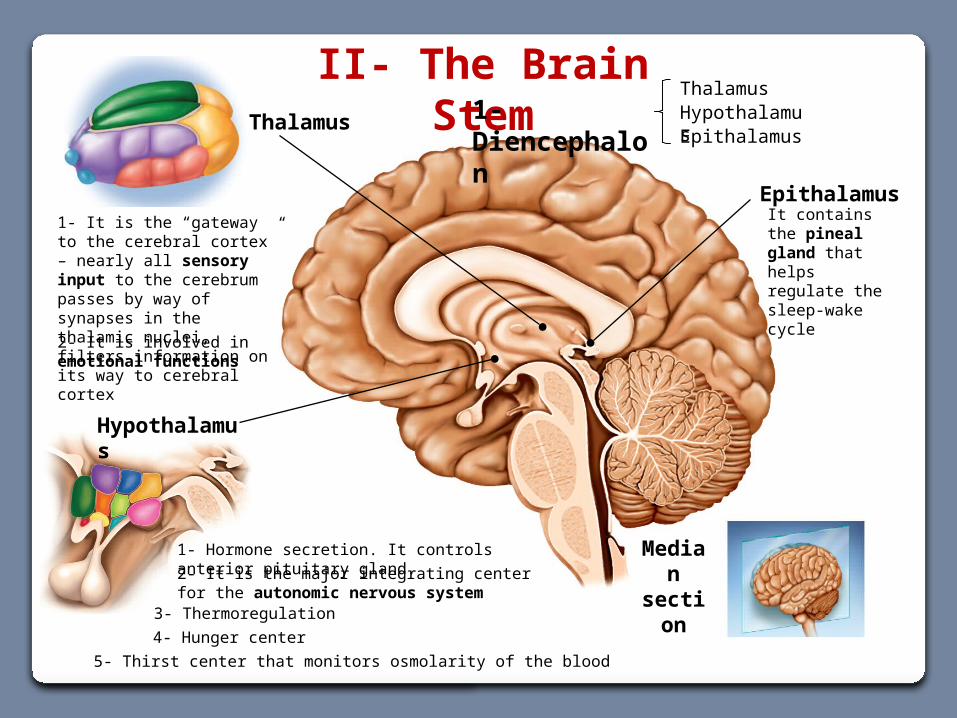
II- The Brain Stem1- Diencephalon
Hypothalamus
1- It is the “gateway to the cerebral cortex” – nearly all sensory input to the cerebrum passes by way of synapses in the thalamic nuclei, filters information on its way to cerebral cortex2- It is involved in emotional functions
Thalamus
1- Hormone secretion. It controls anterior pituitary gland.
ThalamusHypothalamusEpithalamus
EpithalamusIt contains the pineal gland that helps regulate the sleep-wake cycle
2- It is the major integrating center for the autonomic nervous system3- Thermoregulation4- Hunger center 5- Thirst center that monitors osmolarity of the blood
Median section
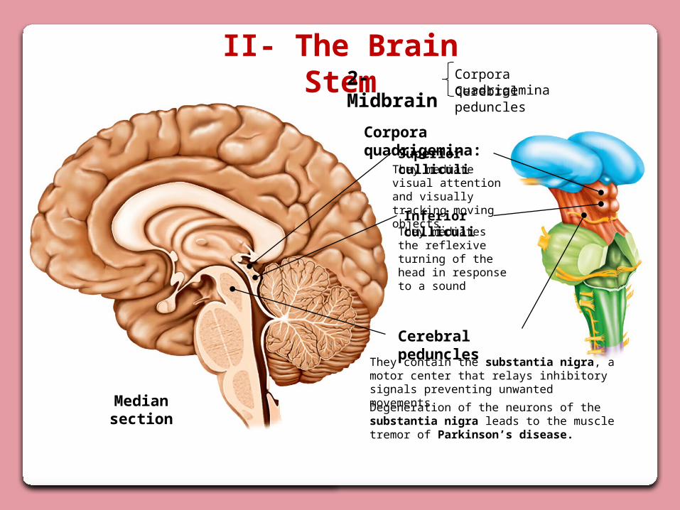
II- The Brain Stem2- Midbrain Cerebral peduncles
Corpora quadrigemina
Corpora quadrigemina:Superior culliculi
They mediate visual attention and visually tracking moving objects.
Inferior culliculiThey mediates the reflexive turning of the head in response to a sound
Cerebral peduncles
They contain the substantia nigra, a motor center that relays inhibitory signals preventing unwanted movements.
Degeneration of the neurons of the substantia nigra leads to the muscle tremor of Parkinson’s disease.
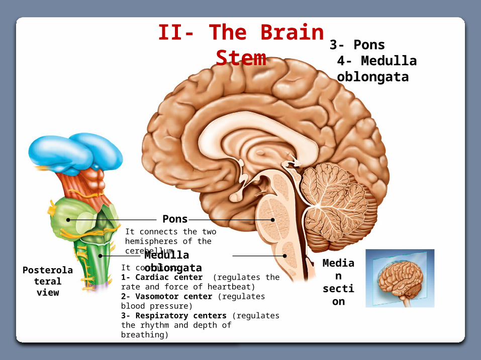
Posterolateral view
Median section
Medulla oblongataIt contains:1- Cardiac center (regulates the rate and force of heartbeat)2- Vasomotor center (regulates blood pressure)3- Respiratory centers (regulates the rhythm and depth of breathing)
PonsIt connects the two hemispheres of the cerebellum
II- The Brain Stem3- Pons4- Medulla oblongata
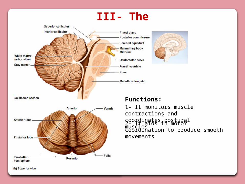
III- The Cerebellum
2- It aids in motor coordination to produce smooth movements
1- It monitors muscle contractions and coordinates postural muscles
Functions:
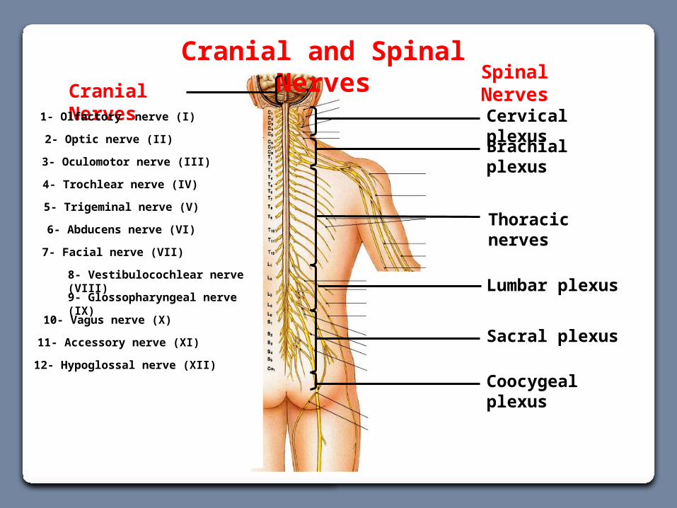
Thoracic nerves
Cranial and Spinal Nerves
Lumbar plexus
Sacral plexus
Brachial plexus
Cervical plexusCranial Nerves
2- Optic nerve (II)
5- Trigeminal nerve (V)
6- Abducens nerve (VI)
7- Facial nerve (VII)
8- Vestibulocochlear nerve (VIII)
9- Glossopharyngeal nerve (IX)
11- Accessory nerve (XI)
12- Hypoglossal nerve (XII)
10- Vagus nerve (X)
3- Oculomotor nerve (III)
1- Olfactory nerve (I)
4- Trochlear nerve (IV)
Spinal Nerves
Coocygeal plexus
Copyright © The McGraw-Hill Companies, Inc. Permission required for reproduction or display.
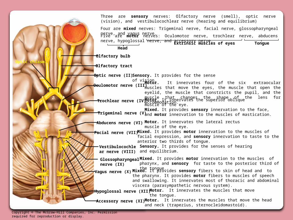
Optic nerve (II)
Trigeminal nerve (V)
Abducens nerve (VI)
Facial nerve (VII)
Vestibulocochlear nerve (VIII)
Glossopharyngeal nerve (IX)
Accessory nerve (XI)
Hypoglossal nerve (XII)
Vagus nerve (X)
Oculomotor nerve (III)
Olfactory tract
Olfactory bulb
Sensory. It provides for the sense of vision.
Motor. It innervates four of the six extraocular muscles that move the eyes, the muscle that open the eyelid, the muscle that constricts the pupil, and the muscle that changes the shape of the lens for accommodation.
Motor. It innervates the superior oblique muscle of the eye.
Mixed. It provides sensory innervation to the face, and motor innervation to the muscles of mastication.
Motor. It innervates the lateral rectus muscle of the eye.
Mixed. It provides motor innervation to the muscles of facial expression, and sensory innervation to taste to the anterior two thirds of tongue.
Sensory. It provides for the senses of hearing and equilibrium.
Mixed. It provides motor innervation to the muscles of pharynx, and sensory for taste to the posterior third of the tongue.
Mixed. It provides sensory fibers to skin of head and to the pharynx. It provides motor fibers to muscles of speech and swallowing. It innervates most of thoracic and abdominal viscera (parasympathetic nervous system).
Optic chiasm
Trochlear nerve (IV)
Motor. It innervates the muscles that move the tongue.
Motor. It innervates the muscles that move the head and neck (trapezius, sternocleidomastoid).
Three are sensory nerves: Olfactory nerve (smell), optic nerve (vision), and vestibulocochlear nerve (hearing and equilibrium)
Four are mixed nerves: Trigeminal nerve, facial nerve, glossopharyngeal nerve, and vagus nerve.
Five are motor nerves: Oculomotor nerve, trochlear nerve, abducens nerve, hypoglossal nerve, and accessory nerve.
HeadExtrinsic muscles of eyes Tongue