Lecture 12b - Johns Hopkins Bloomberg School of Public Health
51
Copyright 2007, The Johns Hopkins University and Ronald Gray. All rights reserved. Use of these materials permitted only in accordance with license rights granted. Materials provided “AS IS”; no representations or warranties provided. User assumes all responsibility for use, and all liability related thereto, and must independently review all materials for accuracy and efficacy. May contain materials owned by others. User is responsible for obtaining permissions for use from third parties as needed. This work is licensed under a Creative Commons Attribution-NonCommercial-ShareAlike License . Your use of this material constitutes acceptance of that license and the conditions of use of materials on this site.
Transcript of Lecture 12b - Johns Hopkins Bloomberg School of Public Health

Copyright 2007, The Johns Hopkins University and Ronald Gray. All rights reserved. Use of these materials permitted only in accordance with license rights granted. Materials provided “AS IS”; no representations or warranties provided. User assumes all responsibility for use, and all liability related thereto, and must independently review all materials for accuracy and efficacy. May contain materials owned by others. User is responsible for obtaining permissions for use from third parties as needed.
This work is licensed under a Creative Commons Attribution-NonCommercial-ShareAlike License. Your use of this material constitutes acceptance of that license and the conditions of use of materials on this site.

Lecture 12b: Congenital Malformations, Stillbirths and
Perinatal Deaths

Congenital Malformations

Definition Congenital Malformations• Anomalies of structure or function of the
fetus which occur before birth• Major Birth Defects
– Anomalies requiring medical/surgical treatment or entailing significant handicaps, diagnosed at birth or during childhood
• Minor Birth Defects– Developmental anomalies present at birth,
but not requiring intervention or causing handicaps.

Childhood Cancers
• Cancers in childhood may have fetal origin and may be a result of intrauterine exposures– Childhood leukemia– Brain cancers– Neuroblastoma

Diagnosis of birth defects
• Diagnosis depends on severity and intensity of investigation:
– External anomalies usually detected at birth– Internal anomalies harder to diagnose
Anomalies threatening survival or causing morbidity in newborn usually diagnosed earlier
– Occult anomalies often not diagnosed before childhood or even adulthood (e.g., cardiovascular anomalies)

Sources of Data
• Birth certificates– variable quality of diagnosis – limited to anomalies discovered at birth, – no information on anomalies diagnosed at
later ages
• Surveillance (e.g., CDC birth defects registry, Hungarian registry etc)
• Specialized studies

Problems of measurement
• Many congenital anomalies result in spontaneous abortion or stillbirth
• Most diagnoses in live born represent selective survival
• Many anomalies do not present until latter age of childhood (e.g., CVD)
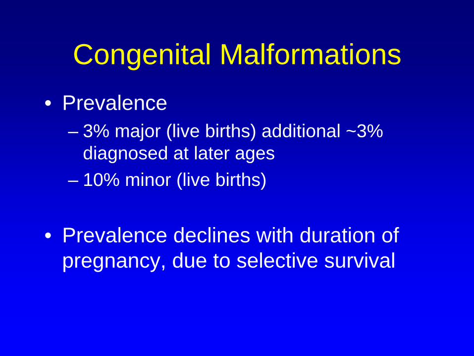
Congenital Malformations• Prevalence
– 3% major (live births) additional ~3% diagnosed at later ages
– 10% minor (live births)
• Prevalence declines with duration of pregnancy, due to selective survival

Distribution of Severity (Czeizel BMJ 1993;306:499)
Hungarian registry data • Lethal
– Rate 0.6%– 9% of anomalies
• Severe– Rate 1.9%– 30% of anomalies
• Mild– Rate 4.0%– 61% of all anomalies
Preventable anomalies ~ 70%?
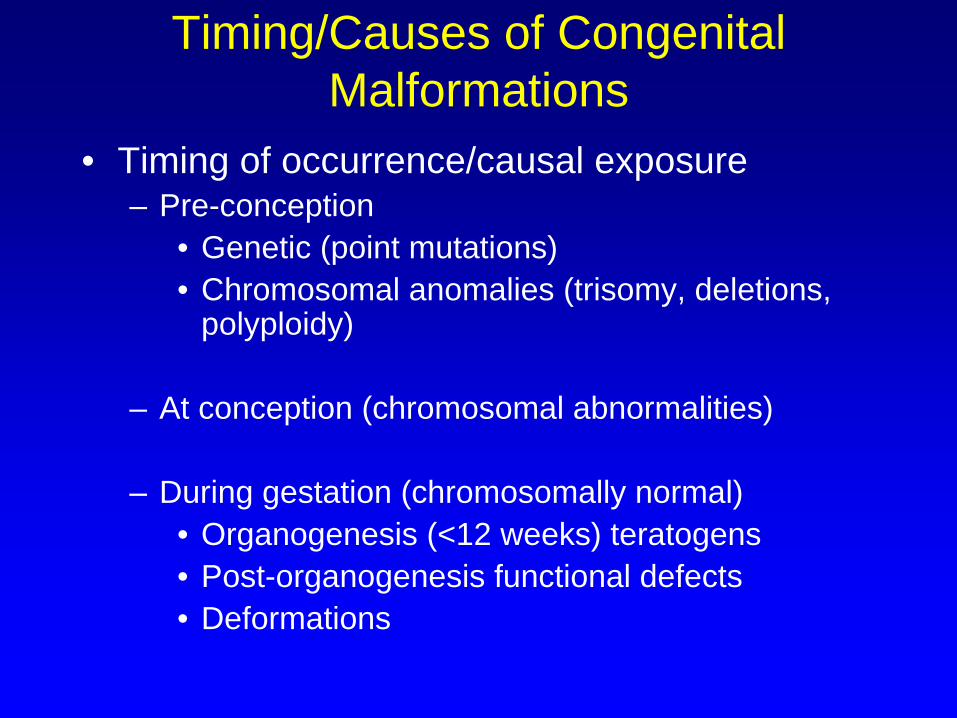
Timing/Causes of Congenital Malformations
• Timing of occurrence/causal exposure– Pre-conception
• Genetic (point mutations)• Chromosomal anomalies (trisomy, deletions,
polyploidy)
– At conception (chromosomal abnormalities)
– During gestation (chromosomally normal)• Organogenesis (<12 weeks) teratogens• Post-organogenesis functional defects• Deformations

Genetic and Chromosomal Defects• Genetic mutations
– rate ~ 2.25/1000 live births– 7.5% of all malformations– Increase with paternal age
• Chromosomal Anomalies– Rate 1.8/1000 live births– 6% of all defects– Increase with maternal age
• Chromosomal anomalies: • 40% spontaneous abortions • 6% stillbirths, • <1% live births
Source: Kalter NEJM 1983

Disorders of Organogenesis “Teratogenesis”
• Agents which disturb organ development are teratogens. • Malformations are multicellular disorders• Critical timing; effects only observed at specific times in
organ development• Most teratogens show a “threshold” or “all or none
phenomenon”– Most teratogenic agents have no effect below a
particular exposure dose– Teratogenesis incidence and severity tend to increase
above threshold in a dose-response manner– Percent of embryos affected increases with dose

Teratogens• Drugs (thalidomide, anticonvulsants, bendectin,
anesthetics)• Medical treatments
– IVF, ICSI• Radiation• Maternal infections (rubella, CMV, Toxoplasma)• Maternal illness (Diabetes, thyroid deficiency)• Environmental/occupational exposures
– Heavy metals (lead, cadmium, mercury)– Organic solvents, pesticides, dioxin
• Personal habits– Alcohol ? Fetal alcohol syndrome?
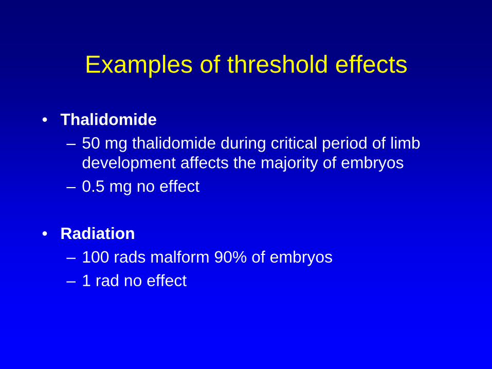
Examples of threshold effects
• Thalidomide – 50 mg thalidomide during critical period of limb
development affects the majority of embryos– 0.5 mg no effect
• Radiation– 100 rads malform 90% of embryos– 1 rad no effect

OrganogenesisTiming of Exposures
• Most organogenesis completed by 12th week in humans• Critical period of susceptibility varies between organ
systems• Dose dependent effects (e.g. embryo/fetal death,
structural defects)
Formation of organ systems• Cell proliferation, migration, differentiation, induction,
programmed cell death (apoptosis), developmental fields, effects on adjacent organ
• Animal models differ from humans (timing, susceptibility), need multiple species studies, and multiple generation studies
Malformation, deformation and disruption
• Malformation is a defect of organ formation which can lead to subsequent deformation or disruption– spina bifida → hip dislocation and club foot
• Deformation– Uterine constraint → club foot
• Disruption– Amniotic rupture → amniotic bands → limb
constriction/amputation

Classifications of Congenital Malformations
• Lumping vs. splitting of outcome• Single defects vs multiple defects (single
defects rarer)• Organ system, • Embryonic layer (mesoderm, ectoderm,
endoderm)• Syndromes (e.g. Downs, one etiology →
multiple organ system effects CNS, CVD)

Animal Studies of Birth defects
Animal Studies (teratogenicity, toxicology)– Can experimentally control dose and timing of
exposure– Can differentiate between maternal toxic
effects and teratogenic effects– Intergenerational effects– Generalizability to human exposure?? (e.g.,
thalidomide in rat or rabbit not teratogenic at human dose)

Toxicity StudiesPossible adverse effects:– Generalized toxicity (e.g. non-specific maternal
effects)– Developmental toxicity in the absence of maternal
toxicity is greatest concern– Adverse effects depend on timing of exposure:
• Embryonic period (< 8 weeks postconception) affect organogenesis, structural defects pregnancy loss)
• Fetal period (intrauterine growth, developmental abnormalities, fetal loss)
• Preconceptional gametogenesis (eg. gene mutation)– Reproductive effects (e.g. hormone exposures,
endocrine disruptors)– Induction of childhood cancers
Human Studies
• Problems– Rare events– Effects may be only on specific defects
(uncommon)– Classification of defects problematic– Easier to detect risks with unusual defects
than with more common defects (e.g., thalidomide)
Exposure measurement and teratogenicity
Refinement of exposure– Dose/timing
• Amount during entire pregnancy?• Amount during critical developmental period?• Total amount, average amount, or maximum
dose?– Problems
• Recall or measurement biases• Medicolegal and media environment (bias)
Congenital Malformations
Refinement of exposure– Stage of gestation
• Early blood glucose control with diabetic mother prevents adverse effects
• Folic acid and neural tube defects• Bendectin and pyloric stenosis
– problem one of the most commonly used drugs in early pregnancy in 1980s, massive publicity, selective recall and other bias, lack of consistency between studies
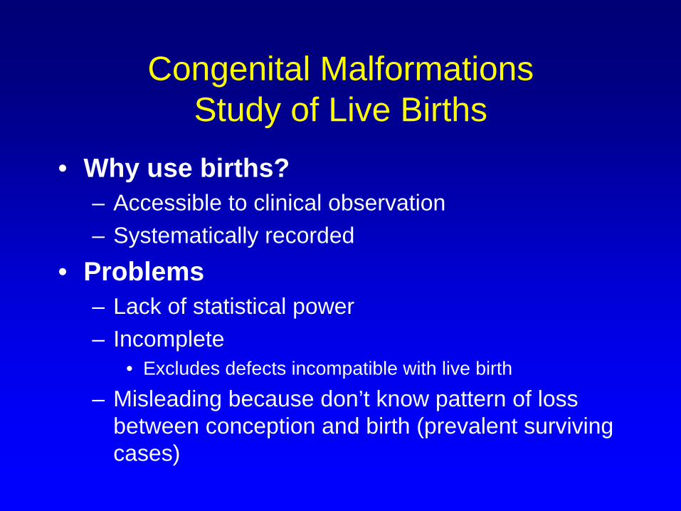
Congenital Malformations Study of Live Births
• Why use births?– Accessible to clinical observation– Systematically recorded
• Problems– Lack of statistical power– Incomplete
• Excludes defects incompatible with live birth
– Misleading because don’t know pattern of loss between conception and birth (prevalent surviving cases)
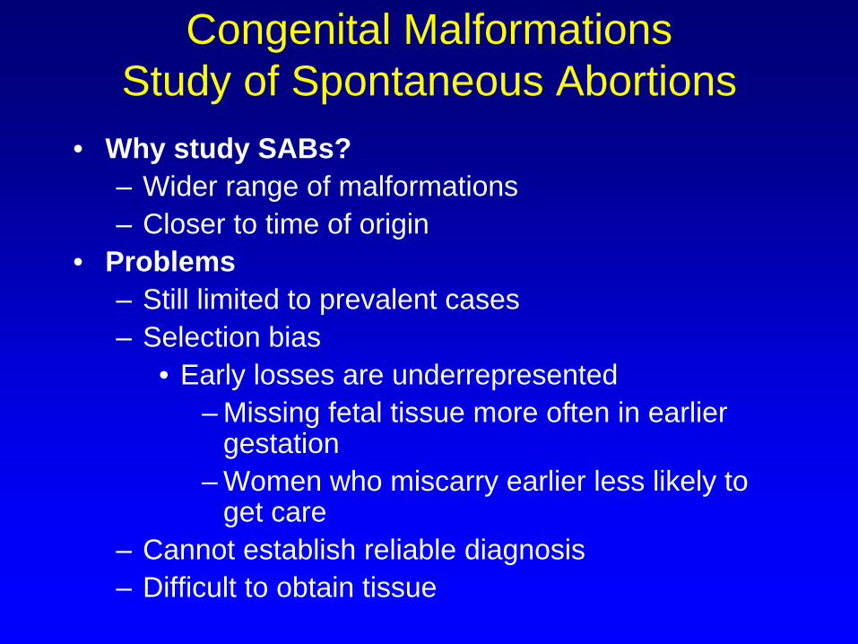
Congenital Malformations Study of Spontaneous Abortions
• Why study SABs?– Wider range of malformations– Closer to time of origin
• Problems– Still limited to prevalent cases– Selection bias
• Early losses are underrepresented– Missing fetal tissue more often in earlier
gestation– Women who miscarry earlier less likely to
get care– Cannot establish reliable diagnosis– Difficult to obtain tissue

0
10
20
30
40
50
60
~14 ~19 ~24 ~29 ~34 ~39 ~44
Maternal age at last menstrual period
Perc
ent
Spontaneous abortions spont abortions + livebirths
Euploid abortions euploid abortions + livebirths
Trisomic abortionsspont abortion + livebirths
Estimated rates (%) of spontaneous abortion, euploidabortion and trisomic abortion by maternal age for public patients
SAB Rates by Age
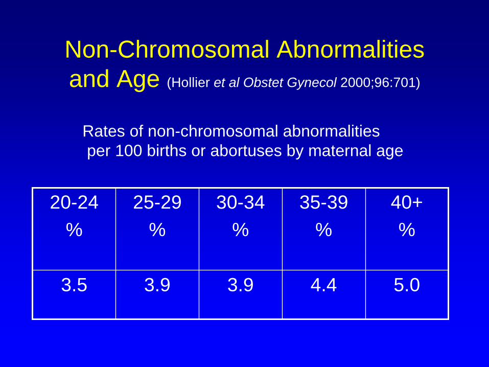
Non-Chromosomal Abnormalities and Age (Hollier et al Obstet Gynecol 2000;96:701)
20-24%
25-29%
30-34%
35-39%
40+%
3.5 3.9 3.9 4.4 5.0
Rates of non-chromosomal abnormalitiesper 100 births or abortuses by maternal age

Sex ratio at birth to infer earlier losses
• Dioxin exposure Seveso, Italy (Mocarelli Lancet 2000)– Paternal dioxin levels associated with reduced
male sex ratio (0.38 CI 0.3-0.5) vs unexposed sex ratio 0.55
• PCB exposures Japan (Occup Env Hlth 2001)
– No effect on sex ratio (0.51)
• Sex ratio may be a marker of of selective toxic exposures in utero
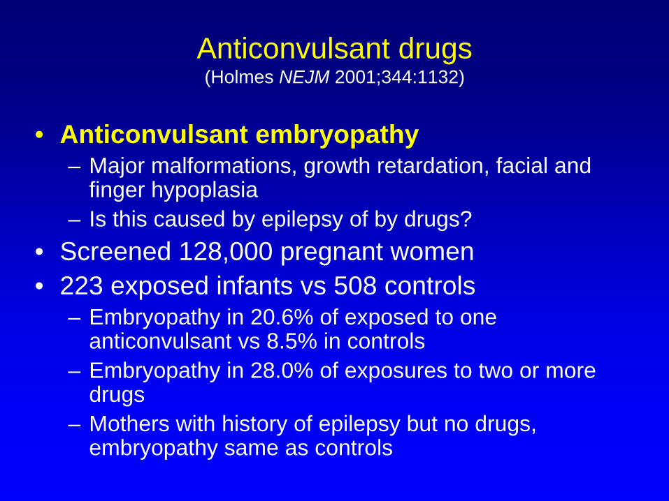
Anticonvulsant drugs (Holmes NEJM 2001;344:1132)
• Anticonvulsant embryopathy– Major malformations, growth retardation, facial and
finger hypoplasia– Is this caused by epilepsy of by drugs?
• Screened 128,000 pregnant women• 223 exposed infants vs 508 controls
– Embryopathy in 20.6% of exposed to one anticonvulsant vs 8.5% in controls
– Embryopathy in 28.0% of exposures to two or more drugs
– Mothers with history of epilepsy but no drugs, embryopathy same as controls
Late onset and intergenerational effects:
• Diethylstilbesterol (DES) Used for prevention of threatened miscarriage– Clear cell vaginal carcinoma in daughters– Increased breast cancer risk in daughters– Increased preterm birth, ectopics and
pregnancy loss in daughters pregnancy– Decreased sperm in sons– Increased testicular cancer in sons?

Ecologic Studies
• Chernobyl– Childhood leukemia in Finland – no effect
observed– Trisomy 21 in Berlin, cluster of 12 cases in
January 1987 (expect 2-3).

Deformations
• Misoprostol (prostaglandin) used for induced abortion in Brazil
• Mobius syndrome: facial paralysis with or without limb constriction defects
• OR = 30 with misoprosotol exposure
• Due to constriction caused by uterine contractions
– Pastuszak NEJM 1998;338:1881

Functional Neurologic Abnormalities (post-organogenesis)
• Cerbral palsy (CP)• Cystic periventricular leukomalacia
cPVL• Mental retardation• Other neurologic abnormalties (e.g.
strabismus)
Neurologic Abnormalities and Immune activation
• Neurologic abnormalities increasing with increased survival of preterm births
• Evidence that immunologic factors particularly fetal response play a role– IL-1 and TNF-a inflammatory cytokines produced by
stress and infections, affect brain by ischaemia/hypotension, cause cell death
• Cystic periventricular leukomalacia (cPVL) in new born (damage to white matter) associated with cerebral palsy and cognitive disorders

Meta-analysis Chorioamnionitis and Cerebral Palsy
(Wu JAMA 2000;284:1417)
• Chorioamnionitis and cerebral palsy RR = 1.9 (1.4-2.5)
• Chorioamnionitis and cystic periventricular leukomalacia (cPVL) RR = 3.0 (2.2-4.0)
• Chorioamnionitis and/or cPVL adjusted for gestational age RR = 1.8 (1.3-2.4)
Case Control Study of Choriomanionitis and Cerebral Palsy (JAMA 2003;290:2677)
• Chorioamnionitis in cerebral palsy (CP) = 14%, control 4%,
• Adj OR = 4.1 (1.6-10.1)

Perinatal Mortality
• Perinatal mortality rate (PNMR) = late fetal deaths plus early neonatal deaths/live and stillbirths– Late fetal deaths > 20 weeks
gestation (US)– Late fetal deaths > 28 weeks (WHO
international comparative data)– Early neonatal deaths < 1 week of life

Why Perinatal mortality
• Indicator of obstetric care and deaths later in pregnancy, intrapartum, post- delivery
• Avoids the problem of differentiating live from stillbirths
• Traditionally associated with “asphyxia”
Causes of Perinatal death
• Preterm delivery LBW• Respiratory distress syndrome• Congenital anomalies• Birth trauma/asphyxia (prolonged labor,
hemorrhage, multiple pregnancies)• Infections (syphilis, HIV)• Sepsis (B hemolytic strep)• Hypothermia

Neonatal Mortality• Neonatal mortality rate (NMR) = deaths
among live born infants prior to the first 28 days of life, per 1000 live births
• Early Neonatal mortality (ENMR) = deaths 0-6 days
• Late Neonatal mortality (LNMR) = deaths 7-28 days

Neonatal deaths world wide Lawn Lancet 2005;365:891
• ~ 4 million deaths• 38% of child deaths• 75% in 1st week of life• 99% in developing countries• Main causes
– Preterm birth (28%)– Infections 26%– Asphyxia 23%– Tetanus 7%

Causes of Non-Infectious Neonatal Deaths During Pregnancy and Delivery
• Birth Outcomes– Low birth weight– Preterm delivery– Birth defects
• Obstetric and Neonatal Complications– Injury/asphyxia– Hypothermia
• Maternal Health Preceding and During Pregnancy

Infectious Causes and Prevention of Neonatal Deaths
• Bacterial Infections– Sepsis (Group B
Streptoccocus)– Respiratory infections– Tetanus– Diarrhea– Meningitis
• Viral Infections– HIV– HSV-2– CMV
• Malaria
• Prevention of infection– immunization– prophylaxis via antibiotics
or antisepsis– Malaria prophylaxis
/control– Cord care (tetanus)– Treatment of infections in
new born– Aspesis/Cleansing of birth
canal– C section
Problems with measurement• Under registration in LDCs• Definition of a live birth problematic.
– Omission of infants who die shortly after birth affect numerator and denominator of NMR
– Variation in definition of “live birth” (e.g., USSR)– Advantage of PNMR
• Cause of death poorly reported– Autopsy in DCs– Verbal autopsy (interview) in LDCs

Neonatal Mortality and Contribution of Infection: Community Studies in Developing Countries
(Stoll Clinic Perinatol 1997;24:1)
• Neonatal Mortality/1000 LB
Africa 9-65/1000S. Asia 19-89/1000
• Proportion due to Infection (%)
9-54%8-64%
Neonatal infection is probably the major preventable cause of neonatal death in less developed countries

Trials to reduced neonatal mortality in developing countries
• India: Improved neonatal care reduced neonatal mortality (Bang Lancet 1999)
• Malawi: GFenital tract cleansing with chlorhexadine reduced neonatal sepsis and mortality (Taha BMJ)
• Nepal: Chlorhexadine washing of cord or baby reduced neonatal sepsis and tetanus

Neonatal tetanus
• Defined syndromically– Normal suckling and crying followed by an
inability to suck or cry; onset 3-10 days of age
– Spasms, stiffness, convulsions• ~ 450,000 cases per year• case-fatality ~75%• Preventable by immunization of mother
and cord care


Neonatal Mortality and Infant Mortality
• Where IMR is high, more deaths occur in the postneonatal period
• As Infant mortality (IMR) declines, NMR and particularly ENMR constitutes a greater proportion of deaths in infancy
• Declines in IMR shift focus of prevention to NMR