Frameshifted APP (APP+1) is a secretory protein and the level of ...

PROTEIN SORTING
Lecture 10 BIOL 266/4
2014-15
Biology Department Concordia University Dr. S. Azam
© 2013 John Wiley & Sons, Inc. All rights reserved.
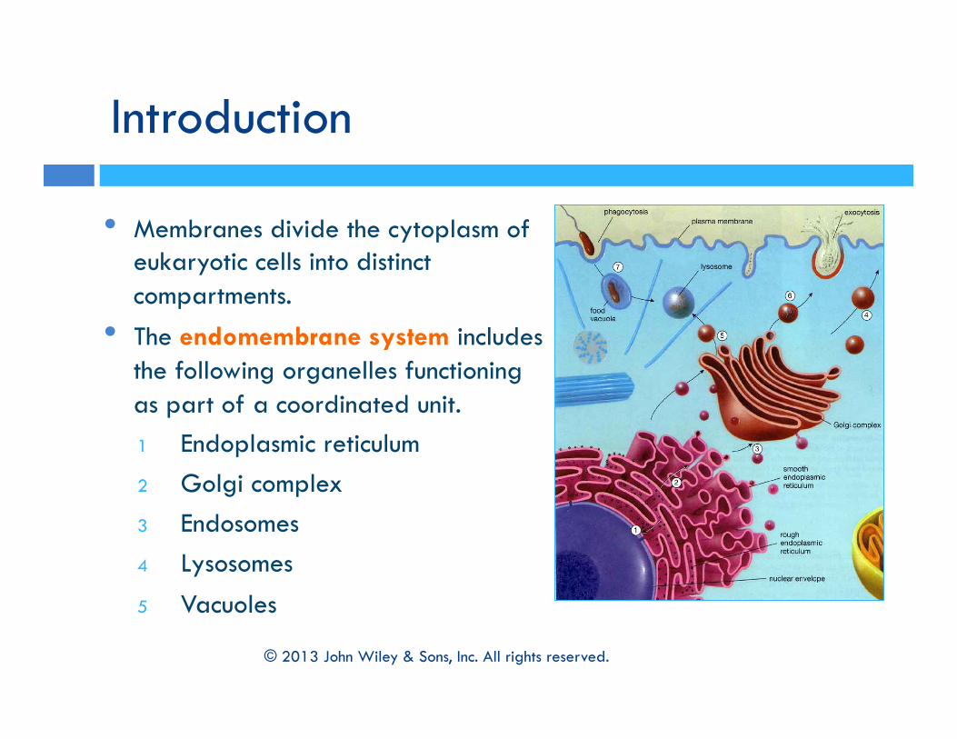
Introduction
• Membranes divide the cytoplasm of eukaryotic cells into distinct compartments.
• The endomembrane system includes the following organelles functioning as part of a coordinated unit. 1 Endoplasmic reticulum 2 Golgi complex 3 Endosomes 4 Lysosomes 5 Vacuoles
© 2013 John Wiley & Sons, Inc. All rights reserved.

• Organelles of the endomembrane system are part of an integrated network in which materials are shuttled back and forth in membrane-bound transport vesicles. • Upon reaching their destination,
the vesicles fuse with the membrane of the acceptor compartment.
Overview of the Endomembrane System
© 2013 John Wiley & Sons, Inc. All rights reserved.

Biosynthetic pathway – synthesis in the ER, modification through the Golgi complex and transport of proteins to the plasma membrane and lysosomes etc.
Also known as the secretory pathway – when proteins are discharged (secreted) from the cell. Constitutive secretion – in a
continuous fashion. Regulated secretion – in response
to a stimulus.
Overview of the Endomembrane System Biosynthetic pathways
© 2013 John Wiley & Sons, Inc. All rights reserved.

Overview of the Endomembrane System Synthesis and transport of secretory proteins
• Secretory pattern of the protein can be studied by using radioactive amino acids
• During regulated secretion, materials to be secreted are stored in large, membrane-bound secretory granules.
For example, secretion of digestive enzymes by pancreatic cells.
3 minute pulse: radioactively labeled amino acid (red)
3 minute pulse: 17 minute chase
3 minute pulse: 117 minute chase
© 2013 John Wiley & Sons, Inc. All rights reserved.

Green fluorescent protein (GFP) – a small protein isolated from jellyfish which emits green fluorescent light.
A GFP-VSV gene chimera allows to observe the protein synthesis in the cell.
By infecting a cell with VSV, massive amounts of VSVG protein are produced in the ER.
Mutant VSVG is unable to leave the ER of the infected cell at 40 degrees
Study of Cytomembranes GFP-based protein tracking
The use of green fluorescent protein (GFP) reveals the movement of proteins within a living cell.
protein stuck in ER proteins can move from ER to Golgi Use of Green Fluorescent Protein
© 2013 John Wiley & Sons, Inc. All rights reserved.

Study of Mutants • Mutants provide insights
about the function of normal gene products.
• Isolation of proteins from yeast has led to the identification of homologous proteins in mammals, pointing to the conserved nature of endomembrane systems.
Study of Cytomembranes Utility of genetic mutants
© 2013 John Wiley & Sons, Inc. All rights reserved.
Muta=ons can be introduced at different steps (1 and 2)

1. The Endoplasmic Reticulum
• The endoplasmic reticulum (ER) comprises a network of membranes that penetrates much of the cytoplasm.
• Like other organelles, the ER is highly dynamic undergoing continual turnover and reorganization.
• Divided into two sub-compartments: 1. Rough endoplasmic reticulum (RER)
2. Smooth endoplasmic reticulum (SER)
Electron micrograph: rough ER
Electron micrograph: smooth ER © 2013 John Wiley & Sons, Inc. All rights reserved.

Rough Endoplasmic Reticulum (RER) • Composed of a network of flattened sacs (cistenae). • Continuous with the outer membrane of the nuclear envelope • Has ribosomes on its cytosolic surface. • Starting point of the biosynthetic pathway • Site of discharge of the protein. • Different types of cells have different ratios of the two types of ER,
depending on activities of the cell.
The Endoplasmic Reticulum
© 2013 John Wiley & Sons, Inc. All rights reserved.
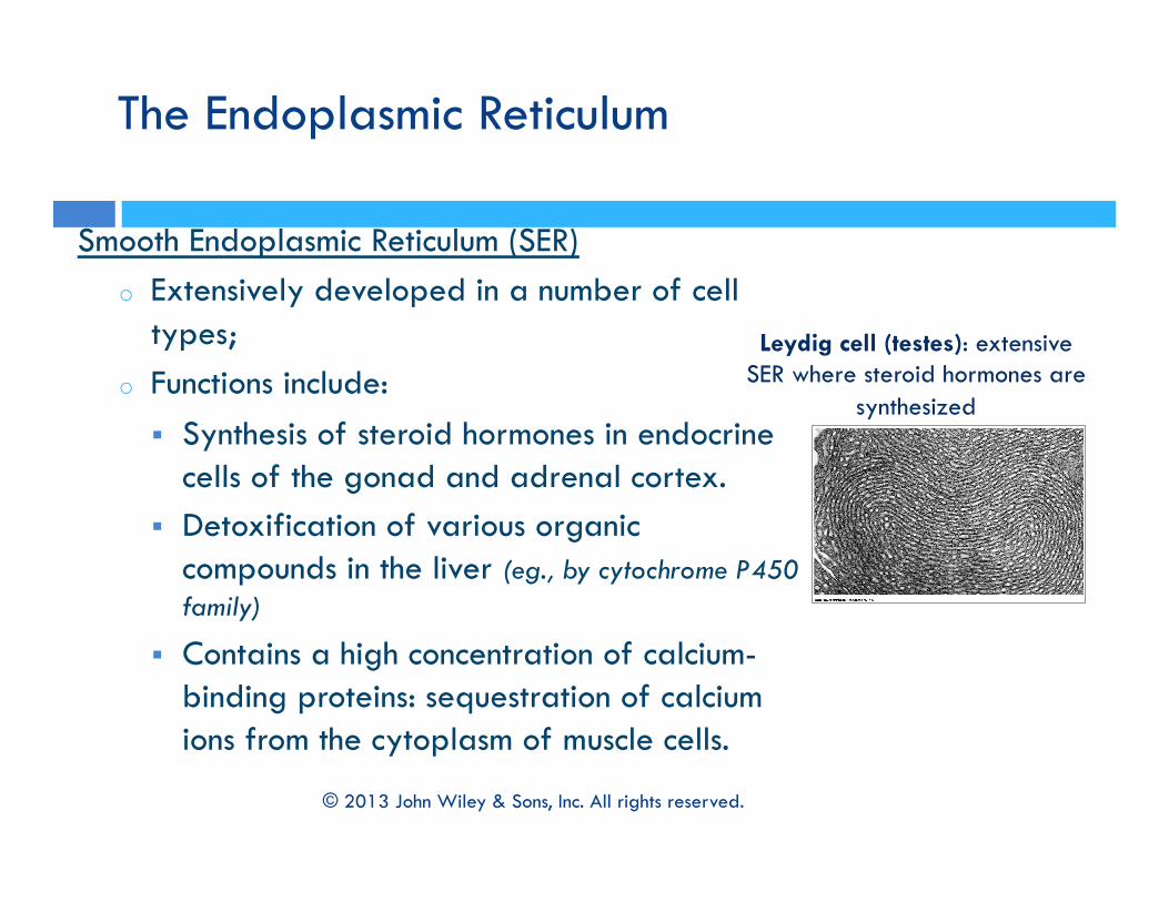
Smooth Endoplasmic Reticulum (SER) o Extensively developed in a number of cell
types; o Functions include:
Synthesis of steroid hormones in endocrine cells of the gonad and adrenal cortex.
Detoxification of various organic compounds in the liver (eg., by cytochrome P450 family)
Contains a high concentration of calcium-binding proteins: sequestration of calcium ions from the cytoplasm of muscle cells.
The Endoplasmic Reticulum
Leydig cell (testes): extensive SER where steroid hormones are
synthesized
© 2013 John Wiley & Sons, Inc. All rights reserved.

The Endoplasmic Reticulum
• Functions of the RER Synthesis of Proteins on Membrane-Bound versus Free Ribosomes • Approximately 1/3 polypeptides encoded by the human
genome are synthesized on ribosomes of RER include secretory proteins and integral membrane proteins • Polypeptides synthesized on “free” ribosomes include
cytosolic proteins, peripheral membrane proteins, nuclear proteins, and proteins incorporated into chloroplasts, mitochondria and peroxisomes.
© 2013 John Wiley & Sons, Inc. All rights reserved.

The Endoplasmic Reticulum Synthesis of protein on membrane-bound ribosome
1. Synthesis of Secretory/ Lysosomal protein on Membrane-Bound Ribosomes
• Messenger RNA binds to free ribosomes on cytosol • Movement through the membrane can occur as it is
being synthesized (co-translationally) or post-translational.
• Secretory proteins synthesized on membrane-bound ribosomes have their signal sequence recognized by a signal recognition particle (SRP)
© 2013 John Wiley & Sons, Inc. All rights reserved.

The Endoplasmic Reticulum Synthesis of protein on membrane-bound ribosome
1. Synthesis of Secretory/ Lysosomal protein on Membrane-Bound Ribosomes • Binding to the ER occurs through two sequential interactions:
1 SRP at N-terminus of the polypeptide must interact with a SRP receptor on the ER membrane.
2 Ribosome interacts with the translocon, which is a protein-lined channel.
• Once the SRP-ribosome-nascent peptide chain complex binds ER, SRP is released.
• Release of SRP requires GTP-binding proteins (G proteins).
© 2013 John Wiley & Sons, Inc. All rights reserved.

The Endoplasmic Reticulum Synthesis of protein on membrane-bound ribosome
A schema=c model of the synthesis of a secretory protein (or a lysosomal enzyme) on a membrane-‐bound ribosome of the RER
© 2013 John Wiley & Sons, Inc. All rights reserved.

2. Synthesis of Integral Membrane Proteins on Membrane-Bound Ribosomes • Integral proteins contain hydrophobic trans-membrane
segments that interfere with transfer into the RER lumen. • The two sequential events: SRP-SRP receptor binding and
ribosome-translocon binding occurs similarly as for the secretory proteins.
• Translocon assists in the proper orientation of transmembrane sequences.
• The arrangement within the membrane is determined by the orientation of the first trans-membrane segment inserted.
The Endoplasmic Reticulum Synthesis of protein on membrane-bound ribosome
© 2013 John Wiley & Sons, Inc. All rights reserved.

The Endoplasmic Reticulum Synthesis of protein on membrane-bound ribosome
A schema=c model for the synthesis of an integral membrane protein
© 2013 John Wiley & Sons, Inc. All rights reserved.
Steps 1-‐3: N terminus is in the lumen and C terminus in the cytosol Entry of the hydrophobic transmembrane sequence into the pore blocks further transloca=on of the polypep=de Lateral gate of the transposon opens and the transmembrane segment is expelled in the lipid bilayer

The Endoplasmic Reticulum Synthesis of protein on membrane-bound ribosome
A schema=c model for the synthesis of an integral membrane protein
© 2013 John Wiley & Sons, Inc. All rights reserved.
Steps 1-4a: C terminus is in the lumen and N terminus in the cytosol Translocon assists in the proper orientation of transmembrane sequences difference in charge between phospholipids of the cytosolic and luminal leaflets of the bilayer is thought to play a role in the protein orientation.

Processing of Newly Synthesized Proteins in the ER • Upon entering the RER lumen, the signal sequence is
cleaved by a signal peptidase. • Carbohydrates are added by the enzyme
oligosaccharyltransferase. • The RER lumen is packed with chaperones to assist in
folding, and also contains protein disulfide isomerase to add disulfide bonds to cysteines.
The Endoplasmic Reticulum Synthesis of protein on membrane-bound ribosome
© 2013 John Wiley & Sons, Inc. All rights reserved.

Maintenance of membrane asymmetry
• Membranes arise from pre-existing membranes.
• Lipids are inserted into existing membranes.
• As the membrane moves from one compartment to the next, its proteins and lipids are modified.
• Membrane asymmetry is established initially and maintained during trafficking.
Membrane Biosynthesis in the ER
© 2013 John Wiley & Sons, Inc. All rights reserved.
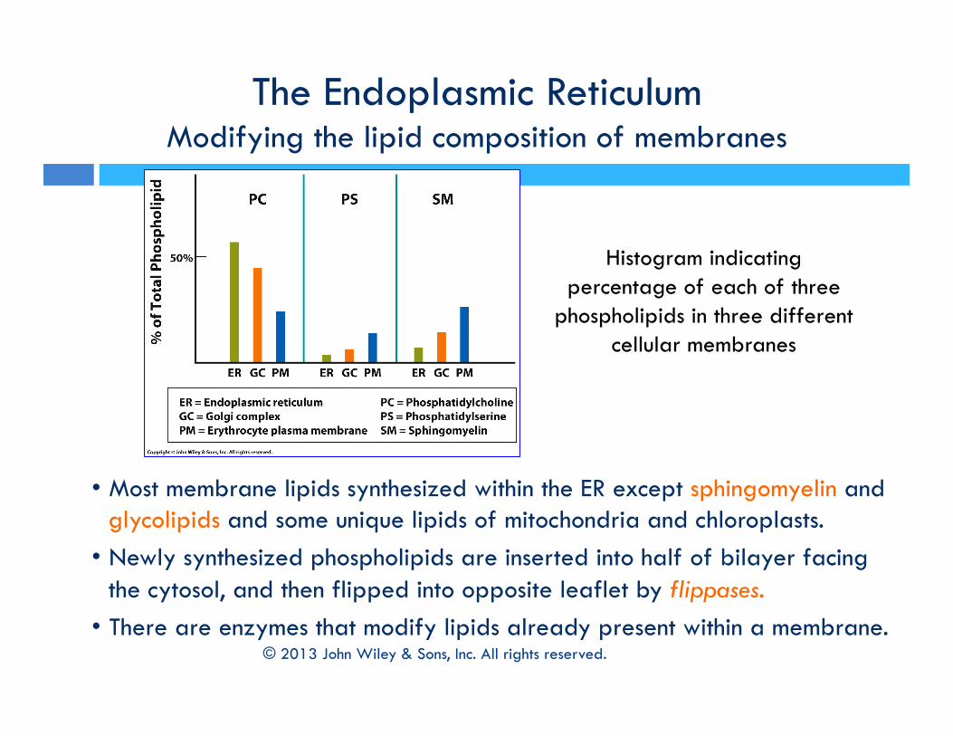
• Most membrane lipids synthesized within the ER except sphingomyelin and glycolipids and some unique lipids of mitochondria and chloroplasts. • Newly synthesized phospholipids are inserted into half of bilayer facing
the cytosol, and then flipped into opposite leaflet by flippases. • There are enzymes that modify lipids already present within a membrane.
The Endoplasmic Reticulum Modifying the lipid composition of membranes
Histogram indicating percentage of each of three
phospholipids in three different cellular membranes
© 2013 John Wiley & Sons, Inc. All rights reserved.
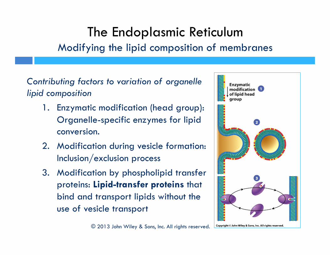
The Endoplasmic Reticulum Modifying the lipid composition of membranes
Contributing factors to variation of organelle lipid composition
1. Enzymatic modification (head group): Organelle-specific enzymes for lipid conversion.
2. Modification during vesicle formation: Inclusion/exclusion process
3. Modification by phospholipid transfer proteins: Lipid-transfer proteins that bind and transport lipids without the use of vesicle transport
© 2013 John Wiley & Sons, Inc. All rights reserved.

Quality control: ensuring misfolded proteins do not proceed forward.
• Soon after transfer to the polypeptide, the oligosaccharide is gradually modified.
• A glycoprotein goes through a system of quality control to determine its fitness for a specific compartment.
• Misfolded proteins are tagged by a terminal glucose and recognized by chaperones for refolding.
• If the protein does not fold correctly, it is translocated to the cytosol and destroyed.
The Endoplasmic Reticulum
Quality control: ensuring that misfolded proteins do not proceed forward.
© 2013 John Wiley & Sons, Inc. All rights reserved.
The Endoplasmic Reticulum
Quality control: ensuring that misfolded proteins do not proceed forward.
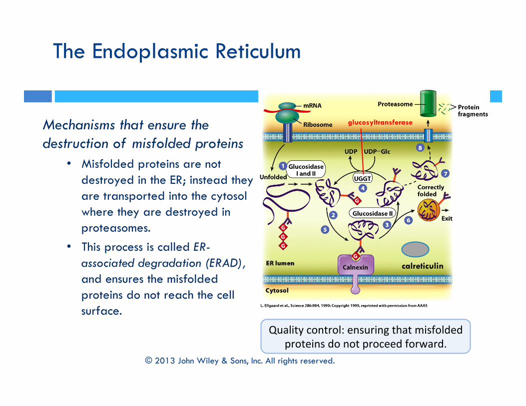
Mechanisms that ensure the destruction of misfolded proteins
• Misfolded proteins are not destroyed in the ER; instead they are transported into the cytosol where they are destroyed in proteasomes.
• This process is called ER-associated degradation (ERAD), and ensures the misfolded proteins do not reach the cell surface.
© 2013 John Wiley & Sons, Inc. All rights reserved.
calreticulin
glucosyltransferase

Destruction of misfolded proteins • Accumulation of misfolded
proteins triggers the unfolded protein response (UPR).
• Sensors in the ER are kept inactive by the chaperone BiP but if misfolded proteins are accumulated, BiP molecules are incapable of inhibiting the sensors.
• Activated sensors send signals to trigger proteins involved in destruction of misfolded proteins.
A model of the mammalian unfolded protein response (UPR)
© 2013 John Wiley & Sons, Inc. All rights reserved.

Put your thinking cap on….
1. Which of the following cells would you expect to be engaged most heavily in bulk-phase endocytosis and exocytosis:
(a) an erythrocyte (b) a pancreatic acinar cell (c) a skeletal muscle cell?
And Why? 2. If you were to add a drug that interfered
with the ability of ribosomes to bind to mRNA, what effect would this be expected to have on the structure of the RER?
© 2013 John Wiley & Sons, Inc. All rights reserved.

Some interesting links
https://www.youtube.com/watch?v=eH5k8XYKycs https://www.youtube.com/watch?v=IyBvn2iql2M https://www.youtube.com/watch?v=4qf1BSXn_tk
© 2013 John Wiley & Sons, Inc. All rights reserved.