Lecture 08:Lecture 08: Review: Basics of the Diffusion ... 08:Lecture 08: Review: Basics of the...
43
BME 42-620 Engineering Molecular Cell Biology Lecture 08: Lecture 08: Review: Basics of the Diffusion Theory The Cytoskeleton (I) 1 BME42-620 Lecture 08, September 22, 2011
Transcript of Lecture 08:Lecture 08: Review: Basics of the Diffusion ... 08:Lecture 08: Review: Basics of the...

BME 42-620 Engineering Molecular Cell Biology
Lecture 08:Lecture 08:
Review: Basics of the Diffusion Theory
The Cytoskeleton (I)
1BME42-620 Lecture 08, September 22, 2011
Outline
• Background: FRAP & SPT
• Review: microscopic diffusion theory
• Review: macroscopic diffusion theoryp y
• An overview of the cytoskeleton• An overview of the cytoskeleton
• Actin and its associated proteins
2

Outline
• Background: FRAP & SPT
• Review: microscopic diffusion theory
• Review: macroscopic diffusion theoryp y
• An overview of the cytoskeleton• An overview of the cytoskeleton
• Actin and its associated proteins
3

Fluorescence Microscopy of Cell Dynamics
http://lippincottschwartzlab.nichd.nih.gov/video/classic/VSVGrelease.mov

Two Frequently Used Methods to Determine Diffusion CoefficientDiffusion Coefficient
• Method 1: Fluorescence recovery after photobleaching
• Method 2: Single particle trackingg p g
5

Fluorescence Recovery After Photobleaching (FRAP)
• FRAP provides a convenientapproach to visualize diffusion.pp
• Diffusion coefficient can beestimated from FRAP.
2wD 1/24D
t
1/2
: radius of a Gaussian profile bleaching beamh lf ti f fl
wt
1) D. Axelrod, D.E. Koppel, J. Schlessinger, E. Elson, and W.W. Webb. MobilityMeasurement by Analysis of Fluorescence Photobleaching Recovery Kinetics.Biophys. J. 1976; 16(9):1055-1069.
1/2: half time of fluorescence recoveryt
6
2) J. Lippincott-Schwartz, N. Altan-Bonnet, G. H. Patterson,Photobleaching and photoactivation: following protein dynamics in living cells.Nature Cell Biology, 2003 Sep;Suppl:S7-14.

Single Particle Tracking (SPT)
D Wi t P ti l t ki i h l f li i ll A RD. Wirtz, Particle-tracking microrheology of living cells, Ann. Rev. Biophys. 38:301-326, 2009.
7
http://web.mit.edu/savin/Public/.Tutorial_v1.2/Introduction.html
Outline
• Background: FRAP & SPT
• Review: microscopic diffusion theory
• Review: macroscopic diffusion theoryp y
• An overview of the cytoskeleton• An overview of the cytoskeleton
• Actin and its associated proteins
8

1D Random Walk in Solution (I)
• Assumptions:(1) A particle i has equal probabilities to walk to the left and to the right.
(2) Particle movement at consecutive time points are independent.
(3) M t f diff t ti l i d d t(3) Movement of different particles are independent.
(4) Each particle moves at a average step size of δ=vx·τ
1i ix n x n i i
9

1D Random Walk in Solution (II)
• Property 1: The mean position of an ensemble of particles undergoing random walk remains at the origin.
• The same holds for a single particle over a sufficiently long period of time (ergodicity). ( g y)
1i ix n x n
1 1 1N N
x n x n x n
1 1
1
1
1 1 1
i ii iN
ii
x n x n x nN N
x n x nN
10

1D Random Walk in Solution (III)
• Property 2: The mean square displacement of an ensemble of particles undergoing random walk increases linearly w r t timeparticles undergoing random walk increases linearly w.r.t. time.
• Again, the same holds for a single particle. g g p
2 2 2 2
1 12 2
1 1 1 2 1N N
i i ii i
x n x n x n x nN N
2 21x n
2 2 2 2tx n n Dt
2 2 2 4r n x n y n Dt
2 2 2 2 6r n x n y n z n Dt
22 V
11
22
2 2xVD

1D Random Walk in Solution (IV)
• Property 3: The displacement of a particle follows a normal distributiondistribution.
! 1 1
! ! 2 2k n k
np k;nk n k
2
2
2 222
1 where and 4 22
k n np k e
2x n k n k k n
2 2 2 2 2 2 2 2 24 4 2k k
2 0x n k n
2 2 2 2 2 2 2 2 24 4 2x n k k n n n n n n n
2
241 where 2xDtp x e n Dt
12
where 24
p x e n DtDt

Application of the Microscopic Theory (I)
Object Distance diffused
1 μm 100 μm 1 mm 1 m1 μm 100 μm 1 mm 1 m
K+ 0.25ms 2.5s 2.5104s(7 hrs)
2.5108s (8 yrs)(7 hrs) (8 yrs)
Protein 5ms 50s 5.0105s (6 days)
5.0109s (150 yrs)
Organelle 1s 104s 108s 1012sg(3 hrs) (3 yrs) (31710 yers)
K+: Radius = 0 1nm viscosity = 1mPa·s-1; T = 25°C; D=2000 μm2/secK+: Radius = 0.1nm, viscosity = 1mPa s ; T = 25 C; D=2000 μm /secProtein: Radius = 3nm, viscosity = 0.6915mPa·s-1; T = 37; D = 100 μm2/secOrganelle: Radis = 500nm, viscosity = 0.8904mPa·s-1; T = 25°C; D = 0.5 μm2/sec
13

Application of the Microscopic Theory (II)
Pure diffusionx2 >
Diffusion with external flow
Pure diffusionac
emen
t <ua
re d
ispl
aM
ean
squ Diffusion in a cage
H. Qian, M. P. Sheetz, E. L. Elson, Single particle tracking: analysis of diffusion and flow in two-dimensional systems, Biophysical Journal 60(4):910 921 1991
t
14
Biophysical Journal, 60(4):910-921, 1991.
Outline
• Background: FRAP & SPT
• Review: microscopic diffusion theory
• Review: macroscopic diffusion theoryp y
• An overview of the cytoskeleton• An overview of the cytoskeleton
• Actin and its associated proteins
15

Macroscopic Theory of Diffusion (I)
• Fick's first equation: net flux is proportional to the spatial gradient of the concentration function.
12N x N x
1
0
2
0
12
12
xF N x N x / A
N x N xA A
lim
lim
CF D
0
1D C x C x
CDx
lim
xF Dx
16
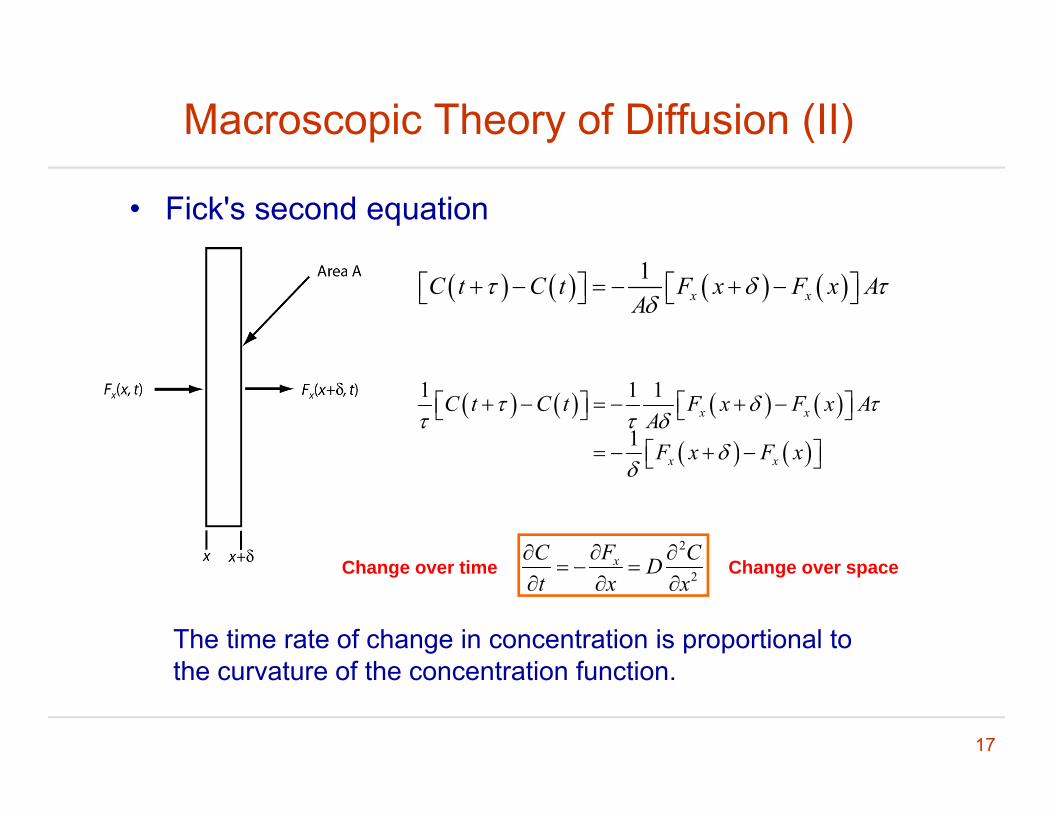
Macroscopic Theory of Diffusion (II)
• Fick's second equation
1x xC t C t F x F x A
A
1 1 1
1x xC t C t F x F x A
AF x F x
x xF x F x
2xFC CD
Ch i Ch2x D
t x x
The time rate of change in concentration is proportional to th t f th t ti f ti
Change over time Change over space
17
the curvature of the concentration function.

Diffusion Coefficient of a Particle
2
2 • Einstein-Smoluchowski Relation
1 12 2
xd
Fv am
22
2
22x x
d
mF mvm kTfv D D
kTDf
f: viscous drag coefficient
• Stokes' relation: the viscous drag coefficient of a sphere moving in an unbounded fluid
6f r : viscousityr: radius
18

An example of D calculation
• Calculation of diffusion coefficient
6kTDr
• k=1.38110-23J/k=1.381 10-17 N·m/k
• T = 273.15 + 25• =0.8904mPa·s=0.8904 10-3 10-12N·m-2·s• r= 500nm=0 5μmr 500nm 0.5μm• D=0.5 m2/s
19

References
• Howard Berg, Random Walks in Biology, Princeton University Press, 1993.
• Jonathon Howard, Mechanics of Motor Proteins and the Cytoskeleton, Sinauer Associated, 2001.
20

Outline
• Background: FRAP & SPT
• Review: microscopic diffusion theory
• Review: macroscopic diffusion theoryp y
• An overview of the cytoskeleton• An overview of the cytoskeleton
• Actin and its associated proteins
21

The Cytoskeleton is Highly Dynamic and Regulated
22
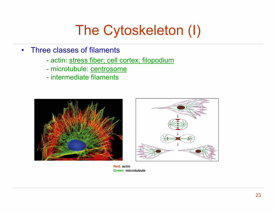
The Cytoskeleton (I)• Three classes of filaments
- actin: stress fiber; cell cortex; filopodiumi b l- microtubule: centrosome
- intermediate filaments
Red: actinGreen: microtubule
23
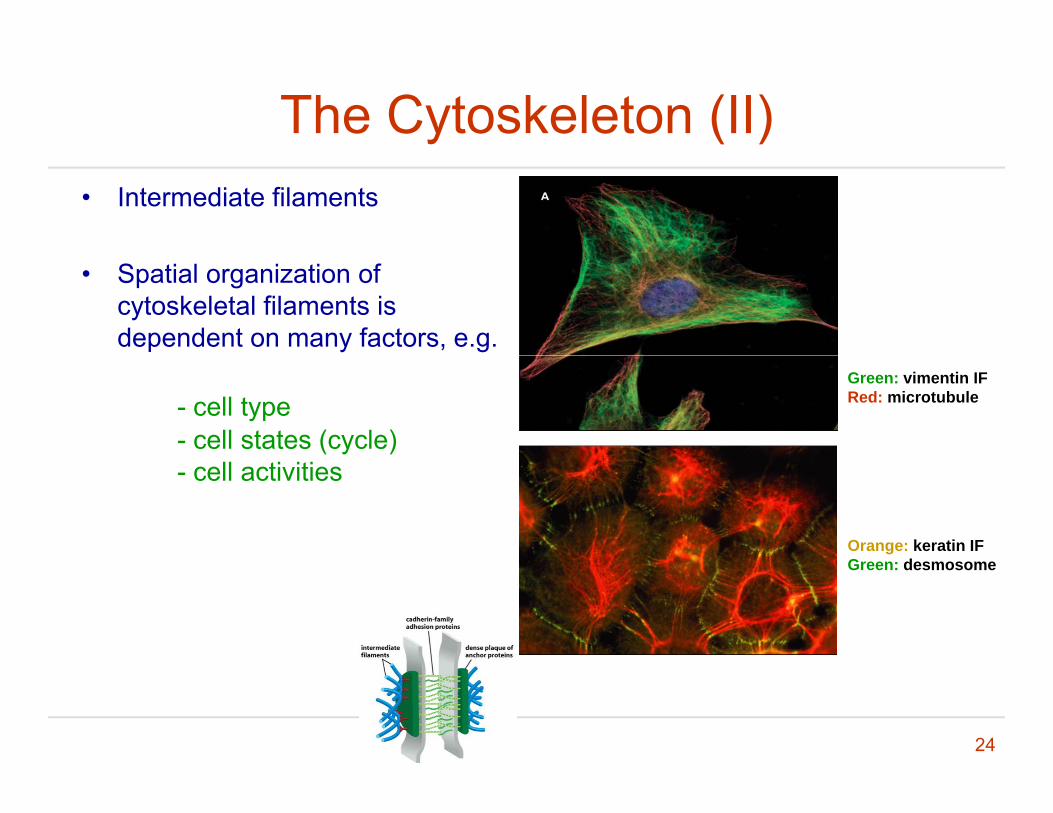
The Cytoskeleton (II)• Intermediate filaments
• Spatial organization of cytoskeletal filaments is dependent on many factors, e.g.
Green: vimentin IFRed: microtubule- cell type
- cell states (cycle)- cell activities
Orange: keratin IFG dGreen: desmosome
24

The Cytoskeleton (III)• The cytoskeleton plays a
critical role in many basic ll l f ticellular functions, e.g.
- structural organization & supportshape control- shape control
- intracellular transport- force and motion generation- signaling integrationg g g
• Highly dynamic and adaptive
25

Overview of Cytoskeletal Filaments
Shape Diameter Subunits Polarized
i bl iactin cable ~6 nm actinmonomer
yes
microtubule tube ~25nm tubulin heterodimer
yes
26
intermediate filament
rope ~10nm Various dimers
no
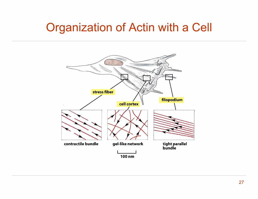
Organization of Actin with a Cell
27

Actin Structure and Function• Each actin subunit is a
globular monomerglobular monomer.
• One ATP binding site per monomer.
• FunctionsFunctions- Cell migration- Cell shape- Used as tracks for myosin Used as t ac s o yosfor short distance transport
28
Pollard & Cooper, Science,326-1208, 2009

Basics Terms of Chemical Reaction Kinetics
• A reversible bimolecular binding reaction
A + B AB
• Rate of association = k+[A][B]
• Rate of disassociation = k-[AB]
• At equilibrium k+[A][B] = k-[AB]
29

Actin Nucleation and Nucleotide Hydrolysis
• Actin polymerizes and depolymerizes substantially faster t th l d (b b d d) that the plus end (barbed end) than
at the minus end (pointed end).
30
Outline
• Background: FRAP & SPT
• Review: microscopic diffusion theory
• Review: macroscopic diffusion theoryp y
• An overview of the cytoskeleton• An overview of the cytoskeleton
• Actin and its associated proteins
31
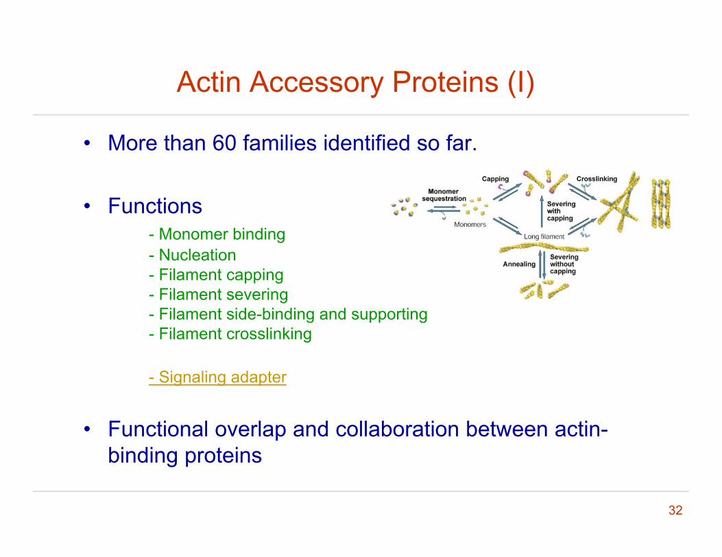
Actin Accessory Proteins (I)
• More than 60 families identified so far.
• Functions- Monomer binding- Nucleation- Filament capping- Filament severing
Filament side binding and supporting- Filament side-binding and supporting- Filament crosslinking
- Signaling adapterg g p
• Functional overlap and collaboration between actin-binding proteins
32
binding proteins

Actin Accessory Proteins (II)
• Monomer binding proteins- profilin: to bind actin monomer and accelerate elongationprofilin: to bind actin monomer and accelerate elongation- thymosin: to bind and lock actin monomer - ADF/cofilin: to bind and destabilize ADP-actin filaments
33

Actin Accessory Proteins (III)
• Actin nucleation- Formins: to initiate unbranched actin filamentsFormins: to initiate unbranched actin filaments- Arp2/3: to bind the side of actin and initiate branching
34

Actin Accessory Proteins (IV)
• Actin capping protein- Blocks subunit addition and disassociationBlocks subunit addition and disassociation
• Actin severing protein
• Three families of proteins perform both functions
- Gelsolin- Fragmin-severin- ADF/cofilin
35

Actin Accessory Proteins (V)
• Actin side-binding proteinstropomyosin nebulin caldesmontropomyosin, nebulin, caldesmon
• Actin crosslinking proteing p- -actinin- filamin- spectrin- ERM
36

Actin Adapter Protein
• Adaptor proteins such as WASP (a branchingas WASP (a branching mediating factor) & VASP (a polymerization mediating factor) servermediating factor) server as connectors between signaling pathways and actin assemblyactin assembly.
37

Actin Regulation
• GTPase: Molecule switch; Family of proteins that areFamily of proteins that are activated by GTP binding and inactivated by GTP hydrolysis and phosphate dissociation.
• Rho GTPase:Rho GTPase: cdc42: its activation triggers actinpolymerization and bundling at filopodia.
Rho: its activation promotes actinbundling.
Rac: its activation promotes
38
Rac: its activation promotes polymerization at the cell periphery.
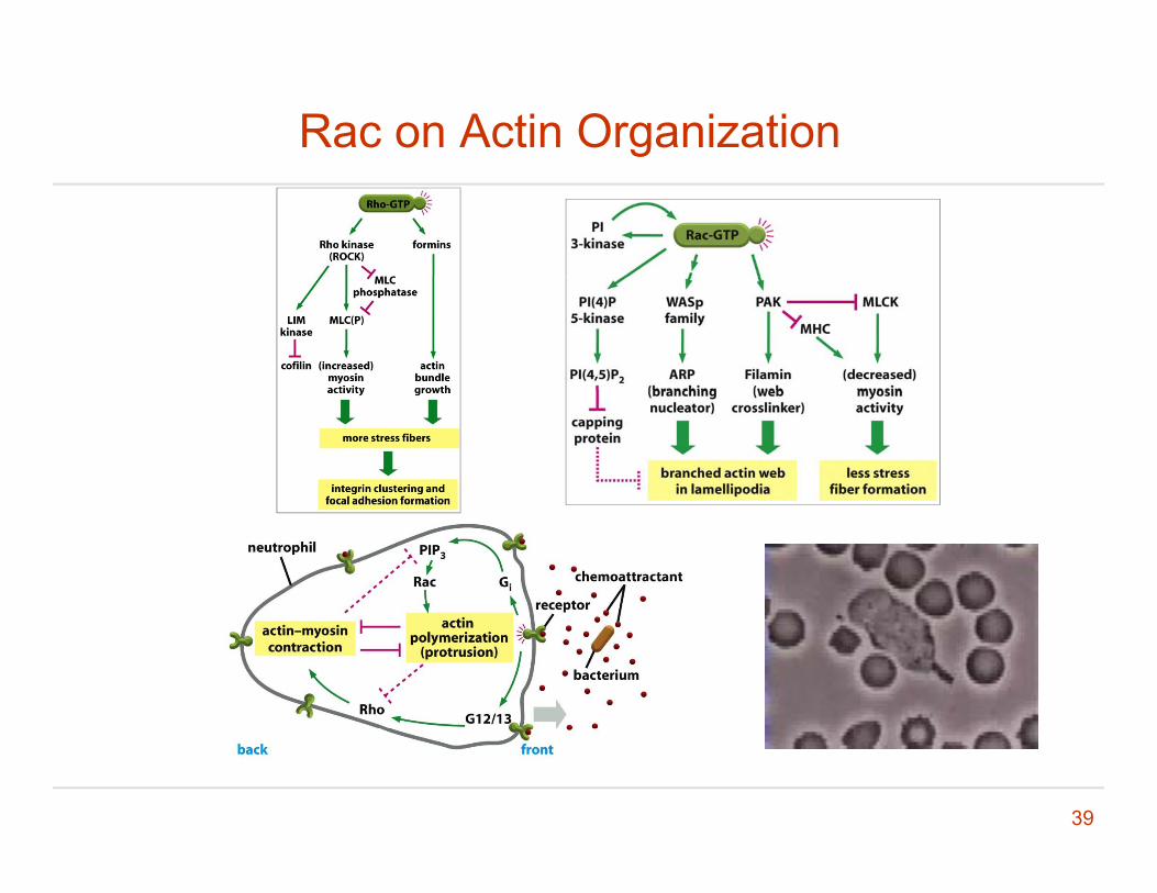
Rac on Actin Organization
39

Summary: actin
• Relatively soft (quantification in following lectures).y (q g )
• Often form bundles; mechanical strength comes mostly from bundling and crosslinking.
• Mostly function to withstand tension rather than• Mostly function to withstand tension rather than compression.
• Relatively stable and easy to work with (biochemically).
40

Summary: actin accessory proteins
• Different proteins have distinct functions.p
• Proteins with multiple functional domains can have lti l f timultiple functions.
• Some of them are essential• Some of them are essential.
• Most of the proteins have functional overlap. p p
41

Required Reading
• Chapter 16p
42

Questions ?
43