
Lectins: tools for the molecular understanding of the...
16
PERSPECTIVE OBC www.rsc.org/obc Lectins: tools for the molecular understanding of the glycocode Moira Ambrosi, a Neil R. Cameron* a and Benjamin G. Davis* b a Department of Chemistry, University of Durham, South Road, Durham, UK DH1 3LE b Department of Chemistry, University of Oxford, Mansfield Road, Oxford, UK OX1 3TA Received 17th September 2004 First published as an Advance Article on the web 11th April 2005 Recent progress in glycobiology has revealed that cell surface oligosaccharides play an essential role in recognition events. More precisely, these saccharides may be complexed by lectins, carbohydrate-binding proteins other than en- zymes and antibodies, able to recognise sugars in a highly specific manner. The ubiquity of lectin–carbohydrate inter- actions opens enormous potential for their exploitation in Moira Ambrosi was born in Florence (Italy) in 1971. She graduated in Chemistry at the University of Florence in 1998 with1st class honours. She then moved to Durham (UK) to undertake a PhD with Dr Neil Cameron, working on glycopolymers, from October 1999 and graduated in September 2002. She currently has a post-doctoral position at the Chemistry Department of the University of Florence, working with Professor Piero Baglioni on derivatives of vitamin C with biomedical applications. She is also collaborating with Procter & Gamble on liquid detergents containing bleaching agents, and with Covercolor (Italy) on the improvement of mechanical properties of ceramic materials. She has recently become “Cultore della Materia” of Physical Chemistry. Neil Cameron was born in Ayr, Scotland, in 1969 and studied chemistry at the University of Strathclyde in Glasgow, graduating with 1st class honours in July 1991. He then remained at Strathclyde to undertake a PhD with Professor David Sherrington, working on highly porous polymer materials (PolyHIPEs), from October 1991 and graduated in 1995. His first post-doctoral position, on nitroxide- mediated polymerisations, was in Eindhoven University of Technology with Professor Anton German, from April 1995 until March 1996, after which he returned to the UK as a post-doctoral associate in the laboratory of Professor Iain Cowie at Heriot-Watt University in Edinburgh, investigating ageing of polymer blends. In October 1997 he joined the academic staff in the Chemistry Department at the University of Durham, where he is currently a Reader (from October 2005). His research interests are in synthetic macromolecular chemistry, with a particular emphasis on materials for use in biological and medical applications. His work to date has led to 50 publications and he has significant current funding from a variety of sources (UK research councils, E.U., industry and charities). In 2003 he became the Associate Director (for Durham) of the Interdisciplinary Research Centre in Polymer Science and Technology (Polymer IRC) and he was awarded the 2003 Young Researchers’ Award by the Macro Group UK, a joint subject group of the Royal Society of Chemistry and the Society for Chemical Industry. Ben Davis got his BA (1993) and DPhil (1996) from the University of Oxford. During this time he learnt the beauty of carbohydrate chemistry under the supervision of Professor George Fleet. He then spent two years as a postdoctoral fellow in the laboratory of Professor Bryan Jones at the University of Toronto exploring protein chemistry and biocatalysis. In 1998 he returned to the UK to take up a lectureship at the University of Durham; in autumn 2001 he moved to the Dyson Perrins Laboratory, University of Oxford and a fellowship at Pembroke College, Oxford. His group’s research centres on chemical biology with an emphasis on carbohydrates and proteins. In particular, their interests encompass synthesis and methodology, inhibitor design, biocatalysis, enzyme mechanism, protein engineering, drug delivery, molecular modelling, molecular biology and glycoscience. This work has been published in 13 patents, 6 books and book chapters and >50 papers and has been recognised by the 1999 RSC Meldola Prize & Medal, a DTI Smart Innovation Award (1999), a Mitzutani Foundation for Glycoscience Award (2000), an AstraZeneca Strategic Research Fund Award (2000), the 2001 RSC Carbohydrate Chemistry Award, a 2002 Phillip Leverhulme Prize for Biochemistry and Molecular Biology and selection for the 2003 MIT TR100. Moira Ambrosi Neil R. Cameron Benjamin G. Davis medicine. Therefore, extraordinary effort is made into the identification of new lectins as well as into the achievement of a deep understanding of their functions and of the precise mechanism of their association with specific ligands. In this review, a summary of the main features of lectins, particu- larly those found in legumes, will be presented with a focus on the mechanism of carbohydrate-binding. An overview of DOI: 10.1039/b414350g This journal is © The Royal Society of Chemistry 2005 Org. Biomol. Chem. , 2005, 3 , 1593–1608 1593
Transcript of Lectins: tools for the molecular understanding of the...
P E R S P E C T I V E
OBC
ww
w.rsc.o
rg/o
bc
Lectins: tools for the molecular understanding of the glycocode
Moira Ambrosi,a Neil R. Cameron*a and Benjamin G. Davis*b
a Department of Chemistry, University of Durham, South Road, Durham, UK DH1 3LEb Department of Chemistry, University of Oxford, Mansfield Road, Oxford, UK OX1 3TA
Received 17th September 2004First published as an Advance Article on the web 11th April 2005
Recent progress in glycobiology has revealed that cellsurface oligosaccharides play an essential role in recognitionevents. More precisely, these saccharides may be complexedby lectins, carbohydrate-binding proteins other than en-zymes and antibodies, able to recognise sugars in a highlyspecific manner. The ubiquity of lectin–carbohydrate inter-actions opens enormous potential for their exploitation in
Moira Ambrosi was born in Florence (Italy) in 1971. She graduated in Chemistry at the University of Florence in 1998 with 1st classhonours. She then moved to Durham (UK) to undertake a PhD with Dr Neil Cameron, working on glycopolymers, from October 1999and graduated in September 2002. She currently has a post-doctoral position at the Chemistry Department of the University of Florence,working with Professor Piero Baglioni on derivatives of vitamin C with biomedical applications. She is also collaborating with Procter& Gamble on liquid detergents containing bleaching agents, and with Covercolor (Italy) on the improvement of mechanical properties ofceramic materials. She has recently become “Cultore della Materia” of Physical Chemistry.
Neil Cameron was born in Ayr, Scotland, in 1969 and studied chemistry at the University of Strathclyde in Glasgow, graduating with1st class honours in July 1991. He then remained at Strathclyde to undertake a PhD with Professor David Sherrington, working onhighly porous polymer materials (PolyHIPEs), from October 1991 and graduated in 1995. His first post-doctoral position, on nitroxide-mediated polymerisations, was in Eindhoven University of Technology with Professor Anton German, from April 1995 until March 1996,after which he returned to the UK as a post-doctoral associate in the laboratory of Professor Iain Cowie at Heriot-Watt University inEdinburgh, investigating ageing of polymer blends. In October 1997 he joined the academic staff in the Chemistry Department at theUniversity of Durham, where he is currently a Reader (from October 2005). His research interests are in synthetic macromolecularchemistry, with a particular emphasis on materials for use in biological and medical applications. His work to date has led to 50publications and he has significant current funding from a variety of sources (UK research councils, E.U., industry and charities). In 2003he became the Associate Director (for Durham) of the Interdisciplinary Research Centre in Polymer Science and Technology (PolymerIRC) and he was awarded the 2003 Young Researchers’ Award by the Macro Group UK, a joint subject group of the Royal Society ofChemistry and the Society for Chemical Industry.
Ben Davis got his BA (1993) and DPhil (1996) from the University of Oxford. During this time he learnt the beauty of carbohydratechemistry under the supervision of Professor George Fleet. He then spent two years as a postdoctoral fellow in the laboratory of ProfessorBryan Jones at the University of Toronto exploring protein chemistry and biocatalysis. In 1998 he returned to the UK to take upa lectureship at the University of Durham; in autumn 2001 he moved to the Dyson Perrins Laboratory, University of Oxford and afellowship at Pembroke College, Oxford. His group’s research centres on chemical biology with an emphasis on carbohydrates andproteins. In particular, their interests encompass synthesis and methodology, inhibitor design, biocatalysis, enzyme mechanism, proteinengineering, drug delivery, molecular modelling, molecular biology and glycoscience. This work has been published in 13 patents, 6 booksand book chapters and >50 papers and has been recognised by the 1999 RSC Meldola Prize & Medal, a DTI Smart Innovation Award(1999), a Mitzutani Foundation for Glycoscience Award (2000), an AstraZeneca Strategic Research Fund Award (2000), the 2001RSC Carbohydrate Chemistry Award, a 2002 Phillip Leverhulme Prize for Biochemistry and Molecular Biology and selection for the2003 MIT TR100.
Moira Ambrosi Neil R. Cameron Benjamin G. Davis
medicine. Therefore, extraordinary effort is made into theidentification of new lectins as well as into the achievementof a deep understanding of their functions and of the precisemechanism of their association with specific ligands. In thisreview, a summary of the main features of lectins, particu-larly those found in legumes, will be presented with a focuson the mechanism of carbohydrate-binding. An overview of
DOI:
10.1
039/
b41
4350
g
T h i s j o u r n a l i s © T h e R o y a l S o c i e t y o f C h e m i s t r y 2 0 0 5 O r g . B i o m o l . C h e m . , 2 0 0 5 , 3 , 1 5 9 3 – 1 6 0 8 1 5 9 3
lectin–carbohydrate interactions will also be given, togetherwith an insight into their energetics. In addition, therapeuticapplications of lectins will be discussed.
1 LectinsLectins (from lectus, the past participle of legere, to select orchoose)1 are defined as carbohydrate binding proteins otherthan enzymes or antibodies2 and exist in most living organisms,ranging from viruses and bacteria to plants and animals. Theirinvolvement in diverse biological processes in many species,3
such as clearance of glycoproteins from the circulatory system,4,5
adhesion of infectious agents to host cells,6 recruitment ofleukocytes to inflammatory sites,7 cell interactions in the immunesystem, in malignancy and metastasis,8 has been shown. Adoctoral thesis from 1888 on the agglutination of red blood cellsby extracts of castor beans (the active component being a proteinnamed ricin) is often cited as the beginning of lectinology,9
however the agglutination of erythrocytes by rattlesnake venom,observed around 1860,10 has in fact been suggested as the firstdemonstrated example of lectin activity. The first pure lectin,concanavalin A (Con A, from jack beans), was isolated in1919 by Sumner,11 who also demonstrated its sugar specificity.12
Subsequently, lectins played a crucial role in elucidating themolecular basis for blood group specificity.13 Recent advancesin carbohydrate chemistry and the study of protein–ligandinteractions mean that lectins are currently the focus of intenseresearch. In particular, the investigation of their role in cellrecognition, as well as their employment as invaluable toolsfor the study of complex carbohydrates in solution and on cellsurfaces, is contributing markedly to advances in glycobiology.
Lectins interact with carbohydrates non-covalently in amanner that is usually reversible and highly specific.14 Classicallectins contain two or more carbohydrate-binding sites; there-fore, their interaction with sugars on the surface of erythrocytesresults in the cross-linking of several blood cells and theirsubsequent precipitation. This phenomenon, known as cellagglutination, is a major attribute of the activity of lectinsand has been used classically and routinely for their detectionand characterisation (however it is now recognised that cellagglutination is not a defining feature of lectins2). Both theagglutination and precipitation processes are inhibited by thecarbohydrate for which the lectin is specific.
According to the monosaccharide ligand toward whichthey exhibit the highest affinity, Sharon classified lectins intofive groups: mannose, galactose/N-acetylgalactosamine, N-acetylglucosamine, fucose and N-acetylneuraminic acid15 (how-
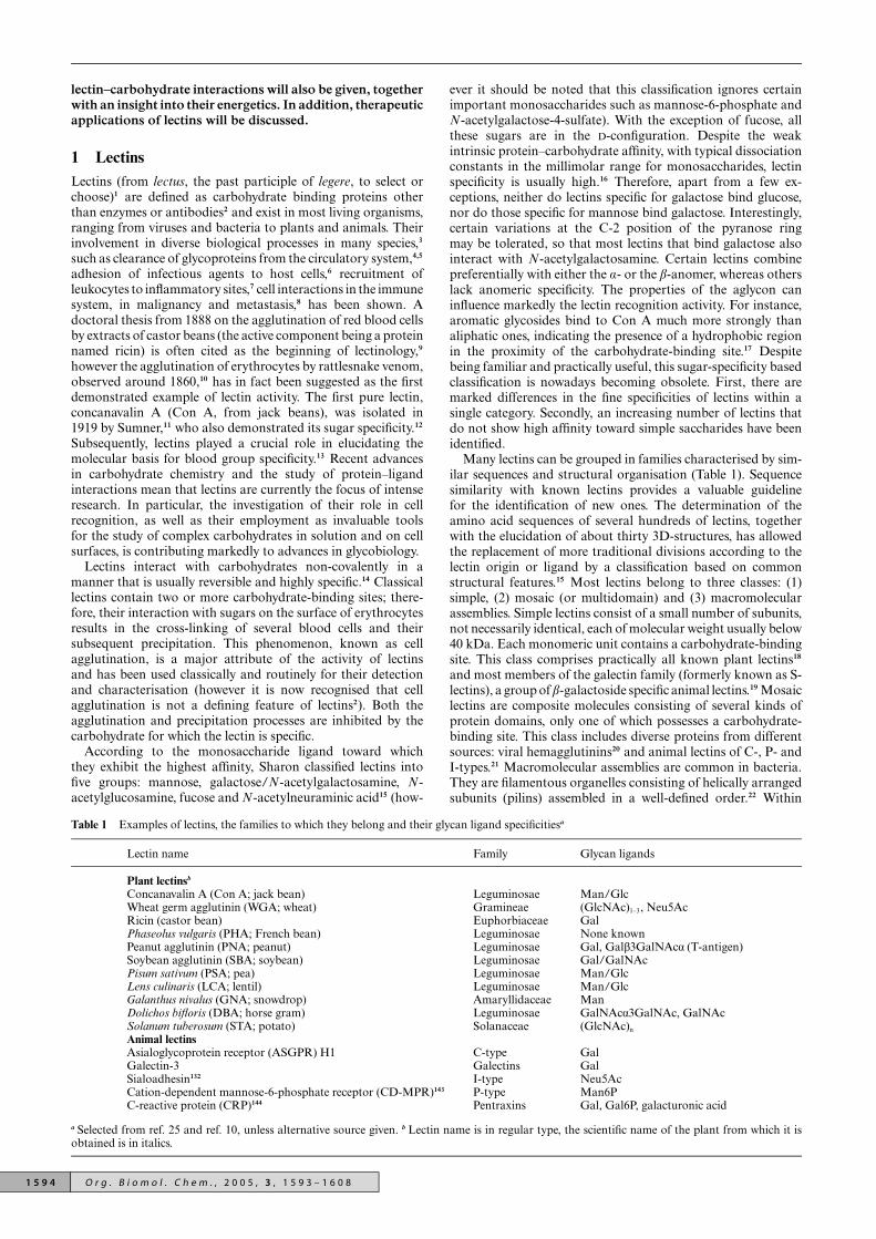
Table 1 Examples of lectins, the families to which they belong and their glycan ligand specificitiesa
Lectin name Family Glycan ligands
Plant lectinsb
Concanavalin A (Con A; jack bean) Leguminosae Man/GlcWheat germ agglutinin (WGA; wheat) Gramineae (GlcNAc)1–3, Neu5AcRicin (castor bean) Euphorbiaceae GalPhaseolus vulgaris (PHA; French bean) Leguminosae None knownPeanut agglutinin (PNA; peanut) Leguminosae Gal, Galb3GalNAca (T-antigen)Soybean agglutinin (SBA; soybean) Leguminosae Gal/GalNAcPisum sativum (PSA; pea) Leguminosae Man/GlcLens culinaris (LCA; lentil) Leguminosae Man/GlcGalanthus nivalus (GNA; snowdrop) Amaryllidaceae ManDolichos bifloris (DBA; horse gram) Leguminosae GalNAca3GalNAc, GalNAcSolanum tuberosum (STA; potato) Solanaceae (GlcNAc)n
Animal lectinsAsialoglycoprotein receptor (ASGPR) H1 C-type GalGalectin-3 Galectins GalSialoadhesin132 I-type Neu5AcCation-dependent mannose-6-phosphate receptor (CD-MPR)143 P-type Man6PC-reactive protein (CRP)144 Pentraxins Gal, Gal6P, galacturonic acid
a Selected from ref. 25 and ref. 10, unless alternative source given. b Lectin name is in regular type, the scientific name of the plant from which it isobtained is in italics.
ever it should be noted that this classification ignores certainimportant monosaccharides such as mannose-6-phosphate andN-acetylgalactose-4-sulfate). With the exception of fucose, allthese sugars are in the D-configuration. Despite the weakintrinsic protein–carbohydrate affinity, with typical dissociationconstants in the millimolar range for monosaccharides, lectinspecificity is usually high.16 Therefore, apart from a few ex-ceptions, neither do lectins specific for galactose bind glucose,nor do those specific for mannose bind galactose. Interestingly,certain variations at the C-2 position of the pyranose ringmay be tolerated, so that most lectins that bind galactose alsointeract with N-acetylgalactosamine. Certain lectins combinepreferentially with either the a- or the b-anomer, whereas otherslack anomeric specificity. The properties of the aglycon caninfluence markedly the lectin recognition activity. For instance,aromatic glycosides bind to Con A much more strongly thanaliphatic ones, indicating the presence of a hydrophobic regionin the proximity of the carbohydrate-binding site.17 Despitebeing familiar and practically useful, this sugar-specificity basedclassification is nowadays becoming obsolete. First, there aremarked differences in the fine specificities of lectins within asingle category. Secondly, an increasing number of lectins thatdo not show high affinity toward simple saccharides have beenidentified.
Many lectins can be grouped in families characterised by sim-ilar sequences and structural organisation (Table 1). Sequencesimilarity with known lectins provides a valuable guidelinefor the identification of new ones. The determination of theamino acid sequences of several hundreds of lectins, togetherwith the elucidation of about thirty 3D-structures, has allowedthe replacement of more traditional divisions according to thelectin origin or ligand by a classification based on commonstructural features.15 Most lectins belong to three classes: (1)simple, (2) mosaic (or multidomain) and (3) macromolecularassemblies. Simple lectins consist of a small number of subunits,not necessarily identical, each of molecular weight usually below40 kDa. Each monomeric unit contains a carbohydrate-bindingsite. This class comprises practically all known plant lectins18
and most members of the galectin family (formerly known as S-lectins), a group of b-galactoside specific animal lectins.19 Mosaiclectins are composite molecules consisting of several kinds ofprotein domains, only one of which possesses a carbohydrate-binding site. This class includes diverse proteins from differentsources: viral hemagglutinins20 and animal lectins of C-, P- andI-types.21 Macromolecular assemblies are common in bacteria.They are filamentous organelles consisting of helically arrangedsubunits (pilins) assembled in a well-defined order.22 Within
1 5 9 4 O r g . B i o m o l . C h e m . , 2 0 0 5 , 3 , 1 5 9 3 – 1 6 0 8

each class, proteins can be grouped into families, with similarsequences and structural properties. Nowadays, as the genomeanalysis of various organisms, including humans, is completed,lectins can also be valuably classified through the conceptof protein (gene) families, whose members show evolutionarykinship.23
The intention of this review is not to provide a comprehensivesurvey of the literature on lectins, but rather to serve as anintroduction to the topic for the reader unfamiliar with the area.The reader is directed to excellent reviews on plant24 and animal10
lectins for further information, and a detailed description ofthe history of lectinology can be found in the recent article byGabius et al.25 In our article, the main features of lectins will besummarised, with an emphasis particularly on legume lectins.The abundance of these proteins in plant seeds, their solubilitiesand their wide range of saccharide specificities make themgood model systems, tools for elucidating protein–carbohydrateinteractions as well as for biomedical and biotechnologicalapplications.
1.1 Legume lectins
Legume lectins represent the largest and most thoroughlystudied family of simple lectins. Around 100 members have beencharacterised, almost all isolated from the seeds of the plants inwhich they are present.18,26 Concanavalin A (Con A), the lectinfrom the jack bean, is the prototype member of the family. Therelative abundance of this protein in jack bean, the ease of itspreparation and the large number of saccharides with which itcan interact, have led to numerous studies on Con A, markedlyaccelerated by the discovery in 1969 that cells transformed byDNA tumour viruses or carcinogens were agglutinated by thelectin more readily than normal cells.27 About 85% of the bindingsites for Con A, which are in a cryptic form on normal cells, werefound to be exposed on the surface membrane of transformedcells. It was therefore hypothesised that the change in structureof the surface membrane, resulting in the exposure of the sites,could produce the change in cellular regulatory mechanismsassociated with transformation.
1.1.1 Structural features. Typically, legume lectins consistof two or four identical or near-identical subunits (protomers)of 25–30 kDa each, which are commonly single polypeptidechains of about 250 amino acids presenting one or two N-linked oligosaccharides. Each protomer typically contains acarbohydrate-combining site, a tightly bound Ca2+ and a transi-tion metal ion, usually Mn2+. Approximately 20% of the aminoacid residues are invariant in all legume lectins and another20% are similar. The conserved amino acids include several ofthose involved in the interaction with the saccharide and almostall the residues that coordinate the metal ions. The resolutionof 3D-structures of about ten legume lectins has shown thateach subunit is constituted largely—nearly 60%—of b-strandsmutually connected by loops. For all legume lectins known sofar, the tertiary structure is made up of two anti-parallel b-sheets,a six-stranded flat “back” and a seven-stranded curved “front”,connected by a five-stranded b-sheet, giving the well known“jellyroll” motif, also referred to as the “lectin fold”28 (Fig. 1a).
The subunit structures of different legume lectins can benearly superimposed, irrespective of the protein specificity.Despite their similarities at the primary, secondary and tertiarystructural monomeric level, legume lectins exhibit considerablevariation in their quaternary structure: small differences in theamino acid sequences at the monomer–monomer interfaces andthe presence/absence of glycosylation affect the monomers’association modes. In the case of lectins with “canonical”quaternary structure, such as Con A, pea lectin, favin and L.ochrus, dimerisation involves anti-parallel side-by-side align-ment of the flat six-stranded b-sheets of the two monomers,resulting in the formation of a continuous 12-stranded sheetthat extends across the dimer interface. A considerable portion
Fig. 1 (a) Representative tertiary structure of a legume lectin monomer;(b) dimerisation in Con A; (c) tetramerisation in Con A. Reprinted withpermission from Elsevier.29
of the surface area is buried in the process: ca. 1000 A2 permonomer (Fig. 1b). Further association of two dimers givesthe tetrameric assembly of Con A observed in physiologicalconditions29 (Fig. 1c). Peanut agglutinin (PNA), the lectinfrom Arachis hypogaea, shows an unusual “open” quaternarystructure, where the homotetramer possesses neither 222 (D2)nor 4-fold (C4) symmetry.30 This structure is a refinement ofearlier low resolution X-ray crystallographic work31 suggestingD2 symmetry, however the conclusion that the PNA tetrameris a dimer of a dimer, with a dimeric association similar tothat observed in lectin IV of Griffonia simplicifolia (GS4),was confirmed.30 The open quaternary association is stabilisedmainly by hydrophobic, hydrogen-bonded and water-mediatedinteractions. While the dimerisation process results in the burialof 1920 A2 surface area of which 71% is non-polar, the percentageof hydrophobic surface area buried during the further formationof the tetramer is relatively low. The dimers interact in sucha way that monomers belonging to two different GS4-likedimers associate in a canonical fashion except that the sheets
O r g . B i o m o l . C h e m . , 2 0 0 5 , 3 , 1 5 9 3 – 1 6 0 8 1 5 9 5

do not interact directly, but by means of six water bridges. As aresult, the formed interface is intrinsically less stable than theanalogous interface in Con A.29 As with most plant lectins,the quaternary structure of PNA depends on the pH. Thelectin is a tetramer at physiological pH,32 which dissociatesreversibly into dimers at pH below 5.1. Below pH 3.4 PNA istotally dimeric.33 Interestingly, it has been found that a partiallyunfolded intermediate of PNA retains carbohydrate bindingability with affinities that are 75–85% of those of native PNA.34
1.1.2 Carbohydrate-binding site. During the past 15 yearsthere has been significant progress in elucidating the features oflectins involved in carbohydrate binding. X-ray crystallographyof the proteins complexed with their ligands, site-directedmutagenesis experiments and molecular modelling have allowedthe identification of the chemical groups belonging to bothinteracting species involved in the binding and of the types ofbond formed. Studies of lectin–oligosaccharide complexes areespecially interesting, providing the basis for the understandingof the proteins’ interaction with natural ligands. Generally,lectins show exquisite specificity for di-, tri- and tetrasaccharides,with association constants significantly higher than those for thecorresponding monosaccharides.
Carbohydrate-binding sites are often shallow depressions onthe surface of the protein. In all cases the combining siteappears to be preformed,35 since few conformational changesoccur upon binding. In all legume lectins, irrespective of theirspecificity, four invariant amino acid residues participate inthe ligand binding: an aspartic acid, an asparagine, a glycine(conserved in all the lectins of the family apart from Con A)and an aromatic amino acid36 or leucine:37 Asp83, Gly104,Asn127 and Tyr125 for PNA.38 Replacement of the asparticacid or asparagine by site-directed mutagenesis results, in severalcases, in the loss of the lectin sugar-binding ability.39 However,despite the conservation of key amino acids involved in thebinding of the carbohydrate, different legume lectins can showdifferent specificity. For instance, while Con A binds mannoseand glucose, PNA, ECorL and SBA bind galactose. Therefore,while the constellation of highly conserved amino acids providesthe framework required for binding, specificity apparently arisesfrom the variability of amino acid residues in other regions ofthe combining pocket. The sugar-combining site is made upby amino acid residues residing in four loops, designated A, B,C and D.40 The invariant aspartic acid and glycine belong to Aand B, respectively, whereas the asparagine and the hydrophobicresidue are in loop C. Additional interactions are provided byamino acids in loop D, suggesting a correlation between thisloop and the lectin specificity. In fact, loop D is highly variable interms of length, sequence and conformation. Thus, for instance,the size of this loop is identical in all mannose-specific lectins.41
The Ca2+ and Mn2+ (or other transition metal) are situ-ated around 4 A apart and in close proximity to the sugar-combining pocket. Although not always directly involved inthe carbohydrate binding, the cations help the positioningof the amino acid residues interacting with the glycoside.The two invariant aspartic acid and asparagine residues alsoparticipate in coordinating Ca2+. A rare cisoid-peptide bondbetween the critical asparagine and the preceding amino acid,usually alanine, confers on the asparagine residue the properorientation.15
Lectins bind carbohydrates through a network of hydrogenbonds and hydrophobic interactions. This is highlighted by theschematic representation of the T-antigenic disaccharide (thecarbohydrate structure for which peanut agglutinin shows thehighest affinity), Galb(1→3)GalNAc (Tant), in the binding siteof PNA shown in Fig. 2.
The four invariant hydrogen bonds are Asp83 Od1-Gal O3,Asp83 Od2-Gal O4, Gly104 N-Gal O3 and Asn127 Nd2-Gal O3.42–45 There is a key stacking interaction between thearomatic ring of Tyr125 and the hydrophobic a-face patch of
Fig. 2 Schematic representation of protein–carbohydrate interactionsin the PNA–Tant complex (reprinted with permission from.15 Copyright(1998) American Chemical Society).
the galactose unit of the disaccharide. Further hydrogen bondsare Asp80 Od2-Gal O6 and that involving Ser211 Oc.
Van der Waals forces, although rather weak (usually afraction of 4.2 kJ mol−1 for each pair of atoms), are frequentlynumerous, contributing significantly to the overall binding.15
The steric disposition of hydroxyl groups in carbohydratescreates hydrophobic patches46 on the sugar surface that caninteract with hydrophobic regions of the protein.47 For examplein the PNA–Tant complex (Fig. 2), about 60 van der Waalscontacts are formed between the disaccharide and amino acidresidues within 4 A of the carbohydrate.44
Contacts between the ligand and the protein are often medi-ated by water molecules. Water acts as a molecular “mortar”;48
its small size and its ability to behave as both hydrogen donorand acceptor make it near-ideal for this function. Tightlybound water molecules can in effect be considered as structural,i.e. an extension of the protein surface. Thus, water plays asignificant role in carbohydrate recognition, imparting in somecases exquisite specificity. Comparison of crystal structuresof PNA complexed with different ligands (T-antigen, Tant,43
methyl-b-galactoside, MeGal,44 N-acetyllactosamine, LacNAc44
and lactose, Lac45) has shown that water bridges involving watermolecules W1 and W2 (Fig. 2) occur in all four complexes.Moreover, major additional interactions between the T-antigenand PNA, compared to the other disaccharides, occur throughtwo water molecules involving the carbonyl O atom of theacetamido group: W3 connects the acetamido O atom to Ile101O, while W4 connects it to Asn41 Nd2 and to Leu212 N. Thesetwo water molecules also exist in the other complexes, butin these cases they interact only with the protein, due to thelack of sugar atoms in their immediate vicinity. Therefore,the additionally high specificity of PNA for the T-antigenicdisaccharide appears, critically, to be generated by W3 and W4.
A detailed study of water molecules in the combining site ofPNA has been performed.44 Interestingly, as already reported forother carbohydrate-binding proteins,49 ordered water moleculescan be found in the unligated form at positions correspondingto hydroxyl groups in the ligated form (for instance, a watermolecule occupies the position of the acetamido carbonyl Oin the GalNAc moiety of the T-antigen). In general, watermolecules in the carbohydrate-binding region mimic the ligandto a substantial extent not only at the primary site, but also inthe regions adjacent to it. Molecular dynamics (MD) simulationscarried out for the PNA–Tant complex revealed that there is aconstant exchange of water molecules between the binding siteand the bulk, especially in the proximity of Asn41, Asn127 andGlu129.50 As evidenced by the short mean residence times and bythe trajectories of water molecules during the formation of waterbridges, these molecules do not stay in the sugar-combiningpocket for long; rather they are in constant motion. MD and
1 5 9 6 O r g . B i o m o l . C h e m . , 2 0 0 5 , 3 , 1 5 9 3 – 1 6 0 8

crystallographic results were comparable. It was found thatthe number of both direct and water-mediated hydrogen bondscalculated by MD were higher than those determined by crystal-lography. Due to the large number of water molecules that haveaccess to the site, the simulation data revealed 23 water-mediatedinteractions, including those present in the crystallographic data.The same authors showed that most intramolecular, water-mediated interactions involving the T-antigen galactose moietyare diminished or even lost upon complexation. By contrast, awater bridge with a long mean lifetime is established within thecomplex between Gal O6 and GalNAc O6. It was hypothesisedthat this very stable water bridge, occurring frequently duringthe simulation, could have a role in maintaining the stretchedconformation of the saccharide. Intramolecular GalNAc O1-W-GalNAc C=O and GalNAc O4-W-GalNAc C=O, pre-existentto complex formation, were still present in the active site withan increased persistence. As was expected, the hydration numberof the T-antigen was significantly reduced upon complexation,going from about 33 to 13. Pratap and co-workers45 carriedout comparative molecular dynamics simulations for PNA–Tant and PNA–Lac complexes. The results showed that thenumber of alternative binding modes is higher for the formerthan for the latter, resulting in the “breaking” of the enthalpy–entropy compensation that is characteristic of the bindingof monovalent sugars to lectins. Such molecular dynamicssimulations provide important contributions that complementthe static view of crystallography, and provide a better sense ofmolecular interactions in the in vivo state.
1.1.3 Physiological functions. Despite their long history,the true physiological role of legume lectins is still not wellunderstood. Many hypotheses have been formulated in thecourse of the years but, at present, no physiological functionfor any legume lectin has been established with certainty.The difficulty in assigning a precise role arises from severalof their features. The defining characteristic of all lectinsis their carbohydrate-binding ability. This activity has beenpreserved during evolution, suggesting that it is essential forthe exploitation of their function. Nevertheless, some lectinspossess additional activities that may be non-secondary indetermining their physiological role. Several legume lectins, forinstance, present adenine-combining sites able to bind activeforms of the cytokinins, a major class of plant hormones.51
The specificity of adenine-binding sites has been maintainedduring evolution despite variations in carbohydrate specificityand tissue distribution. Therefore, it may be possible that in suchcases proteins have a primary role unrelated to carbohydrates,e.g. hormone-binding, and that the carbohydrate-combiningactivity plays a regulatory or transducing function.52
In general, a single legume plant can contain a variety oflectins that may have evolved by gene duplication and becomespecialised for different roles in the plant.53 Furthermore, despitebeing concentrated in plant seeds, lectins are also present indifferent tissues, where their function probably requires lowerconcentrations. One of the most credited theories concerninglegume lectins’ physiological role, extensible to all plant lectins,considers them as defence agents against predators.54 An essen-tial feature of any active defence agent is the ability to recognisespecifically the pathogen. Based on their carbohydrate-bindingspecificity and also considering their abundance in plant seedsand bark, lectins seem to possess all the necessary characteristicsto exploit this function. Early investigators noted the similaritiesof lectins to antibodies and hypothesised that lectins mightfunction as plant antibodies.55 One of the early events in thedefence response of legumes is the production of phytoalexins,56
whose synthesis is stimulated by the release of elicitors, manyof which are oligosaccharides that arise from the breakdownof plant or pathogen cell wall components.57 Therefore, lectinscould participate in this step of the defence process either actingas receptors for elicitors or organising the elicitors in structures
required by their receptors. It has been shown that the levels ofDolichos biflorus stem and leaf lectin, DB58, increase upon plantwounding.52
Again, based on their carbohydrate-binding ability, lectinshave also been thought to be involved in the establishmentof symbiosis between nitrogen-fixing bacteria and plants.3,58
Legumes are able to associate specifically and form symbioseswith soil bacteria of the rhizobia family, a phenomenon thatmakes them independent from soil nitrogen supplies. The ideathat lectins are responsible for this association was advancedover 20 years ago and it has been the subject of much controversy.The nitrogen-fixing symbiosis is a multistep process that requiresthe formation of the root nodule, followed by the adhesionof the bacteria to the roots and, finally, the internalisation ofthe bacteria into the nodule.59 Initiation of nodulation dependson the production of Nod factor signals by the rhizobia.Nod factors are lipochitooligosaccharides that are modifiedin different rhizobial strains, so generating the basis for host–strain specificity. The finding that a legume binds to a specificrhizobial species and not to bacteria that are symbionts for otherlegumes suggests that the interaction occurs between lectins inthe roots of the plant and carbohydrates on the bacteria surface.DB46, the Dolichos biflorus root lectin present on the surface ofroot hairs, has been found to bind with several Nod factors.52
Despite the fact that molecular genetics experiments supportthis hypothesised role of lectins as receptors for oligosaccharidesproduced during the symbiosis processes, several inconsistenciescan be pointed out. First of all, for most host–symbiont systemsthere is no proof of the presence of lectins and of the respectiveligands on the two interacting species. Secondly, the correlationbetween the sugar specificity of legume lectins and their abilityto recognise bacteria appears not to be particularly strong.Furthermore, and most strikingly, mutants of soybean lackingthe lectin are still nodulated by the rhizobial symbiont.15
2 Lectin–carbohydrate interactions:towards therapyThe idea that lectins and carbohydrates are excellent as cellrecognition markers originates from the findings that bothclasses of compound are commonly present on the cell surfaceand that sugars possess tremendous coding capacity. The abilityof lectins to distinguish between subtle variations of oligosac-charide structure makes them perfectly suitable as decoders forsuch carbohydrate-encoded information. In other words, whilstsugars are able to carry the biological information, lectins arecapable of deciphering this “glycocode”.
2.1 Multivalency
The most striking features of lectin–monosaccharide interac-tions are that they are relatively weak, with dissociation con-stants usually in the millimolar range for monosaccharides,16,60
and that they may show relaxed specificity, when compared tothe strict nature of enzyme–substrate associations. The reasonfor this weakness lies in the solvent-exposed nature of thelectin binding-sites, which are shallow pockets making fewdirect contacts with the ligands.61 There is, in fact, a significantdifference in affinity between these shallow sites and deep sites, asis well illustrated by the influenza haemagglutinin lectin, whichbinds sialic acids with an approximately 1000-fold lower affinitythan a neuraminidase found in the same virus.62 Nevertheless,lectins exhibit both high affinity and exquisite specificity foroligosaccharide structures of glycoproteins and glycolipids onthe cell surface. If this was not the case, lectins could not actas recognition molecules in biological processes. It has thereforebeen suggested that multiple protein–carbohydrate interactionsare involved in the recognition event, giving the required highaffinity and specificity.60 Thus, polyvalent associations occurthroughout biology, showing a number of characteristics that
O r g . B i o m o l . C h e m . , 2 0 0 5 , 3 , 1 5 9 3 – 1 6 0 8 1 5 9 7
monovalent interactions do not exhibit.63 In most cases, biologi-cal systems seem to use polyvalent interactions rather than a verystrong single one. Therefore, multivalency is employed in Naturenot only to achieve the necessary high affinity, but also to ensurethe correct functioning of the cells through high specificity.For example, asialoglycoprotein receptor-mediated clearanceof erythrocytes occurs only when the density of galactosemoieties reaches a critical level.64 Moreover, multivalent lectin–carbohydrate interactions can also lead to the formation of sur-prisingly homogeneous complexes and lattices. These have beensuggested to be involved in certain physiological processes suchas T cell death, the signal for which is induced by glycoproteinclustering in the presence of galectin-1.65 Furthermore, protein–carbohydrate interactions may imply significant conformationalchanges that can represent biological signals. Decavalent bindingof IgM to the bacterial surface initiates the complement cascadeleading to the death of the microorganism.63
All classes of antibodies have multiple equivalent receptorsites. Multivalency leads to high affinity binding to surfaces thatshow repeated epitopes, such as almost all invading pathogens.Mannose residues on the tail (the Fc portion) of the antibodyinteract with mannose receptors (the Fc receptors) on the surfaceof a macrophage. The interaction of a single Fc portion withits receptor seems to be too weak to induce a response by themacrophage, whereas multiple antibodies bound to the surfaceof the bacterium can cross-link the Fc receptors, triggering aninternal signal in the macrophage to ingest the infective agent.66
2.2 Energetics
A full understanding of the mechanism of protein–carbohydrateassociation at a molecular level requires the elucidation ofboth structural and energetic aspects of the process. As brieflydiscussed in the previous sections, a large amount of structuraldata is now available. However, the link between structure andenergetic properties remains too obscure to allow a descriptionof the energetics of the interaction from structural information.48
Historically, the strength of protein–ligand complexation hasbeen determined by hemagglutination and precipitation inhibi-tion assays, although several spectroscopic techniques, affinitychromatography and equilibrium dialysis have also been utilised.The so obtained values of the binding constant, K, and of the freeenergy of binding, DG, give little information about the forcesinvolved in the complex formation. Moreover, the enthalpy andentropy of association are derived only indirectly from van’tHoff analyses of the temperature dependence of the free energy:
d ln KdT
= DHRT 2
(1)
The employment of this equation requires the assumption thatthe enthalpy of binding, DH, is not a function of the temperature,i.e. DCP = 0, since
(∂H∂T
)P
= DCP. However, the heat-capacitychange, DCP, for ligand binding in aqueous media is typicallynon-zero, making the accuracy of the calculated values doubtful.
The accessibility of commercial high-resolution mi-crocalorimeters has provided a direct method of evaluationof accurate thermodynamic data that can complement thestructural information. Isothermal titration microcalorimetry,ITC, is the only technique that allows direct determinationof K and DH in a single experiment. Values of DCP are alsoobtained performing titrations at different temperatures. DCP
is a powerful indicator of solvent behaviour during binding67
and provides invaluable information about the driving forcesfor association. Accurate values of DCP are available only bymicrocalorimetry.
2.2.1 Monovalent ligands. Calorimetrically determinedthermodynamic constants for lectin–carbohydrate interactionsare listed in Table 2.
In almost all cases the enthalpy of binding is more negativethan the free energy. It is noteworthy that this pattern isgenerally found for association in aqueous solution and is not aspecial feature of lectin–ligand complexation.68,69 It has alreadybeen pointed out that water molecules are strongly involvedin protein–sugar interactions. Watson et al.70 have reportedthat in the case of complexation of glucose with glycogenphosphorylase, water-mediated hydrogen bonds are as strongas the direct ones, reinforcing the concept of structural water asan extension of the protein surface. However, the displacementof a large number of water molecules during binding can alsobe observed. The effect of this release of water has long beendebated. Numerous models have been formulated in orderto explain the thermodynamic properties of apolar organicmolecules in water, particularly for the surprising cases where themost significant term opposing dissolution is enthalpic ratherthan entropic.48 It has been proposed that although the strengthof hydrogen bonds in the hydration shell of the protein is higherthan in bulk solvent, the fraction of broken bonds is also higher,due to geometric constraints imposed on water by the solute.Thus, the return of water of solvation to bulk water might beenthalpically driven, providing a favourable contribution to theenthalpy of complexation.71 Monte Carlo simulations, carriedout to investigate the structure of water near the surface of thelectin binding site, have shown highly disordered water in theproximity of the protein surface. The phenomenon has beenexplained by assuming that the protein surface could not becomplementary to any low-energy structure that the water couldadopt.72 Chervenak and co-workers67 have reported a calori-metric evaluation of the thermodynamic binding parameters forseveral systems in light and heavy water. In all cases desolvationwas found to contribute to a significant fraction of the bindingenthalpy: ca. 25% for lectin–sugar association. To date, the roleof protein–ligand hydrogen bonding vs. solvent reorganisationas contributors to the enthalpy of binding remains the mostpolemic area of discussion.
Another common feature of lectin–carbohydrate interactionsis the strong linear enthalpy–entropy compensatory behaviour.This offset has been interpreted both in terms of changes in thedegrees of freedom of the ligand upon binding73,74 and in termsof solvent reorganisation.75 Chervenak et al.76 have determinedthe configurational and solvation entropies for the formation ofa series of complexes with Con A and DGL (Table 3).
The total entropy of complexation can be separated into termsaccounting for changes in solvation and losses of configura-tional, rotational and translational degrees of freedom.
DS = DSsolv + DSconfig + DSrot + DStrans (2)
The sum of the first two terms has been referred to as the unitarycontribution, while the sum of rotational and translationalentropies has been defined as the cratic contribution.77 Knowingthat:
(∂S∂T
)P
= DCP
T(3)
DSsolv can be expressed as follows:
DSsolv = DS∗solv + DCp ln
(TT ∗
)(4)
where T* is the temperature at which there is no solvent con-tribution and is equal to 385.15 K. DSsolv* contains protonationand electrostatic contributions and for lectin–ligand interactionscan be considered equal to zero. Murphy et al.78 have proposedthat for the formation of a 1 : 1 complex with 1 M as thestandard state, the entropic cratic contribution, DSrot + DStrans,is equal to −33.5 J mol−1 K−1. Thus, accurate measurements ofDCP provide DSsolv and, consequently, DSconfig. For the systems
1 5 9 8 O r g . B i o m o l . C h e m . , 2 0 0 5 , 3 , 1 5 9 3 – 1 6 0 8

Table 2 Calorimetrically derived thermodynamic parameters of lectin–carbohydrate interactions
Lectin Carbohydrate 10−3 × K/M−1 DG/kJ mol−1 DH/kJ mol−1 DS/J mol−1 K−1 DCP/J mol−1 K−1 Reference
WBAIa Gal 1.2 −17.5 −24.3 −23 — 145GalNAc 7.2 −22.0 −28.0 −20 —MeaGal 6.6 −21.8 −23.5 −6 —MebGal 1.0 −17.1 −19.7 −9 —
Con A MeaMan — −22.2 −27.6 −18.3 −251 68Mana1,6(Mana1,3)Manb — −30.1 −41.0 −36.5 −460
Con A MeaMan 11.9 −22.9 −29.2 −22 — 74Man 2.1 −18.6 −23.9 −18 —MeaGlc 2.7 −19.2 −18.1 +4 —Glc 0.6 −15.4 −17.1 −6 —
Pea MeaMan 1.9 −18.3 −27.3 −31 —Man 0.9 −16.5 −24.8 −29 —MeaGlc 0.6 −15.5 −13.1 +8 —
Con A MeaMan 8.2 — −34.3 −40.7 — 146Mana1,6(Mana1,3)Man 49.0 — −60.3 −92.7 —
DGL c MeaMan — −20.5 −32.6 −40.7 −234 76Mana1,3Man — −26.4 −47.7 −71.6 −167Mana1,6Man — −21.0 −36.0 −50.5 −92Mana1,6(Mana1,3)Man — −34.3 −54.4 −67.4 −402
Con A MeaMan — −22.2 −28.5 −21.0 −209Mana1,3Man — −25.2 −31.0 −19.6 −460Mana1,6Man — −22.2 −28.9 −22.5 −184Mana1,6(Mana1,3)Man — −31.0 −42.7 −39.3 −389
SBA d MebGal 0.5 — −44.4 −96.2 −393GalNAc 9 — −39.7 −57.2 −418MebGalNAc 22 — −58.1 −111.6 —LacNAc 0.7 — −34.3 −60.0 —
ECorL e MebGal 0.4 — −18.4 −11.2 — 86GalNAc 1.2 — −29.7 −40.4 —MebGalNAc 1.3 — −28.5 −34.9 —LacNAc 4.2 — −45.6 −82.3 +393
Gal-1 f LacNAc 6.2 — −27.6 −19.5 −377C2S-Gal-1g LacNAc 2.9 — −11.7 +27.1 —N-Gal-1h LacNAc 8.7 — −2.5 +66.5 —
Gal 1.6 −18.2 −13.7 +15.3 —MeaGal 1.4 −18.1 −21.6 −12.0 —
ECorL MebGal 0.7 −16.3 −18.2 −6.6 — 80GalNAc 1.3 −17.9 −23.0 −17.1 —Lactose 1.9 −18.8 −41.2 −75.4 —LacNAc 9.7 −22.7 −47.1 −83.2 —2′-FL i 3.7 −20.3 −18.0 +7.7 —MeaDNSGalN j 351.5 −31.7 −23.1 +30.0 —Fucose 0.5 −15.2 −4.7 +35.2 —Galb1,4Glc 6.4 −21.7 −20.9 0 —Galb1,4Fruc 8.2 −22.4 −34.4 −40 —
Gal-1 k Galb1,4Man 11.3 −23.0 −35.2 −45 — 88Galb1,4Ara 5.4 −21.3 −36.8 −52 —Galb1,4GlcNAc 22.2 −24.7 −35.9 −38 —Galb1S1bGal 11.6 −23.1 −46.4 −78 —
PNA l Galb1,3GalNAc 20.6 −24.7 −59.0 −115.5 — 45
a Basic lectin from winged bean. b 3,6-Di-O-(a-D-mannopyranosyl)-a-D-mannopyranoside. c Lectin from Dioclea grandiflora. d Soybean agglutinin.e Lectin from Erythrina corallodendron. f From Chinese hamster ovary cells. g Cys to Ser mutant of Gal-1. h Monomeric mutant of Gal-1. i 2′-Fucosyllactose. j Methyl-a-N-dansylgalactosaminide. k From bovine spleen. l Peanut agglutinin.
Table 3 Entropic contributions to the binding of mannose oligosaccharides to Con A and DGL
System DCP/J mol−1 K−1 DS/J mol−1 K−1 DSsolv/J mol−1 K−1 DSconfig/J mol−1 K−1
Con AMeaMan −209 −20.5 +53.6 −40.6Mana1,6Man-OMe −184 −3.8 +47.3 −17.6Mana1,3Man −460 −20.1 +118.0 −104.6Mana1,6(Mana1,3)Man −389 −51.9 +118.0 −136.4DGLMeaMan −234 −41.1 +60.2 −67.8Mana1,6Man −92 −50.3 +23.4 −40.2Mana1,3Man −167 −72.0 +42.7 −81.2Mana1,6(Mana1,3)Man −402 −110.5 +102.9 −179.9
O r g . B i o m o l . C h e m . , 2 0 0 5 , 3 , 1 5 9 3 – 1 6 0 8 1 5 9 9
reported in Table 3, both solvation and configurational entropiesroughly scale with the size of the ligand, as a result of both thenumber of water molecules released and the number of degreesof freedom restricted during binding. Moreover, unfavourableconfigurational entropies are responsible for the observedenthalpy–entropy compensation (shown in Table 2). The morefavourable entropy of association of the 1→3 dimannoside toCon A than to DGL has been attributed to a greatly reducedsolvation contribution in the latter case.
It is now clear that the heat-capacity change contains impor-tant information about the mechanism of protein–carbohydratecomplexation. The factors affecting DCP have been discussedextensively and, at present, the term is usually thought toreflect solvent reorganisation effects and, to a lesser extent,changes in the protein vibrational modes.79 For lectin–sugarinteractions, DCP values are usually small (≤400 J mol−1 K−1)and negative. The significantly larger heat-capacity changesaccompanying antibody–saccharide associations (compared tolectin–sugar complexations) can be interpreted in terms ofstructural differences between the binding sites of the twoclasses of biological polymers.80 The antibody combining sitecontains several aromatic amino acids and the burial of thesehydrophobic residues represents the driving force for protein–antigen binding.81 However, the entropy of binding is stillnegative, offsetting the enthalpic gain. Mutagenesis studies haverevealed that part of the entropic loss may be associated withthe reduced mobility of amino acid side chains in the antibodycombining site.82 In this case, therefore, the enthalpy–entropycompensation is also a function of protein structure, albeit witha different underlying molecular mechanism. Considering againthe data reported in Table 1 and 2,76 considerably differentmechanisms of association can be observed for the bindingof the same oligosaccharide to two different lectins. Small butsignificant differences in the primary sequences of two proteinsmay affect the flexibility of the loops forming binding sites.Although lectin binding sites are greatly preorganised, severalamino acid side chains move considerably to achieve the optimalhydrogen bonding and van der Waals contacts with the ligand.Thus, an enhanced flexibility would favour hydrogen bonding,but, at the same time, a more severe entropic loss would beobserved. However, since the values of DCP are typically small,little change in overall protein conformation is usually involved,as confirmed by X-ray crystallographic determinations.
The larger −DH values obtained for the binding of 3,6-di-O-(a-D-mannopyranosyl)-a-D-mannopyranoside to Con A andDGL compared to methyl-a-D-mannopyranoside indicate thepresence of extended binding sites. The trimannoside is, infact, the minimum carbohydrate epitope that completely fillsthe combining pocket, giving rise to the maximum numberof hydrogen bonds and van der Waals interactions.83 Thisoligosaccharide represents a core branching structure in all N-linked glycoproteins.76
Despite the general feature of being enthalpically drivenwith an unfavourable entropic contribution, a few lectin–carbohydrate systems show positive entropy changes. As re-ported in Table 2, most of the carbohydrates that bind to ECorLshow negative DS, but positive entropic changes are observedfor galactose, fucose, methyl-a-N-dansylgalactosaminide and 2′-fucosyllactose.80 Favourable entropies are associated with non-polar contacts, for example between the aromatic dansyl moietyat C-2 of MeaDNSGalN and Trp 135 in the binding site ofthe lectin. From X-ray crystallographic studies,49 the increaseof entropy associated with the binding of galactose is explainedby the release of tightly bound water molecules, not still fullycompensated by opposing entropically disadvantaged factors.
Differences in the thermodynamics of binding for homol-ogous lectins, for which most of the amino acid residues inthe binding sites are conserved, might indicate the indirect butcritical role of non-conserved residues away from the combiningpocket.84 Effects of single-site mutations on the conformation of
lectins have been investigated by Siebert and co-workers.85 Themutation of sites far removed from the carbohydrate bindingsite in galectin-1 (Gal-1) from CHO cells strongly affects itsthermodynamics of complexation with LacNAc.86 As shownin Table 2, the two Gal-1 mutants C2S-Gal-1 and monomericN-Gal-1 exhibit significantly lower −DH values than Gal-1,whereas the entropic contributions become favourable.
Importantly, studies carried out with monodeoxy analoguesof 3,6-di-O-(a-D-mannopyranosyl)-a-D-mannopyranoside haveshown that K and −DH for the analogues are lower than forMana1,6(Mana1,3)Man.87 The sum of the DDH and DDG valuesfor the hydroxyl groups of the trimannoside, obtained fromthe monodeoxy analogues, do not correspond to the measuredDH and DG for this ligand. This means that the magnitude ofDDH and DDG represents not only the loss of the hydrogenbonds involved, but also differences in the solvent and proteincontributions to the binding of Mana1,6(Mana1,3)Man and ofthe deoxy analogues.
No correlation was found between the calorimetrically de-rived thermodynamic parameters of a series of ligands forthe bovine spleen galectin-1 and the number of protein–sugarclose contacts.88 Interestingly, binding enthalpies calculatedfrom changes in the solvent-accessible areas of the galectin-1binding site upon complexation showed poor agreement withthe calorimetric values, again reflecting the importance of factorsother than the burial of hydrophobic surface area.
Everything mentioned above shows the complex balanceof forces before and after the binding event. It is thereforeclear why both detailed structural and energetic informationis essential in order to design ligand mimics. Attempts toovercome certain thermodynamic barriers through the planningof the glycoconjugate structure may not always result in theexpected high-affinity association, due to the offset of differentcontributions.89
2.2.2 Multivalent ligands. The “cluster glycoside effect”,defined as “the enhancement in the activity of a multivalentligand beyond what would be expected due to the increase insugar local concentration (statistical effect) alone”90 is nowadaysgenerally accepted. Despite the numerous observations of thephenomenon, a molecular interpretation of the effect is difficultand, at present, its physical origin is still not well understood.Efforts to provide a molecular basis for the cluster glycosideeffect have been complicated by two issues: 1) polyvalentligands are often polydisperse and structurally ill-defined and 2)methodologies used to evaluate protein–carbohydrate bindingmeasure several phenomena, including protein–carbohydrateassociation. Moreover, there may exist a relationship betweenthe measured magnitude of the cluster glycoside effect and theassay utilised for the determination.91 In a first approximation,a trend of increased enhancement with increasing valency canbe drawn.
In principle, at least two models of association can bedescribed: an intramolecular, or chelate, binding and an in-termolecular aggregative process. In the former a multivalentligand spans a number of binding sites on a single protein,while in the latter the spanned binding sites belong to differentreceptor molecules, resulting in aggregates that may or may notprecipitate.
Considering, for simplicity, a bivalent ligand, the thermo-dynamic parameters describing a chelate complexation can beexpressed as follows:92
DJbi = 2DJmono + DJ i (5)
where: DJbi = change of any thermodynamic parameter for biva-lent complexation, DJmono = corresponding term for monovalentassociation, DJ i = interaction term. Interaction energies havebeen traditionally considered in entropic terms, where transla-tional and rotational savings and conformational penalties haveto be taken into account. As the translational and rotational
1 6 0 0 O r g . B i o m o l . C h e m . , 2 0 0 5 , 3 , 1 5 9 3 – 1 6 0 8

entropies of a molecule are, respectively, directly and indirectlyproportional to the logarithm of the molecular weight, thebinding of two (or more) ligands produces a multivalent ligandwith translational and rotational entropy roughly equivalentto that of the monovalent ligand. Therefore, the binding of abivalent ligand is characterised by a favourable contributionto the entropic term equal to the translational and rotationalentropy of the corresponding monovalent ligand. This termpresumably accounts for a large part of the interaction freeenergy. On the contrary, the loss of conformational degrees offreedom of the ligand upon binding results in an unfavourablecontribution to the overall entropy. Enthalpy also plays animportant role in determining the overall affinity of a multivalentligand. Alteration of the ligand position within the bindingsite would result in an unfavourable contribution, while thecontribution of the linker may be favourable due to its favourableinteractions with the protein surface (at the periphery of thebinding site or over the space separating the spanned bindingsites) or unfavourable as a result of disadvantaged steric inter-action. This latter aspect can result in a significant unfavourablecontribution, due to the lock of linker dihedral angles intogauche or eclipsed orientations, which can, in turn, precludethe complexation by an intramolecular mode. Considering whathas just been said and combining it with the fact that distancesbetween binding sites on the receptor molecule are generally ofthe order of tens of Angstroms (65 A in the canonical dimer ofconcanavalin A), it appears clear that the achievement of chelatecomplexation is challenging.
Most of the multivalent ligands reported so far reach theobserved affinity enhancement by means of an aggregative pro-cess, where the formed cross-linked complexes may be stabilisedby a range of forces, including protein–protein interactions.Alternatively, a diminished solubility of the aggregate and itsprecipitation would lead to an apparently enhanced affinitythrough a coupled equilibrium.93 In addition, surface plasmon
resonance (SPR) studies have indicated that certain multivalentinteractions have higher affinity constants due to reduceddissociation (kdiss) rather than increased association (kass) rateconstants.94 A redox-switchable aggregation in a snowdrop lectinmutant has recently allowed the separation of direct bindingfrom aggregation in an effort to understand further these twoprocesses.95
Few calorimetric studies of multivalent carbohydrate–proteininteractions (indeed few for any multivalent ligand) have beenreported so far.93,96–101 Moreover, ITC has been limited to smallor dendritic glycoconjugates possessing a maximum number ofcarbohydrate residues equal to six. Only very recently ITC hasbeen used to determine the enthalpy of coaggregation betweentwo oral bacterial pairs.102 After each injection, the numberof free and bound streptococci was evaluated microscopicallyby means of a Petroff–Hausser chamber. Experiments wereperformed both with a coaggregating and a non-coaggregatingspecies, the latter used as a control. Comparison of the heatreleased upon coaggregation with the enthalpy of binding cor-responding to lectin–carbohydrate interactions indicates that thenumber of binding sites involved in the formation of a bacterialcoaggregate is relatively huge. It is worthwhile to note that nomodel was assumed for the determination of the coaggregationconstant. Simply, the heat flow measured directly was divided bythe number of bound streptococci determined microscopicallyassuming that all streptococci injected reached the ampoule.Brewer et al.98 have recently reported the determination ofthe thermodynamics of binding of small multivalent ligands(with structural valency up to 4) to Con A and DGL. Thestructures of those that showed an affinity enhancement relativeto the corresponding monovalent ligands are reported in Fig. 3.The thermodynamic parameters determined using Con A asreceptor protein are reported in Table 4 (italic), together withthose reported by Toone et al.97 (regular) for the interactionof the two series of ligands (dendritic compounds containing
Fig. 3 Multivalent ligands used by Brewer et al.101 The corresponding thermodynamic binding parameters are reported in italics in Table 4.
O r g . B i o m o l . C h e m . , 2 0 0 5 , 3 , 1 5 9 3 – 1 6 0 8 1 6 0 1
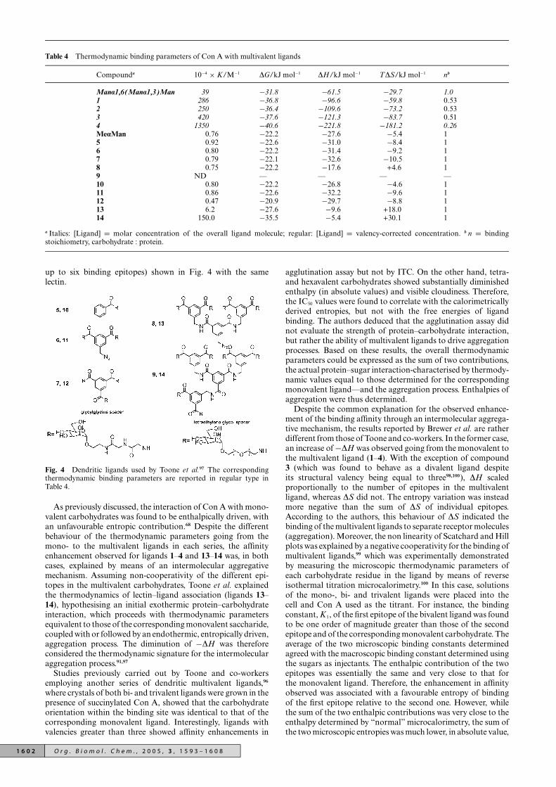
Table 4 Thermodynamic binding parameters of Con A with multivalent ligands
Compounda 10−4 × K/M−1 DG/kJ mol−1 DH/kJ mol−1 TDS/kJ mol−1 nb
Mana1,6(Mana1,3)Man 39 −31.8 −61.5 −29.7 1.01 286 −36.8 −96.6 −59.8 0.532 250 −36.4 −109.6 −73.2 0.533 420 −37.6 −121.3 −83.7 0.514 1350 −40.6 −221.8 −181.2 0.26MeaMan 0.76 −22.2 −27.6 −5.4 15 0.92 −22.6 −31.0 −8.4 16 0.80 −22.2 −31.4 −9.2 17 0.79 −22.1 −32.6 −10.5 18 0.75 −22.2 −17.6 +4.6 19 ND — — — —10 0.80 −22.2 −26.8 −4.6 111 0.86 −22.6 −32.2 −9.6 112 0.47 −20.9 −29.7 −8.8 113 6.2 −27.6 −9.6 +18.0 114 150.0 −35.5 −5.4 +30.1 1
a Italics: [Ligand] = molar concentration of the overall ligand molecule; regular: [Ligand] = valency-corrected concentration. b n = bindingstoichiometry, carbohydrate : protein.
up to six binding epitopes) shown in Fig. 4 with the samelectin.
Fig. 4 Dendritic ligands used by Toone et al.97 The correspondingthermodynamic binding parameters are reported in regular type inTable 4.
As previously discussed, the interaction of Con A with mono-valent carbohydrates was found to be enthalpically driven, withan unfavourable entropic contribution.68 Despite the differentbehaviour of the thermodynamic parameters going from themono- to the multivalent ligands in each series, the affinityenhancement observed for ligands 1–4 and 13–14 was, in bothcases, explained by means of an intermolecular aggregativemechanism. Assuming non-cooperativity of the different epi-topes in the multivalent carbohydrates, Toone et al. explainedthe thermodynamics of lectin–ligand association (ligands 13–14), hypothesising an initial exothermic protein–carbohydrateinteraction, which proceeds with thermodynamic parametersequivalent to those of the corresponding monovalent saccharide,coupled with or followed by an endothermic, entropically driven,aggregation process. The diminution of −DH was thereforeconsidered the thermodynamic signature for the intermolecularaggregation process.91,97
Studies previously carried out by Toone and co-workersemploying another series of dendritic multivalent ligands,96
where crystals of both bi- and trivalent ligands were grown in thepresence of succinylated Con A, showed that the carbohydrateorientation within the binding site was identical to that of thecorresponding monovalent ligand. Interestingly, ligands withvalencies greater than three showed affinity enhancements in
agglutination assay but not by ITC. On the other hand, tetra-and hexavalent carbohydrates showed substantially diminishedenthalpy (in absolute values) and visible cloudiness. Therefore,the IC50 values were found to correlate with the calorimetricallyderived entropies, but not with the free energies of ligandbinding. The authors deduced that the agglutination assay didnot evaluate the strength of protein–carbohydrate interaction,but rather the ability of multivalent ligands to drive aggregationprocesses. Based on these results, the overall thermodynamicparameters could be expressed as the sum of two contributions,the actual protein–sugar interaction-characterised by thermody-namic values equal to those determined for the correspondingmonovalent ligand—and the aggregation process. Enthalpies ofaggregation were thus determined.
Despite the common explanation for the observed enhance-ment of the binding affinity through an intermolecular aggrega-tive mechanism, the results reported by Brewer et al. are ratherdifferent from those of Toone and co-workers. In the former case,an increase of −DH was observed going from the monovalent tothe multivalent ligand (1–4). With the exception of compound3 (which was found to behave as a divalent ligand despiteits structural valency being equal to three98,100), DH scaledproportionally to the number of epitopes in the multivalentligand, whereas DS did not. The entropy variation was insteadmore negative than the sum of DS of individual epitopes.According to the authors, this behaviour of DS indicated thebinding of the multivalent ligands to separate receptor molecules(aggregation). Moreover, the non linearity of Scatchard and Hillplots was explained by a negative cooperativity for the binding ofmultivalent ligands,99 which was experimentally demonstratedby measuring the microscopic thermodynamic parameters ofeach carbohydrate residue in the ligand by means of reverseisothermal titration microcalorimetry.100 In this case, solutionsof the mono-, bi- and trivalent ligands were placed into thecell and Con A used as the titrant. For instance, the bindingconstant, K1, of the first epitope of the bivalent ligand was foundto be one order of magnitude greater than those of the secondepitope and of the corresponding monovalent carbohydrate. Theaverage of the two microscopic binding constants determinedagreed with the macroscopic binding constant determined usingthe sugars as injectants. The enthalpic contribution of the twoepitopes was essentially the same and very close to that forthe monovalent ligand. Therefore, the enhancement in affinityobserved was associated with a favourable entropy of bindingof the first epitope relative to the second one. However, whilethe sum of the two enthalpic contributions was very close to theenthalpy determined by “normal” microcalorimetry, the sum ofthe two microscopic entropies was much lower, in absolute value,
1 6 0 2 O r g . B i o m o l . C h e m . , 2 0 0 5 , 3 , 1 5 9 3 – 1 6 0 8

than the one reported when the ligand was injected into the lectinsolution. Hemagglutination inhibition measurements correlatedperfectly with the results obtained by ITC. The authors providedsome hypotheses to explain the differences between their resultsand those reported by Toone and co-workers. For instance, theyunderlined the difference in the method adopted to express theconcentration of the ligands. While Toone et al. considered thenumber of epitope equivalents, Brewer et al. referred to the molarconcentration of the ligand molecule. Their arguments againstthe use of valency-corrected concentrations were the observednegative cooperativity of binding and the determined differencebetween the structural and functional valency.99
Most examples of intramolecular/chelate binding involveeither bacterial toxins or polymeric ligands. Studies have shownthat the enhancement in binding affinity is much higher whendifferent carbohydrate residues of the ligand bind to sites on thesame receptor molecule. In 1983, Lee et al.103 reported that a syn-thetic tetraantennary undecasaccharide showed an inhibitionconstant for the hepatic Gal/GalNAc receptor 106 times greaterthan an equivalent monoantennary trisaccharide, despite theonly 4-fold statistical increase in absolute galactose concentra-tion. It was therefore pointed out that the number of Gal residuesper cluster and their branching mode were major determinantsof the binding affinity of a ligand to the mammalian hepaticlectin. A striking example of the affinity enhancement achievablethrough multivalency (10 orders of magnitude compared tomonovalent ligand), for a non-carbohydrate system, was givenby Rao et al.104
Most of the currently synthesised polymeric and dendriticmultivalent ligands show a “random” multivalency, whichrarely allows an affinity enhancement higher than 1,000-fold.Moreover, as already pointed out, the reasons underlying thisactivity gain are poorly understood.63 In a different approach,structural information about the spatial arrangement of thetarget binding sites are taken into account for the design ofligands ideal for maximising the interaction with the receptor.
The modular synthesis of multivalent ligands of the heat-labile enterotoxin (LT) from E. coli was recently reported byFan and co-workers.105 The five B subunits of the toxin presenta 5-fold symmetric configuration. In this study, pentavalentligands constituted by a semirigid “core” which can adopta conformation close to 5-fold symmetry, flexible “linkers”that project toward the receptor binding sites and “fingers”represented by 1-b-amidated D-galactose, were synthesised. Dif-ferent lengths of the linker were tested and the receptor–ligandinteraction analysed by an enzyme-linked immunosorbent assay,ELISA. All pentavalent ligands led to significant affinity gainscompared to the corresponding monovalent ones. The best oneshowed an IC50 105-fold lower than galactose, approaching theaffinity of the oligosaccharide portion of the natural gangliosideGM1. The pentavalent ligand also showed a 104-fold affinitygain compared to the corresponding monovalent ligand, or2000-fold on a valency-corrected basis. Dynamic light scatteringruled out the possibility of an aggregative process, supportingthe expected formation of a 1 : 1 toxin–ligand complex. Theanalysis of the effect of linker-length on the affinity showed thatthe greatest enhancement was detected for the ligand whoselinker effective length106 best matched the distance betweennonadjacent binding sites.
One of the most striking examples of affinity enhancementwas recently reported by Kitov et al.107 Shiga-like toxins SLT-Iand SLT-II were inhibited by a decavalent ligand, designatedSTARFISH, whose structure was complementary to that of thereceptor. The SLTs are AB toxins constituted by an enzymatic(A) component and a cell-binding (B) part. The A-subunit issituated on one face of the B-component, which is a pentamerof identical subunits. In the absence of the A-subunits, the B-subunits still form pentamers that are functionally equivalentto the overall toxin in their attachment to the host cell. Thein vivo cytotoxicity of SLTs has been correlated with their
binding affinity toward the glycolipid globotriaosylceramide(Gb3). The crystal structure of SLT-I B-pentamer complexedwith a Gb3 analogue revealed three Gb3-binding sites per B-subunit108 (Fig. 5).
Fig. 5 View along the 5-fold axis of the SLT-I B-pentamer boundto the Pk-MCO trisaccharides. The surface toward the viewer is thesugar binding surface (reprinted with permission from ref. 108, copyright(1998) American Chemical Society).
As determined by means of an ELISA protocol, STARFISHexhibited more than 106-fold increase in inhibition over the Pktrisaccharide. STARFISH was designed to achieve high affinitythrough the simultaneous binding of all five peripheral bridgedPk dimers to the ten sites 1 and 2 of the B-pentamer (with eachtrisaccharide dimer bridging sites 1 and 2 of the monomer).Crystallographic studies of the formed complex revealed adifferent mode of binding (Fig. 6). One STARFISH molecule,in fact, bound two B-subunit monomers from separate toxinmolecules, with the saccharide residues occupying only site 2.Each trisaccharide interaction was identical to that seen for thecorresponding univalent ligand. A higher affinity of binding ofsite 2 compared to 1 had been already highlighted by previousstudies carried out on toxin mutants. Therefore, the observedmixture of aggregative and chelate mechanisms through whichSTARFISH exerted its activity could be explained by thestronger interactions of the carbohydrate within site 2, possiblycoupled with unfavourable entropic and enthalpic contributionsof the bridging.
Recent mutation studies have shown that site 2 of the SLT1B subunit is the most important site for binding of free Pktrisaccharides, however the STARFISH ligand also requires anintact site 3. This suggests that site 3 is specifically involved inbinding of the pentavalent ligand, the individual trisaccharidesof which bind at sites 1 and 2.109
A calorimetric study of the interaction of bivalent glycopep-tides with SLT-I was carried out by Toone and co-workers.93 Thetwo ligands used differed in the nature of the peptidic linker. Bothligands showed affinity gain, but the mechanism through whichthis was achieved depended on the nature of the linker domain:while the compound characterised by a hydrophobic linkerseemed to work through a chelate mechanism, the hydrophilicone bound by means of an intermolecular aggregative process.Again the binding enthalpy of the latter appeared to be signif-icantly lower (in absolute value) than that of the monovalentcompound, a trend that this group already recognised as thesignature for an intermolecular binding process. The affinityenhancement reached through the intramolecular mechanism
O r g . B i o m o l . C h e m . , 2 0 0 5 , 3 , 1 5 9 3 – 1 6 0 8 1 6 0 3

Fig. 6 Diagrams of STARFISH ligand bound to the SLT. Reprinted with permission from Nature Publishing Group.107
was slightly higher than that of the hydrophilic compound.Peptidic spacers were chosen in the search for possible favourableadditional interactions of the ligand with the protein surface.These interactions were given as an explanation for the higheraffinity of the ligand with the hydrophobic linker, and for itsintramolecular mechanism. For the hydrophilic linker, eitherthere were no favourable contacts or there were in fact repulsiveinteractions of the linker with hydrophobic regions on theprotein surface. Therefore, the affinity was lower and the bindingproceeded via an intermolecular mechanism.
The greatest increases in activity on a valency-corrected basisare reported with polymeric ligands.91 Nevertheless, the onlypolymeric ligands synthesised so far which seem to bind throughan intramolecular mechanism are those prepared by Kanaiet al.110 (Fig. 7).
Fig. 7 Scheme for the synthesis of neoglycopolymers by ROMP: (a)MeOH–H2O–DCM (6 : 1 : 5) then add H2O, room temperature; (b)H2O–DCM (2 : 1), DTAB, room temperature; (c) TsNHNH2, H2O,100 ◦C, 56% (n = 10), 91% (n = 25), 58% (n = 52), 100% (n = 143).110
Several ligands with different backbone lengths were synthe-sised and tested as inhibitors for Con A–carbohydrate inter-action. The maximum inhibitory activity seemed to coincidewith an average length sufficient to span two lectin bindingsites belonging to the same protein molecule, supporting thehypothesis of a chelate mechanism of binding. The enhancementin affinity determined for polymers too short to bridge twobinding sites was attributed to a high local concentration ofsugar moieties (statistical effect), which would perturb the rate ofdissociation of the formed complex. The authors concluded thatthe observed dependence of the inhibition activity on the poly-mer length was largely due to a combination of statistical andchelation effects. Moreover, despite the differences in backboneflexibility, the most potent ligands of each series, 2 and 3, hadapproximately the same efficacy. This latter observation pointsout an interesting aspect of multivalent ligand–protein binding.It is believed that rigid linkers would favour the interaction,due to a reduction of the entropic penalties accompanying thebinding.105,107,111 However, the great enhancements achieved withflexible linkers and the negligible gain in activity observed withdendritic ligands characterised by rigid arms, have recently ledToone and co-workers to suggest that entropic concerns might beless severe than was previously thought and probably less severethan those related to unfavourable enthalpic contributions.91
2.3 Diagnostic and therapeutic applications
There is considerable evidence that lectins are involved in manyphysiological events (several examples are given in Table 5).The enormous developments that the possibility to inhibit,activate or exploit protein–carbohydrate interactions could
Table 5 Examples of the functions of animal lectinsa
Function Examples
Intracellular routing of glycoconjugates P-type lectins, ERGIC-53, VIP-36Molecular chaperones during glycoprotein synthesis Calnexin, calreticulinMediation of endocytosis Asialoglycoprotein receptors, macrophage mannose receptorCellular growth regulation Galectins, sarcolectin, cytokinesExtracellular molecular bridging Geodia cydonium galectin, other galectins, interleukin-2Cell–cell interactions for homing and trafficking Selectins, CD22, CD31, CD44Cell–matrix interactions Galectins, heparin- and hyaluronic acid binding lectinsScavenging of cellular debris; anti-inflammatory action Galectin-9
a Selected from ref. 10 and ref. 25.
1 6 0 4 O r g . B i o m o l . C h e m . , 2 0 0 5 , 3 , 1 5 9 3 – 1 6 0 8

bring, especially in medicinal chemistry, push the study oflectins and carbohydrates to the forefront of research. Forexample, the cellular protein glycosylation pattern is influencedby several physiological changes, such as the occurrence ofdisease. Thus, the altered glycoform population of a givenglycoprotein may be diagnostic of the disease responsible for thealteration itself. Abnormal glycosylation has been detected incancer development.112 Both quantitative and qualitative lectin-binding differences were observed for cytosolic glycoproteinsin benign and malignant thyroid neoplasms: in the major-ity of carcinomas lectin-binding was weaker in comparisonwith adenomas and non-neoplastic specimens.113 Changes inprostate-specific antigen (PSA) glycosylation during malignanttransformation may be used for the diagnosis of prostate cancerat an early stage. The quantitative precipitation method ofCon A–carbohydrate interaction114 was used by Basu and co-workers115 for differentiation between prostate cancer (PC) andbenign prostatic hyperplasia (BPH). The carbohydrate contentin the precipitate after binding of Con A with serum PSA of PCwas significantly lower than that of BPH. It was concluded that aserum value <3.0 lg ml−1 of the carbohydrate content of Con A–PSA precipitate indicates strong suspicion for prostate cancer,reducing the rate of unnecessary biopsies in men with total PSAvalue between 4.0 and 10.0 lg ml−1. More recently, wheat germagglutinin (WGA) has been shown to induce rapid apoptosisin malignant cells via a novel mechanism.116 Studies indicatedthat GlcNAc-containing glycoconjugates were also involved inWGA-mediated cell death, and that the mechanism probablyinvolved the mitochondrial pathway.
Such valuable diagnostic and potential therapeutic applica-tions highlight the need to gain further insights into lectin-specific ligand interactions and into the physiological processesthat these associations initiate and/or regulate. Novel lectinsare therefore continuously isolated, such as, in 2003, the N-acetylglucosamine-specific lectin from fresh sclerotia of theedible mushroom Pleurotus tuberregium,117 and the two mannan-and L-fucose-binding lectins from the green alga Enteromorphaprolifera.118 At the same time, a deeper understanding of thefunctioning of known systems is achieved. Loris and co-workers119 have recently reported the crystal structure of thePseudomonas aeruginosa fucose-specific lectin (LecB) in itsmetal-bound and metal-free state as well as complexed withfucose, mannose and fructopyranose. The higher protein affinityfor fucose was ascribed to favourable interactions involving C6and O1 of the saccharide, absent for the other ligands, due to thepartial shielding of a carboxylate group and one or two hydroxylgroups. A recent crystallographic study upon the lung surfactantprotein D (SP-D), a protein capable of interacting directly withcarbohydrate residues on pulmonary pathogens and allergens,of stimulating immune cells and of intervening directly inthe lungs’ immune response, has revealed that a previouslyunreported calcium ion is essential for biological activity,ensuring the right structural requirements for the establishedeffector mechanism.120 The lectin from Vatairea macrocarpaseeds (VML) was used to induce neutrophil migration in ratsin order to explore the pathways by which lectins cause aninflammatory response.121 The results obtained suggested thatthe lectin-induced effects do not involve the usual mediators,such as PAF mediator, rather it acts through an indirectmechanism.
One of the most well-known physiological processes initiatedby protein–carbohydrate interactions is the clearance of olderythrocytes from the blood.5 With age the red blood cellsbecome progressively desialylated. When the density of galactosemoieties so exposed on the surface is high enough, the cellis removed from the circulation and destroyed through therecognition of the galactosyl residues by hepatocyte receptors(so-called asialoglycoprotein receptors). Clues to the role ofgalactose in this process were first obtained in 1968 from exper-iments with rabbits that showed that desialylated ceruloplasmin
(which also has terminal galactose residues) was cleared rapidlyfrom the serum by the liver.122 The carbohydrate recognitiondomain (CRD) of the mammalian asialoglycoprotein receptorof hepatocytes is the prototype for the C-lectins,5,123 a largefamily of Ca2+-dependent animal proteins. Members of thisgroup are important components of the rapidly-respondinginnate immune system, including the mannose-binding proteins,MBPs. The role of MBPs in host defence against viral pathogenshas been demonstrated by several studies.124 Like all othercollectins—a group of soluble C-type lectins—MBPs have anN-terminal collagenous domain and a globular C-terminalportion, containing the CRD.125 Thus, while the C-terminaldomain mediates the binding to various microorganisms, the N-terminal one interacts with the cell, triggering the carbohydrate-dependent complement activation, which then results either inthe killing of the pathogen126 or in the deposition of complementcomponents on the surface of the microorganism (opsonisation)which is then cleared by phagocytic cells like macrophagesand neutrophils. MBPs have been shown to prevent humanimmunodeficiency virus (HIV) infection of H9 cells, probablyby binding to oligomannose units of the envelope gp120 of thevirus.127 A deficiency in MBPs has been claimed to accountfor a significant number of immunodeficiency cases in children,where the immune system is still not fully capable of mountingan efficient adaptive response.128 Serum MBP levels are largelyaffected by polymorphisms of the MBP gene. The presence ofthe minority allele can result in either a significant reductionof the serum protein concentration or in its almost completeabsence. These conditions have been suggested to be associatedwith recurrent infections.129 Recent studies have also shown thatindividuals with the minority alleles of the MBP gene are atrisk from autoimmune diseases; serum MBP may also bind toapoptotic cells and so contribute to the clearance of potentialautoantigens.130
Siglecs (sialic acid-binding immunoglobulin-like lectins), animportant subset of the Ig superfamily, act as endogenous recep-tors of sialic acid residues. Recent advances in the understandingof the function of known siglecs and the identification of newmembers of the family have contributed significantly to theelucidation of their physiological roles. Thus, a participation ininflammation processes, indicated by the expression of the lectinon inflammatory macrophages found in rheumatoid arthritis,was suggested for sialoadhesin (Siglec-1).131 CD22 (Siglec-2), awell-characterised B cell inhibitory receptor, has been found tomodulate leukocyte activation; in particular, it has been shownthat lectin trans-interactions with a-2,6-linked sialic acids in N-glycans can dampen B cell activation.132 All new family membersrecently identified are highly related to CD33 (Siglec-3), a markerof myeloid cells. Their expansion in the innate immune systemsuggests an important role of these proteins in host defence.Similar to the role proposed for CD22, CD33-related siglecsmay regulate the autoreactivity of myeloid cells.132
Selectins, a subfamily of C-type lectins so called because oftheir ability to mediate selective contacts between cells, play akey role in the recruitment of leukocytes to inflammatory sites.10
Selectins represent the first group of mammalian lectins shownto mediate cell–cell contact, in particular they provide rapidand reversible cell adhesion under hydrodynamic flow.133 Thedynamics of the selectin–sialyl Lewis X (sLeX) interaction havebeen probed by single molecule dynamic force spectroscopy.134
Unbinding was determined to involve at least two activationbarriers; that which determines the response of the complexunder high shear is determined by Ca2+-mediated binding of thefucose residue of sLeX to the lectin. During the inflammatoryresponse, damage in tissues surrounding a blood vessel causesthe influx of signalling molecules. Selectins are thus rapidlyexpressed on the inner surface of blood vessels (E-selectins)and on platelets (P-selectins). Highly specific binding of theseproteins to sugars on white blood cell surfaces causes the leuko-cytes to adhere to the vessel walls, which, at this stage, express
O r g . B i o m o l . C h e m . , 2 0 0 5 , 3 , 1 5 9 3 – 1 6 0 8 1 6 0 5

L-selectins. By interaction with L-selectins, leukocytes can nowroll toward the site of damage, where they pass through to thesurrounding tissue.135 Whilst this is a well-controlled process inhealthy individuals, in excess it is a cause of arthritis, asthma andmyocardial infarction. Due to their ability to mediate cell–celladhesion, selectins play a critical role in tumour metastasis.136
For instance, it has been suggested that carcinoma cells caninteract with platelets, leukocytes and endothelia through theselectins. Thus, the blood platelets may form complexes aroundthe tumour cells and facilitate metastasis by masking them fromthe host defence.137 Due to their implication in a large number ofdiseases, suitable selectin inhibitors represent a class of efficienttherapeutic agents. P-selectin glycoprotein ligand (PSGL-1) hasshown good results in phase II clinical trials to prevent ischemiareperfusion injury and it seems to be a promising candidate as acancer therapeutic.137
Galectins are a growing family of b-galactoside-specific pro-teins of which 15 members have been identified in mammals.138
Many tumours, such as colon, thyroid and breast carcinomas,express both galectin-1 and -3, and it has been suggested thatgalectin-3 is a metastasis marker.139 Among the degenerativeprocesses that they can mediate, galectins may favour metastasisby binding glycoconjugates of the extracellular matrix and sub-sequent intravasation of the tumour cell into the blood vessels.A modified citrus pectin (MCP), a complex highly branchedpolysaccharide rich in galactoside residues, has shown very en-couraging results as an anti-cancer agent, significantly reducinglung colonisation of B16-F1 melanoma cells.140 More recently,3′-aromatic amide derivatives of 3′-amino-N-acetyllactosaminehave been found to be potent inhibitors of galectin-3 (IC50 downto 4.4 lM).141
ConclusionsMultivalent protein–carbohydrate interactions regulate a myr-iad of vital cellular events. Lectin–carbohydrate associationsare usually weak when compared to others in Nature, withKD values of the order of mM for monosaccharides. However,when multivalent saccharides of the right type are clusteredtogether with the right geometry, the interaction becomesstrong and highly specific. This increase is more than whatwould be expected on the basis of the increased sugar localconcentration. The reasons of this phenomenon, termed “clusterglycoside effect”, have not been rigorously determined yet, butits implications are extraordinary.
It has been pointed out repeatedly that a thorough under-standing of the lectin–carbohydrate interaction is essential inorder for it to be exploited for biomedical applications.61,63,142
A deep knowledge of both structural and energetic aspects ofthe process is thus necessary. Indeed, the failure of a numberof carbohydrate-based drugs may be attributed to a poor un-derstanding of their supposed mechanism of action rather thanto any inherent deficiency of carbohydrate therapeutics.61 Whengood understanding is achieved, the results are impressive.105,107
AcknowledgementsEPSRC is thanked for a PhD studentship to M.A.
References1 W. C. Boyd and E. Shapleigh, Science, 1954, 119, 419.2 S. H. Barondes, Trends Biochem. Sci., 1988, 13, 480.3 N. Sharon and H. Lis, Science, 1989, 246, 227.4 G. Ashwell and A. G. Morell, Adv. Enzymol., 1974, 41, 99.5 G. Ashwell and J. Harford, Annu. Rev. Biochem., 1982, 51, 531.6 J. C. Paulson in The Receptors, ed. P. M. Conn, Academic Press,
New York, 1985, p. 131; D. C. Wiley and J. J. Skehel, Annu. Rev.Biochem., 1987, 56, 365; N. Sharon, FEBS Lett., 1987, 217, 145.
7 J. Melrose, N. Tsurushita, G. Liu and E. L. Berg, J. Immunol., 1998,161, 2457; N. Watanabe, H. Kawashima, Y.-F. Li and M. Miyasaka,J. Biochem., 1999, 125, 826.
8 H. J. Gabius, Angew. Chem., Int. Ed. Engl., 1988, 27, 1267; M.Monsigny, A. C. Roche, C. Kieda, P. Midoux and A. Obrenovitch,Biochimie, 1988, 70, 1633.
9 N. Sharon and H. Lis, Trends Biochem. Sci., 1987, 12, 488.10 D. C. Kilpatrick, Biochim. Biophys. Acta, 2002, 1572, 187.11 J. B. Sumner, J. Biol. Chem., 1919, 37, 137.12 J. B. Sumner and S. F. Howell, J. Bacteriol., 1936, 32, 227.13 W. M. Watkins and W. T. J. Morgan, Nature, 1952, 169, 825; W. T. J.
Morgan and W. M. Watkins, Br. J. Exp. Pathol., 1953, 34, 94.14 I. E. Liener, N. Sharon, and I. J. Goldstein, The Lectins: Properties,
Functions and Applications in Biology and Medicine, AcademicPress, Orlando, 1986; N. Sharon and H. Lis, Lectins, Chapmanand Hall, London, 1989.
15 H. Lis and N. Sharon, Chem. Rev., 1998, 98, 637.16 I. J. Goldstein and R. D. Poretz, in The Lectins: Properties,
Functions and Applications in Biology and Medicine, ed. I. E. Liener,N. Sharon, and I. J. Goldstein, Academic Press, Orlando, 1986,p. 33.
17 R. D. Poretz and I. J. Goldstein, Biochem. Pharmacol., 1971, 20,2727.
18 A. Pusztai, Plant Lectins, Cambridge University Press, Cambridge,1991.
19 Y. D. Lobsanov, M. A. Gitt, H. Leffler, S. H. Barondes and J. M.Rini, J. Biol. Chem., 1993, 268, 27034.
20 N. K. Sauter, J. E. Hanson, G. D. Glick, J. H. Brown, R. L. Crowther,S.-J. Park, J. J. Skehel and D. C. Wiley, Biochemistry, 1992, 31, 9609.
21 K. Drickamer, J. Biol. Chem., 1988, 263, 9557.22 I. Ofek and R. J. Doyle, Bacterial Adhesion to Cells and Tissues,
Chapman and Hall, London, 1994.23 J. Hirabayashi, Trends Glycosci. Glycotechnol., 1997, 9, 45.24 R. Loris, T. Hamelryck, J. Bouckaert and L. Wyns, Biochim.
Biophys. Acta, 1998, 1383, 9.25 H. J. Gabius, H. C. Siebert, S. Andre, J. Jimenez-Barbero and H.
Rudiger, ChemBioChem, 2004, 5, 741.26 Y. Konami, K. Yamamoto, T. Osawa and T. Irimura, Glycoconju-
gate J., 1995, 12, 128.27 M. Inbar and L. Sachs, Nature, 1969, 223, 710.28 N. Srinivasan, S. D. Rufino, M. B. Pepys, S. Wood and T. L. Blundell,
Chemtracts: Biochem. Mol. Biol., 1996, 6, 149.29 V. R. Srinivas, G. B. Reddy, N. Ahmad, C. P. Swaminathan, N. Mitra
and A. Surolia, Biochim. Biophys. Acta, 2001, 1527, 102.30 R. Banerjee, S. C. Mande, V. Ganesh, K. Das, V. Dhanaraj, S. K.
Mahanta, K. Suguna, A. Surolia and M. Vijayan, Proc. Natl. Acad.Sci. USA, 1994, 91, 227; R. Banerjee, K. Das, R. Ravishankar, K.Suguna, A. Surolia and M. Vijayan, J. Mol. Biol., 1996, 259, 281.
31 D. M. Salunke, M. J. Swamy, M. Islam Khan, S. C. Mande, A.Surolia and M. Vijayan, J. Biol. Chem., 1985, 260, 13576.
32 W. W. Fish, L. M. Hamlin and R. L. Miller, Arch. Biochem. Biophys.,1978, 190, 693.
33 M. Decastel, H. De Boeck, Y. Goussault, C. K. De Bruyne, F. G.Loontiens and J.-P. Frenoy, Arch. Biochem. Biophys., 1985, 240,811.
34 G. B. Reddy, V. R. Srinivas, N. Ahmad and A. Surolia, J. Biol.Chem., 1999, 274, 4500.
35 W. I. Weis and K. Drickamer, Annu. Rev. Biochem., 1996, 65, 441.36 N. Sharon, Trends Biochem. Sci., 1993, 18, 221; R. Adar and N.
Sharon, Eur. J. Biochem., 1996, 239, 668.37 A. Imberty, F. Casset, C. V. Gegg, M. E. Etzler and S. Perez,
Glycoconjugate J., 1994, 11, 400.38 R. Banerjee, K. Das, R. Ravishankar, K. Suguna, A. Surolia and
M. Vijayan, J. Mol. Biol., 1996, 259, 281.39 P. Adhikari, K. Bachhawat-Sikder, C. J. Thomas, R. Ravishankar,
A. A. Jeyaprakash, V. Sharma, M. Vijayan and A. Surolia, J. Biol.Chem., 2001, 276, 40734.
40 N. M. Young and R. P. Oomen, J. Mol. Biol., 1992, 228, 924.41 V. Sharma and A. Surolia, J. Mol. Biol., 1997, 267, 433.42 D. M. Salunke, M. Islam Khan, A. Surolia and M. Vijayan, J. Mol.
Biol., 1982, 154, 177; D. M. Salunke, M. Islam Khan, A. Suroliaand M. Vijayan, FEBS Lett., 1983, 156, 127.
43 R. Ravishankar, M. Ravindran, K. Suguna, A. Surolia and M.Vijayan, Curr. Sci., 1997, 72, 855; R. Ravishankar, M. Ravindran,K. Suguna, A. Surolia and M. Vijayan, Curr. Sci., 1999, 76, 1393.
44 R. Ravishankar, K. Suguna, A. Surolia and M. Vijayan, ActaCrystallogr., Sect. D, 1999, 55, 1375.
45 J. V. Pratap, G. M. Bradbrook, G. B. Reddy, A. Surolia, J. Raftery,J. R. Helliwell and M. Vijayan, Acta Crystallogr., Sect. D, 2001, 57,1584.
46 B. G. Davis, J. Chem. Soc., Perkin Trans. 1, 1999, 3125.47 J. M. Rini, Annu. Rev. Biophys. Biomol. Struct., 1995, 24, 551.48 E. J. Toone, Curr. Opin. Struct. Biol., 1994, 4, 719.49 S. Elgavish and B. Shaanan, J. Mol. Biol., 1998, 277, 917.
1 6 0 6 O r g . B i o m o l . C h e m . , 2 0 0 5 , 3 , 1 5 9 3 – 1 6 0 8

50 E. R. Caffarena, J. R. Grigera and P. M. Bisch, J. Mol. Graphics,2002, 21, 227.
51 C. V. Gegg, D. D. Roberts, I. H. Segel and M. E. Etzler, Biochemistry,1992, 31, 6938.
52 M. E. Etzler, Trends Glycosci. Glycotechnol., 1998, 10, 247.53 M. E. Etzler, in Glycoconjugates. Composition, Structure and Func-
tions, ed. H. J. Allen and E. C. Kisailus, Marcel Dekker Inc., NewYork, 1992, p. 521.
54 W. J. Peumans and E. J. M. Van Damme, in Lectins. BiomedicalPerspectives, ed. A. Pusztai and S. Bardocz, Taylor & Francis,London, 1995, p. 1; J. W. Kijne, Chemtracts Biochem. Mol. Biol.,1996, 6, 180.
55 G. W. G. Bird, Vox Sang., 1959, 4, 307; W. C. Boyd, Vox Sang., 1963,8, 1.
56 R. A. Dixon and N. L. Paiva, Plant Cell, 1995, 7, 1085.57 F. Cote and M. G. Hahn, Plant Mol. Biol., 1994, 26, 1379.58 M. E. Etzler, in The Lectins: Properties, Functions and Applications
in Biology and Medicine, ed. I. E. Liener, N. Sharon, and I. J.Goldstein, Academic Press, Orlando, 1986, p. 371.
59 P. Mylona, D. Pawlowski and T. Bisseling, Plant Cell, 1995, 7, 869.60 Y. C. Lee and R. T. Lee, Acc. Chem. Res., 1995, 28, 321.61 B. G. Davis, Chem. Rev., 2002, 102, 579.62 M. N. Janakiraman, C. L. White, W. G. Laver, G. M. Air and M.
Luo, Biochemistry, 1994, 33, 8172.63 M. Mammen, S.-K. Choi and G. M. Whitesides, Angew. Chem., Int.
Ed. Engl., 1998, 37, 2754.64 M. Yehuda and M. Moshe, Glycoconjugate J., 2002, 19, 1.65 C. F. Brewer, M. C. Miceli and L. G. Baum, Curr. Opin. Struct.
Biol., 2002, 12, 616.66 Z. K. Indik, J. G. Park, S. Hunter and A. D. Schreiber, Blood, 1995,
86, 4389.67 M. C. Chervenak and E. J. Toone, J. Am. Chem. Soc., 1994, 116,
10533.68 B. A. Williams, M. C. Chervenak and E. J. Toone, J. Biol. Chem.,
1992, 267, 22907.69 M. Kaul and D. S. Pilch, Biochemistry, 2002, 41, 7695.70 K. A. Watson, E. P. Mitchell, L. N. Johnson, J. C. Son, C. J. F.
Bichard, M. G. Orchard, G. W. J. Fleet, N. G. Oikonomakos, D. D.Leonidas, M. Kontou and A. Papageorgioui, Biochemistry, 1994,33, 5745.
71 N. Muller, Acc. Chem. Res., 1990, 23, 23.72 H. Beierbeck, L. T. J. Delbaere, M. Vandonselaar and R. U.
Lemieux, Can. J. Chem., 1994, 72, 463.73 J. E. Leffler and E. Grunwald, Rates and Equilibria of Organic
Reactions, John Wiley & Sons, New York, 1963.74 F. P. Schwarz, K. D. Puri, R. G. Bhat and A. Surolia, J. Biol. Chem.,
1993, 268, 7668.75 G. I. Makhatadze and P. L. Privalov, J. Mol. Biol., 1993, 232, 639;
P. L. Privalov and G. I. Makhatadze, J. Mol. Biol., 1993, 232, 660.76 M. C. Chervenak and E. J. Toone, Biochemistry, 1995, 34, 5685.77 W. Kauzmann, Adv. Protein Chem., 1959, 14, 1.78 K. P. Murphy, D. Xie, K. S. Thompson, L. M. Amzel and E. Freire,
Proteins: Struct., Funct., Genet., 1993, 18, 63.79 J. M. Sturtevant, Proc. Natl. Acad. Sci. USA, 1977, 74, 2236.80 A. Surolia, N. Sharon and F. P. Schwarz, J. Biol. Chem., 1996, 271,
17697.81 R. F. Kelley and M. P. O’Connell, Biochemistry, 1993, 32, 6828.82 D. A. Brummell, V. P. Sharma, N. N. Anand, D. Bilous, G.
Dubuc, J. Michniewicz, C. R. MacKenzie, J. Sadowska, B. W.Sigurskjold, B. Sinnott, N. M. Young, D. R. Bundle and S. A.Narang, Biochemistry, 1993, 32, 1180.
83 C. F. Brewer, L. Bhattacharyya, R. D. I. Brown and S. H. Koenig,Biochem. Biophys. Res. Commun., 1985, 127, 1066.
84 T. K. Dam, B. S. Cavada, T. B. Grangeiro, C. F. Santos, V. M.Ceccato, F. A. M. de Susa, S. Oscarson and C. F. Brewer, J. Biol.Chem., 2000, 275, 16119.
85 H. C. Siebert, R. Adar, R. Arango, M. Burchert, H. Kaltner, G.Kayser, E. Tajkhorshid, C. W. von der Lieth, R. Kaptein, N. Sharon,J. F. Vliegenthart and H.-J. Gabius, Eur. J. Biochem., 1997, 249, 27.
86 D. Gupta, M. Cho, M. D. Cummings and C. F. Brewer, Biochem-istry, 1996, 35, 15236.
87 D. Gupta, T. K. Dam, S. Oscarson and C. F. Brewer, J. Biol. Chem.,1997, 272, 6388.
88 F. P. Schwarz, H. Ahmed, M. A. Bianchet, L. M. Amzel and G. R.Vasta, Biochemistry, 1998, 37, 5867.
89 N. Navarre, N. Amiot, A. van Oijen, A. Imberty, A. Poveda, J.Jimenez-Barbero, A. Cooper, M. A. Nutley and G.-J. Boons, Chem.Eur. J., 1999, 5, 2281.
90 Y. C. Lee, in Neoglycoconjugates: Preparation and Applications, ed.Y. C. Lee and R. T. Lee, Academic Press, London, 1994, p. 3.
91 J. J. Lundquist and E. J. Toone, Chem. Rev., 2002, 102, 555.
92 W. P. Jencks, Proc. Natl. Acad. Sci. USA, 1981, 78, 4046.93 J. J. Lundquist, S. D. Debenham and E. J. Toone, J. Org. Chem.,
2000, 65, 8245.94 E. Duverger, N. Frison, A. C. Roche and M. Monsigny, Biochimie,
2003, 85, 167.95 R. E. McDonald, D. Hughes and B. G. Davis, Angew. Chem., Int.
Ed., 2004, 43, 3025.96 S. M. Dimick, S. C. Powell, S. A. McMahon, D. N. Moothoo, J. H.
Naismith and E. J. Toone, J. Am. Chem. Soc., 1999, 121, 10286.97 J. B. Corbell, J. J. Lundquist and E. J. Toone, Tetrahedron:
Asymmetry, 2000, 11, 95.98 T. K. Dam, R. Roy, S. K. Das, S. Oscarson and C. F. Brewer, J. Biol.
Chem., 2000, 275, 14223.99 T. K. Dam, R. Roy, D. Page and C. F. Brewer, Biochemistry, 2002,
41, 1351.100 T. K. Dam, R. Roy, D. Page and C. F. Brewer, Biochemistry, 2002,
41, 1359.101 T. K. Dam and C. F. Brewer, Chem. Rev., 2002, 102, 387.102 F. Postollec, W. Norde, H. C. van der Mei and H. J. Busscher,
J. Microbiol. Methods, 2003, 55, 241.103 Y. C. Lee, R. R. Townsend, M. R. Hardy, J. Lonngren, J. Arnarp,
M. Haraldsson and H. Lonn, J. Biol. Chem., 1983, 258, 199.104 J. Rao, J. Lahiri, L. Isaacs, R. M. Weis and G. M. Whitesides,
Science, 1998, 280, 708.105 E. Fan, Z. Zhang, W. E. Minke, Z. Hou, C. L. M. J. Verlinde and
W. G. J. Hol, J. Am. Chem. Soc., 2000, 122, 2663.106 R. H. Kramer and J. W. Karpen, Nature, 1998, 395, 710.107 P. I. Kitov, J. M. Sadowska, G. Mulvey, G. D. Armstrong, H. Ling,
N. S. Pannus, R. J. Read and D. R. Bundle, Nature, 2000, 403, 669.108 H. Ling, A. Boodhoo, B. Hazes, M. D. Cummings, G. D. Arm-
strong, J. L. Brunton and R. J. Read, Biochemistry, 1998, 37, 1777.109 A. M. Soltyk, C. R. MacKenzie, V. M. Wolski, T. Hirama, P. I.
Kitov, D. R. Bundle and J. L. Brunton, J. Biol. Chem., 2002, 277,5351.
110 M. Kanai, K. H. Mortell and L. L. Kiessling, J. Am. Chem. Soc.,1997, 119, 9931.
111 H. S. Park, Q. Lin and A. D. Hamilton, J. Am. Chem. Soc., 1999,121, 8.
112 J. W. Dennis, S. Laferte, C. Waghorne, M. L. Breitman and R. S.Kerbel, Science, 1987, 236, 582; J. Roth, Chem. Rev., 2002, 102,285.
113 A. Krzeslak, L. Pomorski, Z. Gaj and A. Lipinska, Cancer Lett.,2003, 196, 101.
114 L. L. So and J. Goldstein, J. Biol. Chem., 1967, 242, 1617.115 P. S. Basu, R. Majhi and S. K. Batabyal, Clin. Biochem., 2003, 36,
373.116 B. Gastman, K. Wang, J. Han, Z. Y. Zhu, X. J. Huang, G. Q. Wang,
H. Rabinowich and E. Gorelik, Biochem. Biophys. Res. Commun.,2004, 316, 263.
117 H. Wang and T. B. Ng, Protein Expression Purif., 2003, 29, 156.118 A. L. Ambrosio, L. Sanz, E. I. Sanchez, C. Wolfenstein-Todel and
J. J. Calvete, Arch. Biochem. Biophys., 2003, 415, 245.119 R. Loris, D. Tielker, K.-E. Jaeger and L. Wyns, J. Mol. Biol., 2003,
331, 861.120 A. K. Shrive, H. A. Tharia, P. Strong, U. Kishore, I. Burns, P. J.
Rizkallah, K. B. M. Reid and T. J. Greenhough, J. Mol. Biol., 2003,331, 509.
121 N. M. N. Alencar, A. M. S. Assreuy, V. B. M. Alencar, S. C. Melo,M. V. Ramos, B. S. Cavada, F. Q. Cunha and R. A. Ribeiro,Int. J. Biochem. Cell Biol., 2003, 35, 1674.
122 A. G. Morell, R. A. Irvine, I. Sternlieb, I. H. Scheinbe and G.Ashwell, J. Biol. Chem., 1968, 243, 155.
123 M. Meier, M. D. Bider, V. N. Malashkevich, M. Spiess and P.Burkhard, J. Mol. Biol., 2000, 300, 857.
124 E. M. Anders, C. A. Hartley and D. C. Jackson, Proc. Natl. Acad.Sci. USA, 1990, 87, 4485; A. J. Tenner, S. L. Robinson and R. A. B.Ezekowitz, Immunity, 1995, 3, 485.
125 W. I. Weis, Structure, 1994, 2, 147.126 M. Ohta, M. Okada, I. Yamashina and T. Kawasaki, J. Biol. Chem.,
1990, 265, 1980.127 T. Feizi and M. Larkin, Glycobiology, 1990, 1, 17.128 M. Sumiya, M. Super, P. Tabona, R. J. Levinsky, T. Arai, M. W.
Turner and J. A. Summerfield, Lancet, 1991, 337, 1569.129 P. Garred, F. Larsen, H. O. Madsen and C. Koch, Mol. Immunol.,
2003, 40, 73.130 A. Tsutsumi, H. Ikegami, R. Takahashi, H. Murata, D. Goto, I.
Matsumoto, T. Fujisawa and T. Sumida, Hum. Immunol., 2003, 64,621.
131 T. Angata and E. C. M. Brinkman-Van der Linden, Biochim.Biophys. Acta, 2002, 1572, 294.
132 P. R. Crocker, Curr. Opin. Struct. Biol., 2002, 12, 609.
O r g . B i o m o l . C h e m . , 2 0 0 5 , 3 , 1 5 9 3 – 1 6 0 8 1 6 0 7
133 R. P. McEver, Curr. Opin. Cell Biol., 2002, 14, 581.134 X. H. Zhang, D. F. Bogorin and V. T. Moy, ChemPhysChem, 2004,
5, 175.135 L. A. Lasky, Annu. Rev. Biochem., 1995, 64, 113.136 P. M. Sass, Cancer Invest., 1998, 16, 322; L. Borsig, R. Wong, J.
Feramisco, D. R. Nadeau, N. M. Varki and A. Varki, Proc. Natl.Acad. Sci. USA, 2001, 98, 3352; I. T. Makagiansar, H. Yusuf-Makagiansar, A. Calcagno, A. Ikesue and T. J. Siahaan, Curr. Top.Pept. Protein Res., 2001, 4, 69.
137 P. Nangia-Makker, J. Conklin, V. Hogan and A. Raz, Trends Mol.Med., 2002, 8, 187.
138 C. A. Gray, D. L. Adelson, F. W. Bazer, R. C. Burghardt, E. N. T.Meeusen and T. E. Spencer, Proc. Natl. Acad. Sci. USA, 2004, 101,7982.
139 Y. Takenaka, T. Fukumori and A. Raz, Glycoconjugate J., 2002, 19,543.
140 D. Platt and A. Raz, J. Nat. Cancer Inst., 1992, 84, 438.141 P. Sorme, Y. Qian, P.-G. Nyholm, H. Leffler and U. J. Nilsson,
ChemBioChem, 2002, 3, 183.142 G. Molema and D. K. F. Meijer, Adv. Drug Delivery Rev., 1994, 14,
25.143 N. M. Dahms and M. K. Hancock, Biochim. Biophys. Acta, 2002,
1572, 317.144 R. T. Lee and Y. C. Lee, Glycobiology, 2003, 13, 11.145 F. P. Schwarz, K. Puri and A. Surolia, J. Biol. Chem., 1991, 266,
24344.146 D. K. Mandal, L. Bhattacharyya, S. H. Koenig, R. D. I. Brown, S.
Oscarson and C. F. Brewer, Biochemistry, 1994, 33, 1157.
1 6 0 8 O r g . B i o m o l . C h e m . , 2 0 0 5 , 3 , 1 5 9 3 – 1 6 0 8


![Legume Lectins: Proteins with Diverse Applications · Legume Lectins: Proteins with Diverse Applications Irlanda Lagarda-Diaz 1, ... [30]. The specificity of legume lectins for some](https://static.fdocuments.net/doc/165x107/5fc6b4c426138432574b638e/legume-lectins-proteins-with-diverse-applications-legume-lectins-proteins-with.jpg)