Lectin histochemistry in the efferent ductules of the ... · Histological and Lectin Histochemical...
17
J. Vet. Anat. Vol 4 No 1, (2011) 33 - 49 33 Histological and Lectin Histochemical Characteri- zation of the Efferent Ductules in the Dromedary (Camelus dromedarius) Shireen A. Hafez 1,2 , Samir M. El-shafey 3 , Thomas Caceci 4 1 Department of Comparative Biomedical Sciences, School of Veterinary Medicine, Lou- isiana State University, Baton Rouge, LA, 70803. 2 Department of Anatomy and Embryology, College of Veterinary Medicine, Alexandria University, El-behera, Egypt. 3 Department of Histology and Cytology, College of Veterinary Medicine, Cairo Universi- ty, Giza, Egypt. 4 Department of Biomedical Sciences and Pathobiology, Virginia-Maryland Regional Col- lege of Veterinary Medicine, Virginia Polytechnic Institute and State University, Black- sburg, VA, 24061. With 5 figures Received April, accepted for publication April 2011 Abstract The ductuli efferentes in the drome- dary were lined with low pseudo- stratified columnar epithelium. Two morphologically distinct types of epi- thelium were detected; both in- cluded ciliated and non-ciliated cells. Close to the rete testis, the epithelium was less vacuolated and the tubules had narrower lumens. The terminal segment epithelium in some areas had regional height var- iations similar in appearance to the initial portion of the epididymal duct but the nuclei maintained a pattern specific to the efferent ductules, not to the initial part of the epididymal duct. Histochemical characteristics of the epithelial cells were investi- gated using an array of six lectins: Con-A, DBA, MAA, SBA, SNA-1, and WGA. Specific sugar residues of MAA, SNA-1, and DBA were not detected. The glycocalyx of both ciliated and non-ciliated cells in both type I and type II epithelia showed binding sites for Con-A, SBA, and WGA. The apical and basal cytop- lasm showed strong binding for Con-A and WGA in both type I and type II epithelia, but not for SBA in type I epithelium. The Golgi zone appeared to be un-stained for Con- A; this was different for WGA and SBA. Basal vacuoles showed differ- ent binding patterns for Con-A, SBA and WGA. These regional variations suggest a high degree of functional compartmentalization of the epithe- lium of the efferent ductules. Key Words Efferent ductules, dromedary, lectin histochemistry, efferent ductules morphology
Transcript of Lectin histochemistry in the efferent ductules of the ... · Histological and Lectin Histochemical...

J. Vet. Anat. Vol 4 No 1, (2011) 33 - 4933
Lectin histochemistry in the efferent ductules of the dromedary Hafez et al.
Histological and Lectin Histochemical Characteri-zation of the Efferent Ductules in the Dromedary (Camelus dromedarius) Shireen A. Hafez1,2, Samir M. El-shafey3, Thomas Caceci4 1Department of Comparative Biomedical Sciences, School of Veterinary Medicine, Lou-isiana State University, Baton Rouge, LA, 70803. 2Department of Anatomy and Embryology, College of Veterinary Medicine, Alexandria University, El-behera, Egypt. 3Department of Histology and Cytology, College of Veterinary Medicine, Cairo Universi-ty, Giza, Egypt. 4Department of Biomedical Sciences and Pathobiology, Virginia-Maryland Regional Col-lege of Veterinary Medicine, Virginia Polytechnic Institute and State University, Black-sburg, VA, 24061. With 5 figures Received April, accepted for publication April 2011
Abstract
The ductuli efferentes in the drome-dary were lined with low pseudo-stratified columnar epithelium. Two morphologically distinct types of epi-thelium were detected; both in-cluded ciliated and non-ciliated cells. Close to the rete testis, the epithelium was less vacuolated and the tubules had narrower lumens. The terminal segment epithelium in some areas had regional height var-iations similar in appearance to the initial portion of the epididymal duct but the nuclei maintained a pattern specific to the efferent ductules, not to the initial part of the epididymal duct. Histochemical characteristics of the epithelial cells were investi-gated using an array of six lectins: Con-A, DBA, MAA, SBA, SNA-1, and WGA. Specific sugar residues
of MAA, SNA-1, and DBA were not detected. The glycocalyx of both ciliated and non-ciliated cells in both type I and type II epithelia showed binding sites for Con-A, SBA, and WGA. The apical and basal cytop-lasm showed strong binding for Con-A and WGA in both type I and type II epithelia, but not for SBA in type I epithelium. The Golgi zone appeared to be un-stained for Con-A; this was different for WGA and SBA. Basal vacuoles showed differ-ent binding patterns for Con-A, SBA and WGA. These regional variations suggest a high degree of functional compartmentalization of the epithe-lium of the efferent ductules.
Key Words Efferent ductules, dromedary, lectin histochemistry, efferent ductules morphology

J. Vet. Anat. Vol 4 No 1, (2011) 33 - 4934
Lectin histochemistry in the efferent ductules of the dromedary Hafez et al.
Introduction
The excurrent duct system of the testis consists of the ductuli effe-rentes, the ductus epididymidis, and the ductus deferens (Roberts, 2010). This duct system serves not only to convey spermatozoa to the outside, but is essential for their functional maturation. The efferent ductules (the Anglicized form of ductuli efferentes) connect the rete testis to the ductus epididymidis. These small ducts are unique be-cause they are the only region of the male reproductive tract that is lined with a truly ciliated epithelium. The lining epithelium consists of co-lumnar principal cells that are ci-liated in places and non-ciliated in others (Setchell et al., 1994). Scat-tered free mononuclear cells that have invaded the basal epithelial area have been interpreted as a third genuine cell type but this is probably not the case (Wrobel and Bergmann, 2006).
The ultrastructural features of the ciliated cells lining the efferent ducts suggest that they have only a small capacity for protein synthesis and secretion (Setchell et al., 1994).The ciliated cells help to move the sper-matozoa toward the epididymal duct, whose lining epithelium has “stereocilia,” (i.e., long microvilli lacking an axoneme and therefore non-motile) but no true ciliation. The
non-ciliated cells have resorptive and secretory functions (Ilio and Hess, 1994).
These general morphological fea-tures of the lining epithelial cells are known to demonstrate some inters-pecies variation: previous morpho-logical and histochemical investiga-tions described species specific features of these cells (Jones et al., 1979; Aire, 1980; Goyal and Hrud-ka, 1981; Jones and Holt, 1981; Lewis-Jones et al., 1982; Aureli et al., 1984; Burkett et al., 1987; Goy-al and Williams, 1988; Nagy, 1990; Vicentini et al., 1990; Goyal et al., 1992; Arrighi et al., 1994; Stoffel and Friess, 1994; Wakui et al., 1996; Orsi et al., 1998; Parillo et al., 1998 and 2009; Aire et al, 2003). But a detailed description of the morphology of the efferent ductules and/or the distribution of cellular glycoconjugates in the efferent duc-tules in the dromedary camel has been lacking.
Dromedaries occupy arid regions of the Middle East through northern India and arid regions in Africa. Camels are of particular economic importance in these regions. They are used as beasts of burden by humans and also provide humans with milk, meat, wool, leather, and fuel from dried manure. In Egypt particularly, they are used for recre-ational purposes for tourists. As is
true of other economically valuable animals (such as horses and cattle) understanding the morphology of the reproductive organs is funda-mental to improve and exploit their reproductive performance.
Lectins are sugar-binding proteins that are highly specific for their sug-ar moieties (Alroy et al., 1984; Brooks and Hall, 2001). This makes lectin histochemistry a powerful me-thod for mapping the presence of glycans in tissues; this allows accu-rate detection not only of structurally integral cellular components such as glycoconjugates, but also func-tionally significant molecules be-cause glycoconjugates can act as cell receptors, tumor markers, or immunomodulators. Histochemistry bridges the gap and clarifies the re-lationship between morphological description and the functional signi-ficance of that variation.
The purpose of this report was to study the microscopic anatomy of the efferent ductules of the drome-dary and to map the cellular differ-ences in glycoconjugates demon-strated by lectin histochemistry.
Materials and Methods
Testicles from six apparently healthy adult male dromedaries were collected immediately after slaughter at the central abattoir in
Cairo, Egypt. Samples of the effe-rent ductules were obtained, fixed in Carnoy’s solution for about 4 hours, and paraffin-embedded. Sections 6.0 µm thick were prepared. For his-tological evaluation, sections were stained with Hematoxylin and Eosin or with Masson’s Trichrome stain using Gurr’s (1956) modification as given in Humason (1979). Weigert’s iron Hematoxylin was replaced by Verhoeff’s stain. This stain combina-tion allows the differentiation of col-lagen fibers (blue), muscle (red) and elastic fibers (black) and renders cell nuclei deep mauve; cytoplasmic elements stain in varying shades of red and mauve.
Lectin histochemistry was perfor-med using biotinylated lectins fol-lowing the protocol of Brooks and Hall (2001). Embedded sections were deparaffinized in xylene and rehydrated in increasingly dilute ethanol/distilled water baths. For antigen retrieval, slides were trypsi-nized for 10 minutes at 37oC (Tryp-sin-EDTA, Media Tech Inc., Manas-sas, VA, USA). Slides were incu-bated in 0.3% hydrogen peroxide in methanol for 30 minutes to block endogenous peroxidase, then washed and incubated with Carbo-Free Blocking solution (Vector La-boratories, Burlingame, CA, USA) for 30 min at room temperature fol-lowed by incubation with the biotiny-
J. Vet. Anat. Vol 4 No 1, (2011) 33 - 4935
Lectin histochemistry in the efferent ductules of the dromedary Hafez et al.
Introduction
The excurrent duct system of the testis consists of the ductuli effe-rentes, the ductus epididymidis, and the ductus deferens (Roberts, 2010). This duct system serves not only to convey spermatozoa to the outside, but is essential for their functional maturation. The efferent ductules (the Anglicized form of ductuli efferentes) connect the rete testis to the ductus epididymidis. These small ducts are unique be-cause they are the only region of the male reproductive tract that is lined with a truly ciliated epithelium. The lining epithelium consists of co-lumnar principal cells that are ci-liated in places and non-ciliated in others (Setchell et al., 1994). Scat-tered free mononuclear cells that have invaded the basal epithelial area have been interpreted as a third genuine cell type but this is probably not the case (Wrobel and Bergmann, 2006).
The ultrastructural features of the ciliated cells lining the efferent ducts suggest that they have only a small capacity for protein synthesis and secretion (Setchell et al., 1994).The ciliated cells help to move the sper-matozoa toward the epididymal duct, whose lining epithelium has “stereocilia,” (i.e., long microvilli lacking an axoneme and therefore non-motile) but no true ciliation. The
non-ciliated cells have resorptive and secretory functions (Ilio and Hess, 1994).
These general morphological fea-tures of the lining epithelial cells are known to demonstrate some inters-pecies variation: previous morpho-logical and histochemical investiga-tions described species specific features of these cells (Jones et al., 1979; Aire, 1980; Goyal and Hrud-ka, 1981; Jones and Holt, 1981; Lewis-Jones et al., 1982; Aureli et al., 1984; Burkett et al., 1987; Goy-al and Williams, 1988; Nagy, 1990; Vicentini et al., 1990; Goyal et al., 1992; Arrighi et al., 1994; Stoffel and Friess, 1994; Wakui et al., 1996; Orsi et al., 1998; Parillo et al., 1998 and 2009; Aire et al, 2003). But a detailed description of the morphology of the efferent ductules and/or the distribution of cellular glycoconjugates in the efferent duc-tules in the dromedary camel has been lacking.
Dromedaries occupy arid regions of the Middle East through northern India and arid regions in Africa. Camels are of particular economic importance in these regions. They are used as beasts of burden by humans and also provide humans with milk, meat, wool, leather, and fuel from dried manure. In Egypt particularly, they are used for recre-ational purposes for tourists. As is
true of other economically valuable animals (such as horses and cattle) understanding the morphology of the reproductive organs is funda-mental to improve and exploit their reproductive performance.
Lectins are sugar-binding proteins that are highly specific for their sug-ar moieties (Alroy et al., 1984; Brooks and Hall, 2001). This makes lectin histochemistry a powerful me-thod for mapping the presence of glycans in tissues; this allows accu-rate detection not only of structurally integral cellular components such as glycoconjugates, but also func-tionally significant molecules be-cause glycoconjugates can act as cell receptors, tumor markers, or immunomodulators. Histochemistry bridges the gap and clarifies the re-lationship between morphological description and the functional signi-ficance of that variation.
The purpose of this report was to study the microscopic anatomy of the efferent ductules of the drome-dary and to map the cellular differ-ences in glycoconjugates demon-strated by lectin histochemistry.
Materials and Methods
Testicles from six apparently healthy adult male dromedaries were collected immediately after slaughter at the central abattoir in
Cairo, Egypt. Samples of the effe-rent ductules were obtained, fixed in Carnoy’s solution for about 4 hours, and paraffin-embedded. Sections 6.0 µm thick were prepared. For his-tological evaluation, sections were stained with Hematoxylin and Eosin or with Masson’s Trichrome stain using Gurr’s (1956) modification as given in Humason (1979). Weigert’s iron Hematoxylin was replaced by Verhoeff’s stain. This stain combina-tion allows the differentiation of col-lagen fibers (blue), muscle (red) and elastic fibers (black) and renders cell nuclei deep mauve; cytoplasmic elements stain in varying shades of red and mauve.
Lectin histochemistry was perfor-med using biotinylated lectins fol-lowing the protocol of Brooks and Hall (2001). Embedded sections were deparaffinized in xylene and rehydrated in increasingly dilute ethanol/distilled water baths. For antigen retrieval, slides were trypsi-nized for 10 minutes at 37oC (Tryp-sin-EDTA, Media Tech Inc., Manas-sas, VA, USA). Slides were incu-bated in 0.3% hydrogen peroxide in methanol for 30 minutes to block endogenous peroxidase, then washed and incubated with Carbo-Free Blocking solution (Vector La-boratories, Burlingame, CA, USA) for 30 min at room temperature fol-lowed by incubation with the biotiny-

J. Vet. Anat. Vol 4 No 1, (2011) 33 - 4936
Lectin histochemistry in the efferent ductules of the dromedary Hafez et al.
lated lectin for 60 min in a humid chamber at room temperature.
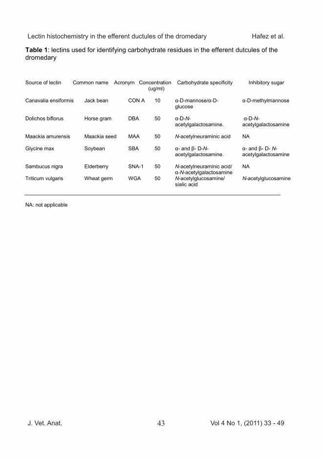
Table (1) lists the lectins (E.Y. La-boratories Inc., San Mateo, CA, USA) used in this study, their source, their abbreviations, the lec-tin concentration used, their major sugar specificities, and their inhibito-ry sugars. Concentrations were se-lected that gave minimal back-ground staining yet were sufficient to detect relatively low levels of specific sugar residues. A final in-cubation, after washing, with ABC (avidin: biotinylated horseradish pe-roxidase complex) reagent (Vector Laboratories) lasted 30 minutes un-der the same conditions. Immuno-reactivity was visualized with 3, 3- diaminobenzidine (DAB substrate kit, Vector Laboratories) in a dark place as outlined in the manufactur-er’s protocol. Slides were counters-tained with hematoxylin, dehy-drated, cleared in xylene, and mounted.
Some sections were treated with neuroaminidase (0.1 units/ml of type VI from Clostridium perfringens, Sigma, USA) (Jones et al., 1995). Controls for non-specific staining included incubation in which the bio-tinylated lectins were replaced with unconjugated lectins, biotinylated bovine serum albumin (E.Y. Labora-tories) or buffer, preincubation of a lectin, except MAA and SNA-1 with
its competing sugar (Vector Labora-tories), or sections to be stained with MAA or SNA-1-were preincu-bated with neuroaminidase (Sigma, USA).
Results
The efferent ductules were lined with low pseudostratified columnar epithelium. There were two types of this epithelium; each type consists of both ciliated and non ciliated principal cells (Fig. 1). The initial segment of each duct was lined with type I epithelium. The segment close to the epididymal duct was lined with type II epithelium. The epithelium of the terminal segment showed in some areas regional height variations from place to place, similar to what is seen in the initial segment of the epididymal duct; but in the efferent ductules this variable appearance was much less pronounced compared to the initial part of the epididymal duct. The ini-tial segment of the epididymal duct and the terminal segments of the efferent ductules could generally be distinguished by the position of the nuclei. The nuclei in the type II epi-thelium were located more or less in the mid portion of the epithelial cells with only a few of them showing ba-sally located nuclei. The terminal segments of the tubules had wider lumens than the other regions; and the epithelium of the terminal seg-
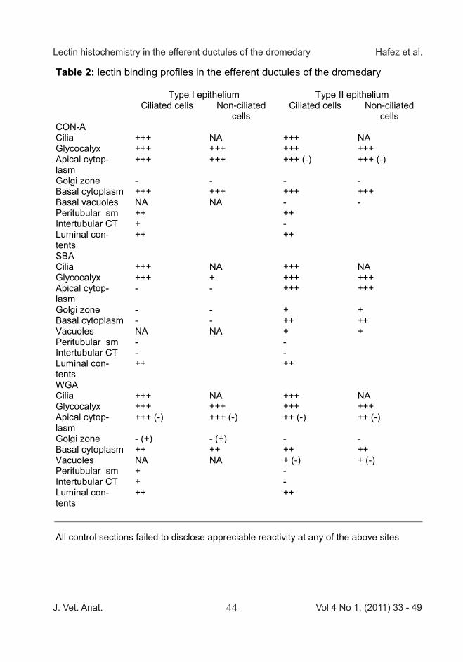
ments was more vacuolated; in ad-dition there was decreased distance between the nuclei and the luminal surface. Delicate peritubular smooth muscle cell layers lined the tubules and a loose connective tissue was dispersed in the space between the adjacent tubules (Fig. 2). There were six lectins studied, but only three gave positive labeling: binding sites for DBA, MAA, and SNA-1 were not detected in the epi-thelium. The lectin binding profile of ciliated and non-ciliated cells of type I and type II epithelia are reported in table (2). Binding sites for Con-A appeared in the glycocalyx, the apical cytoplasm, and the basal cy-toplasm of both ciliated and non-ciliated cells in both type I and type II epithelia (Fig. 3). The intensely stained glycocalyx was interrupted by the less-strongly stained cilia of the ciliated cells. The apical cytop-lasm of the epithelial cells of type II epithelium showed Con-A positively stained granules and some un-stained areas. No binding sites for Con-A were detected in the Golgi zone of either cells of type I or type II epithelia; but in the lateral cytop-lasm (on either side of the Golgi zone) there appeared to be posi-tively stained granules in type I cells. This was not seen in the type II epithelium. Con-A staining was positive in the cilia of both type I and
type II epithelia. Basal vacuoles of type II epithelium did not show bind-ing sites for Con-A. Figure (4) shows that SBA strongly bound to the glycocalyx of ciliated cells in type I epithelium, but the non-ciliated cells were weakly stained. Neither the apical and the basal cytoplasm nor the Golgi zone of the type I epithelial cells had binding sites for SBA. The glyco-calyx and the apical and basal cy-toplasm of the epithelial cells of type II epithelium showed binding sites for SBA. In the type II cells, the Gol-gi zone and the basal vacuoles ap-peared only weakly stained. SBA bound with cilia in both types of epi-thelia.
WGA binding was detected in the glycocalyx, the apical cytoplasm, and the basal cytoplasm in both types of epithelia (Fig. 5). WGA showed more intense binding with glycocalyx in some areas than in other areas in type I epithelium. The apical cytoplasm of both types con-tained both unstained areas and positively stained areas. In general the Golgi zone of neither type showed binding sites for WGA ex-cept in a subgroup of type I cells and a few positively stained gra-nules were detected in the basal vacuoles of type II epithelium. WGA bound with cilia in both type I and type II epithelia.
J. Vet. Anat. Vol 4 No 1, (2011) 33 - 4937
Lectin histochemistry in the efferent ductules of the dromedary Hafez et al.
lated lectin for 60 min in a humid chamber at room temperature.
Table (1) lists the lectins (E.Y. La-boratories Inc., San Mateo, CA, USA) used in this study, their source, their abbreviations, the lec-tin concentration used, their major sugar specificities, and their inhibito-ry sugars. Concentrations were se-lected that gave minimal back-ground staining yet were sufficient to detect relatively low levels of specific sugar residues. A final in-cubation, after washing, with ABC (avidin: biotinylated horseradish pe-roxidase complex) reagent (Vector Laboratories) lasted 30 minutes un-der the same conditions. Immuno-reactivity was visualized with 3, 3- diaminobenzidine (DAB substrate kit, Vector Laboratories) in a dark place as outlined in the manufactur-er’s protocol. Slides were counters-tained with hematoxylin, dehy-drated, cleared in xylene, and mounted.
Some sections were treated with neuroaminidase (0.1 units/ml of type VI from Clostridium perfringens, Sigma, USA) (Jones et al., 1995). Controls for non-specific staining included incubation in which the bio-tinylated lectins were replaced with unconjugated lectins, biotinylated bovine serum albumin (E.Y. Labora-tories) or buffer, preincubation of a lectin, except MAA and SNA-1 with
its competing sugar (Vector Labora-tories), or sections to be stained with MAA or SNA-1-were preincu-bated with neuroaminidase (Sigma, USA).
Results
The efferent ductules were lined with low pseudostratified columnar epithelium. There were two types of this epithelium; each type consists of both ciliated and non ciliated principal cells (Fig. 1). The initial segment of each duct was lined with type I epithelium. The segment close to the epididymal duct was lined with type II epithelium. The epithelium of the terminal segment showed in some areas regional height variations from place to place, similar to what is seen in the initial segment of the epididymal duct; but in the efferent ductules this variable appearance was much less pronounced compared to the initial part of the epididymal duct. The ini-tial segment of the epididymal duct and the terminal segments of the efferent ductules could generally be distinguished by the position of the nuclei. The nuclei in the type II epi-thelium were located more or less in the mid portion of the epithelial cells with only a few of them showing ba-sally located nuclei. The terminal segments of the tubules had wider lumens than the other regions; and the epithelium of the terminal seg-
ments was more vacuolated; in ad-dition there was decreased distance between the nuclei and the luminal surface. Delicate peritubular smooth muscle cell layers lined the tubules and a loose connective tissue was dispersed in the space between the adjacent tubules (Fig. 2). There were six lectins studied, but only three gave positive labeling: binding sites for DBA, MAA, and SNA-1 were not detected in the epi-thelium. The lectin binding profile of ciliated and non-ciliated cells of type I and type II epithelia are reported in table (2). Binding sites for Con-A appeared in the glycocalyx, the apical cytoplasm, and the basal cy-toplasm of both ciliated and non-ciliated cells in both type I and type II epithelia (Fig. 3). The intensely stained glycocalyx was interrupted by the less-strongly stained cilia of the ciliated cells. The apical cytop-lasm of the epithelial cells of type II epithelium showed Con-A positively stained granules and some un-stained areas. No binding sites for Con-A were detected in the Golgi zone of either cells of type I or type II epithelia; but in the lateral cytop-lasm (on either side of the Golgi zone) there appeared to be posi-tively stained granules in type I cells. This was not seen in the type II epithelium. Con-A staining was positive in the cilia of both type I and
type II epithelia. Basal vacuoles of type II epithelium did not show bind-ing sites for Con-A. Figure (4) shows that SBA strongly bound to the glycocalyx of ciliated cells in type I epithelium, but the non-ciliated cells were weakly stained. Neither the apical and the basal cytoplasm nor the Golgi zone of the type I epithelial cells had binding sites for SBA. The glyco-calyx and the apical and basal cy-toplasm of the epithelial cells of type II epithelium showed binding sites for SBA. In the type II cells, the Gol-gi zone and the basal vacuoles ap-peared only weakly stained. SBA bound with cilia in both types of epi-thelia.
WGA binding was detected in the glycocalyx, the apical cytoplasm, and the basal cytoplasm in both types of epithelia (Fig. 5). WGA showed more intense binding with glycocalyx in some areas than in other areas in type I epithelium. The apical cytoplasm of both types con-tained both unstained areas and positively stained areas. In general the Golgi zone of neither type showed binding sites for WGA ex-cept in a subgroup of type I cells and a few positively stained gra-nules were detected in the basal vacuoles of type II epithelium. WGA bound with cilia in both type I and type II epithelia.

J. Vet. Anat. Vol 4 No 1, (2011) 33 - 4938
Lectin histochemistry in the efferent ductules of the dromedary Hafez et al.
Peritubular smooth muscle cells of type I epithelium showed binding sites for WGA. The peritubular smooth muscle of both types showed binding sites for Con-A. SBA did not bind with peritubular smooth muscle cells of either type. Con-A, WGA, and SBA bound with the luminal contents in both types of epithelia (Figs. 3, 4, and 5). The in-tertubular connective tissue showed binding sites for Con-A and WGA only in regions of type I epithelium.
Discussion
Con-A is specific for trimannoside core, which is common for N gly-cans. Binding sites for Con-A were detected readily in various com-partments in both type I and type II epithelia. The presence of Con-A binding sites in the basal cytoplasm may correspond to the binding at the cisternae of the rough endop-lasmic reticulum (RER). This finding agreed with both ultrastructural in-vestigation of this cytoplasmic site by Con-A (Chan and Wong, 1992) and at the light microscope level by Con-A (Parillo et al., 2009). It has been reported that mannose moie-ties present in the RER may be as-sociated with the initial assembly of the N-linked oligosaccharides of glycoproteins, which is completed in the Golgi stacks (Parillo et al., 2009).
WGA has been used in this study to detect sialoglycoconjugates, and the detailed sialic acid binding pattern to internal galactose can be recog-nized using SNA and MAA; SNA detects N-acetylneuraminic acid and α-N-acetylgalactosamine, and MAA detects N-acetylneuraminic acid. Sialic acid residues were detected in some areas in both type I and type II epithelia as demonstrated by binding to WGA. Detailed sialic acid binding was not detected due to lack of binding sites for SNA and MAA. Differences in staining of the same population of cells as in some cases of WGA binding indicate vari-ous stages of secretion as well as cellular compartmentalization.
No binding sites for DBA and SNA were detected in our study. Both DBA and SNA react to α N-acetylgalactosamine. SBA reacts to both α and β N-acetylgalactosa-mine. Binding sites for SBA were detected in some compartments in both type I and type II epithelia, these same areas lacked binding sites for DBA and SNA. This indi-cates that these areas showing binding sites for SBA contain β N-acetylgalactosamine and not α N-acetylgalactosamine.
The Golgi zone did not appear to be stained for most of the lectins used in this study with the exception of some weak staining in the case of
SBA and WGA. If the cells had been actively involved in the synthesis and secretion of glycoconjugates, the Golgi zone should reveal strong binding sites. Camels are seasonal breeders with a relatively short breeding season during the cooler months from December to April (Ha-fez and Hafez, 2001). The speci-mens were collected outside the rutting season; this might explain the lack of strong staining in the Golgi zone.
The glycocalyx of both ciliated and non-ciliated cells in both type I and type II epithelia showed binding sites for Con-A, SBA, and WGA; this indicates the functional com-plexity of the glycocalyx in the two types of epithelium.
Lectin staining at the apical cytop-lasm might indicate the presence of glycoconjugates preceding their ex-trusion. However, cellular blebs were not detected on the luminal surface, which either indicates that apocrine mode of secretion is not detected in the efferent ductules of the dromedary, or high degree of secretion was not detected during the time when the specimens were collected.
While the results of this study may not be of immediate application to dromedary production, it is worth noting that baseline data on varia-
tions of reproductive system histol-ogy have in the past been shown to be significant in other species. The regional differences in the epithe-lium we have demonstrated may be related to the male dromedary’s ability to fertilize a receptive female in the proper season of the year. Had these specimens been col-lected at a different time of the year, the pattern might have been differ-ent. While further work is needed to determine whether this is the case, it seems unlikely that distinct re-gional differences at the molecular level are present without some func-tional significance, and it would be worth knowing whether there may be temporal ones as well.
Conclusion
This study demonstrates that mor-phological and histochemical fea-tures of the efferent ductules of the dromedary provide evidence for the existence of two different types of epithelium (Type I, II), and suggest a high degree of functional com-partmentalization of the epithelium of the efferent ductules.
Acknowledgment
The authors are in great debt to Ms. Sherry Ring at LSU College of Vete-rinary Medicine for technical assis-tance; and Dr. Marxa Figureido at the University of Texas Medical

J. Vet. Anat. Vol 4 No 1, (2011) 33 - 4939
Lectin histochemistry in the efferent ductules of the dromedary Hafez et al.
Peritubular smooth muscle cells of type I epithelium showed binding sites for WGA. The peritubular smooth muscle of both types showed binding sites for Con-A. SBA did not bind with peritubular smooth muscle cells of either type. Con-A, WGA, and SBA bound with the luminal contents in both types of epithelia (Figs. 3, 4, and 5). The in-tertubular connective tissue showed binding sites for Con-A and WGA only in regions of type I epithelium.
Discussion
Con-A is specific for trimannoside core, which is common for N gly-cans. Binding sites for Con-A were detected readily in various com-partments in both type I and type II epithelia. The presence of Con-A binding sites in the basal cytoplasm may correspond to the binding at the cisternae of the rough endop-lasmic reticulum (RER). This finding agreed with both ultrastructural in-vestigation of this cytoplasmic site by Con-A (Chan and Wong, 1992) and at the light microscope level by Con-A (Parillo et al., 2009). It has been reported that mannose moie-ties present in the RER may be as-sociated with the initial assembly of the N-linked oligosaccharides of glycoproteins, which is completed in the Golgi stacks (Parillo et al., 2009).
WGA has been used in this study to detect sialoglycoconjugates, and the detailed sialic acid binding pattern to internal galactose can be recog-nized using SNA and MAA; SNA detects N-acetylneuraminic acid and α-N-acetylgalactosamine, and MAA detects N-acetylneuraminic acid. Sialic acid residues were detected in some areas in both type I and type II epithelia as demonstrated by binding to WGA. Detailed sialic acid binding was not detected due to lack of binding sites for SNA and MAA. Differences in staining of the same population of cells as in some cases of WGA binding indicate vari-ous stages of secretion as well as cellular compartmentalization.
No binding sites for DBA and SNA were detected in our study. Both DBA and SNA react to α N-acetylgalactosamine. SBA reacts to both α and β N-acetylgalactosa-mine. Binding sites for SBA were detected in some compartments in both type I and type II epithelia, these same areas lacked binding sites for DBA and SNA. This indi-cates that these areas showing binding sites for SBA contain β N-acetylgalactosamine and not α N-acetylgalactosamine.
The Golgi zone did not appear to be stained for most of the lectins used in this study with the exception of some weak staining in the case of
SBA and WGA. If the cells had been actively involved in the synthesis and secretion of glycoconjugates, the Golgi zone should reveal strong binding sites. Camels are seasonal breeders with a relatively short breeding season during the cooler months from December to April (Ha-fez and Hafez, 2001). The speci-mens were collected outside the rutting season; this might explain the lack of strong staining in the Golgi zone.
The glycocalyx of both ciliated and non-ciliated cells in both type I and type II epithelia showed binding sites for Con-A, SBA, and WGA; this indicates the functional com-plexity of the glycocalyx in the two types of epithelium.
Lectin staining at the apical cytop-lasm might indicate the presence of glycoconjugates preceding their ex-trusion. However, cellular blebs were not detected on the luminal surface, which either indicates that apocrine mode of secretion is not detected in the efferent ductules of the dromedary, or high degree of secretion was not detected during the time when the specimens were collected.
While the results of this study may not be of immediate application to dromedary production, it is worth noting that baseline data on varia-
tions of reproductive system histol-ogy have in the past been shown to be significant in other species. The regional differences in the epithe-lium we have demonstrated may be related to the male dromedary’s ability to fertilize a receptive female in the proper season of the year. Had these specimens been col-lected at a different time of the year, the pattern might have been differ-ent. While further work is needed to determine whether this is the case, it seems unlikely that distinct re-gional differences at the molecular level are present without some func-tional significance, and it would be worth knowing whether there may be temporal ones as well.
Conclusion
This study demonstrates that mor-phological and histochemical fea-tures of the efferent ductules of the dromedary provide evidence for the existence of two different types of epithelium (Type I, II), and suggest a high degree of functional com-partmentalization of the epithelium of the efferent ductules.
Acknowledgment
The authors are in great debt to Ms. Sherry Ring at LSU College of Vete-rinary Medicine for technical assis-tance; and Dr. Marxa Figureido at the University of Texas Medical

J. Vet. Anat. Vol 4 No 1, (2011) 33 - 4940
Lectin histochemistry in the efferent ductules of the dromedary Hafez et al.
Branch for the use of her lab equipments.
References
Aire, T. A. (1980): The ductuli effe-rentes of the epididymal re-gion of birds. J Anat 130 (Pt 4), 707-723.
Aire, T. A., and M. van der Merwe (2003): The ductuli effe-rentes testis of the greater cane rat ( Thryonomys swin-derianus). Anat Embryol (Berl) 206 (5), 409-417.
Alroy J, Ucci AA, and M. E. A. Pe-reira (1984): Lectins: histo-chemical probes for specific carbohydrate residues. In Diagnostic immunochemi-stry, edited by DeLellis, R. A. New York, 67-88.
Arrighi, S., M. G. Romanello, and C. Domeneghini (1994): Ultra-structure of the epithelium that lines the ductuli effe-rentes in domestic equidae, with particular reference to spermatophagy. Acta Anat (Basel) 149 (3), 174-184.
Aureli, G., S. Arrighi, and M. G. Romanello (1984): Ultra-structural and cytochemical study on the epithelium lin-ing ductuli efferentes in Equus asinus. Basic Appl Histochem 28 (2), 101-115.
Brooks, S. A., and D. M. S. Hall (2001): Lectin Histochemi-
stry to Detect Altered Glyco-sylation in Cells and Tis-sues. In Metastasis Re-search Protocols: Volume I: Analysis of Cells and Tis-sues, edited by Brook, S. and Schumacher, U., 49-65.
Burkett, B. N., B. A. Schulte, and S. S. Spicer (1987): Histochem-ical evaluation of glycocon-jugates in the male repro-ductive tract with lectin-horseradish peroxidase con-jugates: I. Staining of prin-cipal cells and spermatozoa in the mouse. Am J Anat 178 (1), 11-22.
Chan, L., and Y. C. Wong (1992): Localization of prostatic gly-coconjugates by the lectin-gold method. Acta Anat (Ba-sel) 143 (1), 27-40.
Goyal, H. O., and F. Hrudka (1981): Ductuli efferentes of the bull-a morphological, experimen-tal and developmental study. Andrologia 13 (4), 292-306.
Goyal, H. O., V. Hutto, and D. D. Robinson (1992): Reexami-nation of the morphology of the extratesticular rete and ductuli efferentes in the goat. Anat Rec 233 (1), 53-60.
Goyal, H. O., and C. S. Williams (1988): The ductuli effe-rentes of the goat: a mor-phological study. Anat Rec 220 (1), 58-67.
Hafez, E. S., and B. Hafez (2001): Reproductive parameters of male dromedary and bac-trian camels. Arch Androl 46 (2), 85-98.
Humason, G. L. (1979): Animal Tis-sue Techniques: W.H. Free-man and company.
Ilio, K. Y., and R. A. Hess (1994): Structure and function of the ductuli efferentes: a review. Microsc Res Tech 29 (6), 432-467.
Jones, C. J., V. Dantzer, and R. W. Stoddart (1995): Changes in glycan distribution within the porcine interhaemal barrier during gestation. Cell Tissue Res 279 (3), 551-564.
Jones, R., D. W. Hamilton, and D. W. Fawcett (1979): Mor-phology of the epithelium of the extratesticular rete testis, ductuli efferentes and ductus epididymidis of the adult male rabbit. Am J Anat 156 (3), 373-400.
Jones, R. C., and W. V. Holt (1981): Studies of the deferent ducts from the testis of the African elephant, Loxodonta afri-cana. III. Ultrastructure and cytochemistry of the ductuli efferentes. J Anat 133 (Pt 2), 247-255.
Lewis-Jones, D. I., R. G. Harrison, and E. W. Macmillan (1982): A reexamination of rat ductu-
li efferentes. Anat Rec 203 (4), 461-462.
Nagy, F. (1990): On the ultrastruc-ture of the male reproductive tract in the Siberian hamster (Phodopus sungorus camp-belli). I. The ductuli effe-rentes. J Submicrosc Cytol Pathol 22 (4), 615-625.
Orsi, A. M., S. M. Matheus, E. A. Gregorio, and C. C. Beu (1998): Morphological inves-tigations of the surface epi-thelium of ductuli efferentes of black isogenic mice (Mus musculus). Anat Histol Em-bryol 27 (4), 215-218.
Parillo, F., G. E. Magi, S. Diverio, and G. Catone (2009): Im-munohistochemical and lec-tin histochemical analysis of the alpaca efferent ducts. Histol Histopathol 24 (1), 1-12.
Parillo, F., G. Stradaioli, A. V. Sup-plizi, and M. Monaci (1998): Lectin-staining pattern in extratesticular rete testis and ductuli efferentes of prepu-bertal and adult horses. His-tol Histopathol 13 (2), 307-314.
Roberts, K. P. (2010). What are the components of the male re-productive system? In Hand-book of Andrology, edited by Robaire B and P. Chan.
J. Vet. Anat. Vol 4 No 1, (2011) 33 - 4941
Lectin histochemistry in the efferent ductules of the dromedary Hafez et al.
Branch for the use of her lab equipments.
References
Aire, T. A. (1980): The ductuli effe-rentes of the epididymal re-gion of birds. J Anat 130 (Pt 4), 707-723.
Aire, T. A., and M. van der Merwe (2003): The ductuli effe-rentes testis of the greater cane rat ( Thryonomys swin-derianus). Anat Embryol (Berl) 206 (5), 409-417.
Alroy J, Ucci AA, and M. E. A. Pe-reira (1984): Lectins: histo-chemical probes for specific carbohydrate residues. In Diagnostic immunochemi-stry, edited by DeLellis, R. A. New York, 67-88.
Arrighi, S., M. G. Romanello, and C. Domeneghini (1994): Ultra-structure of the epithelium that lines the ductuli effe-rentes in domestic equidae, with particular reference to spermatophagy. Acta Anat (Basel) 149 (3), 174-184.
Aureli, G., S. Arrighi, and M. G. Romanello (1984): Ultra-structural and cytochemical study on the epithelium lin-ing ductuli efferentes in Equus asinus. Basic Appl Histochem 28 (2), 101-115.
Brooks, S. A., and D. M. S. Hall (2001): Lectin Histochemi-
stry to Detect Altered Glyco-sylation in Cells and Tis-sues. In Metastasis Re-search Protocols: Volume I: Analysis of Cells and Tis-sues, edited by Brook, S. and Schumacher, U., 49-65.
Burkett, B. N., B. A. Schulte, and S. S. Spicer (1987): Histochem-ical evaluation of glycocon-jugates in the male repro-ductive tract with lectin-horseradish peroxidase con-jugates: I. Staining of prin-cipal cells and spermatozoa in the mouse. Am J Anat 178 (1), 11-22.
Chan, L., and Y. C. Wong (1992): Localization of prostatic gly-coconjugates by the lectin-gold method. Acta Anat (Ba-sel) 143 (1), 27-40.
Goyal, H. O., and F. Hrudka (1981): Ductuli efferentes of the bull-a morphological, experimen-tal and developmental study. Andrologia 13 (4), 292-306.
Goyal, H. O., V. Hutto, and D. D. Robinson (1992): Reexami-nation of the morphology of the extratesticular rete and ductuli efferentes in the goat. Anat Rec 233 (1), 53-60.
Goyal, H. O., and C. S. Williams (1988): The ductuli effe-rentes of the goat: a mor-phological study. Anat Rec 220 (1), 58-67.
Hafez, E. S., and B. Hafez (2001): Reproductive parameters of male dromedary and bac-trian camels. Arch Androl 46 (2), 85-98.
Humason, G. L. (1979): Animal Tis-sue Techniques: W.H. Free-man and company.
Ilio, K. Y., and R. A. Hess (1994): Structure and function of the ductuli efferentes: a review. Microsc Res Tech 29 (6), 432-467.
Jones, C. J., V. Dantzer, and R. W. Stoddart (1995): Changes in glycan distribution within the porcine interhaemal barrier during gestation. Cell Tissue Res 279 (3), 551-564.
Jones, R., D. W. Hamilton, and D. W. Fawcett (1979): Mor-phology of the epithelium of the extratesticular rete testis, ductuli efferentes and ductus epididymidis of the adult male rabbit. Am J Anat 156 (3), 373-400.
Jones, R. C., and W. V. Holt (1981): Studies of the deferent ducts from the testis of the African elephant, Loxodonta afri-cana. III. Ultrastructure and cytochemistry of the ductuli efferentes. J Anat 133 (Pt 2), 247-255.
Lewis-Jones, D. I., R. G. Harrison, and E. W. Macmillan (1982): A reexamination of rat ductu-
li efferentes. Anat Rec 203 (4), 461-462.
Nagy, F. (1990): On the ultrastruc-ture of the male reproductive tract in the Siberian hamster (Phodopus sungorus camp-belli). I. The ductuli effe-rentes. J Submicrosc Cytol Pathol 22 (4), 615-625.
Orsi, A. M., S. M. Matheus, E. A. Gregorio, and C. C. Beu (1998): Morphological inves-tigations of the surface epi-thelium of ductuli efferentes of black isogenic mice (Mus musculus). Anat Histol Em-bryol 27 (4), 215-218.
Parillo, F., G. E. Magi, S. Diverio, and G. Catone (2009): Im-munohistochemical and lec-tin histochemical analysis of the alpaca efferent ducts. Histol Histopathol 24 (1), 1-12.
Parillo, F., G. Stradaioli, A. V. Sup-plizi, and M. Monaci (1998): Lectin-staining pattern in extratesticular rete testis and ductuli efferentes of prepu-bertal and adult horses. His-tol Histopathol 13 (2), 307-314.
Roberts, K. P. (2010). What are the components of the male re-productive system? In Hand-book of Andrology, edited by Robaire B and P. Chan.
J. Vet. Anat. Vol 4 No 1, (2011) 33 - 4942
Lectin histochemistry in the efferent ductules of the dromedary Hafez et al.
Lawrence, KS: Allen Press, Inc., 1-5.
Setchell, B. P., S. Maddocks, and D. E. Brooks (1994): Anatomy, Vasculature,Innervation, and Fluids of the Male Reproduc-tive Tract. In The Physiology of Reproduction, edited by E. Knobil and J. D. Neill. New York: Raven Press, 1063-1176.
Stoffel, M. H., and A. E. Friess (1994): Morphological cha-racteristics of boar efferent ductules and epididymal duct. Microsc Res Tech 29 (6), 411-431.
Vicentini, C. A., A. M. Orsi, and E. A. Gregorio (1990): Fine structure of the ductuli effe-rentes of the hamster (Me-socricetus auratus). Gegen-baurs Morphol Jahrb 136 (1), 111-118.
Wakui, S., M. Furusato, H. Takaha-shi, M. Motoya, and S. Ushi-gome (1996): Lectin histo-chemical evaluation of gly-coconjugates in dog efferent ductules. J Anat 188 ( Pt 3), 541-546.
Wrobel, K., and M. Bergmann (2006): Male Reproductive System. In Dellmann's Text-book of Veterinary Histology, edited by Eurell J and F. B. Iowa: Blackwell Publishing.
________________
Corresponding author: Shireen Hafez [email protected]
Table 1: lectins used for identifying carbohydrate residues in the efferent dutcules of the dromedary
Source of lectin Common name Acronym Concentration Carbohydrate specificity Inhibitory sugar (ug/ml) Canavalia ensiformis
Jack bean
CON A
10
α-D-mannose/α-D-glucose
α-D-methylmannose
Dolichos biflorus
Horse gram DBA 50 α-D-N- acetylgalactosamine.
α-D-N- acetylgalactosamine
Maackia amurensis
Maackia seed MAA 50 N-acetylneuraminic acid NA
Glycine max
Soybean
SBA 50 α- and β- D-N-acetylgalactosamine.
α- and β- D- N- acetylgalactosamine
Sambucus nigra
Elderberry
SNA-1 50 N-acetylneuraminic acid/ α-N-acetylgalactosamine
NA
Triticum vulgaris
Wheat germ
WGA
50 N-acetylglucosamine/ sialic acid
N-acetylglucosamine
NA: not applicable
J. Vet. Anat. Vol 4 No 1, (2011) 33 - 4943
Lectin histochemistry in the efferent ductules of the dromedary Hafez et al.
Lawrence, KS: Allen Press, Inc., 1-5.
Setchell, B. P., S. Maddocks, and D. E. Brooks (1994): Anatomy, Vasculature,Innervation, and Fluids of the Male Reproduc-tive Tract. In The Physiology of Reproduction, edited by E. Knobil and J. D. Neill. New York: Raven Press, 1063-1176.
Stoffel, M. H., and A. E. Friess (1994): Morphological cha-racteristics of boar efferent ductules and epididymal duct. Microsc Res Tech 29 (6), 411-431.
Vicentini, C. A., A. M. Orsi, and E. A. Gregorio (1990): Fine structure of the ductuli effe-rentes of the hamster (Me-socricetus auratus). Gegen-baurs Morphol Jahrb 136 (1), 111-118.
Wakui, S., M. Furusato, H. Takaha-shi, M. Motoya, and S. Ushi-gome (1996): Lectin histo-chemical evaluation of gly-coconjugates in dog efferent ductules. J Anat 188 ( Pt 3), 541-546.
Wrobel, K., and M. Bergmann (2006): Male Reproductive System. In Dellmann's Text-book of Veterinary Histology, edited by Eurell J and F. B. Iowa: Blackwell Publishing.
________________
Corresponding author: Shireen Hafez [email protected]
Table 1: lectins used for identifying carbohydrate residues in the efferent dutcules of the dromedary
Source of lectin Common name Acronym Concentration Carbohydrate specificity Inhibitory sugar (ug/ml) Canavalia ensiformis
Jack bean
CON A
10
α-D-mannose/α-D-glucose
α-D-methylmannose
Dolichos biflorus
Horse gram DBA 50 α-D-N- acetylgalactosamine.
α-D-N- acetylgalactosamine
Maackia amurensis
Maackia seed MAA 50 N-acetylneuraminic acid NA
Glycine max
Soybean
SBA 50 α- and β- D-N-acetylgalactosamine.
α- and β- D- N- acetylgalactosamine
Sambucus nigra
Elderberry
SNA-1 50 N-acetylneuraminic acid/ α-N-acetylgalactosamine
NA
Triticum vulgaris
Wheat germ
WGA
50 N-acetylglucosamine/ sialic acid
N-acetylglucosamine
NA: not applicable
J. Vet. Anat. Vol 4 No 1, (2011) 33 - 4944
Lectin histochemistry in the efferent ductules of the dromedary Hafez et al.
Table 2: lectin binding profiles in the efferent ductules of the dromedary
Type I epithelium Type II epithelium Ciliated cells Non-ciliated
cells Ciliated cells Non-ciliated
cells CON-A Cilia +++ NA +++ NA Glycocalyx +++ +++ +++ +++ Apical cytop-lasm
+++ +++ +++ (-) +++ (-)
Golgi zone - - - - Basal cytoplasm +++ +++ +++ +++ Basal vacuoles NA NA - - Peritubular sm ++ ++ Intertubular CT + - Luminal con-tents
++ ++
SBA Cilia +++ NA +++ NA Glycocalyx +++ + +++ +++ Apical cytop-lasm
- - +++ +++
Golgi zone - - + + Basal cytoplasm - - ++ ++ Vacuoles NA NA + + Peritubular sm - - Intertubular CT - - Luminal con-tents
++ ++
WGA Cilia +++ NA +++ NA Glycocalyx +++ +++ +++ +++ Apical cytop-lasm
+++ (-) +++ (-) ++ (-) ++ (-)
Golgi zone - (+) - (+) - - Basal cytoplasm ++ ++ ++ ++ Vacuoles NA NA + (-) + (-) Peritubular sm + - Intertubular CT + - Luminal con-tents
++ ++
All control sections failed to disclose appreciable reactivity at any of the above sites
Fig (1): Ductuli efferentes of Camelus dromedarius stained with hematoxylin and eosin. The epithelial lining is a low pseudostratified columnar type containing two populations of cells, in which both ciliated and non-ciliated varieties occur. A. Type I epithelium showing apical cytoplasmic granules and a greater distance between the nuclei and the luminal surface; B. Type II epithelium showing basal vacuoles. Bar = 200 µm.

J. Vet. Anat. Vol 4 No 1, (2011) 33 - 4945
Lectin histochemistry in the efferent ductules of the dromedary Hafez et al.
Table 2: lectin binding profiles in the efferent ductules of the dromedary
Type I epithelium Type II epithelium Ciliated cells Non-ciliated
cells Ciliated cells Non-ciliated
cells CON-A Cilia +++ NA +++ NA Glycocalyx +++ +++ +++ +++ Apical cytop-lasm
+++ +++ +++ (-) +++ (-)
Golgi zone - - - - Basal cytoplasm +++ +++ +++ +++ Basal vacuoles NA NA - - Peritubular sm ++ ++ Intertubular CT + - Luminal con-tents
++ ++
SBA Cilia +++ NA +++ NA Glycocalyx +++ + +++ +++ Apical cytop-lasm
- - +++ +++
Golgi zone - - + + Basal cytoplasm - - ++ ++ Vacuoles NA NA + + Peritubular sm - - Intertubular CT - - Luminal con-tents
++ ++
WGA Cilia +++ NA +++ NA Glycocalyx +++ +++ +++ +++ Apical cytop-lasm
+++ (-) +++ (-) ++ (-) ++ (-)
Golgi zone - (+) - (+) - - Basal cytoplasm ++ ++ ++ ++ Vacuoles NA NA + (-) + (-) Peritubular sm + - Intertubular CT + - Luminal con-tents
++ ++
All control sections failed to disclose appreciable reactivity at any of the above sites
Fig (1): Ductuli efferentes of Camelus dromedarius stained with hematoxylin and eosin. The epithelial lining is a low pseudostratified columnar type containing two populations of cells, in which both ciliated and non-ciliated varieties occur. A. Type I epithelium showing apical cytoplasmic granules and a greater distance between the nuclei and the luminal surface; B. Type II epithelium showing basal vacuoles. Bar = 200 µm.

J. Vet. Anat. Vol 4 No 1, (2011) 33 - 4946
Lectin histochemistry in the efferent ductules of the dromedary Hafez et al.
Fig (2): Ductuli efferentes of Camelus dromedarius stained with Masson’s Trichrome and Verhoeff’s stains showing that the tubules were surrounded by delicate peritubular smooth muscle cell layers (red color) with a loose collagenous connective tissue (bluish color) dispersed in the space between adjacent tubules. Bar = 100 µm
Fig (3): Con-A binding in the glycocalyx and the apical and basal cytoplasm in type I epithelium (A) and type II epithelium (B). No binding sites for Con-A were detected in the Golgi zone of epithelial cells of either type I or type II epithelium (arrows); however, the lateral cytoplasm on either side of Golgi zone appeared to have positively stained gra-nules in type I (arrow heads in A), but not in type II epithelium. Con-A bound with Cilia in A and B, and did not bind to basal vacuoles in B. Bar = 40 µm.

J. Vet. Anat. Vol 4 No 1, (2011) 33 - 4947
Lectin histochemistry in the efferent ductules of the dromedary Hafez et al.
Fig (2): Ductuli efferentes of Camelus dromedarius stained with Masson’s Trichrome and Verhoeff’s stains showing that the tubules were surrounded by delicate peritubular smooth muscle cell layers (red color) with a loose collagenous connective tissue (bluish color) dispersed in the space between adjacent tubules. Bar = 100 µm
Fig (3): Con-A binding in the glycocalyx and the apical and basal cytoplasm in type I epithelium (A) and type II epithelium (B). No binding sites for Con-A were detected in the Golgi zone of epithelial cells of either type I or type II epithelium (arrows); however, the lateral cytoplasm on either side of Golgi zone appeared to have positively stained gra-nules in type I (arrow heads in A), but not in type II epithelium. Con-A bound with Cilia in A and B, and did not bind to basal vacuoles in B. Bar = 40 µm.

J. Vet. Anat. Vol 4 No 1, (2011) 33 - 4948
Lectin histochemistry in the efferent ductules of the dromedary Hafez et al.
Fig (4): SBA binding in the glycocalyx of ciliated cells and non-ciliated cells (with less intensity, arrow) in type I epithelium (A). The glycocalyx and the apical and basal cytop-lasm of the epithelial cells of type II epithelium (B) showed binding sites for SBA. Golgi zone and the basal vacuoles of type II epithelium appeared weekly stained. SBA bound with cilia in both types of epithelia. Bar = 40 µm.
Fig (5): WGA binding in the glycocalyx, the apical cytoplasm, and the basal cytoplasm in type I epithelium (A) and type II epithelium (B). The apical cytoplasm showed un-stained areas in addition to positively stained areas. The Golgi zone (arrows) in neither type of epithelia showed binding sites with WGA except in a subgroup of cells (arrow heads) in type I epithelium. Some positively stained granules were detected in the basal vacuoles of type II epithelium. WGA bound with cilia in type I and type II epithelia. Bar = 40 µm.

J. Vet. Anat. Vol 4 No 1, (2011) 33 - 4949
Lectin histochemistry in the efferent ductules of the dromedary Hafez et al.
Fig (4): SBA binding in the glycocalyx of ciliated cells and non-ciliated cells (with less intensity, arrow) in type I epithelium (A). The glycocalyx and the apical and basal cytop-lasm of the epithelial cells of type II epithelium (B) showed binding sites for SBA. Golgi zone and the basal vacuoles of type II epithelium appeared weekly stained. SBA bound with cilia in both types of epithelia. Bar = 40 µm.
Fig (5): WGA binding in the glycocalyx, the apical cytoplasm, and the basal cytoplasm in type I epithelium (A) and type II epithelium (B). The apical cytoplasm showed un-stained areas in addition to positively stained areas. The Golgi zone (arrows) in neither type of epithelia showed binding sites with WGA except in a subgroup of cells (arrow heads) in type I epithelium. Some positively stained granules were detected in the basal vacuoles of type II epithelium. WGA bound with cilia in type I and type II epithelia. Bar = 40 µm.