Water Spinach (Ipomoea aquatica, Convolvulaceae) A food gone wild
Upload
truongkienCategory
view
223download
0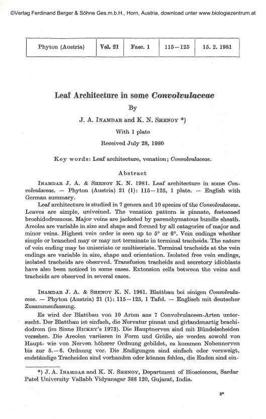
Phyton (Austria) Vol. 21 Fasc. 1 115-125 15. 2. 1981
Leaf Architecture in some ConvolvulaceaeBy
J. A. INAMDAR and K. N. SHENOY *)
With 1 plate
Received July 28, 1980
Key words: Leaf architecture, venation; Gonvolvulaceae.
Abstract
IN AMD AR. J. A. & SHENOY K. N. 1981. Leaf architecture in some Con-volvulaceae. — Phyton (Austria) 21 (1): 115 — 125, 1 plate. — English withGerman summary.
Leaf architecture is studied in 7 genera and 10 species of the Gonvolvulaceae.Leaves are simple, univeined. The venation pattern is pinnate, festoonedbrochidodromous. Major veins are jacketed by parenchymatous bundle sheath.Areoles are variable in size and shape and formed by all catagories of major andminor veins. Highest vein order is seen up to 5° or 6°. Vein endings whethersimple or branched may or may not terminate in terminal tracheids. The natureof vein ending may be uniseriate or multiseriate. Terminal tracheids at the veinendings are variable in size, shape and orientation. Isolated free vein endings,isolated tracheids are observed. Transfusion tracheids and secretory idioblastshave also been noticed in some cases. Extension cells between the veins andtracheids are observed in several cases.
INAMDAE J. A. & SHENOY K. N. 1981. Blattbau bei einigen Gonvolvula-ceae. — Phyton (Austria) 21 (1): 115—125, 1 Tafel. — Englisch mit deutscherZusammenfassung.
Es wird der Blattbau von 10 Arten aus 7 Convolvulaceen-Arten unter-sucht. Der Blattbau ist einfach, die Nervatur pinnat und girlandenartig brachi-dodrom (im Sinne HIOKEY'S 1973). Die Hauptnerven sind mit Bündelscheidenversehen. Die Areolen variieren in Form und Größe, sie werden sowohl vonHaupt- wie von Nerven höherer Ordnung gebildet, es kommen Nebennervenbis zur 5.-6. Ordnung vor. Die Endigungen sind einfach oder verzweigt,endständige Tracheiden sind vorhanden oder können fehlen, die Enden sind ein-
*) J. A. IN AMD AR and K. N. SHENOY, Department of Biosciences, SardarPatel University Vallabh Vidyanagar 388 120, Gujarat, India.
©Verlag Ferdinand Berger & Söhne Ges.m.b.H., Horn, Austria, download unter www.biologiezentrum.at

116
oder mehrreihig, die endständigen Tracheiden variieren in Größe, Form undOrientierung. Isolierte freie ISTervenendigungen und isolierte Tracheiden wurdenbeobachtet. In einigen Fällen finden sich Transfusionstracheiden und sekre-torische Idioblasten, desgleichen „Extensionszellen", d. s. Zellen zwischen denNerven und Tracheiden.
(Ed. transl., abbrev. and compl.)
IntroductionLeaves are highly polymorphic organs and provide sets of diverse
features. The veins and veinlets which form the vasculature, called the'venation', is an important feature of mature leaves. LEVIN (1929) andSTRAIN (1933) discussed the taxonomic significance of veinislet areas andvein endings. FOSTER (1950a, b) studied the venation pattern in Quiinaacutangula, Touroulia guianensis and Foresia tricarpapires. GUPTA (1961)studied the absolute veinlet termination numbers and absolute veinisletnumbers in leaves of some Solanaceous plants. HICKEY (1973), HICKEY &WOLFE (1975) and MELVILLE (1976) classified the architecture of dicotyle-donous and angiospermous leaves respectively. DILCHER (1974) reviewed itin the approaches to the identification of angiosperm leaf remains. PATEL &SHAH (1974) in their foliar studies of brinjal and chilli described the venationof lamina particularly vein endings. COLEMAN & GREYSON (1976) studiedthe leaf ontogeny in tomato and narrated the development of major andminor venation patterns. Leaf architecture and venation pattern studied indifferent families of dicotyledons are: Berberidaceae (SINGH et dl. 1978),Betulaceae (FRANK 1979), Bignoniaceae (JAIN 1978), Compositae (BANERJEE& DESHPANDE 1973), Euphorbiaceae (SEHGAL & PALIWAL 1974), Labiatae(TYAGI & KUMAR 1978), Rosaceae (MERRILL 1978), Scrophulariaceae (VER-GHESE 1969) and Solanaceae (INAMDAR & MURTHY 1978). The present workis undertaken to give a comprehensive account of the venation pattern andleaf architecture in 7 genera and 10 species of the Convolvulaceae as noreport exists on the subject.
Material and Methods
Material of 7 genera and 10 species: Colonyction speciosum CHOISY,Convolvulus arvensis L., Convolvulus microphyllus (ROTH) SIEB., Erycibepaniculata ROXB., Evolvulus alsinoides (L.) L., Evolvuhis nummularius (L.)L,. Hewittia sublobata (L. f.) 0. K., Jacquemontia paniculata (BURM. f.)HALL, f., Jacquemontia pentantha (JACQ.) G. DON and Porana paniculataROXB. were collected from Gujarat and Kerala states. The mature leaves"were cleared in (i) 10% aqueous sodium hydroxide solution followed bysodium hypochlorite solution (PAYNE, 1969) and (ii) solution of trichloroa-cetic acid and phenol crystals mixed in 2 : 1 (by weight) at 60° G (MOHANRAM & NAYYAR 1978). Cleared leaves were stained in fast green or Kores
©Verlag Ferdinand Berger & Söhne Ges.m.b.H., Horn, Austria, download unter www.biologiezentrum.at
Explanation to plate figures (A —S) showing cleared leaves, venationpattern, vein endings and other features of:
A. Hewittia sublobata ( X 2); B. Evolvulus nummularius ( X 4); C. Calonyctionspeciosum ( X 2); D. Evolvulus alsinoides ( x 4 ) ; E . Jacquemontia pentantha ( X 36);F. Erycibe paniculata ( X 4); G. Convolvulus arvensis ( X 30); H. Evolvulus num-mularius ( x 10); I. Convolvulus microphyllus ( x 220); J. Evolvulus nummularius(X85); K. Jacquemontia pentantha (x250); L. Calonyction speciosum (X155);M. Calonyction speciosum ( X 800); N". Calonyction speciosum x 680); O. Colony-ction speciosum x 600); P. Hewittia sublobata ( x 900); Q. Calonyction speciosum
( X 300); R. Hewittia sublobata ( x 540); S. Erycibe paniculata ( x 175).
©Verlag Ferdinand Berger & Söhne Ges.m.b.H., Horn, Austria, download unter www.biologiezentrum.at

©Verlag Ferdinand Berger & Söhne Ges.m.b.H., Horn, Austria, download unter www.biologiezentrum.at
117
stamp pad purple ink (manufactured by Kores India Ltd. Bombay, 400 018).Staining in Kores pad ink (modified from MOHAN RAM & NAYYAR, 1978)retains the stain for a considerable period. Photographs were taken withZEISS photomicroscope-I, using yellow filter and ORWO NP 15 film. Leafsize was measured using graph paper. Areole size and number of vein endingswere taken from five different fields of different leaves. Terminologies asdefined by HICKBY (1973) and HICKEY & WOLFE (1975) are adopted todescribe the leaf architecture.
Abbreviations used in the manuscript, plate figures and Table:
AA Acute AcuteAO Acute ObtuseAR Acute RightBr Branched secondary veinBs Bundle sheathEc Extension cellIFV Isolated free vein endingIt Isolated tracheidRA Right AcuteRR Right RightSI Secretory idioblastsSn SinuousTt Transfusion tracheids
Observations
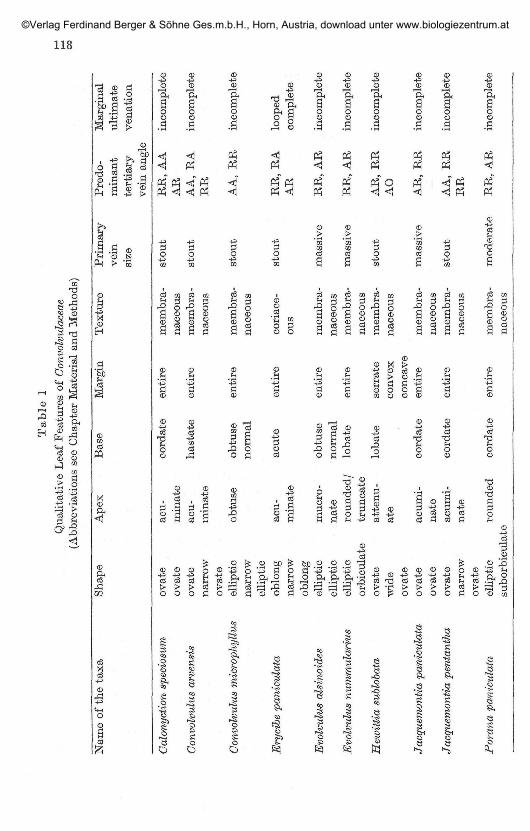
Leaves are basically simple and alternate in all cases. Lamina is moreor less symmetrical. The apex is acuminate (Fig. C) or mucronate (Fig. D)or rounded/truncate (Fig. B) or obtuse or attenuate (Fig. A). The margin isentire in all cases except in Hewittia sublobata, where it is slightly lobed(Fig. A). The texture is membranaceous or coriaceous, varying in differentspecies. The venation is pinnate where a single primary vein serves as theorigin for the higher order venation. The first, second and third degreeveins are considered as major and the higher order veins, the minor venationpatterns.
Major venation pat tern:The primary vein is the thickest vein of the leaf and its thickness
decreases gradually towards the apex and it gives off other degree veins oneither side. In all cases, a single strand enters the base of the lamina fromthe petiole and forms the primary vein which after travelling a shortdistance branches laterally. Major veins are generally jacketed by paren-chymatous sheath called 'Bundle sheath' (Fig. G). The thickness of thesheath may vary. Primary vein may be straight or sinuous (Fig. G). Some-times, even 2° and 3° veins are sinuous (Fig. E, G). The primary vein is
©Verlag Ferdinand Berger & Söhne Ges.m.b.H., Horn, Austria, download unter www.biologiezentrum.at
Tab
le 1
Qua
lita
tive
Lea
f F
eatu
res
of C
onvo
lvul
acea
e(A
bbre
viat
ions
see
Cha
pter
Mat
eria
l an
d M
etho
ds)
Nam
e of
the
tax
a
Cal
onyc
tion
spe
cios
um
Con
volv
ulus
arv
ensi
s
Con
volv
ulus
mic
roph
yllw
ä
Ery
cibe
pan
icul
ata
Evo
lvul
us a
lsin
oide
s
Evo
lvul
us n
umm
ular
ius
Hew
itti
a su
blob
ata
Jacq
uem
onti
a pa
nicu
lata
Jacq
uem
onti
a pe
ntan
tha
Par
ana
pani
cula
ta
Shap
e
ovat
eov
ate
ovat
ena
rrow
ovat
eel
liptic
narr
owel
liptic
oblo
ng-
narr
owob
long
ellip
ticel
liptic
ellip
ticor
bicu
late
ovat
ew
ide
ovat
eov
ate
ovat
eov
ate
narr
owov
ate
ellip
ticsu
borb
icul
&te
Ape
x
acu-
min
ate
acu-
min
ate
obtu
se
acu-
min
ate
muc
ro-
nate
roun
ded/
trun
cate
atte
nu-
ate
acum
i-na
teac
umi-
nate
roun
ded
Bas
e
cord
ate
hast
ate
obtu
seno
rmal
acut
e
obtu
seno
rmal
loba
te
loba
te
cord
ate
cord
ate
cord
ate
Mar
gin
enti
re
enti
re
enti
re
enti
re
enti
re
enti
re
serr
ate
conv
exco
ncav
een
tire
enti
re
enti
re
Tex
ture
mem
bra-
nace
ous
mem
bra-
nace
ous
mem
bra-
nace
ous
cori
ace-
ou
s
mem
bra-
nace
ous
mem
bra-
nace
ous
mem
bra-
nace
ous
mem
bra-
nace
ous
mem
bra-
nace
ous
mem
bra-
nace
ous
Pri
mar
yve
insi
ze
stou
t
stou
t
stou
t
stou
t
mas
sive
mas
sive
stou
t
mas
sive
stou
t
mod
erat
e
Pred
o-m
inan
tte
rtia
ryve
in a
ngle
RR
, A
AA
RA
A,
RA
RR
AA
, R
R
RR
, R
AA
R
RR
, A
R
RR
, A
R
AR
, R
RA
O
AR
, R
R
AA
, R
RR
R
RR
, A
R
Mar
gina
lul
tim
ate
vena
tion
inco
mpl
ete
inco
mpl
ete
inco
mpl
ete
loop
edco
mpl
ete
inco
mpl
ete
inco
mpl
ete
inco
mpl
ete
inco
mpl
ete
inco
mpl
ete
inco
mpl
ete
©Verlag Ferdinand Berger & Söhne Ges.m.b.H., Horn, Austria, download unter www.biologiezentrum.at
119
mostly stout occasionally massive or moderate. The second degree veinsare strongly brochidodromous. The secondaries have a set of secondaryloops outside the main brochidodromous and thus forms 'festooned brochi-dodromous' type (Fig. A—D). The number of second degree veins on eitherside of the primary vein vary from 5 to 10. Intersecondary veins are observedin all cases. Rarely in Evolvulus nummularius, one of the secondary veins ispeculiarly bifurcated at the point of origin from the primary. Eventuallyboth the branches fuse to form common strand of secondary vein (Fig. H).
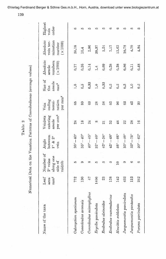
Minor venation pat tern:The highest order veins is identified up to 5 degree in most cases, but in
some up to 6 degrees. The qualitative leaf features and numerical data onthe venation pattern are charted in Table 1 and 2. Marginal ultimatevenation is incomplete in all the species studied, except in Erycibe paniculatawhere it is looped and complete (Fig. F). The areoles are 'imperfect' and theshape may be triangular, orbicular, polygonal or rectangular. The size ofthe areole is not constant, varies in different species and even in the samespecies. The plasticity of venation characters is shown by the variations inareole size, number of veinlets per areole and organisation of terminal veinendings in different species.
Vein endings:The ultimate veins of the leaf are either simple or branched. Simple
vein endings may be linear or curved. The branched ones may dividedichotomously once or twice and branches may be symmetrical or asym-metrical. The veinlets are mostly uniseriate, biseriate or multiseriate. Theymay be long and thin or thick and short. Usually a large number of veinendings are present in a big areole (Fig. F, G). But in most of the caseswhere areoles are devoid of vein endings, a loop like structure is seen whichis formed due to the union of veins or tracheids (Fig. E). Loop formation thusdecreases the distance between the veins and help in transporting system.
Tracheids:Tracheids are seen terminally at the vein endings. They vary in size,
shape and orientation. The tracheids may be uniseriate or biseriate. Uni-seriate tracheids are dilated and isodiametric (Fig. N) or elongated (Fig. 0).They occur singly or in groups (Fig. R). Uniseriate tracheids may be jux-taposed or superimposed (Fig. R). Biseriate tracheids may be either iso-diametric or elongated. A group of uniseriate tracheids wherein a group ofjuxtaposed dilated tracheids lie at right angles to an elongated tracheids isseen in Hewittia sublobata (Fig. P). A group of biseriate tracheids is seen inErycibe paniculata (Fig. S). A group of both biseriate and uniseriate tracheidslying opposite to each other is noticed in Calonyction speciosum (Fig. Q).
©Verlag Ferdinand Berger & Söhne Ges.m.b.H., Horn, Austria, download unter www.biologiezentrum.at
Tab
le 2
Num
eric
al D
ata
on t
he V
enat
ion
Pat
tern
s of
Con
volv
ulac
eae
(ave
rage
val
ues)
Nam
e of
the
tax
a
Calo
nyc
tion s
pec
iosu
m
Convo
lvulu
s arv
ensi
s
Convo
lvulu
s m
icro
ph
yllu
s
Ery
cibe
panic
ula
ta
Evo
lvulu
s als
inoid
es
Evo
lvulu
s num
mula
riu
s
Hew
itti
a s
ublo
bata
Jacq
uem
onU
a p
an
icu
lata
Jacq
uem
onU
a p
enta
nth
a
Pora
na p
anic
ula
ta
Lea
fare
am
m2
772
130 47
1406 2
3
138
142
432
112
242
Nu
mb
er
of2°
vei
ns
alo
ng
one
sid
e of
mid
rib 5 9 8 5 5 5 10
9 6 6
Angle
bet
wee
n1°
& 2
°v
ein
30
°-4
0°
35
°-4
0°
40
°-4
5°
32
°-4
0°
42
°-5
4°
42
°-4
8°
58
°-6
6°
35
°_4
0°
55
°-7
5°
50
°-6
2°
Vei
nle
tsen
teri
ng
areo
lep
er m
m2
28 18 24 8 21 32 46 22 28 16
Vei
nen
din
gte
rmi-
nati
on
per
mm
2
65 80 63 28 54 52 96 62 42 20
Siz
e of
the
areo
lem
m2
1.0
0.5
0.3
3
1.0
0.3
3
0.5
0.5
0.5
1.0
O.o
Ab
solu
tear
eole
nu
mb
ers
( X
1000)
0.7
7
0.2
6
0.1
4
1.4
0.6
9
0.2
8
0.2
8
0.8
6
0.1
1
0.4
8
Ab
solu
tev
ein
ter-
min
ati
on
nu
mb
er
(X1
00
0)
50.1
8
10.4
2.9
6
39.3
7
1.24
7.17
13.6
3
10.7
8
4.7
0
4.8
4
Hig
hest
vein
orde
r 6 5 5 5 5 6 6 5
; 5 5
©Verlag Ferdinand Berger & Söhne Ges.m.b.H., Horn, Austria, download unter www.biologiezentrum.at
121
Isolated tracheids:These are tracheids either uniseriate or biseriate, lying free in the
areole (Fig. L). Sometimes tracheids are connected with the free vein endingby extension cells which are parenchymatous in nature. Such tracheids maybe regarded as isolated, as they are not connected with the vein ending bytracheary elements.
Isolated vein endings:These are vein endings either uniseriate or multiseriate with terminal
tracheids lying free and disjunct in the areole.
Isolated free vein endings:These are freely existing isolated vein endings in the areole without the
terminal tracheids (Fig. K).
Extension cells:These are parenchymatous cells which adjoin two veins or isolated
tracheids with a vein (Fig. M). Extension parenchymatous cells have failedto differentiate into either sieve or tracheary elements and may be uniseriateor biseriate.
Transfusion tracheids:These are relatively short, squarish box shaped cells with spiral or
reticulate or pitted walls which occur along the borders of veins and veinendings (Fig. I). They are abundant at the apex and margin, graduallydeclining towards the remaining parts of the lamina. They have beenobserved in Evolvulus alsinoides, E. nummularius, Convolvulus microphyllus,and Colonyction speciosum.
Secretory idioblasts:These are specific more or less variously elongated or branched cells,
clearly distinguished from the other cells of the mesophyll tissue by theirsize and structure. These secretory idioblasts lie disjunct (Fig. J), and areseen in the mesophyll between veinlets. Their frequency is however, moretowards the base and apex of the lamina.
Discussion
Leaf venation in angiosperm varies both in pattern (HICKEY 1973) andregularity (HIOKEY & DOYLE 1972). According to PEAY (1954), the veinsof first, second and third order form major venation pattern and those ofsubsequent orders constitute minor venation patterns. The venation patternstudied in 10 species of the Convolvulaceae conforms to festooned brochido-
©Verlag Ferdinand Berger & Söhne Ges.m.b.H., Horn, Austria, download unter www.biologiezentrum.at
122
dromous type of HICXEY & WOLFE (1975). According to HICKEY & WOLFE(1975), leaves of Polemoniales are basically simple, margin entire, venationpinnate, secondary veins are strongly brochidodromous and tending toform an intramarginal vein. The present observations are in accordancewith those of HICKEY & WOLFE (1975) except the formation of intra-marginal veins. The plasticity of venation varies in different species andsometimes in the same species by variations in areole size and shape;number of vein endings and other qualitative features. NICELY (1965)reported significant variations within the same leaf as regards the size andshape of areoles and number of vein endings in each veinislet. SEHGAL &PALIWAL (1974) on the basis of their study of venation pattern of 150species of Euphorbia, have concluded that the size of the areole cannot beof much significant value particularly when there are large number ofspecies in a genus, as the areole size overlaps considerably in several species.Since diversity of opinions exist regarding the size of the areole and numberof vein endings, the statistical data will not be of any use. The number ofvein endings are in no way connected to the size of the areole, as the nearbyareoles even though more or less equal in size vary in their number of veinendings. Loop formation is a common feature in the areoles where there arefew vein endings or none. Marginal ultimate venation is generally in-complete. HICKEY (1973) classified the vein endings into simple and branch-ed. Branched ones divide once, twice or thrice dichotomously. In Erycibepaniculata, the veinlets are thick and multiseriate. In Porana panicvlata,Jacquemontia paniculata and J. pentantha, they are long and uniseriate.Mostly tracheids at the vein endings increase the cell diameter and areextraordinarily variable in size, shape and orientation. Both uniseriate andbiseriate tracheids are observed in different species. In the same areoleboth uniseriate and biseriate tracheids are observed at the veinendings.KASAPLIGIL (1951), FOSTER & ARNOTT (1960), HERBST (1972) have reportedthe occurrence of isolated veins in dicotyledonous leaves. SEHGAL &PALIWAL (1974) described the tracheidal elements lying free in the areole as'free vein ending'. But there is no clear cut distinction between isolatedveins, isolated free vein endings and isolated tracheids. FOSTER (1956) usedthe terms tracheary idioblasts (for dilated enlarged terminal tracheids atthe vein endings) and tracheoidal idioblasts (for isolated tracheids that liefree and disjunct in the areole), though these structures donot show muchdifference. The term enlarged terminal tracheary idioblasts is used for suchmodified cellular entities occuring at the termination and in intimatecontinuation with the underlying normal or conventional tracheary elements.The term tracheoidal idioblasts is applied to those elements that are foundisolated from and independent of the veins and vein endings (see TUCKER1964). GOVINDARAJALU (1972) observed in 15 species of Alangium, theoccurrence of tracheoidal idioblasts in varying proportion and combination.SEHGAL & PALIWAL (1974) designated uni, bi and tri veined leaves on
©Verlag Ferdinand Berger & Söhne Ges.m.b.H., Horn, Austria, download unter www.biologiezentrum.at
123
the basis of number of strands entering the base of the petiole and servesas the origin for the higher order venation. HARA (1962) reported someelongated cells termed as extension cells between one vein and vein endingsof another vein. Such cells have been observed in all the species studied.The primary veins are covered by a parenchymatous bundle sheath and infew cases even second and third degree veins. FOSTER (1972) described theoccurrence of transfusion tracheids in the two species of Ephedra. Earlier,SCHEIT (1883) also reported such cells in Ephedra altissima, but called them'tracheid border'. Occurrence of transfusion tracheids in the same specieswas also recorded by THOMPSON (1912). During the course of presentobservations, transfusion tracheids have been seen in Calonyction speciosum,Convolvulus microphyllus, Evolvulus alsinoides and E. nummularius. Theoccurrence of transfusion tracheids perhaps attributed to adaptive features.However, their presence in specific leaves alone indicates the localised needto compensate and assist the water distributing system. Secretory idioblastshave also been reported as isolated cells between palisade and spongyparenchyma in the Convolvulaceae by METCALPE & CHALK (1950).
Acknowledgment
One of us (K. JN". SHENOY) thanks the Management of Bhandarkars' Arts &Science College, Coondapur South Kanara for deputation and the UniversityGrants Commission for awarding a Teacher Fellowship.
References
BANERJEE G. & DESHPANDE B. D. 1973. Foliar venation and leaf histology ofcertain members of Compositae. — Flora 162: 529 — 532.
COLEMAN W. K. & GREYSON P. I. 1976. The growth and development of leaf intomato (Lycopersicon esculentum). II. Leaf ontogeny. — Can. J. Bot.54: 2704-2717.
DILCHER D. L. 1974. Approaches to the identification of angiosperm leafremains. — Bot. Rev., 40: 1 — 157.
FOSTER A. S. 1950 a. Morphology and venation of the leaf in Quiina acutangulaDUCKE. — Amer. J. Bot. 37: 159 — 171.
— 1950 b. Venation and histology of leaflets in Touroulia guianensis ATJBLand Foresia tricarpapires. — Amer. J. Bot. 37: 848 — 862.
— 1956. Plant idioblasts remarkable example of cell specialisation. —Protoplasma. 46: 184-193.
— 1972. Venation patterns in the leaves of Ephedra. — J. Am. Arb. 53:364-378.
— & AENOT H. J. 1960. Morphology and dichotomous vasctilature of theleaf of Kingdonia uniflora. — Amer. J. Bot. 47: 684 — 698.
FRANK D. H. 1979. Development of vein pattern in leaves of Ostrys virginiana{Betulaceae). — Bot. Gaz. 140: 77-83.
GOVINDARAJAIAT E. 1972. The comparative morphology of the Alangiaceae.V. Terminal idioblasts in leaves. — Proc. Ind. Acad. Sei. 75, No. 5,sec. B. .
©Verlag Ferdinand Berger & Söhne Ges.m.b.H., Horn, Austria, download unter www.biologiezentrum.at
124
GUPTA R. 1961. Correlation of tissues in leaves. I. Absolute veinislet numbersand absolute veinlet termination numbers. — Ann. Bot. 25: 65 — 70.
HARA N. 1962. On the types of foliage venation of Daphne mezereum A, GRAY. —Bot. Mag. Tokyo. 75: 107-113.
HERBST D. 1972. Ontogeny of foliar venation in Euphorbia forbesii. — Amer. J.Bot. 59: 843-850.
HICKEY L. J. 1973. Classification of the architecture of dicotyledonous leaves. —Amer. J. Bot. 60: 17 — 33.
— and DOYLE J. A. 1972. Fossil evidence on the evolution of angiospermleaf venation. — Amer. J. Bot. 59: 661 (Abstract).
— and WOLFE J. A. 1975. The bases of angiosperm phylogeny. Vegetativemorphology. — Ann. Misso. Bot. Gard. 62: 538 — 589.
INAMDAR J. A. & MUBTHY G. S. R. 1978. Leaf architecture in some Solanaceae. —Flora 167: 265 — 272.
JAIN D. K. 1978. Studies in Bignoniaceae. III. Leaf architecture. — J. IndianBot. Soc. 57: 369-386.
KASAPLIGIL B. 1951. Morphological and ontogenetic studies of Unbellulariacalifornica NTJTT and Laurus nobilis L. — Uni. Calif. Pub. Bot. 25: 115 —240.
LEVIN F. A. 1929. The taxonomic value of veinislet areas based upon a study ofthe genera Berosma, Cassia, Erythroxylon and Digitalis. — J. Pharm.Pharmacol. 2: 17-43.
MELVILLE R. 1976. The terminology of leaf architecture. — Taxon. 25: 549 — 561.MERRILL E. K. 1978. Comparision of mature leaf architecture of three types in
Sorbus L. (Bosaceae). — Bot. Gaz. 139: 447 — 453.METCALFE C. R. & CHALK L. 1950. Anatomy of the Dicotyledons. Vol. II. —
Clerendon Press.MOHAN RAM H. Y. & NAYYAE Vijaylaxmi 1978. Leaf clearing technique with
a wide range of applications. — Proc. Indian. Acad. Sei. Vol. 87 B,(Plant sciences — 2), No. 5, pp. 125 — 127.
NICELY K. A. 1965. A monographic study of the Calycanthaceae. — Gastanea30: 38-81.
PATEL J. D. & SHAH J. J. 1974. Developmental studies on leaf epidermis ofbrinjal (Solanum melongena L.) and chilli (Capsicum annuutn L.). —Proc. Indian Acad. Sei., 80: 197 — 206.
PAYNE W. W. 1969. A quick method for clearing leaves. — Wards Bulletin.61 (8): 4—5.
PRAY T. R. 1954. Foliar venation of Angiosperms. I. Mature venation ofLiriodendron. — Amer. J. Bot., 41: 663 — 670.
SCHEIT M. 1883. Die Tracheidensäume der Blattbündel der Coniferen mit ver-gleichenden Ausblicken auf die übrigen Gefasspflanzen, besonders dieCycadeen und Gnetaceaen. — Jenaische Zeitschr. Naturwiss. 16: 615 —636.
SEHGAL L. & PALIWAL G. S. 1974. Studies on the leaf anatomy of. Euphorbia. II .Venation pattern. — Bot. J. Linn. Soc. 68: 173 — 208.
SINGH V., JAIN D. K. & MEENA SHARMA 1978. Leaf architecture in Berberidaceaeand its bearings on circumscription of the family. — J. Indian Bot. Soc.57: 272-281.
©Verlag Ferdinand Berger & Söhne Ges.m.b.H., Horn, Austria, download unter www.biologiezentrum.at
125
STRAIN R. W. 1933. A study of vein endings in leaves. — An. Midland Naturalist.14: 367-375.
THOMPSON W. P. 1912. The anatomy and relationships of Gnetales. I. The genusEphedra. Ann. Bot. 26c 1077-1104.
TUCKER, S. C. 1964. The terminal idioblasts in magnoliaceous leaves, Amer. J.Bot., 51: 1051 — 1062.
TYAGI, S. and KUMAE, V. 1978. Venation pattern in the tribe Ocimoideae(Labiatae). — J. Ind. bot. Soc. Abstracts. V-17.
VERGHESE, T. M. (1969). A contribution on the foliar venation of Scrophularia-ceae. Recent Advances in the Anatomy of Tropical Seed plants. Choud-hary, K. A. (ed.) 253-266.
©Verlag Ferdinand Berger & Söhne Ges.m.b.H., Horn, Austria, download unter www.biologiezentrum.at