Leading Edge SARS-COV-2 Testing - Whitehat Com
56
Leading Edge SARS-COV-2 Testing SCIENTIFIC EXCHANGE Jeff Andrews, MD, FRCSC Worldwide Medical Director, Integrated Diagnostic Solutions, BD Life Sciences
Transcript of Leading Edge SARS-COV-2 Testing - Whitehat Com

Leading Edge SARS-COV-2 TestingSCIENTIFIC EXCHANGE
Jeff Andrews, MD, FRCSC
Worldwide Medical Director, Integrated Diagnostic Solutions, BD Life Sciences

COVID-19 disease & SARS-CoV-2 An epidemic of acute respiratory syndrome (Covid-19) started in humans in Wuhan in 2019, and became a pandemic.
◦ SARS-CoV-2 is a member of Coronaviridae, a family of enveloped, positive-sense, single-stranded RNA viruses that infect a broad range of vertebrates.
◦ A SARS-CoV-2 variant carrying the spike protein amino acid change G614 has become the most prevalent clade in the global pandemic; with G614 replacing D614 [Korber B, et all Cell 2020;182:812-27]
◦ patients infected with G614 shed more viral nucleic acid compared with those with D614, and G614-bearing viruses show significantly higher infectious titers in vitro than their D614 counterparts.
SARS-CoV-2 is an enveloped, single-stranded, and positive-sense RNA virus, belonging to the beta-CoV genera in the family Coronaviridae. ◦ The genome of this and other emerging pathogenic human CoVs encodes four major structural proteins [spike (S), envelope (E), membrane (M), and
nucleocapsid (N)], approximately 16 nonstructural proteins (nsp1–16), and five to eight accessory proteins. [Jiang S, et al. Trends Immun 2020:Apr 24]
◦ Among them, the S protein plays an essential role in viral attachment, fusion, entry, and transmission. It comprises an N-terminal S1 subunit responsible for virus–receptor binding and a C-terminal S2 subunit responsible for virus–cell membrane fusion. S1 is further divided into an N-terminal domain (NTD) and a receptor-binding domain (RBD). [Huang Y, et al. Acta Pharm Sinica 2020;41:1141-9]
◦ SARS-CoV-2 attaches to angiotensin-converting enzyme 2 (ACE2). ACE2 is a membrane-anchored carboxypeptidase highly expressed by airway epithelial and type I and II alveolar epithelial cells, found to be the virus cell entry receptor previously during SARS-CoV outbreak. [Perrotta F, et al. Respir Med 2020;168:105996]
◦ During infection, CoV first binds the host cell through interaction between its S1-RBD and the cell membrane receptor, triggering conformational changes in the S2 subunit that result in virus fusion and entry into the target cell. [Huang Y, et al. Acta Pharm Sinica 2020;41:1141-9]
A vaccine to prevent infection has been crucial to obtain, and the claims of 95% effective are encouraging. Vaccination may begin with the highest prioritized individuals in December and by April the general population may be able to be vaccinated.

Korber B, et al. Cell 2020;182(4):812-27
The Malaysian Institute for Medical Research, Malaysia Health Ministry, The Star Media Group, Kuala Lumpur

The structure of the SARS-CoV-2 virion
SARS-CoV-2 is a spherical, enveloped virus, with three structural proteins present in the lipid bilayer:
◦ spike glycoprotein
◦ membrane protein
◦ envelope protein
The nucleocapsid protein is associated with the membrane protein and is complexed with the viral RNA genome.
SARS-CoV-2=severe acute respiratory syndrome coronavirus 2.
Poland GA, et al. Lancet 2020; Oct 13

A. Genomic organization of COVID-19. The COVID-19 genome contains 6-11 ORFs where the two-thirds of viral genome is contained in the first ORF (ORF1a/b), which codes for 2 polyproteins (pp1a and pp1ab) and 16 Nsps. The genome of COVID-19 is organized in the order of 5′-orf1/ab (replicase)-structural proteins (SG-SEP-MP-NCP)-3′.
B. Structural organization of COVID-19: COVID-19 exists in round, elliptic, and pleomorphic morphology with average diameter of 60 to 140 nm. The structural and accessory proteins including SG, SEP, NCP, and MP. ORF, Open reading frame; Nsp, non-structural protein; SG, spike glycoprotein; SEP, small envelope protein; MP, matrix protein; NCP, nucleocapsid protein.
Thankam FG, et al. JVCTS 2020; June 6

Pathology of COVID-19 infection.
SG mediates the attachment of COVID-19 to its receptor, ACE2, in the plasma membrane of alveolar cells. The binding is facilitated by the sheddases (ADAM metallopeptidase domain-17/CTSL/transmembrane protease/serine subfamily member 2), leading to the membrane fusion between COVID-19 and host cells.
The S1 subunit of SG is involved in the high-affinity binding of COVID-19 to the ACE2 receptor, whereas the S2 subunit facilitates the membrane fusion by harboring essential mediators. Following membrane fusion, the viral genome is released to the cytosol, where uncoated viral genome initiates the translation of pp1a and pp1ab.
The pp1a and pp1ab code for non-structural proteins required for the assembly of RTC in DMV. Immediately following the assembly, the RTC initiates the replication of viral RNA to synthesize a battery of sgRNAs encoding the structural and accessory proteins.
Finally, employing the host ER and Golgi machinery, the newly formed viral genome and proteins assemble to form VPBs. VPBs are virioncontaining vesicles that ultimately fuse with the host cell plasma membrane to release virus.
COVID-19, Coronavirus disease 2019; SG, spike glycoprotein; ACE2, angiotensin-converting enzyme 2; sgRNA, subgenomic RNA;RTC, replication transcription complex; DMV, double-membrane vesicle; ER, endoplasmic reticulum; VPB, viral particle buds; MP, matrix protein;SG, spike glycoprotein; NCP, nucleocapsid protein; SEP, small envelope protein; ERGIC, ER-Golgi intermediate compartment.
Thankam FG, et al. JVCTS 2020; June 6

Life Cycle of Highly Pathogenic Human Coronaviruses (CoVs) and Specific Neutralizing Antibodies (nAbs) against These Coronaviruses.
(A) SARS-CoV-2 enters host cells by first binding to their respective cellular receptors [angiotensin-converting enzyme 2 (ACE2) for severe acute respiratory syndrome on the membranes of host cells expressing ACE2 (e.g., pneumocytes, enterocytes) or DPP4 (e.g., liver or lung cells including Huh-7, MRC-5, and Calu-3) via the surface spike (S) protein, which mediates virus–cell membrane fusion and viral entry. ▪ Viral genomic RNA is released and translated into viral polymerase proteins.
▪ The negative-sense genomic RNA is synthesized and used as a template to form subgenomic or genomic positive-sense RNA.
▪ Viral RNA and nucleocapsid (N) structural protein are replicated, transcribed, or synthesized in the cytoplasm, whereas other viral structural proteins, including S, membrane (M), and envelope (E), are transcribed then translated in the endoplasmic reticulum (ER) and transported to the Golgi.
▪ The viral RNA–N complex and S, M, and E proteins are further assembled in the ER–Golgi intermediate compartment (ERGIC) to form a mature virion, then released from host cells.
(B) Potential targets of nAbs against SARS-CoV-2 and other pathogenic human CoVs.
(a) Human CoV receptor binding and membrane fusion process. The CoV first binds a viral receptor (ACE2 or DPP4) through the receptor-binding domain (RBD) in the S protein, followed by fusion of the virus with cell membranes via the formation of a six-helix bundle (6-HB) fusion core. NTD, N-terminal domain.
(b) Potential targets of nAbs on the S protein of human CoVs. Monoclonal antibody (mAb), antigen-binding fragment (Fab), single-chain variable region fragment (scFv), or single-domain antibody [nanobody (Nb) or VHH derived from camelid heavy chain antibody (HcAb)] binds to the RBD, S1 subunit (non-RBD, including NTD), or S2 of the viral S protein, blocking binding between the RBD and the respective receptor (for RBD-targeting nAbs), interfering with the conformational change of S (for S1-targeting nAbs), or hindering S2-mediated membrane fusion (for S2-targeting nAbs), leading to the inhibition of infection with pathogenic human CoVs in the host cells.
Jiang S, Hillyer C, Du L. Neutralizing antibodies against SARS-CoV-2 and other human coronaviruses Trends Immunol 2020;41(5):355-9Jiang S, et al. Trends Immun 2020;41(5):355-9

Acute respiratory COVID-19SARS-CoV-2 infection mainly results in pneumonia and upper/lower respiratory tract infection.
Fever and cough are two major clinical symptoms, but others include shortness of breath, muscle pain (myalgia), fatigue, confusion, headache, sore throat, and even acute respiratory distress syndrome, leading to respiratory or multi-organ failure.
For elderly people with underlying comorbidities such as diabetes, hypertension, or cardiovascular disease, SARS-CoV-2 infection may result in severe and fatal respiratory diseases.
The virus can be transmitted through respiratory droplets, or aerosolized form, or close contact with infected surfaces.
Since early September, stronger clinical evidence has emerged to support the efficacy and safety of a small number of therapeutic interventions against SARS-CoV-2.
CDC. Scientific Brief. Updated 10/5/20 https://www.cdc.gov/coronavirus/2019-ncov/more/scientific-brief-sars-cov-2.html
IDSA. Guidelines on the Treatment and Management of Patients with COVID-19. Updated 11/18/20. https://www.idsociety.org/practice-guideline/covid-19-guideline-treatment-and-management/
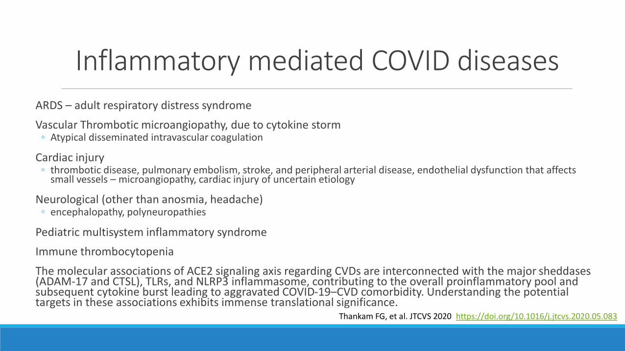
Inflammatory mediated COVID diseases ARDS – adult respiratory distress syndrome
Vascular Thrombotic microangiopathy, due to cytokine storm◦ Atypical disseminated intravascular coagulation
Cardiac injury◦ thrombotic disease, pulmonary embolism, stroke, and peripheral arterial disease, endothelial dysfunction that affects
small vessels – microangiopathy, cardiac injury of uncertain etiology
Neurological (other than anosmia, headache)◦ encephalopathy, polyneuropathies
Pediatric multisystem inflammatory syndrome
Immune thrombocytopenia
The molecular associations of ACE2 signaling axis regarding CVDs are interconnected with the major sheddases(ADAM-17 and CTSL), TLRs, and NLRP3 inflammasome, contributing to the overall proinflammatory pool and subsequent cytokine burst leading to aggravated COVID-19–CVD comorbidity. Understanding the potential targets in these associations exhibits immense translational significance.
Thankam FG, et al. JTCVS 2020 https://doi.org/10.1016/j.jtcvs.2020.05.083

ACE2 signaling in CVD versus COVID-19 infection.
ACE2 proteolytically inactivates Ang I and Ang II and acts on Ang I to form Ang 1-9 peptide, which is the precursor of the vasodilator Ang 1-7. The binding of Ang II with type-1 receptors (AT1R and AT2R) initiates a series of biochemical events leading to vasoconstriction, oxidative stress, fibrosis, and retention of electrolytes.
In contrast, ACE2–Ang 1-7 axis via G-protein coupled protein receptor (Mas) acts as counter-regulator by promoting vasodilation, antioxidant responses, and antifibrotic reactions.
The decline of ACE2 expression following the progression of CVDs enhances the myocardial dysfunction and induces inflammatory burden.
The decreased ACE2 activity results in the activation of myocardial NADPH oxidase system, leading to superoxide-activated oxidative stress and hyperactivationof MMPs, which subsequently aggravate CVD pathology.
The COVID-19 SG increases the susceptibility of SG to undergo proteolytic cleavage by sheddases, including ADAM-17, CTSL, and TMPRSS2, which is essential for coronavirus entry to the host cells.
The shedding of ACE2 results in the suppression of downstream signaling, leading to increased Ang II level and subsequent aggravation of CVD pathology. sACE2 is associated with aggravated CVD pathology.
Ang, Angiotensin; ACE2, angiotensin-converting enzyme 2; COVID-19, coronavirus disease 2019; SG, spike glycoprotein; ADAM-17, ADAM metallopeptidase domain-17; CTSL, cysteine protease cathepsin L; TMPRSS2, transmembrane protease/serinesubfamilymember 2; MMP, matrix metallopeptidase; NADPH, nicotinamide adenine dinucleotide phosphate.
Thankam FG, et al. JVCTS 2020; June 6

ADAM-17 activates atherosclerotic plaque rupture and vascular inflammation. ACE2 inhibits the DAMPs, including HMGB1 released from the infected and ischemic/necrotic cells due to membrane damage. The decreased levels of ACE2 lead to increased DAMPs, especially OxLDL, HMGB1, AGEs, and ROS. These mediators trigger NLRP3 inflammasome via TLR2, TLR4, RAGE, and/or TREM1 axes in cardiovascular system. The upregulation of such DAMPs in the ACE2-depleted environment is detrimental, resulting in aggravated COVID-19–CVD comorbidity. ADAM-17, ADAM metallopeptidase domain-17; OxLDL, oxidized low-density lipoprotein; AGEs, advanced glycation end products; HMGB1, high mobility group box 1; TLR, Toll-like receptor; ROS, reactive oxygen species; ACE2, angiotensin-converting enzyme 2; TNF-α, tumor necrosis factor-α; COVID-19, coronavirus disease 2019; TRIF, TIR-domain-containing adaptor protein including IFN-β; RIG-1, retinoic acid-inducible gene 1; MDA5, melanoma differentiation-associated gene 5; STING, stimulator of interferon genes protein; MP, matrix protein; MAV, mitochondrial antiviral-signaling protein;TRAF3, TNF receptor-associated factor; PAMPs, pathogen-associated molecular patterns; NF-κB, nuclear factor-κB; IRF3, interferon regulatory factor 3; ORF, open reading frame; SEP, small envelope protein; NLRP3, Nod-like receptor protein 3; IL, interleukin; IFN, interferon; CVD, cardiovascular disease.
Proposed molecular mechanism underlying aggravated inflammatory response in COVID-19–CVD comorbidity.
The components of COVID-19, including the RNA and proteins, act as intracellular PAMPs, which are recognized by conventional pattern recognition receptors, especially TLRs, RIG-I-like receptors (RLRs), and NLRP3 inflammasome. The TLRs, including TLR3, TLR7, TLR8, and TLR9, detect viral genome in the endosomal vesicles. In addition, the viral genome in the cytosol are recognized by the cytosolic receptors including RIG-1 and MDA5. The binding of viral ligands with the receptors initiates the recruitment and assembly of adaptor proteins including TRIF, MAVS, and STING, which trigger the activation of the transcription factor NF-κB and IRF3 via MyD88 adapter. IRF3 triggers the expression of type I IFNs, whereas NF-κB stimulates the expression of a battery of proinflammatory cytokines leading to cytokine burst. IL-1β is generated by the proteolytic activation of pro–IL-1β by caspase-1 following the activation of NLRP3 inflammasome. The active NLRP3 inflammasome upregulates the transcription of pro–IL-1β gene and subsequent activation by caspase-1. The COVID-19 proteins including MP, SEP, and ORF3a activate NLRP3 via TRAF3 and subsequent IL-1β and ORF3a activate NF-κB and downstream cytokine burst. The apoptotic/necrotic cells following virus infection upregulate ADAM-17, the major sheddase for ACE2. IL-1β and TNF-α enhance the ACE2 shedding. The resultant sACE2 is a potent mediator for vascular inflammation and CVD pathology. Thankam FG, et al. JVCTS 2020; June 6

https://covid19.healthdata.org/united-states-of-america?view=infections-testing&tab=trend&test=infections
IHME model led by Chris Murray projects an Increase in cases as we progress to February, followed by a decline
The light blue dotted line is the IHME projection on Aug 30,2020

Worldometer
Simple extrapolation of current decline in new cases
Solid line = Oct 21 projection
Dotted line = Sept 20 projection
Dashed line = prior projection Aug 20
https://www.worldometers.info/coronavirus/country/us/

COVID-19 Tracker - STAT
Simple extrapolation of current decline in new cases
Solid line = Oct 21 projection
Dotted line = Sep 20 projection
Dashed line = Aug 30 projection
https://www.statnews.com/2020/03/26/covid-19-tracker/

Solid line = Oct 21 projection
Dotted line = Sep 20 projection
Dashed line = Aug 30 projection
CDC projection to mid-Dec, with extrapolation
Jan 1
https://www.cdc.gov/coronavirus/2019-ncov/covid-data/forecasting-us.html

Fighting COVID-19 disease & SARS-CoV-2
Prevention – Infection Control• Face masks• Physical distancing• Wash hands• Outside better than inside• Quarantine 14d after exposure• Isolate 10d after diagnosis• Contact trace & test• Vaccines
Diagnostics • Rapid POC tests• Antigen, isothermal PCR, other• RT-PCR, CRISPr, others• Culture• Serology• Chest Xray• CT Scan• other
Therapeutics• Donor plasma• Monoclonal antibodies• Repurposed drugs (Remdesivir)• Corticosteroids• Anticoagulants• New drugs• Oxygen• Positioning• Ventilator

Bairi KE, et al. European J Cancer 2020;141:40-61

Figure 1Virologic aspects, life-cycle and targets for drug development. (A) CoVs have a long, capped and poly-adenylated RNA genome, which contains between 8 to 10 open-reading-frames (ORFs), allowing structural, non-structural and accessory viral protein synthesis [87
]. SARS-CoV2 is 29,903 base-long and contains 6 majors ORFs, as well as additional accessory genes; the reference sequence is registered in GenBank with ID: MN908947.3 [1
]. (A and B) Up to 28 different polypeptides are potentially produced in fine from the different ORFs and after polyprotein processing by viro-encoded proteases [87
]. If the RNA genome contained in virions can already serve, after cell entry, as a template for the synthesis of non-structural proteins, which are involved in the early phase of virus replication (mainly by forming the replicase complex), subgenomic messenger RNAs (sgmRNAs) are also produced in the late phase of the cycle to allow the synthesis of structural proteins (e.g. spike (S), envelope (E), membrane (M) and nucleocapsid (N) proteins), as well as other accessory polypeptides. Another main replication intermediate is the complementary minus-sens RNA, which is used by the viro-encoded RNA-dependent RNA-polymerase (RdRp), within replicase complex, to amplify the full-length genome, which is then capped and polyadenylated by both viral and host enzymes before being incorporated into virus progeny. (B) After entry into ACE2-positive (entry receptor) and TMPRSS2-positive (co-factor for entry) cells, and the membrane fusion (i.e. uncoating process), a full-length genome is released in the cytoplasm of cells. This full-length polycistronic RNA is directly used to efficiently encode a polyprotein from the first ORFs present on the molecule, starting from 5’ extremity, i.e. ORF1a and ORF1b; the latter is read after a frame-shift from ribosomal scanning of ORF1a. (A and B) The polyprotein is then processed by two viro-encoded proteases, a papain-like cysteine protease (PLpro/Nsp3) and a chemotrypsin-like protease (3CLpro/Nsp5; also known as main protease (Mpro)), into 16 proteins/polypeptides (Nsp1 to 16). (B) These non-structural proteins/polypeptides are important for the early stageof infection, as they allow in particular the formation of the replicase complex around the RdRp enzymatic activity, which is involved in the synthesis of negative-sense full-length RNA, as well as sgmRNAs by a discontinuous transcription strategy [87
]. The latters enable an efficient and stochiometric synthesis of all other viral proteins/polypeptides, which are important for virus assembly and release of progeny virions.
Jiang S, et al. Trends Immun 2020;41(5):355-9

Presymptomatic infectious period
Post-inflammatory phase illness
Latent pre- infectious period
Asymptomatic infectedWithout symptoms or signs
Attribution: Dr. Melvin Sanicas, Zurich, Switzerland@Vaccinologist and TEDedhttps://ed.ted.com/search?qs=SanicasModified by Dr. Jeff Andrews 19OCT2020

Reproduction number = R0 or R nought
Attribution: Counotte MJ, et al. PLOS Medicinedoi: https://doi.org/10.1371/journal.pmed.1002611.g001Modified by Dr. Jeff Andrews 19OCT2020

What is the evidence regarding severity of disease and viral load?
Early reports, circa March 2020, suggested that milder disease was correlated with lower viral load and severe disease was correlated with higher viral load
◦ However, the evidence of the relationship was limited by the poor quality of many of the studies, the retrospective nature of the studies, small sample sizes, and the potential problem of selection bias.
The more recent scientific study reports (August-October, 2020) indicate that there is not a statistically significant difference in viral loads between mild and severe disease
Liu Y, et al. Lancet 2020;20(6):656-7Heneghan C, et al. CEBM March 26, 2020Zheng S, et al. BMJ 2020;369:m1443

Figure 2a. The distribution of Ct values corresponding to the 38 specimens that were positive by the Lyra assay (from specimens collected from participants, 0-7 DSO) following stratificationby number of symptoms. Ct score distribution for specimens matched to 1 symptom is shown in blue while those matched to ≥2 symptoms are shown in orange; the pink color indicates blue/orange overlap
Figure 2b. The mean Ct values (and standard deviation) are shown for the ≥2 symptom specimens (n=31; mean=22.10, standard deviation=5.63) and the 1 symptom specimens (n= 7; mean=25.56, standard deviation=3.90). A two-sample t-test (2-tailed) analysisindicated non-significant difference between the means (p-value = 0.077; mean difference of 3.46; [95% CI: -0.43, 7.36]).
Young S, et al. J Clin Micro 2020. DOI: 10.1128/JCM.02338-20

Figure 3a. The distribution of Ct values corresponding to the 38 specimens that were positive by the Lyra assay (from specimens collected from participants, 0-7 DSO). Plotted along the fitted distribution line are the 29 true positive Veritor results (orange circles) and the nine participant designations (letters superimposed onto blue circles), corresponding to those in Table 3, that represent the Veritor false negative results matched to Lyra assay Ct value.
Figure 3b. The mean Ct values (and standard deviation) are shown for the 29 true positive (20.76 and 4.21, respectively) and the 9 false negative (29.12 and 4.11, respectively) Veritor test results. A two-sample t-test (2-tailed) analysis indicated a significantly higher mean Lyra assay Ct value for specimens matched to the 9 Veritor test false negative results compared to those matched to the 29 true542 positive results (p<0.001; mean difference of 8.36; [95% CI: 4.95, 11.77]).
Young S, et al. J Clin Micro 2020. DOI: 10.1128/JCM.02338-20

What is the evidence regarding asymptomatic infection and viral load?
Early reports, circa March 2020, suggested that asymptomatic infected individuals had lower viral load than symptomatic patients
◦ However, the evidence was limited by very small sizes (1-4 asymptomatic individuals in study reports), the poor quality of many of the studies, the retrospective nature of the studies, and the potential problem of selection bias.
The more recent scientific study reports (July-October, 2020) indicate that there is not a statistically significant difference in viral loads between asymptomatic infected individuals and symptomatic patients
◦ The difference is that symptomatic patients have a day of onset or symptoms and that date may be used to associate with viral load changes over time, whereas asymptomatic individuals have no onset of symptoms day, and often are uncertain about the timing of exposure
Current estimates about the proportion of population with asymptomatic infection, compared to symptomatic diagnosed with COVID-19
◦ CDC, USA – 40%
◦ South Korea, study of a congregate setting – 20%Liu Y, et al. Lancet 2020;20(6):656-7Heneghan C, et al. CEBM March 26, 2020Zheng S, et al. BMJ 2020;369:m1443 Lee S, et al. JAMA Intern Med 2020; Aug 6
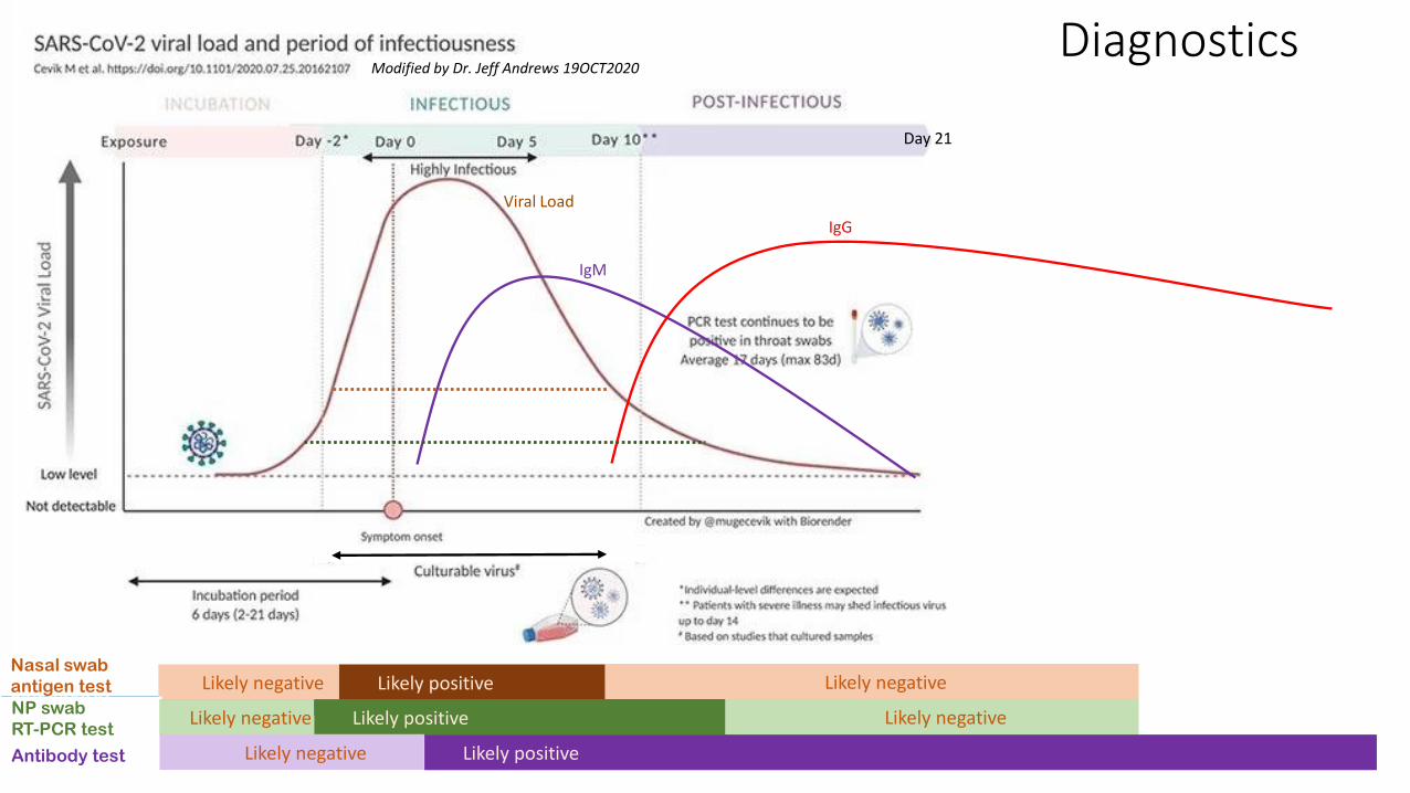
Nasal swab
antigen test Likely negative Likely positive Likely negativeNP swab
RT-PCR testLikely negative Likely positive Likely negative
Day 21
IgM
IgG
Viral Load
Antibody test Likely negative Likely positive
DiagnosticsModified by Dr. Jeff Andrews 19OCT2020

Figure 1A. The 38 RT-PCR assay positive specimens are plotted by Log10 copies/mL (y-axis) and are stratified by the SARS-CoV-2 live culture results (negative, n=10; positive, n=28). The median and inter-quartile range values, respectively, for the RT-PCR-positive/SARS-CoV-2 TMPRSS2 culture-negative were 4.21 and 1.37; the median and inter-quartile range values, respectively, for the RT-PCR-positive/SARS-CoV-2 TMPRSS2 culture negative were 7.39 and 1.66. The mean values for the SARS-CoV-2 TMPRSS2 culture-negative and SARS-CoV-2 TMPRSS2 assay-positive specimen groups were significantly different (4.01 versus 7.16, respectively; p-value <0.001 based on two-sample t-test [2-tailed]). Antigen test positive results are indicated as red data points (n=29) and the antigen test negative results (n=9) are indicated by the green data points.

Figure 1B. Probit models linking viral load to the probability of positive result of RT-PCR (Lyra), antigen test (Veritor), SARS-CoV-2 TMPRSS2 culture and SARS-CoV-2 VeroE6 culture (refer to Huang et al 2020). Viral load levels at which there is a 5% chance of positive result: 1.6, 2.6, 4.5, and 5.75 log10 cp/ml for RT-PCR, antigen, SARS-CoV-2 TMPRSS2 culture, and SARS-CoV-2 VeroE6 culture, respectively.

Figure S2. Relation of the RT-PCR Ct scores for the BD MAX assay, the RT-PCR assay, and the RT-PCR method used in Huang et al (2020) to viral load. Empirical equation for the RT-PCR assay Ct = 42.69 - 3.14 Log10 copies/mL. Empirical equation for the E target in Huang et al: Log10 copies/mL = 12.377 – 0.052 Ct – 0.005 Ct.

Diagnostics: viral RNA target
RT-PCR◦ quantitative real‐time RT‐PCR (RT‐qPCR) is one of the commonly used techniques for virus detection, which
has high sensitivity, rapid detection, and other desirable characteristics.
Isothermic PCR◦ reverse transcription loop‐mediated isothermal amplification (RT‐LAMP) performed in one step at 63°C
isothermal conditions, and the results are obtained within 15–40 min, by targeting the ORF1ab, spike (S), envelope (E) or/and N gene of SARS‐CoV‐2
◦ RT‐LAMP result can be evaluated using real‐time turbidimeter, electrophoresis or fluorescent, which is faster and more convenient for clinical diagnosis of SARS‐CoV‐2
CRISPR◦ Clustered regularly interspaced short palindromic repeats (CRISPR)‐based diagnostic platforms have also
been developed for point‐of‐care nucleic acid detection, such as SHERLOCK or DETECTR
◦ combines recombinase polymerase amplification with CRISPR‐Cas enzymology for specific recognition of targeted DNA or RNA sequences
Li C, et al. Transbound Emerg Dis 2020 Jul;67(4):1485-1491

FDA EUA approved viral testsOctober 21, 2020
Technology
real-
time,
basic
not
real-
time
with
home
collection
with
screening
claim
with
pooling
claim
with
saliva
specimen
with
multi-
analyte total
RT-PCR 103 14 18 2 5 10 6 158
isothermal/LAMP 8 2 10
sequencing 4 4
CRISPER 2 2
TMA luminescense 1 2 1 4
other 8 8
total tests 186
home collection kit 5
saliva collection kit 1
Source: https://www.fda.gov/medical-devices/coronavirus-disease-2019-covid-19-emergency-use-authorizations-medical-devices/vitro-diagnostics-euas
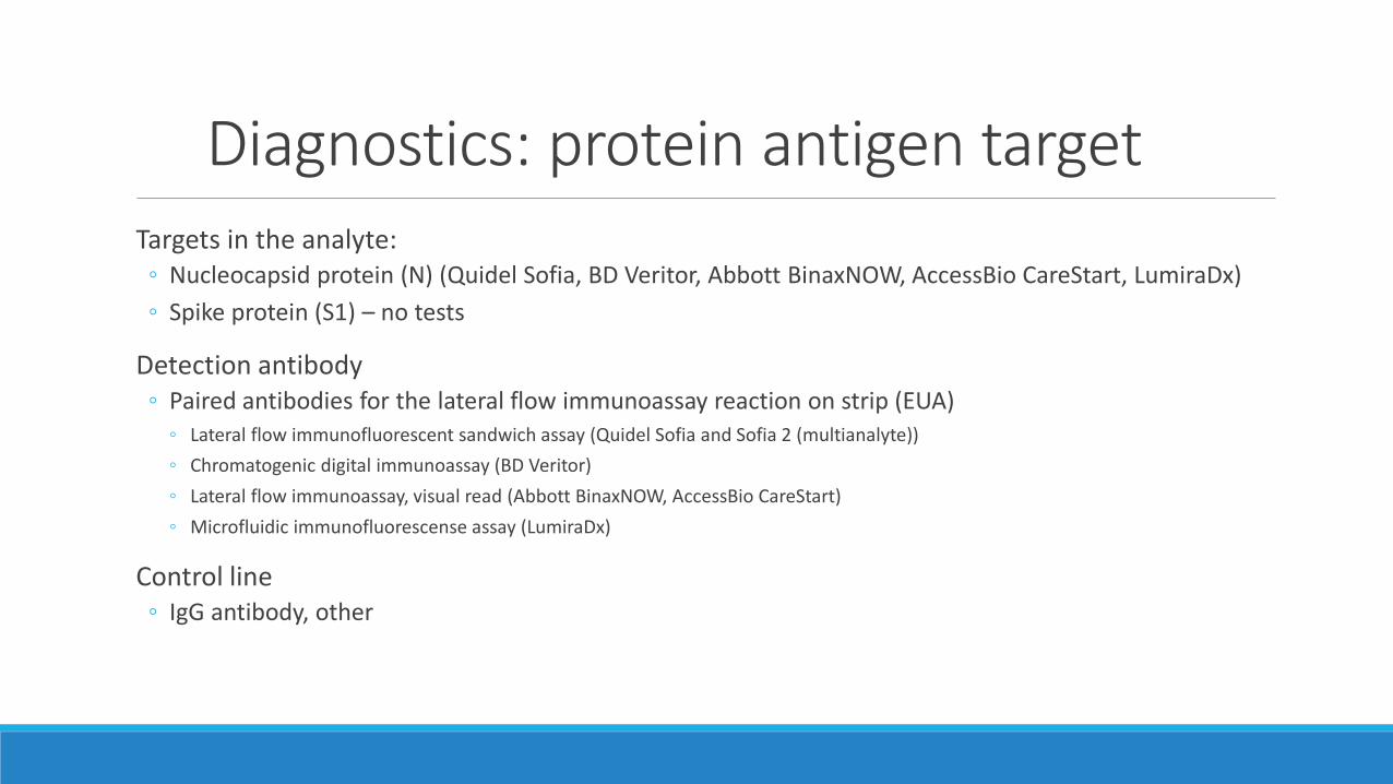
Diagnostics: protein antigen targetTargets in the analyte:
◦ Nucleocapsid protein (N) (Quidel Sofia, BD Veritor, Abbott BinaxNOW, AccessBio CareStart, LumiraDx)
◦ Spike protein (S1) – no tests
Detection antibody◦ Paired antibodies for the lateral flow immunoassay reaction on strip (EUA)
◦ Lateral flow immunofluorescent sandwich assay (Quidel Sofia and Sofia 2 (multianalyte))
◦ Chromatogenic digital immunoassay (BD Veritor)
◦ Lateral flow immunoassay, visual read (Abbott BinaxNOW, AccessBio CareStart)
◦ Microfluidic immunofluorescense assay (LumiraDx)
Control line◦ IgG antibody, other

Specimen types for virus and antigenMixed results on RT-PCR sensitivity – larger sample size studies have shown statistically significant lower sensitivity for Nasal vs NP. Smaller studies did not find a difference in sensitivity. All studies found lower Ct values with NP compared to nasal.
Viral load is lower on nasal swab vs NP. RT-PCR is able to correct for this through amplification cycles and often yields positive concordance.
Indirectly, lower viral load likely means lower protein load and therefore the antigen test (unable to make up for low load by amplification) will likely have discordance between nasal and NP (negative nasal specimen and positive NP specimen)
Comparing a nasal swab antigen versus NP PCR increases the sensitivity bias in favor of the reference, thus decreasing antigen sensitivity
Combining a nasal with a pharyngeal swab may achieve viral load concordance with NP.
Oropharyngeal swab yields inferior viral load and sensitivity, compared to NP.
Saliva yields inferior viral load and sensitivity compared to oropharyngeal swab
For many studies, the swab type or transport media are not specified.
Péré H, et al. J Clin Microbiol. 2020;58(6):e00721-20. Tu YP, Jet al. P medRxiv 2020.04.01.20050005 Berenger BM, et al. medRxiv 2020.05.05.20084889
Callahan C, et al. medRxiv 2020 Jun 14:2020.06.12.20128736Pinninti S, et al. Clinical Infectious Diseases 2020, ciaa882, https://doi.org/10.1093/cid/ciaa882 Vlek ALM, et al. Eur J Clin Microbiol Infect Dis. doi: 10.1007/s10096-020-03972-y

Diagnostics: antibody serology tests
U.S. Food and Drug Administration (FDA) has authorized >35 antibody tests under Emergency Use Authorization (EUA)
◦ A number of these tests have been recalled due to changes in performance specification criteria.
ELISAs for detecting anti-trimer spike antibodies (IgG and IgA) and anti-nucleocapsid antibodies (IgG)
SARS-CoV-2 has four structural proteins, and among them the spike (S1) and the nucleocapsid (N) are considered the main immunogens and are widely used in immunoassays.
◦ The nucleocapsid is a protein with a small size that can easily be produced and purified in prokaryotic or eukaryotic hosts in vast quantities.
◦ anti-nucleocapsid antibodies appear earlier than the spike antibodies
◦ current commercial serology tests target anti-nucleocapsid antibodies
◦ current vaccine research serology tests target anti-spike antibodies

Specimen types and tests for antibody
specimen types: blood draw, finger prick, saliva
Type of test Time to result
Comment
Rapid diagnostic test (RDT)
<30mins
Qualitative result, no info about whether antibodies are able to inhibit virus growth
Chemiluminescentimmunoassay
<2 hours Quantitative, no info about whether antibodies are able to inhibit virus growth
Enzyme-linked immunosorbent assay (ELISA)
<5 hours Quantitative, no info about whether antibodies are able to inhibit virus growth
Neutralization assay 3-5 days The presence of active antibodies in patient serum that are able to inhibit virus growth ex vivo, in a cell culture system, but may miss antibodies to viral proteins that are not involved in replication.

The 2X2 table of diagnostic statistics
False negative rate = 100%-Sensitivity
False positive rate = 100%-Specificity
Values change with prevalence
Values do not change with prevalence
Created by Dr. Jeff Andrews, Oct 21, 2020

The 2X2 table of diagnostic statistics
Reference test
Percent Positive Agreement
Percent Negative Agreement
Probability that person with positive index will also have positive reference test
Probability that person with negative index will also have negative reference test
Positive Agreement
Negative AgreementPutative False Negative
Putative False Positive
False negative rate = 100%-Sensitivity
False positive rate = 100%-Specificity
Values change with prevalence
Values change with prevalence
Values do not change with prevalence
Created by Dr. Jeff Andrews, Oct 21, 2020
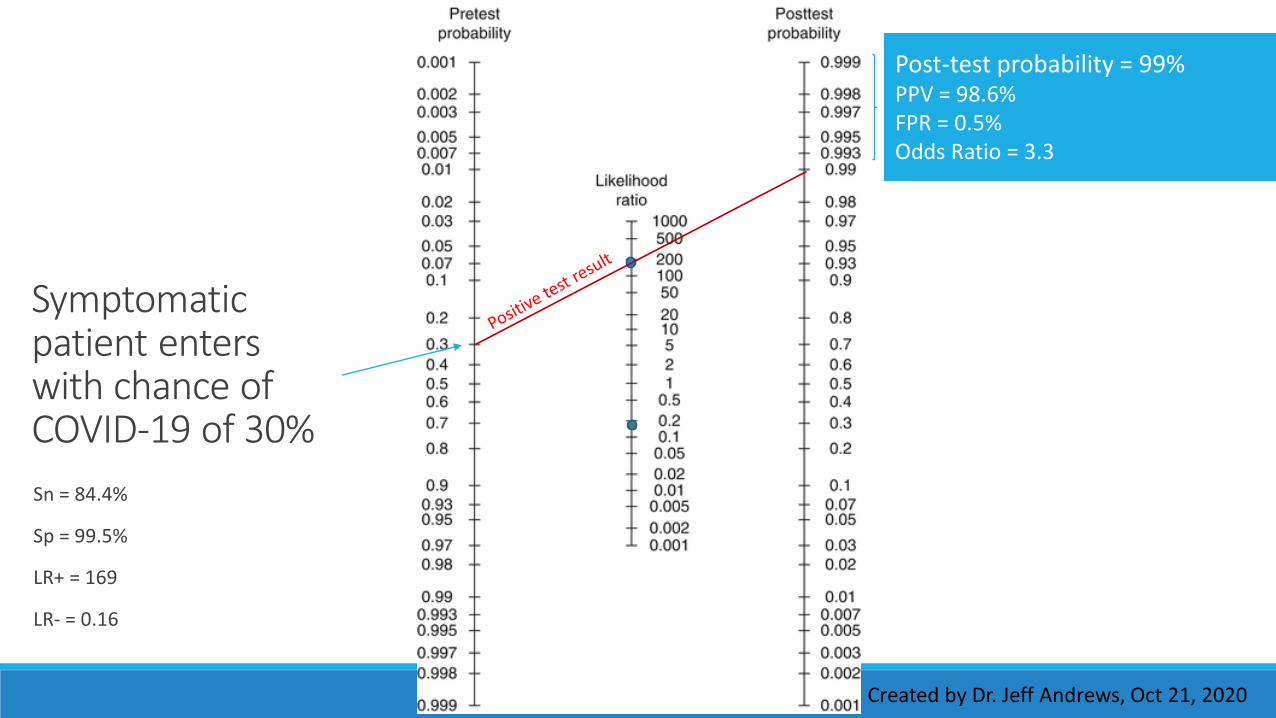
Symptomatic patient enters with chance of COVID-19 of 30%
Sn = 84.4%
Sp = 99.5%
LR+ = 169
LR- = 0.16
Post-test probability = 99%PPV = 98.6%FPR = 0.5%Odds Ratio = 3.3
Created by Dr. Jeff Andrews, Oct 21, 2020

Post-test probability = 95%PPV = 95%FPR = 0.5%Odds ratio = 9.5
Asymptomatic individual, known exposure to COVID-19, enters with chance of COVID-19 of 10%
Sn = 84.4%
Sp = 99.5%
LR+ = 169
LR- = 0.16
Created by Dr. Jeff Andrews, Oct 21, 2020

Post-test probability = 60%PPV = 60%FPR = 0.5%Odds ratio = 60
Asymptomatic individual, no known exposure to COVID-19, enters with chance of COVID-19 of 1%
Created by Dr. Jeff Andrews, Oct 21, 2020
Sn = 84.4%
Sp = 99.5%
LR+ = 169
LR- = 0.16

Post-test probability = 14%PPV = 14.3%FPR = 0.5%Odds ratio = 140
Asymptomatic individual, no known exposure to COVID-19, low prevalence community, enters with chance of COVID-19 of 0.1%
Sn = 84.4%
Sp = 99.5%
LR+ = 169
LR- = 0.16
Created by Dr. Jeff Andrews, Oct 21, 2020

Post-test probability = 6.3%NPV = 93.7%FNR = 15.6%Inverse odds ratio = 4.8
Symptomatic patient enters with chance of COVID-19 of 30%
Sn = 84.4%
Sp = 99.5%
LR+ = 169
LR- = 0.16
Created by Dr. Jeff Andrews, Oct 21, 2020

Post-test probability = 1.7%NPV = 98.3%FNR = 15.6%Inverse odds ratio = 5.9
Asymptomatic individual, known exposure to COVID-19, enters with chance of COVID-19 of 10%
Sn = 84.4%
Sp = 99.5%
LR+ = 169
LR- = 0.16
Created by Dr. Jeff Andrews, Oct 21, 2020

Post-test probability = 0.2%NPV = 99.8%FNR = 15.6%Inverse odds ratio = 5
Asymptomatic individual, no known exposure to COVID-19, enters with chance of COVID-19 of 1%
Sn = 84.4%
Sp = 99.5%
LR+ = 169
LR- = 0.16
Created by Dr. Jeff Andrews, Oct 21, 2020

Asymptomatic individual, no known exposure to COVID-19, low prevalence community, enters with chance of COVID-19 of 0.1%
Post-test probability = 0.02%NPV = 99.98%FNR = 15.6%Inverse odds ratio = 5
Sn = 84.4%
Sp = 99.5%
LR+ = 169
LR- = 0.16
Created by Dr. Jeff Andrews, Oct 21, 2020

Symptomatic patient enters with chance of COVID-19 of 30%
Post-test probability = 99%PPV = 98.6%FPR = 0.5%
Interpretation: confirmation of COVID-19
Created by Dr. Jeff Andrews, Oct 21, 2020

Post-test probability = 95%PPV = 95%FPR = 0.5%
Asymptomatic individual, known exposure to COVID-19, enters with chance of COVID-19 of 10%
Interpretation: confirmation of COVID-19. After 222 tests, would have to repeat 19 tests to find 1 false positive.
Created by Dr. Jeff Andrews, Oct 21, 2020

Asymptomatic individual, no known exposure to COVID-19, enters with chance of COVID-19 of 1% Interpretation: probable
COVID-19. Isolate. Follow-on with RT-PCR. After 400 tests, would have to repeat 5 tests to find 3 true positives.
Post-test probability = 60% PPV = 60%FPR = 0.5%
Created by Dr. Jeff Andrews, Oct 21, 2020

Post-test probability = 14% PPV = 14.3%FPR = 0.5%
Asymptomatic individual, no known exposure to COVID-19, low prevalence community, enters with chance of COVID-19 of 0.1%
Interpretation: possible COVID-19. Isolate. Follow-on with RT-PCR. After 1200 tests, would have to repeat 7 tests to find 1 true positive.
Created by Dr. Jeff Andrews, Oct 21, 2020

Post-test probability = 6.3%NPV = 93.7%FNR = 15.6%
Symptomatic patient enters with chance of COVID-19 of 30% Interpretation: possible COVID-19.
Quarantine away from others. Follow-on with RT-PCR. Have to repeat 16 tests to find 1 false negative.
Created by Dr. Jeff Andrews, Oct 21, 2020

Post-test probability = 1.7%NPV = 98.3%FNR = 15.6%
Asymptomatic individual, known exposure to COVID-19, enters with chance of COVID-19 of 10%
Interpretation: Unlikely COVID-19. Continue working, with infection control precautions. In high-risk setting, may need to quarantine. Would have to repeat 60 tests to find 1 false negative.
Take note of time since exposure. May need to repeat test within 48-72 hours to cover early incubation period.
Created by Dr. Jeff Andrews, Oct 21, 2020

Post-test probability = 0.2%NPV = 99.8%FNR = 15.6%
Asymptomatic individual, no known exposure to COVID-19, enters with chance of COVID-19 of 1%
Interpretation: Rules out COVID-19. Normal activity. Would have to repeat 500 tests to find 1 false negative.
Created by Dr. Jeff Andrews, Oct 21, 2020

Asymptomatic individual, no known exposure to COVID-19, low prevalence community, enters with chance of COVID-19 of 0.1%
Post-test probability = 0.02%NPV = 99.98%FNR = 15.6%
Interpretation: Rules out COVID-19. Normal activity. Would have to repeat 5000 tests to find 1 false negative.
Created by Dr. Jeff Andrews, Oct 21, 2020

Influenza
CDC reports November 20 that seasonal influenza activity in the USA remains low
Past daily new cases of influenza in USA ranged 6-14/100,000 during peak days
The peak number of daily new cases of influenza in the USA during last season was lower than the daily new cases of coronavirus in the USA today
Mask-wearing will decrease the overall incidence of influenza this season
Healthcare provider outpatient visits related to influenza are 50% of 2019 baseline.
In this season, the clinicians will not be able to determine if symptoms of flu are due to influenza, COVID-19, or some other diagnosis
Therefore, they will want to use rapid multi-analyte tests for symptomatic patients, if available
If there are test kits shortages, for adults, they will prefer SARS-CoV-2 first (due to higher mortality), and then reflex to Influenza if SARS-CoV-2 negative
If there are test kits shortages, for young children, they will prefer influenza first (due to higher mortality), and then reflex to SARS-CoV-2 if influenza negative
https://www.cdc.gov/flu/weekly/index.htm

Vaccine prioritization
Vaccine administration might begin in Dec-Jan with general population vaccination beginning after Mar-Apr
ADM Giroir said priorities for vaccine will be:◦ Healthcare workers
◦ Nursing home staff and residents
◦ First responders
◦ Vulnerable older populations and those with pre-existing conditions
◦ Teachers
These priorities target a reduction in deaths (not new cases)
Since the individuals who are currently spreading disease are not priority targets of vaccination, the new case rate will not be impacted until months after the general population is vaccinated (at earliest)

Insights
Test ordering for SARS-CoV-2 likely to remain very strong for at least 12 more months (due to sustained new case rates) and probably for several years
Vaccine impact on need for testing unlikely to be notable prior to October 2021 (given the vaccination timeline of CDC & ADM Giroir & need for 2 doses & unknown effectiveness or duration of effect)
Interest in multi-viral tests likely to be moderately-strong throughout the traditional flu season, and likely to repeat in 2021-2022 season and beyond
COVID-19 is here to stay, and the future depends on a lot of unknowns, including whether people develop lasting immunity to the virus, whether seasonality affects its spread, and —perhaps most importantly — the choices made by governments and individuals.
To end the pandemic, the virus must either be eliminated worldwide — which most scientists agree is near-impossible because of how widespread it has become — or people must build up sufficient immunity through infections or a vaccine. It is estimated that 55–80% of a population must be immune for this to happen, depending on the country

Thank You!