Health Effects of Lead Chapter 2 Lead Abatement for Workers Course.
Upload
truongcongCategory
view
228download
3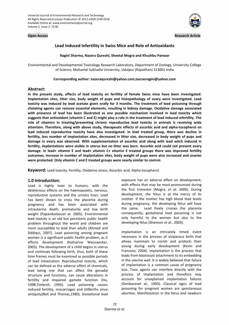
Universal Journal of Environmental Research and Technology
All Rights Reserved Euresian Publication © 2012 eISSN 2249 0256
Available Online at: www.environmentaljournal.org
Volume 2, Issue 2: 72-82
Open Access Research Article
72
Sharma et al.
Lead Induced Infertility in Swiss Mice and Role of Antioxidants
Ragini Sharma, Nazera Qureshi, Sheetal Mogra and Khushbu Panwar
Environmental and Developmental Toxicology Research Laboratory, Department of Zoology, University College
of Science, Mohanlal Sukhadia University, Udaipur (Rajasthan) 313001 India
Corresponding author: [email protected],[email protected]
Abstract: In the present study, effects of lead toxicity on fertility of female Swiss mice have been investigated.
Implantation sites, litter size, body weight of pups and histopathology of ovary were investigated. Lead
toxicity was induced by lead acetate given orally for 3 months. The treatment of lead poisoning through
chelating agents can remove essential elements, resulting in kidney damage. Oxidative damage associated
with presence of lead has been illustrated as one possible mechanism involved in lead toxicity which
suggests that antioxidant (vitamin C and E) might play a role in the treatment of lead induced infertility. The
role of vitamins in treating/preventing chronic reproductive lead toxicity in animals is receiving wide
attention. Therefore, along with above study, therapeutic effects of ascorbic acid and alpha-tocopherol on
lead induced reproductive toxicity have also investigated. In lead treated group, there was decline in
fertility, less number of implantation sites, decreased in litter size, decreased in body weight of pups and
damage in ovary was observed. With supplementation of ascorbic acid along with lead witch induced in
fertility, implantations were visible in uterus but no litter was born. Ascorbic acid could not prevent ovary
damage. In lead+ vitamin E and lead+ vitamin C+ vitamin E treated groups there was improved fertility
outcomes, increase in number of implantation sites, body weight of pups were also increased and ovaries
were protected. Only vitamin C and E treated groups were nearly similar to control.
Keyword: Lead toxicity, Fertility, Oxidative stress, Ascorbic acid, Alpha-tocopherol.
1.0 Introduction: Lead is highly toxic to humans, with the
deleterious effects on the haemopoietic, nervous,
reproductive systems and the urinary tract. Lead
has been shown to cross the placenta during
pregnancy and has been associated with
intrauterine death, prematurity and low birth
weight (Papanikolaouet al. 2005). Environmental
lead toxicity is an old but persistent public health
problem throughout the world and children are
more susceptible to lead than adults (Ahmed and
Siddiqui, 2007). Lead poisoning among pregnant
women is a significant public health problem, as it
effects development (Katharine Weizsaecker,
2003). The development of a child begins in uterus
and continues following birth, thus, both of these
time frames must be examined as possible periods
of lead intoxication. Reproductive toxicity, which
can be defined as the adverse effect of chemicals,
lead being one that can affect the gonadal
structure and functions, can cause alterations in
fertility and impaired gamete function (Hu,
1998;Timbrell, 1995). Lead poisoning causes
reduced fertility, miscarriages and stillbirths since
antiquity(Bell and Thomas,1980). Gestational lead
exposure has an adverse effect on development;
with effects that may be most pronounced during
the first trimester (Mogra, et al. 2009). During
development, the fetus is at the mercy of its
mother. If the mother has high blood lead levels
during pregnancy, the developing fetus will have
the same. Lead freely crosses the placenta
consequently; gestational lead poisoning is not
only harmful to the women but also to the
developing fetus (Shannon et al., 2003).
Implantation is an intricately timed event
necessary in the process of viviparous birth that
allows mammals to norish and protects their
young during early development (Kevin and
Franceso, 2004). Implantation is the process that
leads from blastocyst attachment to its embedding
in the uterine wall. It is widely believed that failure
of implantation is a common cause of pregnancy
loss. Toxic agents can interfere directly with the
process of implantation and therefore may
account for unexplained implantation failures
(Genbacevet al., 1993). Classical signs of lead
poisoning for pregnant women are spontaneous
abortion. Manifestation in the fetus and newborn

Universal Journal of Environmental Research and Technology
73
Sharma et al.
include prematurity, fetal hypotrophy and
malformations (Klein et al., 1994). Lead is one of
the strong teratogens which cause most of its
congenital effect at the time of organogenesis
during embryonic development. The ovarian
follicle is the functional unit of the ovary. It
contains the oocyte that may eventually ovulate,
undergo fertilization and form an embryo. It also
provides the steroid and protein hormones
required for maintenance of the ovarian cycle, the
secondary sex characteristics and preparation of
the uterus for implantation (Findlayet al., 2009).
ROS affect multiple physiological processes from
oocyte maturation to fertilization. Ovulation-
induced oxidative base damage to DNA of the
ovarian epithelium can be prevented by
antioxidant (Agarwalet al. 2005). Antioxidative
properties of vitamin E is believed to prevent
reproductive disease associated with oxidative
stress (Brigelius-Floheet al. 2002). Vitamin C was
shown to act as a chain breaking scavenger for
peroxy radicals and also to act as synergist with
vitamin E (Stocker and Feri, 1991). Ascorbic acid
may also prevent gametes from damage by free
radicals during reproduction and fertilization.
2.0 Materials and Methods: Inbred, healthy, female Swiss mice in the age
group of 5-6 weeks, with 22-28 gm body weight
were used for the experiment and divided in to VII
groups containing ten animals in each. These
groups were treated orally with (160mg/kg/day)
dose of lead acetate, vitamin C (200mg/ kg/day)
and vitamin E (160mg/kg/day) for 3 months
according to following schedule:
Group I (Control group) - This group served as
control, and was given distilled water through
Canula.
Group II (L) - Treated with lead acetate
(160mg/kg/day).
Group III (LC) - Treated with lead acetate + vitamin
C (160 + 200mg/kg/day, respectively).
Group IV (LE) - Given lead acetate + vitamin E (160
+ 160mg/kg/day, respectively).
Group V (LCE) - Treated with lead acetate +
vitamin C + vitamin E (160+200+160mg/kg/day,
respectively).
Group VI (Only Vitamin C) - Given only vitamin C
(200mg/kg/day).
Group VII (Only Vitamin E) - Given only vitamin E
(160mg/kg/day).
In each group there were total 12 animals (10
females, 2 males). Total VII group were selected
for this study. In all the groups after two months,
male was introduced in the breeding cages. Each
animal of the group was checked in the morning
for vaginal plug. The animals showing vaginal plugs
were separated marked and put for further
observations. After termination of the experiment
animals were sacrificed by cervical dislocation.
Immediately after sacrificing animals, ovaries were
fixed in Bouin’s fixative for histopathological
observations (Drury and Wallington, 1967). Uterus
were immersed in 2% Na OH for 1 min. to observe
implantation sites and resorption.
3.0 Results and Discussion:
3.1 Group I (Control): In this group after 19-20 days females delivered
pups.
All the pubs were healthy and normal.
Litter size 8-10 animal.
In this group all the follicles were normal in
structure and distribution pattern of various
components. Germinal epithelium (Ge), cortex (C)
and inner medullary region (M) were apparent.
3.2 Group II (Lead treated): In lead treated group the number of implantation
sites was less than controls.
The number of pups was less than number of
implantation sites.
Numbers of resorption were increased.
Reduced litter size.
Body weight was reduced.
Young ones were eaten by mothers.
In this group animals showed damaging pattern in
this structure and distribution of in various
follicles. There was evident damage in germinal
epithelium, cortex and inner medullary region.
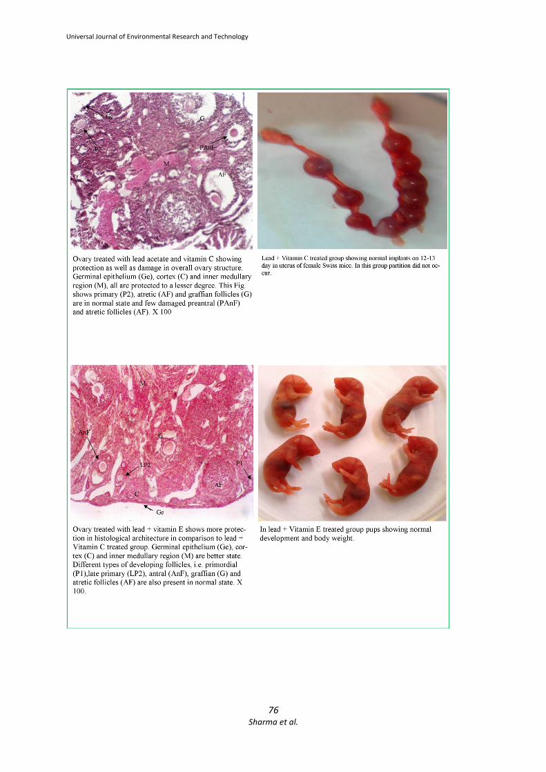
3.3 Group III (Lead + Vitamin C) and
Group V (Lead + Vitamin C + Vitamin E): In lead with vitamin C treated group pregnancies
were continued for approximately thirteen days
but may be due to resorption, the gestation period
was not completed. In lead + vitamin C +vitamin E
treated group the pregnancy continued till mid
gestation period and resorption occur after mid
gestation period.
No parturition was occurred.
No litters were born.
Administration of lead with vitamin C induced the
toxic effects in the ovary, however appreciable
protection was also observed. Germinal
epithelium, cortex and inner medullary regions
were also protected. Ovary of lead treated
animals, with co-administration of vitamin C and
vitamin E showed minor protection in germinal
epithelium, cortex and inner medullary region.
Universal Journal of Environmental Research and Technology
74
Sharma et al.
Both damage as well as protection can be seen in
all components.
3.4 Group IV (Lead + Vitamin E)-
Implantation sites with few resorption.
Litter size was small.
Pups were healthy.
Body weight of pups was less than control.
The ovary of the mice treated with vitamin E along
with lead showed more protection in comparison
to lead + vitamin C treated animals. Germinal
epithelium, cortex and inner medullary regions
were preserved.
3. 5 Group VI (Only Vitamin C) and Group
VII (Only Vitamin E): Female delivered the pups,
Pups were healthy.
Litter size was normal.
Body weight of pups was less than controls.
In these group follicles were normal in their
structure and distribution pattern of various
components in ovary was also normal.
Effects of lead and vitamins on reproduction and body weight of pups
S.No
.
Group
s
No. of
Animals
Treatment No.of
pregnat
female
after
2monts
Littersiz
e
(Numbe
r of
pups)
Average
weight
of single
pup
1. Group
I
12
10(Female)
+
2(Male)
Control 6+1
8-10 1.78
gm/each
2. Group
II
12 Only Lead 3 Preg.
3 Px
4-6 Abnorma
l
3. Group
III
12 Lead+VitaminC 2 Preg.
2 Px
- -
4. Group
IV
12 Lead+VitaminE 4Preg.
→
2 Px
6-7 1.22
gm/each
5. Group
V
12 Lead+VitaminC+Vitami
nE
3Preg→
2Px
- -
6. Group
VI
12 Only Vitamin C 6Preg→ 8-9 1.25
gm/each
7. Group
VII
12 Only Vitamin E 6Preg→ 9-10 1.27
gm/each
Px: No pregnancy
Preg→: Pregnancy persist

Universal Journal of Environmental Research and Technology
75
Sharma et al.
Universal Journal of Environmental Research and Technology
76
Sharma et al.

Universal Journal of Environmental Research and Technology
77
Sharma et al.
The present study shows that exposure of lead
caused post implantation loss in female Swiss
mice because it might be possible that metal are
directly bind to critical membrane sites and/ or
intracellular ligands, including protein and nucleic
acid, that may trigger inhibition of development
and death prior to metal-associated oxidative
damage. It seems that a deficiency in the
progesterone levels is directly implied in this
inhibition (Jacquet, 1976). Lead treatment was
found to reduce significantly the incidence of
pregnancies and increase the post implantation

Universal Journal of Environmental Research and Technology
78
Sharma et al.
loss in the pregnant females (Jacquet, 1975).
Previously, Tang and Zhu (2003) also noted that
occupational lead exposure of female workers
could lead to the impairment of the functions of
reproductive system.
In the present investigation, lead treated females
showed reduced number of pregnancies, small
litter size and decreased body weight with
spontaneous abortion. Wide (1985) studied the
exposure to lead at a time of early organogenesis,
caused fertility decrease by interfering with the
development of the female germ cells. The effects
of 200 and 400 ppm lead acetate in drinking water
on reproduction and development as well as on
renal and hepatic parameters of rats at different
life stages, from gestation to 3 months post
weaning were studied by Teijonet al. (2006). They
concluded a dose dependent effect on
reproduction with variations in the number of
births and in pups’ weight. Hamilton et al. (1994)
reported that dietary calcium and lead exposure
influenced pup body weight and lengths. The lead
induced reduction in body weight and lengths was
greatest in pups of dams fed with the low calcium
diet, probably as a result of the grater organ lead
accumulation and toxicity caused by this diet and/
or the reduced weight gain of the dams during
pregnancy. The later may be due to the result of
the reduced food intake that has been associated
with lead toxicity. Results suggest that litter size,
gender and fetal position in the uterus can
influence fetal weight in rats. In the present
investigation, animals treated with lead showed
vaginal plug but no pregnancies were observed in
these females and body weights of females were
also decreased. Animals treated with lead revealed
reduce fertility as compared to control group
animals, the maternal transfer of lead during
gestation are very efficient. It is also apparent that
lead uptake is grater during the fetal/ neonatal
period. Gestational exposure adversely affects the
postnatal development of pups. It reduces the pup
body weight, length and delays their physical
development.
The pathogenesis of adverse pregnancy outcomes
including preeclampsia and fetal growth restriction
(Myatt and Cui, 2004) and a number of neonatal
outcomes (Saugsted, 2001) have been showed to
be associated with oxidative stress. Lead
potentially induced oxidative stress and evidence
was accumulating to support the role of oxidative
stress in the pathophysiology of lead toxicity. It
was capable of inducing oxidative damage to brain,
heart, kidneys and reproductive organs. The
mechanisms for lead induced oxidative stress
include, the effects of lead on membranes, DNA
and antioxidant defense systems of cells
(Vassalloet al. 2008). Antioxidant (vitamin Cand E)
might play a role in the treatment of lead induced
infertility (Gureret al., 2001). Naturally occurring
antioxidants have been extensively studied for
their capacity to protect organisms and cells from
damage induced by oxygen reactive species
(Cozziet al., 1997). The role of vitamins in treating
chronic lead toxicity in animals is receiving wide
attention (Anitraand Frei, 1999). It had become
clear that high to moderate doses of lead exposure
induce generation of free radicals resulting in
oxidative damage to critical biomolecules, lipids,
proteins and DNA (Ahamedet al., 2007). The
present study indicates that, ovaries of treated
group showed increased number of atretic follicles
and congestion in stromal tissue, compared to
controls. There was a dose dependent reduction in
the number of follicles at different stages of
maturation. It was also observed that ovarian
physiology and rate of ovulation might also altered
in females exposed to high lead level, because
corpora lutea was rarely seen in the treated
females.
From the findings of present study it is clear that
the lead also affects the developing follicles but
the extent of damage increases with time. Thecal
cells may also be targets for heavy metal injury.
Lead is known to disturb the normal profile of
reproductive hormones in animals, both at
hypothalamic pituitary and at the gonadal levels
(Roniset al., 1996; Roniset al., 1998). Similarly
Shirotaet al. (2003) and McMurryet al. (1995)
noted that lead causes reduction in the number of
primordial follicles and decrease the number of
follicles that enter the growing phase. Lead
adversely affects pituitary hypothalamus axis, and
the balance of gonadotropin. Form the present
study it is clear that both the time of exposure and
amount of teratogens affects the development
process, but the mechanism through teratogens
causing teratogenicity is very important. Lead
toxicity leads to free radical damage via two
separate, although related pathways: (i) the
generation of reactive oxygen species (ROS),
including hydroperoxides, singlet oxygen, and
hydrogen peroxide and (ii) the direct depletion of
antioxidant reserves. In any biological system,
oxidative stress (OS) can arise as result of
excessive production of free radicals. The oxidant
status can influence early embryo development by
modifying the key transcription factors (Dennery,
2004). Another aspect which affects the
Universal Journal of Environmental Research and Technology
79
Sharma et al.
development may be hormonal imbalance. The
reproductive axis is particularly sensitive to lead
during specific developmental period. The
mechanisms underlying this appear to involve lead
actions on both LH release and gonadal function
(Roniset al. 1998). The inhibition of implantation
caused by lead seems to be due to mainly an
action of this metal on the hormonal balance of
the exposed mother (Jacquet, 1978). The
implantation failure may be due to an effect of
lead on uterine responsiveness to ovarian steroids
(Wide, 1980).
Agarwal et al. (2005) reported that ROS (reactive
oxygen species) affect multiple physiological
processes from oocyte maturation to fertilization,
embryo development and pregnancy. During the
last 90, years since the discovery of vitamin E,
research has focused on different properties of
this molecule, the focus often depending on the
specific techniques and scientific knowledge
present at each time. Originally discovered as a
dietary factor essential for reproduction in rats,
vitamin E has revealed in the meantime many
more important molecular properties, such as the
scavenging of reactive oxygen and nitrogen species
associated with many diseases (Zingg, 2007). In
the present study, order of effectiveness of
vitamins was found to be as follows: vitamin C plus
vitamin E> vitamin E> vitamin C. In the present
investigation lead and vitamins (vitamin C and E)
revealed protective role on the reproduction of
female Swiss mice. In lead + vitamin C treated
group the animals showed vaginal plugs and the
pregnancy were continued for approximately
thirteen days but may be due to resorption, the
gestation period was not completed and no litters
were born.
In lead + vitamin E treated group, the animals
showing vaginal plugs were separated, and young
ones were born and they were healthy. In lead +
vitamin C + vitamin E treated group, the animals
exhibited vaginal plugs, in these females
pregnancy continued till mid gestation period but
parturition did not occur. In the present study the
follicular growth and regression of atretic follicles
were observed in control animals. Antral cavities
with dead granulosa cells can be easily identified in
ovaries of controls. But the dynamics of follicular
growth and formation of atretic follicles were
altered with the lead exposure. The lead exerts
inhibitory effect on follicular growth but stimulates
the atretic follicle formation. Evans et al. (1997)
have shown that the ovarian hormones secreted
by follicles may play an important role in the
regulation of FSH and follicular dynamics. The
microscopical examination of ovary in the present
study revealed that there was apparent damage
and reduction in the number of primordial follicles
while the number of atretic follicles increases
markedly. From the findings of present study it is
clear that the low levels of lead affects the follicle
and the extent of damage increase with the
concentration of lead.
In the present investigation lead treated animals
showed reduced primary follicles, the granulosa
cells gathered in the centre of the follicle and
oocyte was not apparent. The reduction in the
number of granulosa cells, and apparent shrinkage
in these cells were also observed. In most of the
follicles the regular structure of granulosa cells
altered. The degeneration in granulosa cells was
apparent in all developing follicles. Zona pellucida
was also less obvious as compared to controls.
Bedaiwy and Falcone (2003) reported that the
control of ovarian stromal cells and germ cell
function is a diverse paradigm and oxidative stress
may be one of the modulators of ovarian germ cell
and stromal cell physiology. The hypothalamic
pituitary gonadal axis present in females, GnRH
release and the timing of changes in the relative
levels of the major gonadotropin, LH and FSH are
linked to ovarian follicular cycle. Disruptions at the
gonad, pituitary or hypothalamus during the
preovulatory stage can cause failure of
folliculogenesis and there will be no ovulation for
the affected cycle. It has been demonstrated that
LH stimulates follicular maturation and induces
follicular atresia (Balasch and Fabregues, 2006).
Recent study suggests that LH is also stimulant for
early stages of follicular growth (Tajima et al.,
2007; Mori et al., 2009). Lead not only exerts
inhibitory effect on follicular growth but also
stimulates the atretic follicle formation.
Reactive oxygen species can cause granulosa cell
death and addition of ascorbic acid has been seen
to reduce this damage (Tilly and Tilly, 1995). Luck
et al. (1995) noted that ascorbic acid has long been
associated with fertility, but no consistent study of
its mechanisms of action in reproductive tissues
has been made. Treatment with metal chelatars is
not recommendable due to their multiple side-
effects. Prevention from the heavy metal sources
and introducing antioxidant vitamins in regular
diet is the only adequate treatment. Knnan and
Flora (2004) also reported that co-administration
of vitamin E or vitamin C may be useful in the
restoration of altered biochemical variables. Since
vitamin C works in cooperation with other

Universal Journal of Environmental Research and Technology
80
Sharma et al.
antioxidants, its administration in disease which
are followed by oxidative stress is more effective
when used in combined preparations.The
treatment of antioxidant vitamin, like vitamin C
and vitamin E were not completely ameliorating
these damages produced by lead, but vitamin C
and vitamin E individually gave positive results for
body weight, body size and development of pups.
Kim et al. (2004) studied the Ischemic tissue
damage in ovarian cortex and to evaluate the
effectiveness of ascorbic acid and antioxidant to
protect ovarian tissue from apoptosis. Murryet al.
(2001) reported that ascorbic acid is necessary for
remodeling the basement membrane during
follicular growth and that the ability of follicles to
uptake ascorbic acid confers an advantage in terms
of granulosa cell survival. In the present
investigation there was complete protection in all
ovary components i.e. germinal epithelium, cortex
and inner medullary region, in the ovary of the
animals treated with lead, vitamin C and vitamin E.
The lead and vitamins (Vitamin C and E) treated
animals showed protection in primordial, primary,
preantral and antral follicles. In these animals the
number of graffian follicles were increased as well
as number of granulosa cells were also increased.
Qureshi et al. (2010) noted that lead toxicity
induced histological alterations in the various
components in the ovary and these changes were
rebalanced with the administration of antioxidant
vitamins. Brigelius-Floheet al. (2002) reported that
vitamin E is necessary for reproduction in female
rats. Antioxidative properties of vitamin E is
believed to prevent disease associated with
oxidative stress. Stocker and Frei (1991) concluded
that vitamin C donates a hydrogen atom to the
vitamin E deliver phenolate radical thus
regenerating its activity. Ascorbic acid also acts to
protect membranes against peroxidation by
enhancing the activity of alpha- tocopherol, the
chief lipid soluble and chain breaking antioxidant.
Ascorbic acid may also prevent gametes from
damage by free radicals during reproduction and
fertilization. Vitamin C removes not only the free
radicals, as supported by a majority of recent
investigations, but also the toxicity of lead or lead-
induced oxidative damage from the human body
(Tariq, 2007).
4.0 Conclusions: We can conclude that the reactive oxygen species
generated by lead is responsible for the ovarian
dysfunction affecting the female reproduction,
with the poor fertility outcomes and reduced body
weight of dams and pups. The beneficial effects of
oral supplementation of antioxidants vitamins in
ovarian dysfunction are also very promising.
Future studies on animal models will provide novel
information on the safely and effectiveness of
antioxidant vitamins in improving the female
fertility. Impaired oxidant / antioxidant balance be
partially responsible for the toxic effects of lead.
Antioxidants vitamin E+C plays an important role
in abating many reproductive hazards of lead.
Vitamins are capable of protecting the follicles at
every stage of their development and in the
improvement of various factors related to fertility.
References: 1) Papanikolaou, N.C., Hayzidaki, E.G., Belivanis,
S.,Tzanakakis, G.N. and Tsatsakis, A.M.(2005)
:Lead toxicity update. A brief
review.MedSciMonit,Oct; 11(10): RA 329-3.
2) Ahamed, M. and Siddiqui, M.K. (2007): Low
level lead exposure and oxidative stress:
current opinions. ClinChimActa,Aug; 383(1-
2):57-64.
3) Katharina Weizsaecker, M.D. (2003): Lead
toxicity during pregnancy. Primary care
update for OB/GYNS, 10: 304-309.
4) Hu, H. (1998): Heavy metal poisoning. In :
Fauci, A.S., Braunwald, E., Jsselbacher, K.J.,
Wilson, J.D., Martin, J.B., Kasper, D.L., Hauser,
S.L., Longo, D.L. Eds. Harrison’s Principales of
Internal Medicine, 14 ed., New York:McGraw-
Hill, p. 2564-2569.
5) Bell, J.U. and Thomas, J.A. (1980): Effects of
lead on mammalian reproduction. In “Lead
Toxicity”.(RL Singhal and JA Thomas, eds.),
pp.169-186. Urban &Schwarzenberg, Munich.
6) Mogra, S., Sharma, R. and Qureshi, N. (2009):
Effects of meternal lead acetate exposure on
prenatal development of Swiss albino mice.
Asian J. Environmental Sci., 4(2): 216-220.
7) Shannon, M. (2003): Severe lead poisoning in
pregnancy. Ambul. Pediatr., 3: 37-39.
8) Kevin, Y.L. and Francesco, J.D. (2004): Animals
models of implantation. Reproduction, 128:
679-695.
9) Genbacev, O., White, T.E., Gavin, C.E. and
Miller, R.K. (1993): Human trophoblast
cultures: Models for implantation and
periimplantation toxicology. ReprodToxicol,
7:75-94.
10) Klein, Y.L., Kaminsky, P., Barbe, F. and Due, M.
(1994): Lead poisoning in pregnancy. Press
Med., 23:576-580.
11) Findlay, J.K., Kerr, J.B., Britt, K., Liew, S.H.,
Simpson, E.R., Rosairo, D. and Dewmmond
(2009): Ovarian physiology: follicle

Universal Journal of Environmental Research and Technology
81
Sharma et al.
development, oocyte and hormonal
relationship. Anim. Reprod., 6(1): 16-19.
12) Agarwal, A., Gupta, S. and Sharma, R.K.
(2005): Role of oxidative stress in female
reproduction. ReprodBiolEndocrinol, Jul 14;
3(1):28.
13) Brigelius- Flohe, R., Kelly, F.J., Salonen, J.T.,
Neuzil, J., Zingg, J.M. and Azzi, A. (2002): The
European perspective on vitamin E: current
knowledge and futureresearch. Am J ClinNutr,
Oct; 76(4):703-16.
14) Stocker, R. and Frei, B. (1991): Endogenous
antioxidant defences in human blood plasma.
In: Oxidative Stress ( Sies, H. ed.), pp.213-243.
Academic Press, London.
15) Drury, R.A.B. and Wallington, E.A. (1967):
Carleton’s histologicaltechaniques. Oxford
University Press: New York, Toronto.
16) Jacquet, P. (1976): Effects of lead
administration during pregnancy to C57BL
mice. C R Seances Soc. BiolFil., 170 : 1319-
1322.
17) Jacquet, P., Leonard, A. and Gerber, G.B.
(1975): Embryonic death in mouse due to lead
exposure. Experientia, 31: 1312-1313.
18) Tang, N. and Zhu, Z.Q. (2003): Adverse
reproductive effects in female workers of lead
battery plants. Internat. J. Occup. Med.
Environ. Health, 16(4): 359-361.
19) Wide, M. (1985): Lead exposure on critical
days of fetal life affects fertility in the female
mouse. Teratology, Dec., 32(3): 375-380.
20) Teijon, C., Olmo, R., Blanco, D., Romero, A.
and Teijon, J.M. (2006): Low doses of lead:
effects on reproduction and development in
rats. Biol. Trace Elem Res, Summer; 111 (1-3):
151-165.
21) Hamilton. D., O’Flaherty, E.J., Ross, R., Shukla,
R., Gartside, P.S. (1994): Structural equation
modeling and nested ANOVA: effects of lead
exposure on maternal and fetal growth in rats.
Environ Res 64: 53-64.
22) Myatt, L. and Cui, X. (2009): Oxidative stress in
the placenta. Histochem. Cell Biol., 122: 369-
382.
23) Saugsted, O.D. (2001): Update on oxygen
radical disease in neonatology.
CurrOpinObstet 542 Gynacol, 13:147.24.
24) Vassallo, D.V., Lebarch, E.C., Moreira, C.M.,
Wiggers, G.A. and Stefanon, I. (2008): Lead
reduces tension development and the myosin
ATPasa activity of the rat right ventricular
myocardium. Braz. J. Med. Biol. Res.; 41(9):
789-795.
25) Gurer, H., Ozygunes, H., Saygin, E. and Erca, I.
N. (2001): Antioxidant effects of taurin against
lead- induced oxidative stress. Archieves of
Environmental Contamination and Toxicology,
41(4): 379-402.
26) Cozzi, R., Ricordy, R., Aglitti, T., Gatta, V.,
Perticone, P. and De Salvia, R. (1997): Ascorbic
acid and Beta – carotene as modulators of
oxidative damage. Carcinogenesis, 18(1): 223-
228.
27) Anitra, C.C. and Feri, B.(1999): Toward a new
recommended dietary allowance for vitamin C
based on antioxidant and health effects in
humans. American J. Clinical Nutrition, 69(6):
1086-1107.
28) Ahmed, M. and Siddiqui, M. K. (2007): Low
level lead exposure and oxidative stress:
current opinions. ClinChimActa, Aug; 383 (1-
2): 57-64.
29) Ronis, M.J., Badger, M.T., Shema, S.J., et al.
(1996): Reproductive toxicity and growth
effects in rats exposed to lead at different
periods during development.
ToxicolApplPharmacol, 136:101-20.
30) Ronis, M.J., Gandy, J. and Badger, T. (1998):
Endocrine mechanisms underlying
reproductive toxicology in the developing rat
chronically exposed to dietary lead. J. Toxicol.
Environ. Health A., 54: 77-99.
31) Shirota, M., Soda, S., Katoh, C., Asai, S., Sato,
M., Ohta, R., Watanabe, G., Taya, K. and
Shirota, K. (2003): Effects of reduction of the
number of primordial follicles on follicular
development to achieve puberty in female
rats. Reproduction, Jan; 125(1): 85-94.
32) McMurry, S.T., Lochmiller, R.L., Chandra, S.A.
and Qualls, C.W. Jr(1995): Sensitivity of
selected immunological, hematological and
reproductive parameters ih the cotton rat
(Sigmodonhispidus) to subchronic lead
exposure. J Wildl Dis, Apr; 31(2): 193-204.
33) Dennery, P.A. (2004): Role of redox in fetal
development and neonatal diseases. Antioxid
Redox Signal, 6: 147-153.
34) Jacquet, P. (1978): Effect of exogenous
progesterone and estradiol on the process of
embryonic implantation in lead intoxicated
female mice. C.R. SeanesSocBiolFil., 172: 1037-
1040.
35) Wide, M. (1980): Interference of lead with
implantation in the mouse: Effect of
exogenous oestradiol and progesterone.
Teratology, 21: 187-191.
36) Agarwal, A., Gupta, S. and Sharma, R. K.
(2005): Role of oxidative stress in female
reproduction. ReprodBiolEndocrinol, Jul 14;
3(1):28.
Universal Journal of Environmental Research and Technology
82
Sharma et al.
37) Zingg, J. M. (2007): Vitamin E: An Overview of
major research diractions. Mol. Aspects Med.,
Oct-Dec; 28 (5-6): 400-422.
38) Evans, A.C.O., Komar, C.M., Wandji, S.A. and
Fortune, J.E. (1997): Changes in androgen
secretion and lutenizing hormone pulse
amplitude are associated with the recruitment
and growth of ovarian follicles during the
luteal phase of the bovine estrous cycle.
BiolReprod, 57,394-40.
39) Bedaiwy, M.A. and Falcone, T. (2003):
Peritoneal fluid environment in
endometriosis. Clinicopathological
implications. Minerva Ginecol, 55: 333-345.
40) Balasch, J. and Fabregues, F. (2006): LH in the
follicular phase: neither too high nor too low.
Reprod Biomed Online, 12: 406-15.
41) Tajima, K.,Orisaka, M., Mori, T. and Kotsuji, F.
(2007): Ovarian theca cells in follicular
function. Reprod Biomed Online, 15(4): 15:
591-609.
42) Mori, T., Nonogauchi, K., Watanabe, H.,
Ishikawa, H., Tamura, I. and Kinoshita, K.
(2009): Morphogenesis of polycystic ovaries as
assessed by pituitary ovarian androgenic
function. Reprod Biomed Online, 18: 635-643.
43) Tilly, J. L. and Tilly, K. I. (1995): Inhibitors of
oxidative stress mimic the ability of follicle
stimulating hormone to suppress apoptosis in
culture rat ovarian follicles. Endocrinology,
136: 242-252.
44) Luck, M.R., Jeyaseelan, I. and Scholes, R.A.
(1995): Ascorbic acid and fertility. BiolReprod,
Feb; 52(2): 262-6.
45) Kannan, G. M. and Flora, S.J. (2004): Chronic
arsenic poisoning in the rat: treatment with
combined administration of succimers and an
antioxidant. Ecotoxicol Environ Sef, May;
58(1): 37-43.
46) Kim, S.S., Yang, H.W., Kang, H.G., Lee, H.H.,
Lee, H.C., Ko, D.S. and Gosden, R.G. (2004):
Quantiative assessment of ischemic tissue
damage in ovarian cortical tissue with or
without antioxidant (ascorbic acid) treatment.
Fertile Steril, Sep., 82(3): 679-85.
47) Murray, A.A., Molinek, M.D.,Baker, S.J.,
Kojima, F.N., Smith, M.F., Hillier, S.G. and
Spears, N. (2001): Role of ascorbic acid in
promoting follicle integrity and survival in
intact mouse ovarian follicles in vitro.
Reproduction, Jan; 121(1): 89-96.
48) Qureshi, N., Sharma, R., Mogra, S. and
Panwar, K. (2010): Amelioration of lead
induced alterations in ovary of Swiss mice, by
antioxidant vitamins. J Herbal Med Toxicol,
4(1): 89-95.
49) Brigelius- Flohe, R., Kelly, F.J., Salonen, J.T.,
Neuzil, J., Zingg, J.M. and Azzi, A. (2002): The
European perspective on vitamin E: current
knowledge and future research. Am J ClinNutr,
Oct; 76(4): 703-13.
50) Stocker,R. and Frei, B. (1991): Endogenous
antioxidant defenses in human blood plasma.
In: Oxidative Stress (Sies, H. ed.) pp. 213-243.
Academic Press, London.
51) Tariq, S.A. (2007): Role of Ascorbic Acid in
Scavenging Free Radicals and Lead Toxicity
from Biosystems. MolBiotechnol, 37:62-65.