Larynx Objectives Describe anatomical structure of larynx. Enlist the cartilages of the larynx. ...
33
ن م ح ر ل ه ا ل ل م ا س ب م ي ح ر ل ا
-
Upload
cora-cleasby -
Category
Documents
-
view
234 -
download
2
Transcript of Larynx Objectives Describe anatomical structure of larynx. Enlist the cartilages of the larynx. ...

الرحمن الله بسمالرحيم
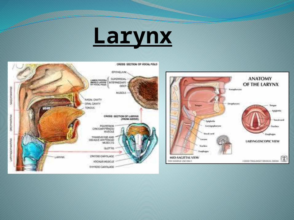
Larynx
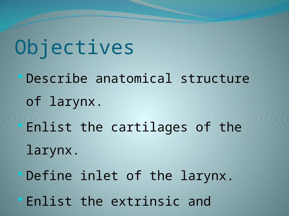
Objectives Describe anatomical structure of larynx.
Enlist the cartilages of the larynx.
Define inlet of the larynx.
Enlist the extrinsic and intrinsic muscles of the
larynx with their nerve supply and actions.
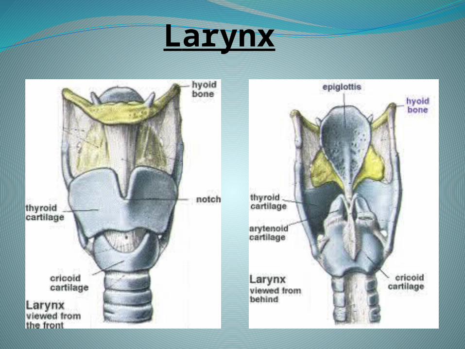
Larynx
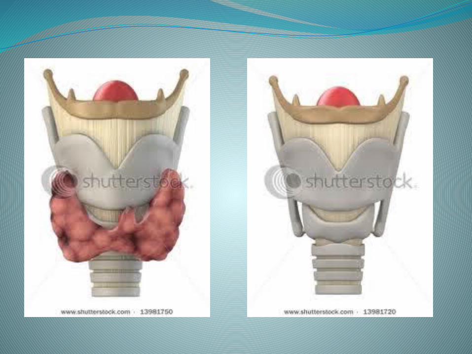
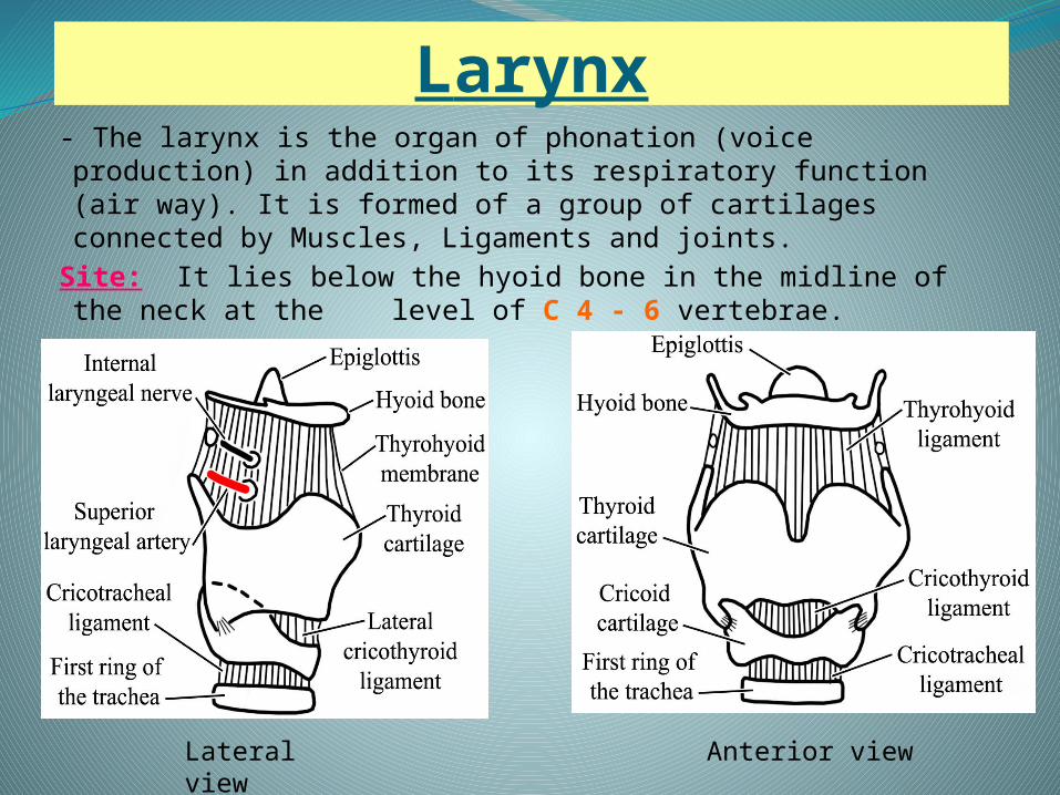
Larynx- The larynx is the organ of phonation (voice production) in
addition to its respiratory function (air way). It is formed of a group of cartilages connected by Muscles, Ligaments and joints.
Site: It lies below the hyoid bone in the midline of the neck at the level of C 4 - 6 vertebrae.
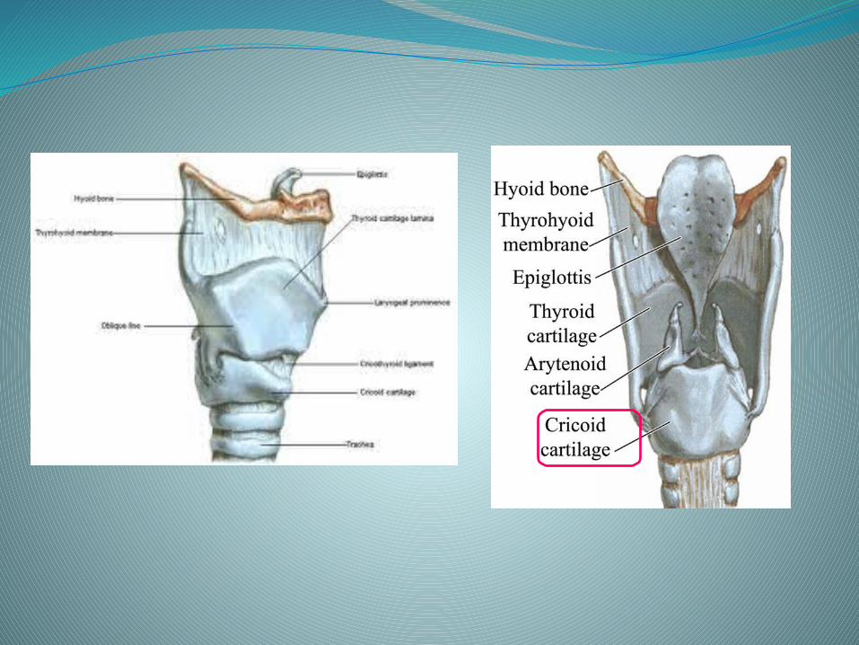
Lateral view Anterior view
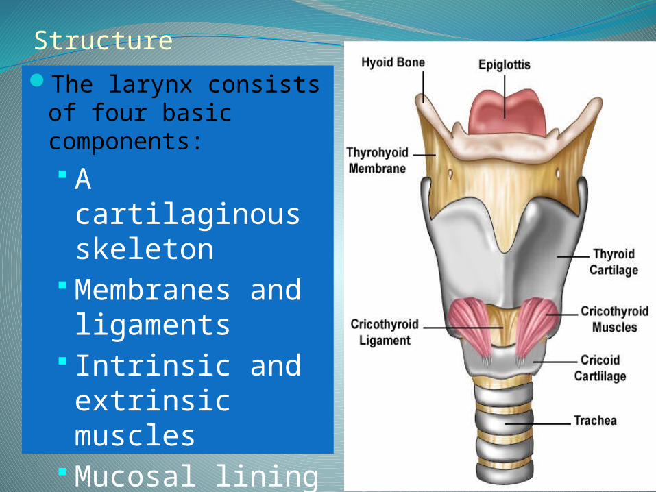
StructureThe larynx consists of
four basic components: A cartilaginous skeleton
Membranes and ligaments
Intrinsic and extrinsic muscles
Mucosal lining
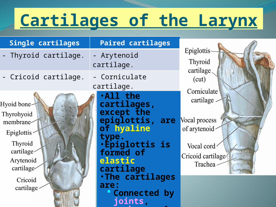
Cartilages of the LarynxSingle cartilages Paired cartilages
- Thyroid cartilage. - Arytenoid cartilage.
- Cricoid cartilage. - Corniculate cartilage.
- Epiglottic cartilage. - Cuneiform cartilage.
•All the cartilages, except the epiglottis, are of hyaline type. •Epiglottis is formed of elastic cartilage •The cartilages are:
Connected by joints, membranes & ligaments
Moved by muscles
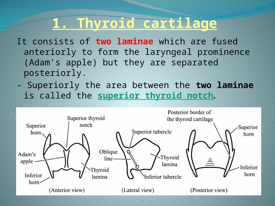
1. Thyroid cartilageIt consists of two laminae which are fused
anteriorly to form the laryngeal prominence (Adam’s apple) but they are separated posteriorly.
- Superiorly the area between the two laminae is called the superior thyroid notch.
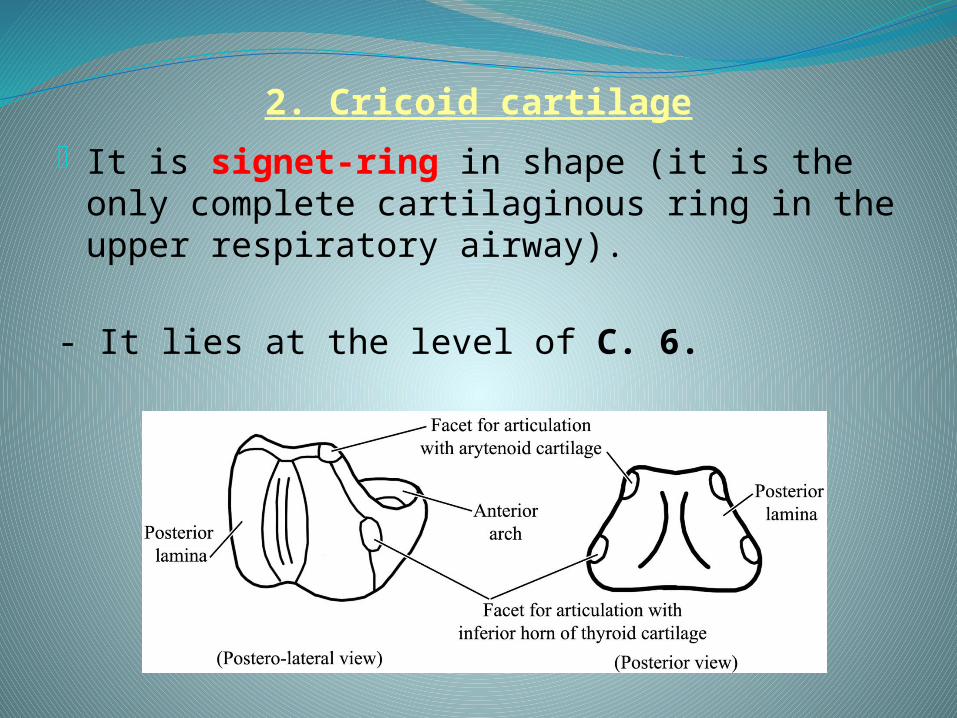
2. Cricoid cartilage- It is signet-ring in shape (it is the only
complete cartilaginous ring in the upper respiratory airway).
- It lies at the level of C. 6.
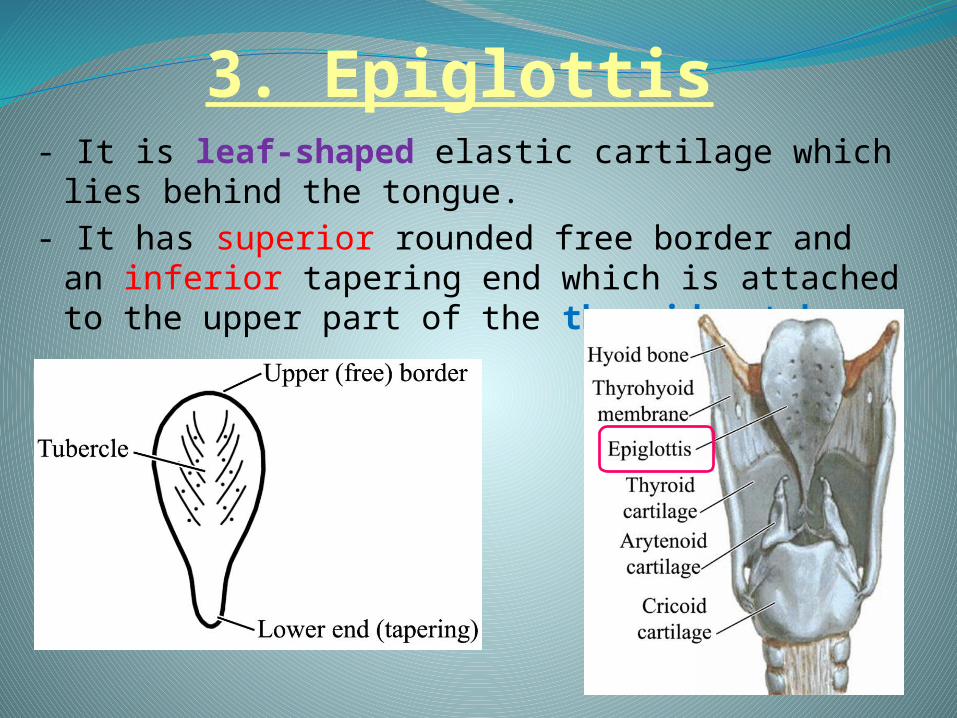
3. Epiglottis - It is leaf-shaped elastic cartilage which lies
behind the tongue.- It has superior rounded free border and an
inferior tapering end which is attached to the upper part of the thyroid notch.
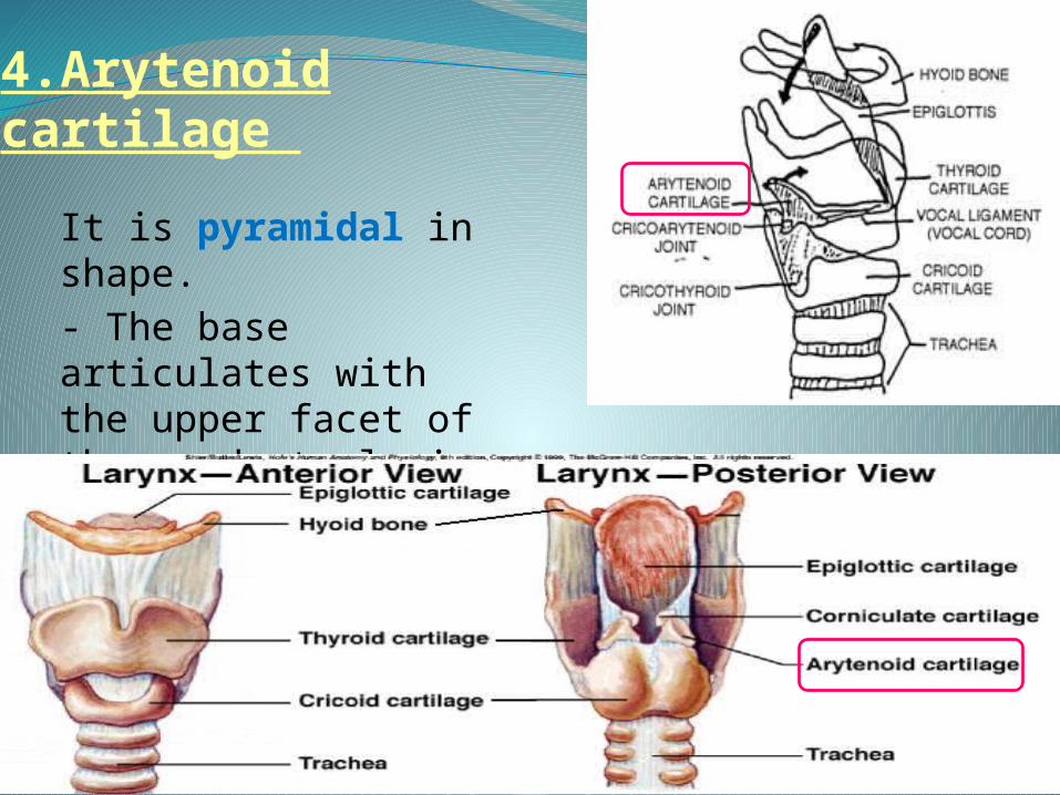
4.Arytenoid cartilage
It is pyramidal in shape. - The base articulates with the upper facet of the quadrate lamina of the cricoid cartilage.
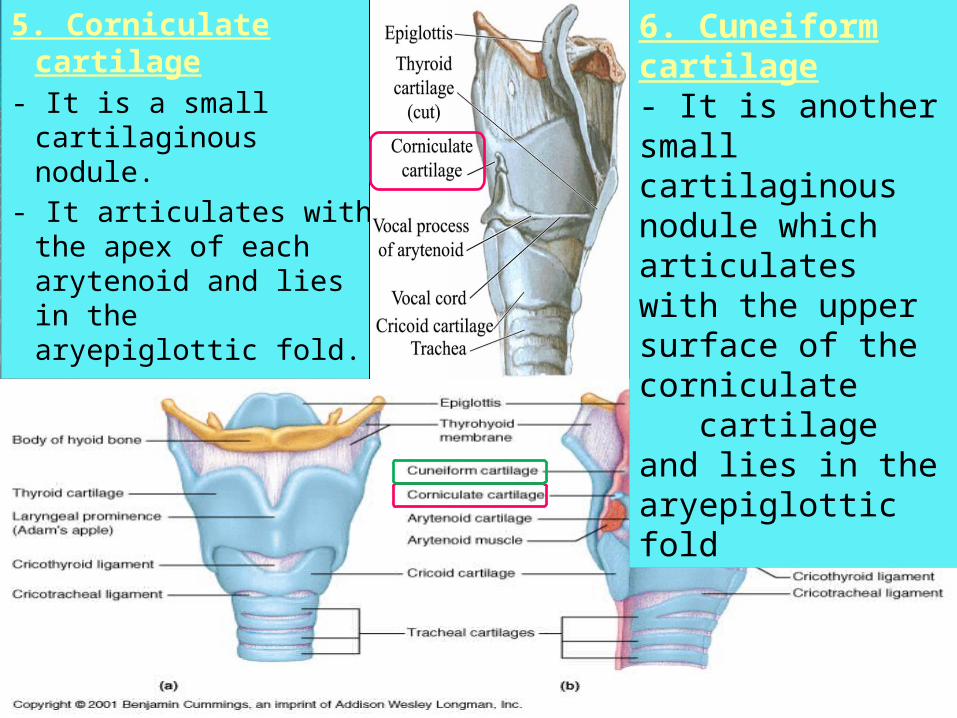
5. Corniculate cartilage
- It is a small cartilaginous nodule.
- It articulates with the apex of each arytenoid and lies in the aryepiglottic fold.
6. Cuneiform cartilage- It is another small cartilaginous nodule which articulates with the upper surface of the corniculate cartilage and lies in the aryepiglottic fold
Ligaments and Membranes
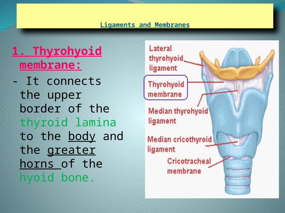
1. Thyrohyoid membrane:
- It connects the upper border of the thyroid lamina to the body and the greater horns of the hyoid bone.
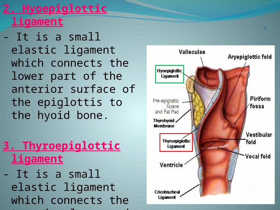
2. Hyoepiglottic ligament
- It is a small elastic ligament which connects the lower part of the anterior surface of the epiglottis to the hyoid bone.
3. Thyroepiglottic
ligament- It is a small elastic
ligament which connects the tapering lower end of the epiglottis to the inner
surface of the thyroid cartilage.
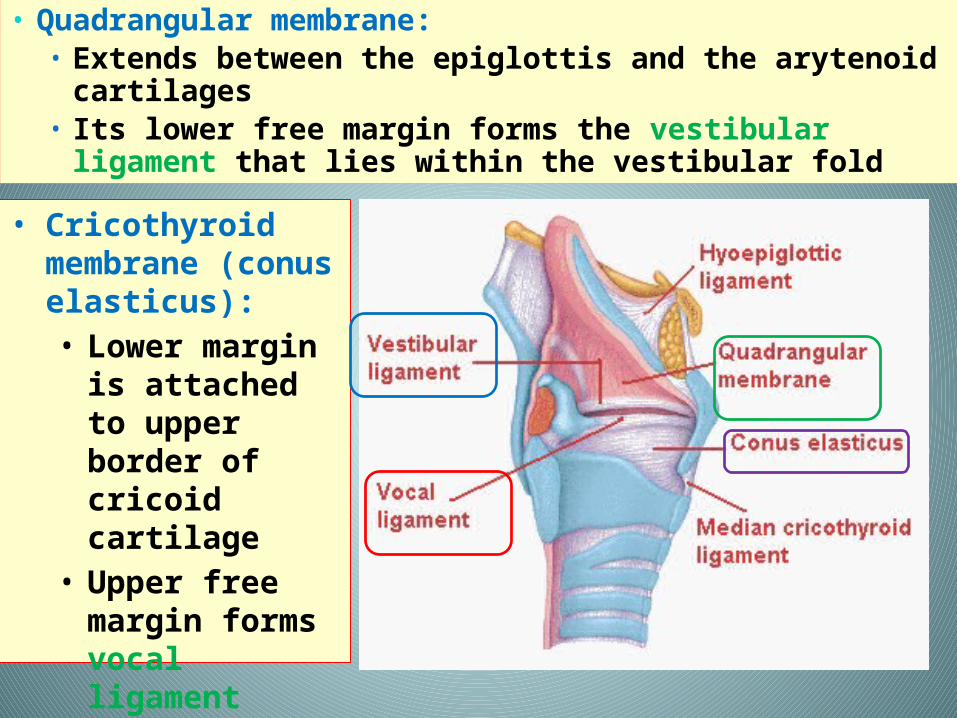
• Quadrangular membrane:• Extends between the epiglottis and the arytenoid
cartilages• Its lower free margin forms the vestibular
ligament that lies within the vestibular fold
• Cricothyroid membrane (conus elasticus): • Lower margin
is attached to upper border of cricoid cartilage
• Upper free margin forms vocal ligament
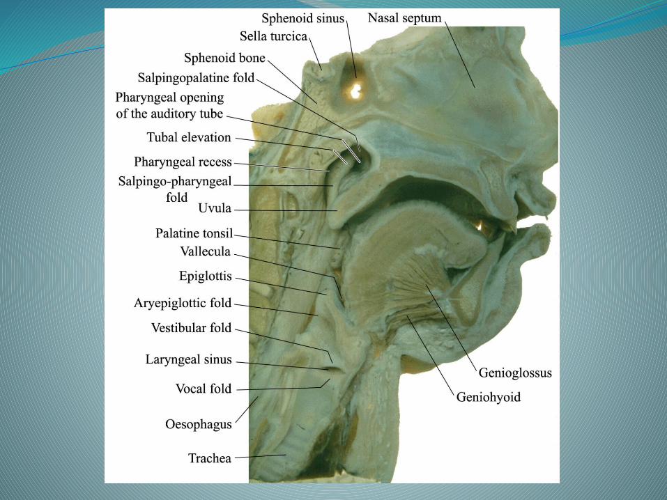
Cavity of larynx
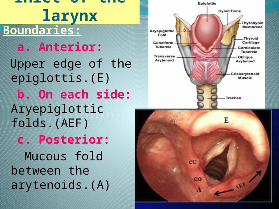
Inlet of the larynxBoundaries: a. Anterior: Upper edge of the
epiglottis.(E) b. On each side:
Aryepiglottic folds.(AEF)
c. Posterior: Mucous fold
between the arytenoids.(A)
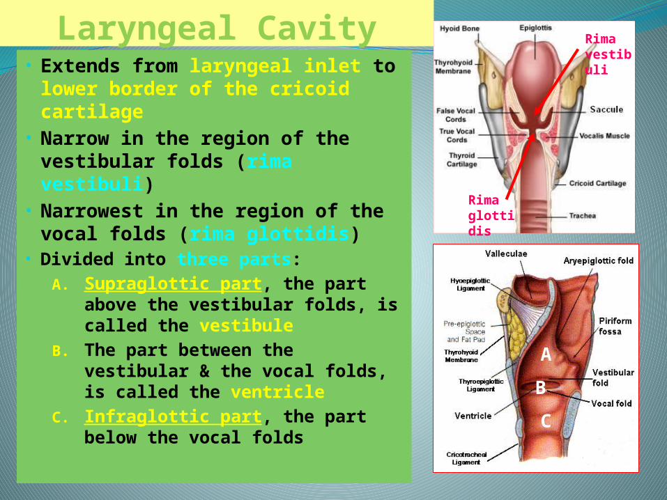
Laryngeal Cavity• Extends from laryngeal inlet to
lower border of the cricoid cartilage
• Narrow in the region of the vestibular folds (rima vestibuli)
• Narrowest in the region of the vocal folds (rima glottidis)
• Divided into three parts:A. Supraglottic part, the part
above the vestibular folds, is called the vestibule
B. The part between the vestibular & the vocal folds, is called the ventricle
C. Infraglottic part, the part below the vocal folds
Rima vestibuli
Rima glottidis
B
A
C
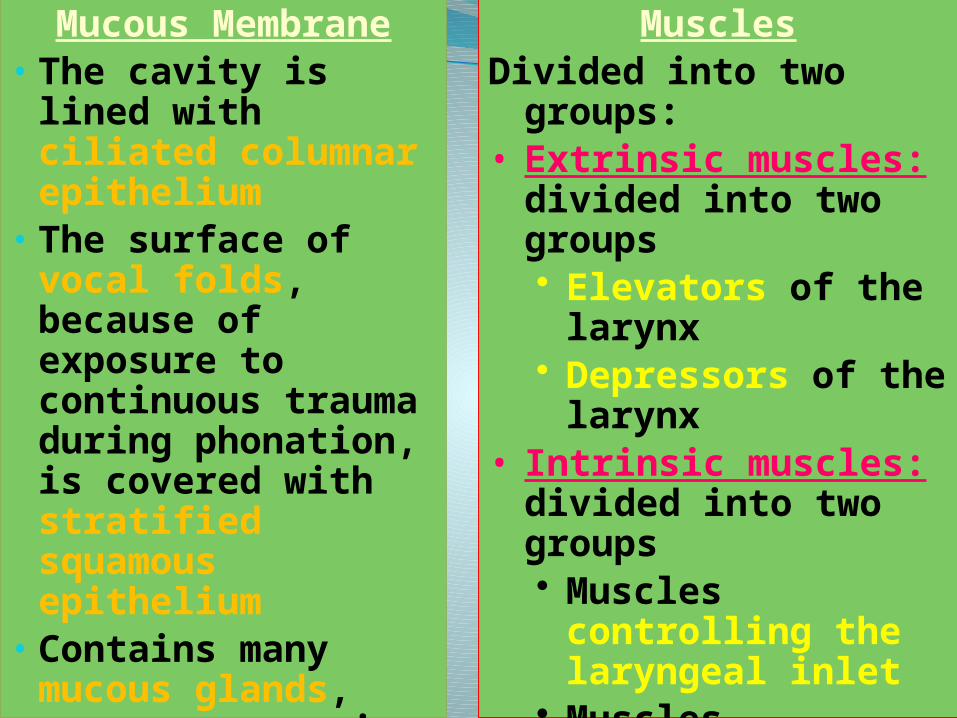
Mucous Membrane• The cavity is lined
with ciliated columnar epithelium
• The surface of vocal folds, because of exposure to continuous trauma during phonation, is covered with stratified squamous epithelium
• Contains many mucous glands, more numerous in the saccule (for lubrication of vocal folds)
MusclesDivided into two
groups:• Extrinsic muscles:
divided into two groups• Elevators of the
larynx• Depressors of the
larynx• Intrinsic muscles:
divided into two groups• Muscles
controlling the laryngeal inlet
• Muscles controlling the movements of the vocal cords
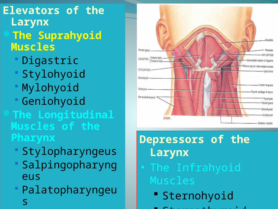
Depressors of the Larynx
• The Infrahyoid Muscles Sternohyoid Sternothyroid Omohyoid
Elevators of the Larynx
The Suprahyoid Muscles Digastric Stylohyoid Mylohyoid Geniohyoid
The Longitudinal Muscles of the Pharynx Stylopharyngeus Salpingopharynge
us Palatopharyngeus
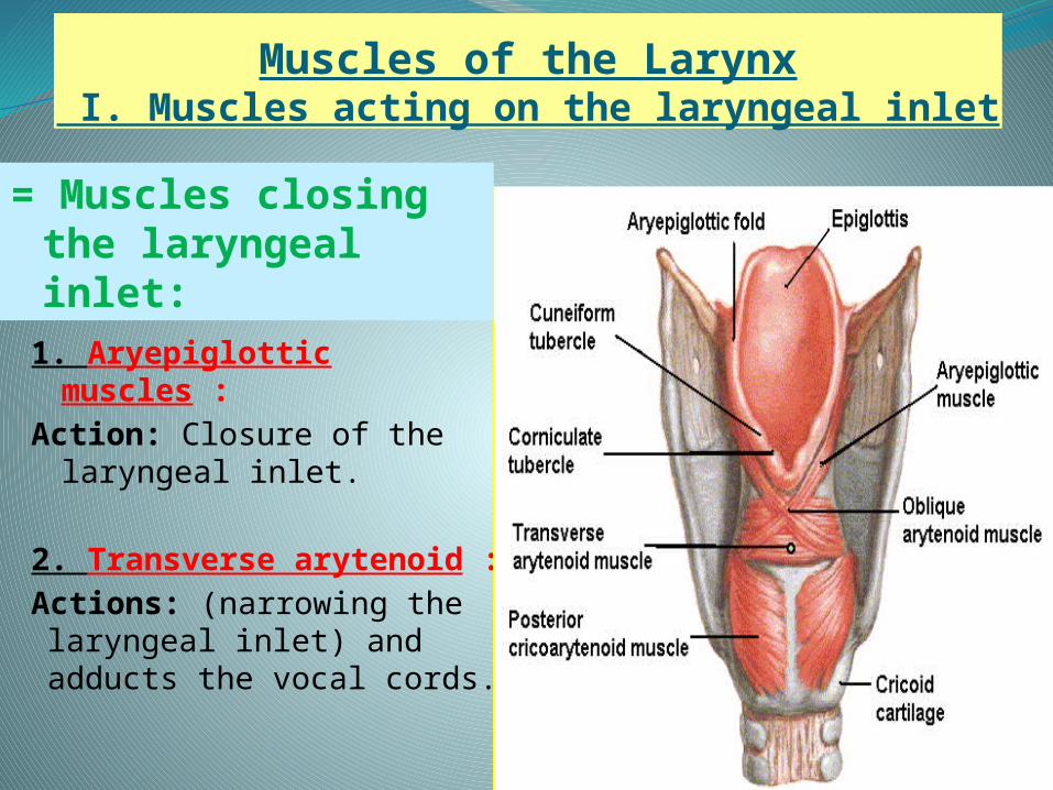
Muscles of the Larynx I. Muscles acting on the laryngeal inlet
1. Aryepiglottic muscles :
Action: Closure of the laryngeal inlet.
2. Transverse arytenoid :
Actions: (narrowing the laryngeal inlet) and adducts the vocal cords.
= Muscles closing the laryngeal inlet:
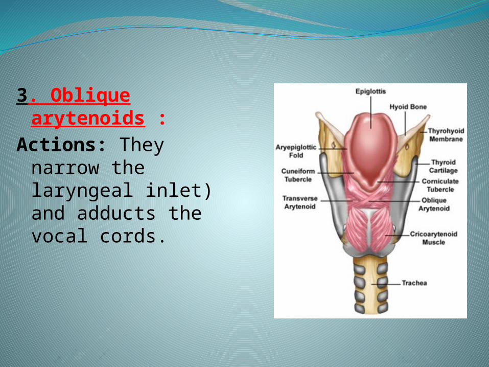
3. Oblique arytenoids :
Actions: They narrow the laryngeal inlet) and adducts the vocal cords.
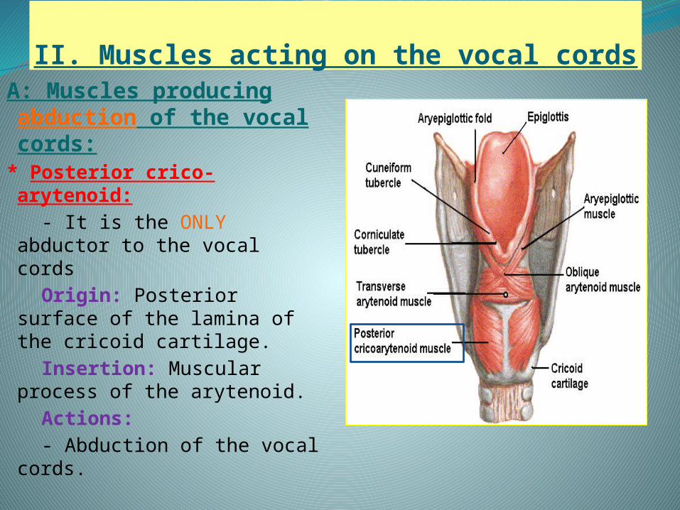
II. Muscles acting on the vocal cordsA: Muscles producing
abduction of the vocal cords:
* Posterior crico-arytenoid:
- It is the ONLY abductor to the vocal cords
Origin: Posterior surface of the lamina of the cricoid cartilage.
Insertion: Muscular process of the arytenoid.
Actions: - Abduction of the vocal
cords.
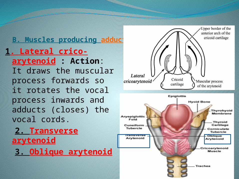
B. Muscles producing adduction of the vocal cords
1. Lateral crico-arytenoid : Action: It draws the muscular process forwards so it rotates the vocal process inwards and adducts (closes) the vocal cords.
2. Transverse arytenoid
3. Oblique arytenoid
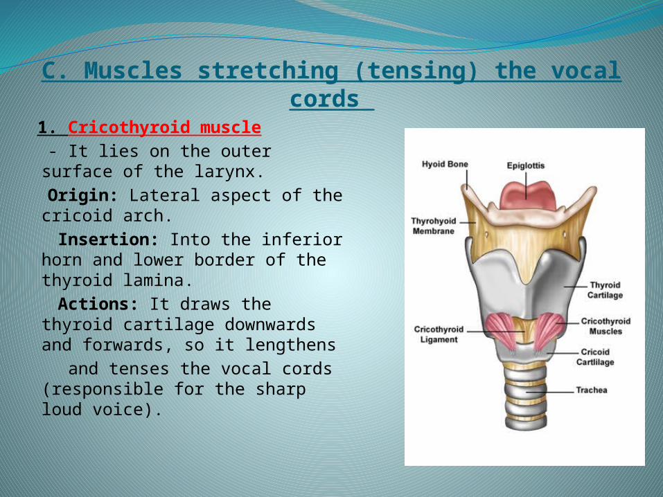
C. Muscles stretching (tensing) the vocal cords 1. Cricothyroid muscle - It lies on the outer surface of
the larynx. Origin: Lateral aspect of the
cricoid arch. Insertion: Into the inferior
horn and lower border of the thyroid lamina.
Actions: It draws the thyroid cartilage downwards and forwards, so it lengthens
and tenses the vocal cords (responsible for the sharp loud voice).
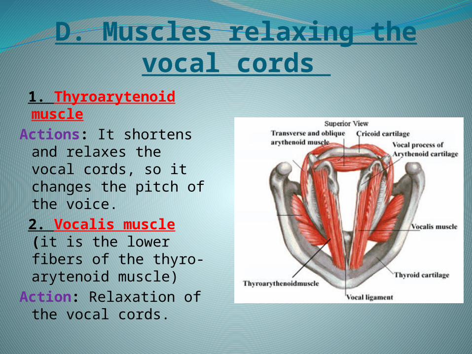
D. Muscles relaxing the vocal cords 1. Thyroarytenoid
muscleActions: It shortens
and relaxes the vocal cords, so it changes the pitch of the voice.
2. Vocalis muscle (it is the lower fibers of the thyro-arytenoid muscle)
Action: Relaxation of the vocal cords.
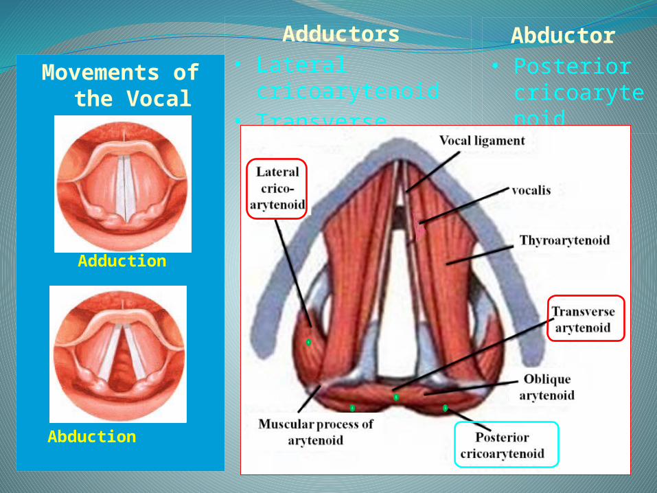
Movements of the Vocal
Cords
Adductio
n
Abduction
Abductor • Posterior
cricoarytenoid
Adductors • Lateral cricoarytenoid• Transverse arytenoid
Nerve supplyA. Motor supply- All intrinsic muscles of the larynx are supplied
by the “recurrent laryngeal nerve” EXCEPT cricothyroid which is supplied by
the external laryngeal nerve.B. Sensory supply- Above the vocal cords ----------------- Internal
laryngeal nerve (from vagus N).- Below the vocal cords ----------------- Recurrent
laryngeal nerve (from vagus N).
Blood supply
Blood supply:A. Arterial supply:1. Above the vocal cords: Superior laryngeal
artery (from the superior thyroid artery). 2. Below the vocal cords: Inferior laryngeal artery
(from the inferior thyroid artery). B. Venous drainage:- It drains its venous blood into the corresponding
superior and inferior thyroid veins respectively.

Thank You