
Laparoscopy for Impalpable Testes - InTech -...
12
7 Laparoscopy for Impalpable Testes Ahmed Khairi Pediatric Surgery Unit, Department of Surgery, Alexandria Faculty of Medicine, Alexandria Egypt 1. Introduction 1.1 Embryology Until the 7-8 weeks gestation embryo (sexual differentiation time), the testis and ovary have the same position. Afterwards, the testis remains close to the future inguinal canal and the ovary moves away from the groin. Both gonads have upper suspensory ligament and lower gubernaculum. The cranial suspensory ligament persists in females and regresses in boys. This is believed to be due to androgenic hormonal control. The gubernaculum enlarges in boys. This is controlled by a non-androgenic hormone mostly mullerian inhibiting substance (MIS). Condensation of mesenchyme around the gubernaculum forms the inguinal canal musculature. A diverticulum of peritoneum grows caudally through the mesenchyme of the gubernaculum to form the processus vaginalis. The caudal part of the gubernaculum remains solid and the proximal part is divided into a central part (attached to the epididymis) and a parietal part (the cremasteric muscle) (Heyns 1987). At the third trimenster, the caudal part of the gubernaculums migrates through the inguinal abdominal wall and across the pubic region to the scrotum. The processus vaginalis grows inside the gubernaculum, with the testis leaving the abdominal cavity within it (Backhouse,1982). Scrotal migration of the testis and gubernaculums is under androgenic control. This is believed to be through the release of calcitonin gene related peptide (CGRP) by the genitofemoral nerve that control gubenaculum migration (Grocock ,1988). Intra-abdominal pressure, through the patent processus vaginalis, is a probable additional factor. This process is complete by the 35 weeks (Huston, 1996). 1.2 Incidence Cryptorchidism is the most common anomaly of the male genital system. Around 4% of infants at birth have undescended testes. At 1 year old, the incidence is around 1%. 20% of undescended testes are impalpable. Blind inguinal exploration of the impalpable undescended testes is unlikely successful. 1.3 Etiology Multifactorial and many possible causes are raised: - Defects in the migratory mechanism itself. www.intechopen.com
Transcript of Laparoscopy for Impalpable Testes - InTech -...

7
Laparoscopy for Impalpable Testes
Ahmed Khairi Pediatric Surgery Unit, Department of Surgery,
Alexandria Faculty of Medicine, Alexandria Egypt
1. Introduction
1.1 Embryology
Until the 7-8 weeks gestation embryo (sexual differentiation time), the testis and ovary have the same position. Afterwards, the testis remains close to the future inguinal canal and the ovary moves away from the groin. Both gonads have upper suspensory ligament and lower gubernaculum. The cranial suspensory ligament persists in females and regresses in boys. This is believed to be due to androgenic hormonal control. The gubernaculum enlarges in boys. This is controlled by a non-androgenic hormone mostly mullerian inhibiting substance (MIS). Condensation of mesenchyme around the gubernaculum forms the inguinal canal musculature. A diverticulum of peritoneum grows caudally through the mesenchyme of the gubernaculum to form the processus vaginalis. The caudal part of the gubernaculum remains solid and the proximal part is divided into a central part (attached to the epididymis) and a parietal part (the cremasteric muscle) (Heyns 1987). At the third trimenster, the caudal part of the gubernaculums migrates through the inguinal abdominal wall and across the pubic region to the scrotum. The processus vaginalis grows inside the gubernaculum, with the testis leaving the abdominal cavity within it (Backhouse,1982). Scrotal migration of the testis and gubernaculums is under androgenic control. This is believed to be through the release of calcitonin gene related peptide (CGRP) by the genitofemoral nerve that control gubenaculum migration (Grocock ,1988). Intra-abdominal pressure, through the patent processus vaginalis, is a probable additional factor. This process is complete by the 35 weeks (Huston, 1996).
1.2 Incidence
Cryptorchidism is the most common anomaly of the male genital system. Around 4% of infants at birth have undescended testes. At 1 year old, the incidence is around 1%. 20% of undescended testes are impalpable. Blind inguinal exploration of the impalpable undescended testes is unlikely successful.
1.3 Etiology
Multifactorial and many possible causes are raised: - Defects in the migratory mechanism itself.
www.intechopen.com

Advanced Laparoscopy
96
- Genitofemoral nerve defects due to lack of gonadotrophin secretion by the pituitary or
the placenta.
- Abnormal location of the genitofemoral nerve with subsequent wrong site migration
(outline the line of descent).
- MIS or testosterone deficiency.
- Some inherited syndromes are associated with undescended testes:
microcephaly: possibly due to pituitary hormone or gonadotrophin deficiency.
Prune-belly syndrome: possibly due to mesodermal defect and possibly decreased
abdominal pressure.
Abdominal wall defects: gastroschisis, omphalocele, bladder extrophy
(Hadziselimovic, 1987).
Neural tube defects as myelomeningocele due to defect of the genitofemoral nerve
motor nucleus and abdominal wall paralysis.
2. Laparoscopy for impalpable testis
2.1 History
- 1976: Cortesi et al published the first case managed by laparoscopy.
- 1991: Bloom performed the first stage of Fowler-Stephens by laparoscopy.
- 1992: Jordan performed single stage laparoscopic orchidopexy.
- 1994: Caldamone reported second stage Fowler-Stephens orchidopexy laparoscopically.
2.2 Differential diagnosis
Impalpable testis may be one of the following:
1. In the inguinal canal but can not be felt (excess fat, small testis)
2. Absent testis: either due to agenesis or intrauterine torsion (vanishing testis).
3. Intra-abdominal testis:
- Peeping: close to the deep inguinal ring and can be manipulated into the canal.
- Low abdominal testis.
- High abdominal testis.
2.3 Age of the procedure
As parenchymal damage is clear under electron microscope at 1 year old and by light
microscope at 18 months, the optimum age for surgery is before 1 year of age.
2.4 Preoperative preparation
Informed consent including the possibility of converting to laparotomy, staged orchidopexy,
orchiectomy, contraletral scrotal orchidopexy.
2.5 Technique
Under general endotracheal anesthesia. Re-examine the boy under anesthesia to confirm the diagnosis of non-palpable testis. Make sure that the stomach is empty. Nasogastric tube might be inserted in small infants. Make sure that the urinary bladder is empty. If full under anesthesia; apply suprapubic pressure (Crede' manuveur) or insert a small catheter. Prep and drape the abdomen and the scrotum
www.intechopen.com

Laparoscopy for Impalpable Testes
97
2.6 Position
The child is supine. The surgeon in the opposite side of the impalpable testis, the monitor is opposite to the surgeon, the assistant on the left hand side of the surgeon and the scrub nurse to the right. The patient is rotated to the contra-lateral side with mild Trendelenberg position on starting intervention.
2.7 Port - placement
For diagnostic laparoscopy, an umbilical port, by either the closed or open technique, is inserted. If the decision is taken for intervention; insert 3 mm trocar at the side of the impalpable testis at the midclavicular line at the umbilical level, and 5 mm trocar at the contra-lateral midclavicular line at a slightly lower level.
2.8 Pneumoperitoneum
Is established at 10-12 mmHg. This can be tolerated even by small infants. Pressure is up to 15 mmHg in older boys.
Fig. 1. Flow chart for the actions that should be taken depending on the findings on laparoscopy.
Impalpable testis
Diagnostic laparoscopy
Blind-ending vas No testis
Vessels entering the deep ring No testis seen
Testis seen
No further exploration
Inguinal exploration
Viable testis orchidopexy
Testicular remnantexcision
Atrophic Teenager Excision
viable
Enough cord lengthorchidopexy
Length not enough Clip the vessels
2nd stage 6 m later
www.intechopen.com

Advanced Laparoscopy
98
2.9 Diagnostic laparoscopy
Using OO telescope, start exploration by looking for the testis at the possible sites. This could be deep to the internal inguinal ring, along its line of descent, or behind the urinary bladder. Plunging testis is detected by the presence of a patent processus vaginalis and by trying to manipulate the testis in and out the deep ring. Sometimes, the colon has to be retracted medially in order to detect a high testis. Further management is depending on what you find out on laparoscopy. See the flow chart for different possible scenarios and management:
2.10 Laparoscopic orchidopexy (Figures 2-15)
The colon is mobilized medially. Dissect the gubernaculum out of the internal ring and divide as distally as possible. Beware of long-loup vas around the gubernaculum. Dissect the peritoneum around the internal ring. Proceed with the dissection around the spermatic vessels high up. Avoid dissection at the area of meeting of the vas with the vessels. The small vessels anastomoses in this area are valuable testicular supply in case in need for clipping the gonadal vessels because of short pedicle (Fowler-Stephens technique). Dissect the bands of adhesions as needed to gain enough length. This could be as high as the origin of the gonadal vessels. Test the gained length by pulling the testis to the contra-lateral internal ring. If this is possible, then the length is enough and it is likely to reach the scrotum without much tension. The testis could be passed to the scrotum in one of three main routes: 1. Through the normal native internal ring through the inguinal canal if the cord length is good. 2. Medial to the inferior epigastric vessels (lateral umbilical fold): in case of short cord length (Prentiss maneuver). 3. Medial to the medial umbilical fold: in case of very short cord length. Prepare a subdartos pouch in the hemiscrotum, push an instrument (long artery forceps or 5-mm trocar) through the scrotum into the abdominal cavity. Some authors pass an instrument antegradely i,e from the abdominal cavity, through the internal ring, guided to the scrotum by palpation over the pubic area and the pass the trocar sheath over the instrument into the abdominal cavity. The testis is grasped from the area of gubernaculums attachment and pulled into the scrotum Deflation of the pneumoperitoneum is helpful in that step. The testis is secured in the subdartos pouch with fine stitches. The pathway might need dilatation in case of large gonads (Jordan et al., 1992). Re-inspect inside again through the laparoscope after re-inflating the peritoneum, if has been deflated, for any residual bands that need be dissected and to ensure hemostasis. The peritoneum over the area of dissection at the internal ring might be closed with stitches or endoclips. Some authors leave this area without closure with no reported complications related to this attitude.
2.11 Staged orchidopexy
If a two-stage procedure is planned, then ligation and division of the testicular vessels proximal or cephalad to the testis is required, followed by a 6-9 month period of time to allow augmentation of the secondary collateral vasculature to the testis. This ligation and division can be performed either with endoscopic cautery, endoscopic laser (KTP) or endoscopic clips (Holcomb, 1994).
www.intechopen.com

Laparoscopy for Impalpable Testes
99
The peritoneum overlying the testicular vessels approximately 2 cm cephalad to the testicle is incised. The testicular vessels are then either coagulated with the cautery or laser or secured with the 5 mm endoscopic clip applier and divided. If the two-staged approach is used, nothing further is performed at this time.
Fig. 2. Left abdominal testis. Note the vas (dotted arrow), spermatic vessels (thick arrow), gubernaculum out of the internal inguinal ring (thin arrow).
Fig. 3. Gubernaculum is pulled out of the internal ring (arrow).
www.intechopen.com
Advanced Laparoscopy
100
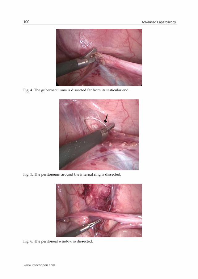
Fig. 4. The gubernaculums is dissected far from its testicular end.
Fig. 5. The peritoneum around the internal ring is dissected.
Fig. 6. The peritoneal window is dissected.
www.intechopen.com

Laparoscopy for Impalpable Testes
101
Fig. 7. The vascular pedicle after being dissected high up to its origin.
Fig. 8. Testing the cord length by pulling the testis towards the contra-lateral internal ring.
Fig. 9. The scrotal crease incision.
www.intechopen.com

Advanced Laparoscopy
102
Fig. 10. The subdartos pouch is developed.
Fig. 11. 5 mm trocar is passed through the scrotal incision to inside the peritoneal cavity. This might be guided over an instrument passed from the peritoneal cavity outwards.
Fig. 12. The port as it is passed inside. Notice the medial relation to the inferior epigastric vessels. This rout is useful in case of relatively short cord.
www.intechopen.com

Laparoscopy for Impalpable Testes
103
Fig. 13. The testis is grasped from the gubernaculum by an atraumatic instrument and pulled to the scrotum; not necessarily inside the sheath but rather following its tract.
Fig. 14. The testis after being pulled and positioned in the subdartos pouch.
Fig. 15. The testis is secured in the scrotum without tension.
www.intechopen.com

Advanced Laparoscopy
104
3. Conclusions
Laparoscopy is the gold standard for management of impalpable testes. Its diagnostic as well interventional potentials make it the routine technique for dealing with that problem.
4. References
Backhouse KM: Embryology of testicular descent and maldescent,Urol Clin North Am 9:315,1982.
Bloom DA .Two-step orchiopexy with pelviscopic clip ligation of spermatic vessels. J Urol 145:1030-33, 1991.
Caldamone AA & Amaral JF. Laparoscopic stage 2 FowlerStephens orchiopexy. J Urol 152:1253, 1994.
Cortesi N et al. Diagnosis of bilateral abdominal cryptorchidism by laparoscopy. Endoscopy 8:33, 1976.
Diamond DA & Caldamone AA. The value of laparoscopy for 106 impalpable testes relative to clinical presentation. J Urol 148:632-34, 1992.
Docimo SG et al. Laparoscopic orchiopexy for the high palpable undescneded testis: preliminary experience. J Urol 154: 1513-15, 1995.
Grocock CA, Charlton HM & Pike MC: Role of the fetal pituitary in cryporchidism induced by exogenous maternal oesterogen during pregnancy in mice. J Reprod Fertil 83:295,1988.
Hadziselimovic F et al. Omphalocele, cryptorchidism and brain malformations, J Pediatr Surg 22:854, 1987.
Heyns CF: The gubernaculum during testicular descent in the human fetus. J Anat 153:93, 1987
Holcomb GW III, Brock JW, Neblett WW et al. Laparoscopy for the nonpalpable testis. Am Surg
1994; 60:143-147. Huston JM: Normal and abnormal testicular distention. In Stephens FD, Smith ED, Huston
JM, editors: Congenital anomalies of urinary and genital tracts, Oxford, 1996, Isis Medical.
Jordan GH, Robey EL, Winslow BH. Lapendoscopic surgical management of the abdominal/transinguinal undescended testicle. J Endourolo 6:159-63, 1992.
www.intechopen.com

Advanced LaparoscopyEdited by Prof. Ali Shamsa
ISBN 978-953-307-674-4Hard cover, 190 pagesPublisher InTechPublished online 30, September, 2011Published in print edition September, 2011
InTech EuropeUniversity Campus STeP Ri Slavka Krautzeka 83/A 51000 Rijeka, Croatia Phone: +385 (51) 770 447 Fax: +385 (51) 686 166www.intechopen.com
InTech ChinaUnit 405, Office Block, Hotel Equatorial Shanghai No.65, Yan An Road (West), Shanghai, 200040, China
Phone: +86-21-62489820 Fax: +86-21-62489821
The present book, published by InTech, has been written by a number of highly outstanding authors from allover the world. Every author provides information concerning treatment of different diseases based on his orher knowledge, experience and skills. The chapters are very useful and innovative. This book is not merelydevoted to urology sciences. There are also clear results and conclusions on the treatment of many diseases,for example well-differentiated papillary mesothelioma. We should not forget nor neglect that laparoscopy is inuse more extensively than before, and in the future new subjects such as use of laparascopy in treatment ofkidney cysts, simple nephrectomy, pyeloplasty, donor nephrectomy and even robotic laparoscopy will beresearched further.
How to referenceIn order to correctly reference this scholarly work, feel free to copy and paste the following:
Ahmed Khairi (2011). Laparoscopy for Impalpable Testes, Advanced Laparoscopy, Prof. Ali Shamsa (Ed.),ISBN: 978-953-307-674-4, InTech, Available from: http://www.intechopen.com/books/advanced-laparoscopy/laparoscopy-for-impalpable-testes

© 2011 The Author(s). Licensee IntechOpen. This chapter is distributedunder the terms of the Creative Commons Attribution-NonCommercial-ShareAlike-3.0 License, which permits use, distribution and reproduction fornon-commercial purposes, provided the original is properly cited andderivative works building on this content are distributed under the samelicense.


![Intech Prostate [DD219]](https://static.fdocuments.net/doc/165x107/5695d0811a28ab9b0292ba48/intech-prostate-dd219.jpg)