Laparoscopic management of fibroids and tissue extraction ... · the fibroid – find the right...
39
Laparoscopic management of fibroids and tissue extraction options Jon I Einarsson MD PhD MPH Director of MIGS Brigham and Women’s Hospital Associate Professor of Obstetrics and Gynecology Harvard Medical School
Transcript of Laparoscopic management of fibroids and tissue extraction ... · the fibroid – find the right...

Laparoscopic management of fibroids and tissue extraction options
Jon I Einarsson MD PhD MPHDirector of MIGSBrigham and Women’s HospitalAssociate Professor of Obstetrics and GynecologyHarvard Medical School

DisclosuresI have no financial relationships with a commercialentity producing health-care related productsand/or services

Myomectomy
Surgical option of choice for women who want to retain their options for future fertility
Laparoscopic myomectomy vs abdominal myomectomy Quicker recovery Shorter hospital stay Decreased blood loss Decreased adhesion formation (30 vs 90%) Comparable pregnancy rate
Bulletti et al. J Am Assoc Gynecol Laparosc. 1996;3:533–536Seracchioli et al. Hum Reprod. 2000;15:2663–2668Palomba et al. Fertil Steril. 2007 Oct;88(4):933-41

Our data – LM vs. RALM
289 women – 02/07-09/09 LM (n=115) RALM (n=174) p
Operative time (min) 118.3 195.1 <.0001
EBL (ml) 85.9 110.0 0.04
Conversions to laparotomy 0 0 NS
Weight of fibroids (g) 201 (1-1473) 159 (8-780) NS
Median n of fibroids 2 (1-21) 3 (1-16) NS
Largest fibroid (cm) 7.5 (2.2-16.5) 7.3(3.1-13.8) NS
Blood transfusions n(%) 1(0.9) 10(5.7) NS
Hospital stay >1 day n(%) 4(3.5) 29(16.9) OR 5.73

Laparoscopic/robotic myomectomy– the standard approach
We looked at all myomectomies at Brigham and Women’s Hospital from 2009-2012
966 patients were identified
There were 731 laparoscopic/robotic cases (76%) and 235 (24%) abdominal cases
Conversion to laparotomy was required in 8 cases (1.09%) mean number in converted cases, 9.75 vs 3.48, p = .003 mean weight in converted cases, 667.9 vs 259.25 g, p = .015
Conversion was significantly associated with a uterine weight over 500 grams
J Minim Invasive Gynecol. 2016 Mar-Apr;23(3):352-7

More details from 2009-12966 women – 2009-2012
AM (n=237) LM (n=385) RALM (n=340) p
Operative time (min) 135 122 190 <.0001
EBL(ml) 185 87 79 <.0001
Any post op complication
12.2% 5.9% 9.4% 0.02
Median n of fibroidsremoved
8.5 2 2 <.0001
Mean total weight of resected fibroids (g)
593.9 302.8 218.4 <.0001
Entered cavity 46% 13.2% 27.1% <.0001
Length of stay (median (d))
2.0 0 1 <.0001

Brief description of our technique Two parallel trocars on surgeon side
Faciliates suturing – especially in the setting of a horizontal hysterotomy
Inject dilute vasopressin subserosally – avoid using more than 10 units every 30 minutes
We like to use large volumes, 20 units of vasopressin in 400 ml of saline – we inject 200 ml (10 units) at a time
RCT just completed comparing blood loss in using 200 vs 60 ml of diluted vasopressin solution No statistically significant difference in blood loss

Step 1- Vasopressin injection

Step 2 – Hysterotomy Carry the incision into
the fibroid– find the right plane We prefer the Harmonic
due to minimal lateral thermal spread
A horizontal incision is preferred for suturing with two ipsilateral trocars
Pick whatever incision direction that works best in that scenario
Avoid fallopian tubes and major vessels

Step 3 – Fibroid extraction Rock and Roll
Needs quite a bit of force
Avoid entering the cavity if possiblewill do this deliberately in
women who have completed their childbearing
easy to pluck out the submucosal fibroids this way

Hysterotomy closure - video
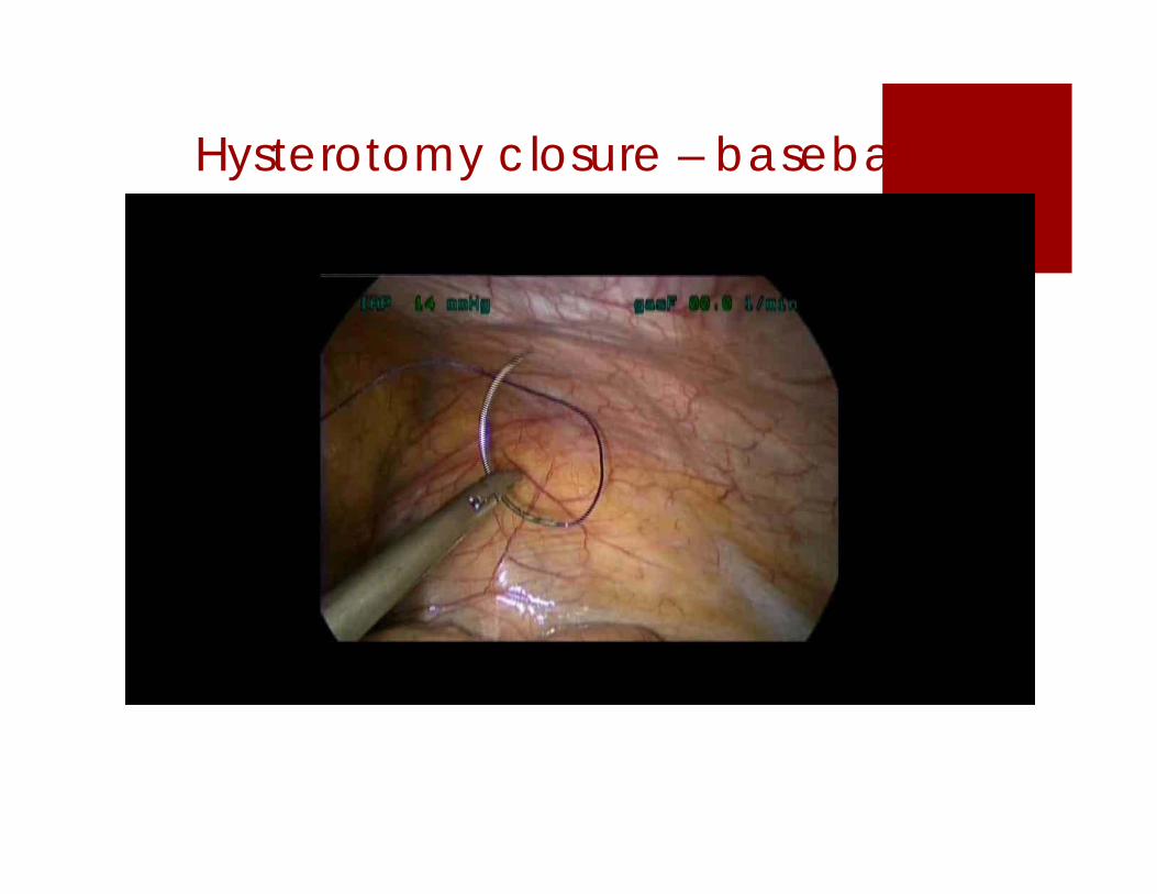
Hysterotomy closure – baseball

Tissue extraction
This has changed drastically in the last several months
In short, we do not use electronic morcellators anymore
ALL tissue extraction methods are contained, whether through the vagina, umbilicus or a minilaparotomy

Tissue extraction More abdominal hysterectomies are being
performed these days
A recent study of 18,299 revealed; utilization of LH had decreased by 4.1% 8 months
after the FDA safety communication Major surgical complications (excluding transfusions)
significantly increased Rate of hospital readmissions within 30 days
significantly increased
Harris et al. Am J Obstet Gynecol. 2016;214(1):98.e1-98

Potentially worse survival with morcellation
Park et al. 2011: 56 consecutive patients treated for early stage uterine leiomyosarcoma at a South Korean referral hospital from 1989-2011
5 year disease free interval 50% vs. 79% morcellated vs intact
5 year overall survival 46% vs. 73% morcellated vs intact Gynecol Oncol. 2011;122(2):255–259.

Park study – morcellation group
Procedures performed (n=25) LAVH (18) VH (1) Myomectomy via minilaparotomy (5) Laparoscopic myomectomy (1)
What this study is showing is that that ANY KIND of uncontained morcellation (tissue disruption) of a LMS may worsen prognosis

LMS 10-year Survival
Tumor Injury (A)
No Injury (B)
62% mortality 38% mortality
LMS treated from 1969-2005
TAH (21) vs. “tumor injury”(16) Abdominal myomectomy (4) LH with morcellator knife (2) Hysteroscopic myomectomy
(4) Subtotal AH (4) TAH, injury with sharp
instrument (2)
Age range 30-74 (mean 50)
Again, this study is not evaluating laparoscopic morcellation
Perri T, Int J Gynecol Cancer 2009;19:257-60
A
B

Recent study from Spain 37 cases of sarcoma in 4014 patients undergoing surgery for presumed
fibroids (0.9%)
Increased disease free survival in laparotomy group vs. morcellation group (70.3 vs. 10.4 months, p=0.18)
Median DFS was 6.3 months in laparoscopic morcellation cases, 11.9 months in vaginal fragmentation cases and 149.9 months in nonmorcellated cases (p<.002)
Median age was 40 (range 26-83 years)
Cusido M et al. JMIG 2015 22, 1068-1074

Identifying at risk patients
Age: Peak age for LMS is at approx age 50 with a wide age range
Clinical Presentation:
- Rapidly enlarging uterine mass, severe pain and heavy bleeding NOT reliable predictors
- New or enlarging uterine mass in postmenopausal woman warrants evaluation

Identifying at risk patients
Imaging;
MRI seems to be the best bet, but far from perfect
2 studiesGoto et al – MRI plus LDH Sato et al – MRI with DWI (diffusion weighted
imaging)

Goto et alProspective study of 227 patients diagnosed with
smooth muscle tumor From 1990-2000 10 patients with LMS (4.4% incidence?) 130 patients with degenerated fibroid 87 with regular leiomyoma
Combination of contrast enhanced MRI and LDH (total plus LDH-3) had a sensitivity and specificity of 100%
However, if the incidence of sarcoma is 1/500, the PPV is only 3%
Goto et al. Int J Gynecol Cancer 2002, 12, 354-61

Sato et al. 10 lesions from 5 patients with LMS
83 uterine fibroids in 76 patients
Classified into low risk and high risk based on findings from DWI
Sensitivity 100%, specificity 94%, PPV 66.7%, NPV 100%
Preliminary data based on very few patients; requires a larger study for a more robust evaluation
Again, if the incidence of sarcoma is 1/500 the PPV is only 4.7%
Sato et al. Am J Obstet Gynecol. 2014 Apr;210(4):368.e1-8.

Specimen removal – all contained
Uterus too large to fit out intact Narrow introitus/poor access – morcellate with
electronic morcellator inside an endobag or via a minilaparotomy We now only use minilaparotomy
Good vaginal access – place specimen in a bag and morcellate vaginally using a 10 blade knife and triple hooks We do this for specimens up to 1500g
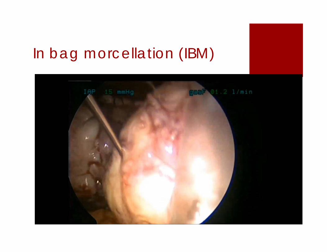
In bag morcellation (IBM)

Contained tissue extraction -minilaparotomy

Contained tissue extraction -transumbilical

Contained tissue extraction -transvaginal

Alexis contained tissue extraction system

Limits
Surgeon experience
Size
Number
Location
What is the ultimate goal of surgery? Fertility preservation? Volume reduction
Blood loss – will the patient accept a blood transfusion?

Surgeon experience Most important factor
Move strategically and control the situation at all times
Gradually build up
Need high volumes (>30/year) to become really good
Rapid suturing is important

Size The largest specimen weight for a myomectomy
in our group is 3080 g
Does not tell the whole story
MUCH easier to remove one large fibroid rather than multiple small ones (raisin bread)
Time for extraction can be excessive – a minilaparotomy may be advisable with manual morcellation with a 10 blade
Also consider hand assisted surgery

Number Have removed over 60 fibroids in one patient,
but our median number is 2 per case.
Important to have a discussion with the patient about limitations. It is not always possible to remove all fibroids. Small ones may be left behind
Preoperative evaluation is very important for mapping

Location Intramural vs submucosal vs intracavitary vs
subserosal
Cervical – watch out for uterines – clip at origin if necessary
Broad ligament – usually pretty easy – open peritoneum and peel out – again stay away from major vessels

Preoperative evaluation MRI is obtained on most
patients
Delineates location, characteristics and size of fibroids
Detects adenomyosis
Helps with preoperative counseling and planning

Tips for limiting blood loss
Use high volume vasopressin – 20 units in 400 ml of saline –inject 200 ml
Use lupron preoperatively to build blood counts – may make dissection of fibroids more difficult IF the fibroids are already necrotic
Be quick
Avoid making an incision close to ascending uterines
Use clips on the uterine arteries
Consider preop embolization
Consider using cell saver

Case in point 39 y/o G0 – Jehovah's witness
Heavy bleeding despite Lupron for 6 months
H/H 9/29 despite repeated iv iron infusions
Wants pregnancy in near future
Multiple fibroids on imaging, overall uterine size 19.5x17.2x8.6cm – 10 cm intracavitary fibroid –total uterine weight approx 1500 grams
EMB benign

Video

In Summary Laparoscopic myomectomy has become the
standard of care for removal of uterine fibroids at our institution
With adequate surgical volume, laparoscopic myomectomy can be performed effectively and safely, even in a large institution with multiple surgeons
Mastering laparoscopic suturing is the most important factor in being able to perform laparoscopic myomectomy

Thank you