Laparoendoscopic Removal of a Benign Gastric Stromal · PDF fileLaparoendoscopic Removal of a...
5
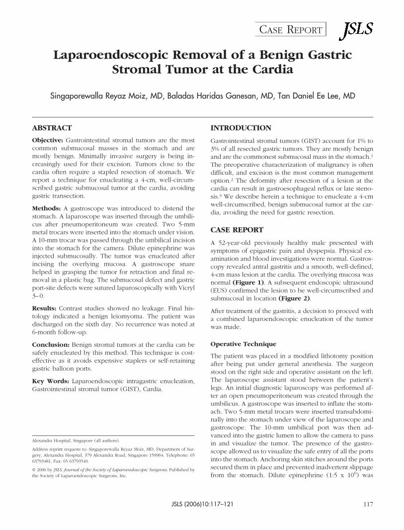
Laparoendoscopic Removal of a Benign Gastric Stromal Tumor at the Cardia Singaporewalla Reyaz Moiz, MD, Baladas Haridas Ganesan, MD, Tan Daniel Ee Lee, MD ABSTRACT Objective: Gastrointestinal stromal tumors are the most common submucosal masses in the stomach and are mostly benign. Minimally invasive surgery is being in- creasingly used for their excision. Tumors close to the cardia often require a stapled resection of stomach. We report a technique for enucleating a 4-cm, well-circum- scribed gastric submucosal tumor at the cardia, avoiding gastric transection. Methods: A gastroscope was introduced to distend the stomach. A laparoscope was inserted through the umbili- cus after pneumoperitoneum was created. Two 5-mm metal trocars were inserted into the stomach under vision. A 10-mm trocar was passed through the umbilical incision into the stomach for the camera. Dilute epinephrine was injected submucosally. The tumor was enucleated after incising the overlying mucosa. A gastroscope snare helped in grasping the tumor for retraction and final re- moval in a plastic bag. The submucosal defect and gastric port-site defects were sutured laparoscopically with Vicryl 3– 0. Results: Contrast studies showed no leakage. Final his- tology indicated a benign leiomyoma. The patient was discharged on the sixth day. No recurrence was noted at 6-month follow-up. Conclusion: Benign stromal tumors at the cardia can be safely enucleated by this method. This technique is cost- effective as it avoids expensive staplers or self-retaining gastric balloon ports. Key Words: Laparoendoscopic intragastric enucleation, Gastrointestinal stromal tumor (GIST), Cardia. INTRODUCTION Gastrointestinal stromal tumors (GIST) account for 1% to 3% of all resected gastric tumors. They are mostly benign and are the commonest submucosal mass in the stomach. 1 The preoperative characterization of malignancy is often difficult, and excision is the most common management option. 2 The deformity after resection of a lesion at the cardia can result in gastroesophageal reflux or late steno- sis. 3 We describe herein a technique to enucleate a 4-cm well-circumscribed, benign submucosal tumor at the car- dia, avoiding the need for gastric resection. CASE REPORT A 52-year-old previously healthy male presented with symptoms of epigastric pain and dyspepsia. Physical ex- amination and blood investigations were normal. Gastros- copy revealed antral gastritis and a smooth, well-defined, 4-cm mass lesion at the cardia. The overlying mucosa was normal (Figure 1). A subsequent endoscopic ultrasound (EUS) confirmed the lesion to be well-circumscribed and submucosal in location (Figure 2). After treatment of the gastritis, a decision to proceed with a combined laparoendoscopic enucleation of the tumor was made. Operative Technique The patient was placed in a modified lithotomy position after being put under general anesthesia. The surgeon stood on the right side and operative assistant on the left. The laparoscope assistant stood between the patient’s legs. An initial diagnostic laparoscopy was performed af- ter an open pneumoperitoneum was created through the umbilicus. A gastroscope was inserted to inflate the stom- ach. Two 5-mm metal trocars were inserted transabdomi- nally into the stomach under view of the laparoscope and gastroscope. The 10-mm umbilical port was then ad- vanced into the gastric lumen to allow the camera to pass in and visualize the tumor. The presence of the gastro- scope allowed us to visualize the safe entry of all the ports into the stomach. Anchoring skin stitches around the ports secured them in place and prevented inadvertent slippage from the stomach. Dilute epinephrine (1:5 x 10 6 ) was Alexandra Hospital, Singapore (all authors). Address reprint requests to: Singaporewalla Reyaz Moiz, MD, Department of Sur- gery, Alexandra Hospital, 379 Alexandra Road, Singapore 159964. Telephone: 65 63793481, Fax: 65 63793540. © 2006 by JSLS, Journal of the Society of Laparoendoscopic Surgeons. Published by the Society of Laparoendoscopic Surgeons, Inc. JSLS (2006)10:117–121 117 CASE REPORT
Transcript of Laparoendoscopic Removal of a Benign Gastric Stromal · PDF fileLaparoendoscopic Removal of a...
Laparoendoscopic Removal of a Benign GastricStromal Tumor at the Cardia
Singaporewalla Reyaz Moiz, MD, Baladas Haridas Ganesan, MD, Tan Daniel Ee Lee, MD
ABSTRACT
Objective: Gastrointestinal stromal tumors are the mostcommon submucosal masses in the stomach and aremostly benign. Minimally invasive surgery is being in-creasingly used for their excision. Tumors close to thecardia often require a stapled resection of stomach. Wereport a technique for enucleating a 4-cm, well-circum-scribed gastric submucosal tumor at the cardia, avoidinggastric transection.
Methods: A gastroscope was introduced to distend thestomach. A laparoscope was inserted through the umbili-cus after pneumoperitoneum was created. Two 5-mmmetal trocars were inserted into the stomach under vision.A 10-mm trocar was passed through the umbilical incisioninto the stomach for the camera. Dilute epinephrine wasinjected submucosally. The tumor was enucleated afterincising the overlying mucosa. A gastroscope snarehelped in grasping the tumor for retraction and final re-moval in a plastic bag. The submucosal defect and gastricport-site defects were sutured laparoscopically with Vicryl3–0.
Results: Contrast studies showed no leakage. Final his-tology indicated a benign leiomyoma. The patient wasdischarged on the sixth day. No recurrence was noted at6-month follow-up.
Conclusion: Benign stromal tumors at the cardia can besafely enucleated by this method. This technique is cost-effective as it avoids expensive staplers or self-retaininggastric balloon ports.
Key Words: Laparoendoscopic intragastric enucleation,Gastrointestinal stromal tumor (GIST), Cardia.
INTRODUCTION
Gastrointestinal stromal tumors (GIST) account for 1% to3% of all resected gastric tumors. They are mostly benignand are the commonest submucosal mass in the stomach.1
The preoperative characterization of malignancy is oftendifficult, and excision is the most common managementoption.2 The deformity after resection of a lesion at thecardia can result in gastroesophageal reflux or late steno-sis.3 We describe herein a technique to enucleate a 4-cmwell-circumscribed, benign submucosal tumor at the car-dia, avoiding the need for gastric resection.
CASE REPORT
A 52-year-old previously healthy male presented withsymptoms of epigastric pain and dyspepsia. Physical ex-amination and blood investigations were normal. Gastros-copy revealed antral gastritis and a smooth, well-defined,4-cm mass lesion at the cardia. The overlying mucosa wasnormal (Figure 1). A subsequent endoscopic ultrasound(EUS) confirmed the lesion to be well-circumscribed andsubmucosal in location (Figure 2).
After treatment of the gastritis, a decision to proceed witha combined laparoendoscopic enucleation of the tumorwas made.
Operative Technique
The patient was placed in a modified lithotomy positionafter being put under general anesthesia. The surgeonstood on the right side and operative assistant on the left.The laparoscope assistant stood between the patient’slegs. An initial diagnostic laparoscopy was performed af-ter an open pneumoperitoneum was created through theumbilicus. A gastroscope was inserted to inflate the stom-ach. Two 5-mm metal trocars were inserted transabdomi-nally into the stomach under view of the laparoscope andgastroscope. The 10-mm umbilical port was then ad-vanced into the gastric lumen to allow the camera to passin and visualize the tumor. The presence of the gastro-scope allowed us to visualize the safe entry of all the portsinto the stomach. Anchoring skin stitches around the portssecured them in place and prevented inadvertent slippagefrom the stomach. Dilute epinephrine (1:5 x 10⁶) was
Alexandra Hospital, Singapore (all authors).
Address reprint requests to: Singaporewalla Reyaz Moiz, MD, Department of Sur-gery, Alexandra Hospital, 379 Alexandra Road, Singapore 159964. Telephone: 6563793481, Fax: 65 63793540.
© 2006 by JSLS, Journal of the Society of Laparoendoscopic Surgeons. Published bythe Society of Laparoendoscopic Surgeons, Inc.
JSLS (2006)10:117–121 117
CASE REPORT

injected into the submucosal plane beneath the tumor toreduce bleeding and facilitate dissection. The mucosaover the tumor was incised with a diathermy hook (Fig-ure 3), and carefully peeling away the overlying mucosaand underlying muscle separated the tumor. An endo-scopic snare through the gastroscope was used to graspthe tumor (Figure 4) and aid in retraction and final re-moval through the oral cavity in a plastic bag. The gastric
submucosal defect (Figure 5) was closed intraluminallyby 3–0 Vicryl sutures. After hemostasis, a nasogastric tubewas placed in the stomach, and the 3 ports were with-drawn from the stomach. The gastric port-site defectswere also closed laparoscopically with continuous Vicryl3–0 sutures. The stomach showed no leakage intraoper-atively when tested with colored dye.
RESULTS
An oral Gastrografin study on the fifth postoperative daydemonstrated no leakage (Figure 6). The patient had an
Figure 1. Gastroscopy shows a stromal tumor at the cardia withnormal mucosa.
Figure 2. Endoscopic ultrasound (EUS) showing a 4-cm, well-circumscribed submucosal tumor.
Figure 3. Mucosa incised over the tumor.
Figure 4. Tumor grasped by endoscopic snare.
Laparoendoscopic Removal of a Benign Gastric Stromal Tumor at the Cardia, Moiz SR et al
JSLS (2006)10:117–121118

uneventful recovery and was discharged on the sixth day.The final histology indicated a benign leiomyoma. Fol-low-up gastroscopy (Figure 7) and EUS (Figure 8) 6months later showed no recurrence. The patient had nodysphagia or symptoms of gastroesophageal reflux.
DISCUSSION
Gastrointestinal stromal tumor (GIST) is the preferredterm for leiomyomas, leiomyoblastomas, and leiomyosar-
comas because they have been shown to originate fromundifferentiated stromal fibroblasts.1 They are the com-monest submucosal masses found in the stomach, com-prising 1% to 3% of all resected gastric neoplasms. Themajority of these are benign and asymptomatic.2–5 Theymay occasionally present as symptoms of obstruction,compression or hemorrhage.6,7 Hemorrhage is the mostcommon symptom of malignancy.6 Malignant lesions ac-count for 20% of all GISTs.6 Malignancy is indicated only
Figure 5. Submucosal defect after excision of tumor.
Figure 6. Contrast study on fifth day showed no leak.
Figure 7. Gastroscopy at 6 months showed no recurrence.
Figure 8. Endoscopic ultrasound (EUS) at 6 months showed noresidual tumor or recurrence.
JSLS (2006)10:117–121 119

by metastasis or local invasion noted at the time of surgeryor after resection.2,3 Histological diagnosis from biopsyspecimens is difficult.4 For further definition, endoscopicultrasound (EUS) and ultrasound-guided needle aspira-tion have been used. EUS characterizes the muscular or-igin of these lesions in the gastric wall and the well-defined margins of benign tumors. Thus in the majority ofcases, the diagnosis is established and FNAC is not war-ranted.6 In our patient, EUS (Figure 2) showed the tumorto be well circumscribed and arising from the laminamuscularis with sparing of the muscularis propria. Thisfinding on EUS is classical of a benign stromal tumor.Histological diagnosis from biopsy specimens is difficultfor these submucosal lesions and therefore no biopsieswere taken during the gastroscopy.
Walsh and Heniford6 suggest that once diagnosed, a GISTof any size should be excised to eliminate the need forserial endoscopy and prevent the progression of symp-toms. Moreover, a size greater than 6cm is more com-monly associated with malignancy. Cheng et al,2 however,believe that surgical intervention is not required for small,clinically insignificant lesions and that resection is indi-cated only for large or symptomatic lesions.
The recommended methods for excision of GIST tumorsinclude local excision with a 2-cm to 4-cm margin,2,3
enucleation8 and stapled gastric resection.5,9 The latteroption is especially indicated if evidence of malignancy ispresent.8,10 All these procedures can be performed bylaparoscopic or open methods.
Minimally invasive methods described in the literaturerange from a purely endoscopic technique,11,12 to a com-bined endolaparoscopic6,7,13,14 to a purely laparoscopicmethod.4,5,8 The technique can be performed intracorpo-really or extracorporeally,15 intragastrically8,14 or transgas-trically.4,5 Position is an important factor in determiningthe approach used. Anterior wall lesions and those situ-ated along the curvatures generally undergo wedge resec-tion. Posterior wall lesions undergo a gastrostomy andresection, whether intragastric or transgastric. For tumorslocated at the cardia, laparoscopic gastric resection usingan exogastric or intragastric approach is difficult.9 Due toits location, resection with sufficient margins is not possi-ble without compromising the Angle of His and the mus-cle layers that construct the antireflux mechanism.14 Thedeformity after resection of a lesion in the gastric fundusand cardia can result in gastroesophageal reflux or steno-sis.16
According to Taniguchi et al,8 enucleation is a satisfactoryprocedure for benign gastric leiomyomas. However, for
gastric leiomyosarcomas, a wider resection of the fullthickness of the gastric wall with adequate margins isnecessary. As partial resection of the stomach with ade-quate margins cannot be applied to tumors close to thecardia, they have been subjected to major surgery, such astotal or proximal gastrectomy.8 Such major surgery wouldbe excessive if the tumor is proven to be benign at post-operative pathological examination. Hence, they recom-mend that for GIST tumors at the cardia that look benignand have low malignant potential, enucleation is a betteroption. If the tumor were proven to be benign, as sus-pected preoperatively, the enucleation would be suffi-cient and give the patient a less-invasive outcome. Thepotential problems of reflux and stenosis are also avoided.If, however, the enucleated tumor is diagnosed as malig-nant, a second-look operation will be necessary, andbased on the full pathological information the optimumgastric resection performed. Concerning the potential fortumor seedings, it is believed that the risks are negligiblebecause almost all the tumors to which this technique isapplied would be low-grade malignancies that appearbenign.
In our patient, the endoscopic ultrasound features werequite classical of a benign stromal tumor. We thereforeproceeded to perform a combined laparoendoscopic in-tragastric enucleation. Since the tumor was only 4 cm andcould be retrieved orally, only 3 openings in the stomachfor the trocars were needed. Furthermore, under the mag-nified view of the laparoscope, the relationship betweenthe tumor and the Z line or the gastric muscle layers couldbe clearly seen. This facilitated the preservation of theantireflux mechanism and avoided stenosis. Follow-upgastroscopy and EUS is recommended for early detectionof recurrence.
This technique can also be applied to lesions situated onthe posterior gastric wall. However, it is not suitable fortumors on the anterior wall or those showing extragastricgrowths. An EUS is mandatory to confirm the origin, andin suspicious cases a guided biopsy may be advisable toconfirm the benign nature of the lesion before proceedingwith this method.
CONCLUSION
Although technically demanding, this is a safe, feasibletechnique for removal of well-circumscribed benign stro-mal tumors at the cardia. By avoiding a gastric resection inthis region, the risks of gastroesophageal reflux or stenosiscan be prevented.
Laparoendoscopic Removal of a Benign Gastric Stromal Tumor at the Cardia, Moiz SR et al
JSLS (2006)10:117–121120

References:
1. Bennet MK. Pathology of esophageal and gastric cancers. In:Griffin SM, Raimes SA, eds. Upper gastrointestinal surgery. Lon-don: WB Saunders; 1997;1–35.
2. Cheng HL, Lee Wj, Lai IR, Yuan RH, Yu SC. Laparoscopicwedge resection of benign gastric tumour. Hepatogastroenterol-ogy. 1999;46:2100–2104.
3. Llorente J. Laparoscopic gastric resection for gastric leiomyo-mas. Surg Endosc. 1994;8:887–889.
4. Rohatgi A, Singh KK. Laparoendoscopic management ofgastrointestinal stromal tumors. J Laparoendosc Adv Surg Tech A.2003;13(1):37–40.
5. Ibrahim IM, Silvestri F, Zingler B. Laparoscopic resection ofa posterior gastric leiomyoma using combined and gastroscopicapproach. Surg Endosc. 1997;11:277–279.
6. Walsh RM, Heniford BT. Laparoendoscopic treatment ofgastric stromal tumors. Semin Laparosc Surg. 2001;8:189–194.
7. Payne WG, Murphy CG, Grossbard LJ. Combined laparo-scopic and endoscopic approach to resection of gastric leiomy-oma. J Laparoendosc Surg. 1995;5:119–122.
8. Taniguchi E, Kamiike W, Yamanishi H, et al. Laparoscopicintragastric surgery for gastric leiomyoma. Surg Endosc. 1997;11:287–289.
9. Tagaya N, Mikami H, Kogure H, Kubota K, Hosoya Y, NagaiH. Laparoscopic intragastric stapled resection of gastric submu-
cosal tumors located near the esophagogastric junction. SurgEndosc. 2002;16:177–179.
10. Seelig MH, Hinder RA, Floch NR, et al. Endo-organ andlaparoscopic management of gastric leiomyomas. Surg LaparoscEndosc Percutan Tech. 1999;9:78–81.
11. Fujasaki J, Mine T, Akimoto K, Yoshidea S, Hasegawa Y,Ogata E. Enucleation of a gastric leiomyoma by combined laserand snare electrocuting technique. Gastrintest Endosc. 1988;34:128–130.
12. Spinelli P, Cenai FG, Cambareri AR, Meroni E, Pizzatti P.Two step endoscopic resection of gastric leiomyomas. SurgEndosc. 1993;7:90–92.
13. Gurbuz AT, Peetz ME. Resection of a gastric leiomyomausing combined laparoscopic and gastroscopic approach. SurgEndosc. 1997;11:285–286.
14. Uchikoshi F, Ito T, Nishida T, Kitagawa T, Endo S, MatsudaH. Laparoscopic Intragastric Resection of gastric stromal tumorlocated at the esophago-cardiac junction. Surg Laparosc EndoscPercutan Tech. 2004;14(1):1–4.
15. Yamashita Y, Bekki F, Kakegawa T, Umetani H, Yatsuka K.Two Laparoscopic techniques for resection of leiomyomas instomach. Surg Laparosc Endosc. 1995;5:38–42.
16. Tangoku A, Yamamoto K, Hirazawa K, et al. Laparoscopicresection of large leiomyomas of the gastric fundus. Surg En-dosc. 1999;13:1050–1052.
JSLS (2006)10:117–121 121