Lactate Shuttles in Neuroenergetics—Homeostasis ... · PDF fileMason...
15
REVIEW published: 02 February 2017 doi: 10.3389/fnins.2017.00043 Frontiers in Neuroscience | www.frontiersin.org 1 February 2017 | Volume 11 | Article 43 Edited by: Andrew Harkin, Trinity College, Dublin, Ireland Reviewed by: Claude Messier, University of Ottawa, Canada Fahmeed Hyder, Yale University, USA *Correspondence: Shayne Mason [email protected] Specialty section: This article was submitted to Neuroenergetics, Nutrition and Brain Health, a section of the journal Frontiers in Neuroscience Received: 31 October 2016 Accepted: 20 January 2017 Published: 02 February 2017 Citation: Mason S (2017) Lactate Shuttles in Neuroenergetics—Homeostasis, Allostasis and Beyond. Front. Neurosci. 11:43. doi: 10.3389/fnins.2017.00043 Lactate Shuttles in Neuroenergetics—Homeostasis, Allostasis and Beyond Shayne Mason * Centre for Human Metabolomics, North-West University, Potchefstroom, South Africa Understanding brain energy metabolism—neuroenergetics—is becoming increasingly important as it can be identified repeatedly as the source of neurological perturbations. Within the scientific community we are seeing a shift in paradigms from the traditional neurocentric view to that of a more dynamic, integrated one where astrocytes are no longer considered as being just supportive, and activated microglia have a profound influence. Lactate is emerging as the “good guy,” contrasting its classical “bad guy” position in the now superseded medical literature. This review begins with the evolution of the concept of “lactate shuttles”; goes on to the recent shift in ideas regarding normal neuroenergetics (homeostasis)—specifically, the astrocyte–neuron lactate shuttle; and progresses to covering the metabolic implications whereby homeostasis is lost—a state of allostasis, and the function of microglia. The role of lactate, as a substrate and shuttle, is reviewed in light of allostatic stress, and beyond—in an acute state of allostatic stress in terms of physical brain trauma, and reflected upon with respect to persistent stress as allostatic overload—neurodegenerative diseases. Finally, the recently proposed astrocyte–microglia lactate shuttle is discussed in terms of chronic neuroinflammatory infectious diseases, using tuberculous meningitis as an example. The novelty extended by this review is that the directionality of lactate, as shuttles in the brain, in neuropathophysiological states is emerging as crucial in neuroenergetics. Keywords: neuroenergetics, lactate, astrocyte-neuron lactate shuttle (ANLS), astrocyte-microglia lactate shuttle (AMLS), neuropathology, traumatic brain injury (TBI), neurodegenerative disease, infectious neuroinflammatory disease INTRODUCTION The human brain represents approximately 2% of total body weight and receives up to 15% of total blood flow, consuming up to 20% of oxygen and 25% of circulating glucose under normal conditions (Pellerin, 2010). The metabolism of this high energy consuming organ involves complex intercellular trafficking of metabolites and compartmentalization of numerous processes. Tight coupling exists between the supply and demand of energy in brain metabolism with changes in cerebral blood flow and glucose utilization in response to neuronal activity (i.e., neurovascular and neurometabolic coupling) (Bélanger et al., 2011) under normal homeostatic conditions. Mechanisms involved in brain energy metabolism adapt during periods of perturbation and trauma. The important role of lactate, and its shuttles, has been overlooked and merits acknowledgement.
Transcript of Lactate Shuttles in Neuroenergetics—Homeostasis ... · PDF fileMason...
REVIEWpublished: 02 February 2017
doi: 10.3389/fnins.2017.00043
Frontiers in Neuroscience | www.frontiersin.org 1 February 2017 | Volume 11 | Article 43
Edited by:
Andrew Harkin,
Trinity College, Dublin, Ireland
Reviewed by:
Claude Messier,
University of Ottawa, Canada
Fahmeed Hyder,
Yale University, USA
*Correspondence:
Shayne Mason
Specialty section:
This article was submitted to
Neuroenergetics, Nutrition and Brain
Health,
a section of the journal
Frontiers in Neuroscience
Received: 31 October 2016
Accepted: 20 January 2017
Published: 02 February 2017
Citation:
Mason S (2017) Lactate Shuttles in
Neuroenergetics—Homeostasis,
Allostasis and Beyond.
Front. Neurosci. 11:43.
doi: 10.3389/fnins.2017.00043
Lactate Shuttles inNeuroenergetics—Homeostasis,Allostasis and BeyondShayne Mason*
Centre for Human Metabolomics, North-West University, Potchefstroom, South Africa
Understanding brain energy metabolism—neuroenergetics—is becoming increasingly
important as it can be identified repeatedly as the source of neurological perturbations.
Within the scientific community we are seeing a shift in paradigms from the traditional
neurocentric view to that of a more dynamic, integrated one where astrocytes are no
longer considered as being just supportive, and activated microglia have a profound
influence. Lactate is emerging as the “good guy,” contrasting its classical “bad guy”
position in the now superseded medical literature. This review begins with the evolution
of the concept of “lactate shuttles”; goes on to the recent shift in ideas regarding normal
neuroenergetics (homeostasis)—specifically, the astrocyte–neuron lactate shuttle; and
progresses to covering the metabolic implications whereby homeostasis is lost—a
state of allostasis, and the function of microglia. The role of lactate, as a substrate
and shuttle, is reviewed in light of allostatic stress, and beyond—in an acute state
of allostatic stress in terms of physical brain trauma, and reflected upon with respect
to persistent stress as allostatic overload—neurodegenerative diseases. Finally, the
recently proposed astrocyte–microglia lactate shuttle is discussed in terms of chronic
neuroinflammatory infectious diseases, using tuberculous meningitis as an example. The
novelty extended by this review is that the directionality of lactate, as shuttles in the brain,
in neuropathophysiological states is emerging as crucial in neuroenergetics.
Keywords: neuroenergetics, lactate, astrocyte-neuron lactate shuttle (ANLS), astrocyte-microglia lactate shuttle
(AMLS), neuropathology, traumatic brain injury (TBI), neurodegenerative disease, infectious neuroinflammatory
disease
INTRODUCTION
The human brain represents approximately 2% of total body weight and receives up to 15%of total blood flow, consuming up to 20% of oxygen and 25% of circulating glucose undernormal conditions (Pellerin, 2010). The metabolism of this high energy consuming organ involvescomplex intercellular trafficking of metabolites and compartmentalization of numerous processes.Tight coupling exists between the supply and demand of energy in brain metabolism withchanges in cerebral blood flow and glucose utilization in response to neuronal activity (i.e.,neurovascular and neurometabolic coupling) (Bélanger et al., 2011) under normal homeostaticconditions. Mechanisms involved in brain energy metabolism adapt during periods of perturbationand trauma. The important role of lactate, and its shuttles, has been overlooked and meritsacknowledgement.
Mason Neuro(patho)physiological Lactate Shuttles
The interconversion of lactate and pyruvate occurs via lactatedehydrogenase, with increased lactate typically being associatedwith anaerobic respiration. Thus, one could postulate thatelevated levels of lactate present in the cerebrospinal fluid (CSF)due to neuroinflammation could be from hypoxia caused byischemia—increased anaerobic respiration (Rossi et al., 2007),or by raised glucose levels and hence elevated flow through theglycolysis pathway. However, in most neuroinflammatory casesthere are typically periods of low levels of glucose in the CSF.Furthermore, several studies have shown no correlation betweenCSF lactate levels and cerebral blood flow (i.e., they are unrelatedto ischemia) (Brodersen and Jorgensen, 1974; DeSalles et al.,1986; De Salles et al., 1987). Thus, an alternative postulate is thatthe elevated levels of lactate in the CSF of neuroinflammatorycases is unlikely to be due to anaerobic respiration but instead ispossibly a product of temporarily increased flux in the glycolysispathway—using glycogen as a supplementary source of glucose.
The notion that lactate is not simply just a by-product ofglycolysis but also a shuttle system was pioneered by Brooks(1985). In June the following year, Brooks provided moreevidence for a “shuttling” system for lactate (Brooks, 1986a);by December Brooks (1986b) formally coined the term “lactateshuttle” in skeletal muscle during rest and exercise underfully aerobic conditions. Brooks further defined lactate shuttlesas being either intracellular (cytosolic to mitochondrial) orcell-to-cell (extracellular) (Brooks, 2000, 2002). Later, Brooks(2009) provided evidence that glycolytic and oxidative pathwaysshould be viewed as linked, as opposed to alternative processes,because lactate, the product of one pathway, is the substrate forthe other (Brooks, 2009). The concept of intracellular lactateshuttles was challenged (Sahlin et al., 2002), and continues tobe so (Laureys et al., 2014). A study by Cruz et al. (2012)discussed these challenges but ultimately reiterated Brooks’sentiment regarding interaction between energy systems—theproduct of one is the substrate of another. Soon after Brooksfirst introduced the concept of lactate shuttles in 1986, severalstudies by Schurr et al. (1988, 1997a,b,c, 1999a,b) providedexperimental evidence that astrocytic lactate is even usedpreferentially over glucose by neurons after episodes of cerebralischemia.
Thus, lactate plays an important role as a shuttle, even beyondthe brain—in numerous systems, for example, in pregnancy (Zuoet al., 2015), in reproduction (Kuchiiwa et al., 2011), and, notably,within the human heart (Cruz et al., 2012; Rakus et al., 2016).This paper provides an overview of brain neuroenergetics and thecrucial roles of lactate shuttles. These roles will be discussed: (1)with regard to the normal physiological function and relationshipbetween astrocytes and neurons (homeostasis)—the astrocyte–neuron lactate shuttle (ANLS) model; (2) the dynamic roleof microglia and their preferential utilization of lactate underperturbed conditions (allostasis); (3) the activity of lactate andits shuttles in neuropathological states (in particular in traumaticbrain injury and neurodegenerative diseases); and (4) thebrain in crisis—in response to a neuroinflammatory infectiousdisease—according to the astrocyte–microglia lactate shuttle(AMLS) model, using tuberculous meningitis (TBM) as anexample.
HOMEOSTATIC NEUROENERGETICS
Shifting Neuroenergetics ParadigmsThe classical view of neuroenergetics is that the blood suppliesoxygen and glucose to the brain (Sokoloff, 1989). Glucose isthe primary source of energy utilized by both neurons andastrocytes. It undergoes complete oxidation via glycolysis, theKrebs cycle and oxidative phosphorylation, which ultimatelyproduces adenosine triphosphate (ATP) for energy-dependentreactions. Thus, glucose is used in the same way by all cell types.Since neurons consume the greatest quantity of energy of allbrain elements, metabolic intermediates (e.g., in the Krebs cycle)are diverted toward neurons. Some of the pyruvate producedby glycolysis is converted to lactate and released into theextracellular space. In this classical view, lactate is considered aby-product with deleterious effects when in excess (Norenberget al., 1987; Siesjö, 1988; Bender et al., 1997). Astrocytes havelong been thought to play a passive role in supporting neuronalfunction, with the neuron being the star of the show. However,the dynamic involvement of astrocytes (Ranjbar andAmiri, 2015)in the forefront of neuroenergetics is now being recognized,shifting paradigms (Haydon and Carmignoto, 2006; Giaumeet al., 2010). The neurocentric view of neuroenergetics is evolvinginto a more integrated one of complementary and co-operativemetabolic interactions between astrocytes and neurons.
Astrocytes–More than a Supporting Role?At the cellular level the human brain consists of the all-important neurons and up to 10 times more glial cells thanneurons (Kimelberg and Norenberg, 1989). There are four typesof glial cells: ependymal cells, oligodendrocytes, astrocytes,and microglia (resident macrophages in the brain). Astrocytesconstitute about 50% of the total human brain volume and areclassically divided into three types based on morphology andspatial organization. These three types are: (1) radial—orientatedperpendicular to ventricular surfaces with long, unbranchedprocesses (end-feet); (2) protoplasmic—displaying bushymorphology with numerous, highly branched, short processes;and (3) fibrous—manifesting stellate shapes with smooth,long processes that are less branched. The cytoarchitecturalorganization of astrocytes is such that, according to Pellerin(2010) particular sections cover 99% of the surface area ofcerebral blood vessels; although, due to tissue shrinkage withchemical fixation (Korogod et al., 2015), this 99% value islikely an inflated estimate. These astrocytes are, however, thepreferential site for glucose uptake from the blood, as well ashaving projections in peri-synaptic areas of neurons, providingclose interaction with neuronal elements and acting as a cellularbarrier between blood and neurons. The unique morphologicaland phenotypic characteristics of astrocytes ideally positionthem to sense and respond dynamically to changes in neuronalactivity (Pellerin, 2010; Bélanger et al., 2011), lending them toconduct numerous critical functions (Chen and Swanson, 2003;Steele and Robinson, 2012), such as glutamate homeostasis(in the glutamate–glutamine cycle), maintaining brain ionicequilibrium (K+ and H+ buffering), the maintenance of reactiveoxygen species (ROS) (in glutathione recycling) and osmotic
Frontiers in Neuroscience | www.frontiersin.org 2 February 2017 | Volume 11 | Article 43
Mason Neuro(patho)physiological Lactate Shuttles
regulation. Astrocytes therefore support neuronal activity viastructural, trophic and metabolic means, suggesting a criticalrole in regulating neuroenergetics and homeostatic functions(Pellerin, 2010). Notably, neurons rely on astrocytes to supplyprecursors of the Krebs cycle intermediates, or their derivatives,as the enzyme pyruvate carboxylase is present in only astrocytesbut not in neurons (Hertz et al., 1999).
Astrocytes exhibit a higher capacity for glucose utilization,as well as greater metabolic plasticity, than neurons;these characteristics are important for homeostatic andneuroprotective functions. The calculated energy needs ofastrocytes only represent about 10–15% of the total brain energyneeds (Attwell and Laughlin, 2001; Gjedde et al., 2002; Rothmanet al., 2003; Shulman et al., 2004). Hence, approximately85% of the glucose in the brain is used in the expenditure ofenergy in neurons via the glycolytic pathway and the Krebscycle leading to the synthesis of ATP (Jueptner and Weiller,1995; Attwell and Laughlin, 2001). The high glycolytic rateof astrocytes suggests a preference for the production andrelease of lactate. The neuroprotective role of lactate has beenexperimentally demonstrated by studies (Cater et al., 2001, 2003).Neuroprotection here is defined as an intervention that preventsthe death of vulnerable neurons and slows disease progression.Hence, evidence that has emerged over the past two decadeshas begun to highlight lactate as a supplementary substrate forneurons, resulting in the (re)emergence of a dynamic nursingrole for astrocytes (Bouzier-Sore et al., 2002).
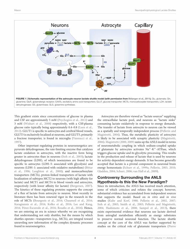
Astrocyte–Neuron Lactate Shuttle (ANLS)HypothesisMagistretti and Pellerin (1996) presented the framework ofa hypothesis that they have since developed and refined tobecome one of the prevailing contemporary viewpoints ofneuroenergetics—the ANLS hypothesis. The hypothesis statesthat astrocytes respond to intensified neuron activity byincreasing their rate of glucose uptake, glycolysis and the releaseof lactate into the extracellular space, as shown schematicallyin Figure 1. At the metabolic level, it begins with glutamatergicactivity, a process whereby increased neuronal activity results inthe release of glutamate, the main excitatory neurotransmitterin the brain, into the extracellular space along the glutamatetransporter EAAT3, which is exclusively located in neurons.Astrocytes sense increased activity at the glutamatergic synapses,followed by glutamate uptake via the glia-specific glutamatetransporters EAAT1 and EAAT2. The transport of glutamateis driven by a sodium gradient (i.e., by a Na+-dependentmechanism), with a stoichiometry of three Na+ ions co-transported with one glutamate, resulting in a significant increasein intracellular Na+ concentrations in astrocytes (Magistrettiand Pellerin, 1999; Pellerin and Magistretti, 2004; Bélangeret al., 2011). Glutamate taken up by astrocytes is convertedto glutamine through an ATP-dependent reaction catalyzed byastrocyte-specific glutamine synthetase. Glutamine is releasedback into the extracellular space and taken up by neurons,where it is converted to glutamate by glutaminase (Magistrettiand Pellerin, 1999; Bélanger et al., 2011). This reaction
thereby replenishes the neurotransmitter pool of glutamate andcompletes the glutamate–glutamine cycle. Glutamate uptakeby astrocytes stimulates glucose uptake with a stoichiometricrelationship of 1:1 between uptake of glutamate and glucose.Increased concentrations of Na+ in astrocytes activate theenzyme Na+-K+-ATPase, particularly the α2 subunit. The resultis the triggering of glycolysis, leading to the production andrelease of lactate into the extracellular space; the lactate isthen taken up as an energy substrate by neurons for oxidative-derived ATP production (Debernardi et al., 1999; Magistrettiand Pellerin, 1999; Pellerin, 2003; Pellerin and Magistretti, 2004;Pellerin et al., 2007; Bélanger et al., 2011). This demonstrates thepresence of open astroglial metabolic networks—an intercellularroute, allowing the trafficking of energy substrates throughastrocytes from their source, which is blood vessels, to the site ofhigh energy demand and use, the neurons (Giaume et al., 2010).
Fox et al. (1988) were the first to show that focal, physiologicalincrease in neuronal activity induced by visual stimulation inhumans was associated with increased glucose uptake and bloodflow in the human visual cortex. This suggested that increasedneuronal functional activity stimulates glycolysis. Prichard et al.(1991) and Sappey-Marinier et al. (1992) further developed thispostulate by demonstrating increased levels of lactate in thehuman visual cortex using nuclear magnetic resonance (NMR)technology, following visual stimulation. Thus, a tight couplingwas demonstrated between neuronal activation and glucoseutilization in astrocytes (Magistretti, 2006), an intrinsic featureof astrocytes not specifically linked to culture conditions or cellorigin (Pellerin et al., 2007). It is interesting to note that glucoseuptake by astrocytes is disproportionately high compared totheir energy requirements. This suggests that sustained astrocyticglycolysis occurs in order to maintain the extracellular lactatepool to meet the energy requirements of neurons. Most recently,data from Angamo et al. (2016) suggest that astrocytic neuronallactate shuttles contribute to the regulation of ion homeostasisand synaptic signaling in the presence of ample glucose.
In addition to blood-borne glucose, the brain also makesuse of an important energy reserve in the form of glycogenstores located almost exclusively in astrocytes. Mobilization ofglycogen occurs without ATP requirement during extendedperiods of limited energy supply (e.g., in hypoglycemia) and leadsto enhanced lactate production and release, thereby maintainingenergetic homeostasis and preserving neuronal function andviability (Wender et al., 2000; Pellerin, 2003; Pellerin andMagistretti, 2004). Glycogen mobilization can thus be viewedas an extension of the ANLS concept instead of a competinghypothesis (Pellerin et al., 2007). But, as stated by Dieneland Cruz (2015), glycogen is very difficult to study in thehuman brain. Roles for glycogen in brain function continue toemerge, revealing increasing complexity of the signaling andregulatory mechanisms that integrate glycogen mobilization withphysiological activities.
It is important to note that the ANLS hypothesis does notpreclude glucose as a source of energy for the brain. Glucoseremains an important energy substrate with concentrationgradients facilitating the transport of glucose across the blood–brain barrier into the brain via glucose transporters (GLUTs).
Frontiers in Neuroscience | www.frontiersin.org 3 February 2017 | Volume 11 | Article 43
Mason Neuro(patho)physiological Lactate Shuttles
FIGURE 1 | Schematic representation of the astrocyte-neuron lactate shuttle model (with permission from Bélanger et al., 2011). Glu, glutamate; Gln,
glutamine; GluR, glutamatergic receptor; EAATs, excitatory amino acid transporters; GLUT, glucose transporter; MCTs, monocarboxylate transporters; LDH, lactate
dehydrogenase; GS, glutaminase; GLS, glutamine synthetase.
This gradient exists since concentrations of glucose in plasmaand CSF are approximately 5 mM (Psychogios et al., 2011) and3 mM (Wishart et al., 2008) respectively, with a CSF:plasmaglucose ratio typically being approximately 0.4–0.8 (Leen et al.,2012). GLUT1 is specific to astrocytes and cerebral blood vessels,GLUT3 is exclusively localized at neurons, and GLUT5, primarilya fructose transporter, is found in microglia (Vannucci et al.,1997).
Other important regulating proteins in neuroenergetics are:pyruvate dehydrogenase, the rate-limiting enzyme that catalyzeslactate oxidation in astrocytes, with the inactive form beinggreater in astrocytes than in neurons (Itoh et al., 2003); lactatedehydrogenase (LDH), of which isoenzymes are found to bespecific to astrocytes (LDH–5: associated with glycolysis) andneurons (LDH–1: associated with oxidative metabolism) (Bittaret al., 1996; Laughton et al., 2000); and monocarboxylatetransporters (MCTs), proton-linked transporters of lactate withlocalization of subtypes MCT2 to neurons (with high affinity forlactate) and MCT1 and MCT4 to blood vessels and astrocytes,respectively (with lower affinity for lactate) (Bergersen, 2007).The kinetics of these regulating proteins supports the conceptof a flux of lactate from astrocyte to neuron. Most recently inliterature there has been renewed vigor in the research in therole of MCTs (Bourgeois et al., 2016; Chaumeil et al., 2016;Karagiannis et al., 2016; Kolko et al., 2016; Lee and Kang,2016; Pérez-Escuredo et al., 2016; Rosafio et al., 2016). Hence,we are entering an era in science whereby we are recognizingthat understanding not only shuttles, but the means by whichshuttles operate—transporters (e.g., MCTs), are integral towardunraveling new information of the complex dynamic processesfound in neuroenergetics.
Astrocytes are therefore viewed as “lactate sources” supplyingthe extracellular lactate pool, and neurons as “lactate sinks”consuming lactate oxidatively in response to energy demands.The transfer of lactate from astrocyte to neuron can be viewedas a spatially and temporally independent process (Pellerin andMagistretti, 2004). Thus, the metabolic plasticity of astrocytesis likely to be associated with synaptic plasticity (Magistretti,2006).Magistretti (2000, 2009) sums up the ANLSmodel in termsof neurometabolic coupling in which sodium-coupled uptakeof glutamate by astrocytes activates Na+-K+-ATPase, whichtriggers glucose uptake and its glycolytic processing. This resultsin the production and release of lactate that is used by neuronsfor activity-dependent energy demands. It has become generallyaccepted that lactate is a pivotal component in neuronal brainenergy homeostasis (Schurr et al., 1999a; Pellerin, 2003, 2010;Gladden, 2004; Schurr, 2006; van Hall et al., 2009).
Controversy Surrounding the ANLSHypothesis–Is this the New Paradigm?Since its introduction, the ANLS has received much attention,some of which criticizes and refutes the concept; however,substantial evidence has accumulated from independent sourcesthat supports the model. Based on in vitro and in vivostudies (Kuhr and Korf, 1988; Pellerin et al., 2002, 2007;Itoh et al., 2003; Smith et al., 2003; Pellerin and Magistretti,2004; Hashimoto et al., 2008; Sampol et al., 2013), thereexists experimental evidence that neurons use lactate derivedfrom astroglial metabolism efficiently as energy substratesto preserve normal neuronal function. The lactate shuttleconcept at the core of the ANLS was further supported bystudies on the critical role of glutamate transporters (Pierre
Frontiers in Neuroscience | www.frontiersin.org 4 February 2017 | Volume 11 | Article 43
Mason Neuro(patho)physiological Lactate Shuttles
et al., 2000; Voutsinos-Porche et al., 2003) and distributionof MCTs in the brain (Pierre et al., 2000; Chiry et al.,2008; Erlichman et al., 2008; Robinet and Pellerin, 2011),particularly for long-term memory formation (Suzuki et al.,2011).
Molecular mechanisms provide further in vitro evidencethat supports the ANLS hypothesis (Bliss et al., 2004; Porraset al., 2004), showing that upregulation of glycolysis inneurons decreased oxidation of glucose through the pentosephosphate pathway, resulting in impaired regeneration ofreduced glutathione, and subsequently oxidative stress andapoptotic death. Thus, neurons downregulate glycolysis inorder to use available glucose to maintain antioxidant status(a neuroprotective mechanism) at the expense of its use forbioenergetics purposes. Neuroenergetic demands can be met byother sources, such as lactate (Tabernero et al., 1996; Herrero-Mendez et al., 2009).
Rouach et al. (2008) demonstrated in vitro the change incharacter of astrocyte metabolic networks in response to localenergy demand and trafficking of energetic metabolites fromblood vessels, through astrocytes, to distal neurons. Using13C-NMR, this metabolic flux of lactate from astrocyte toneuron can be measured noninvasively in the human brain(Bouzier-Sore et al., 2003; Gallagher et al., 2009; Boumezbeuret al., 2010). Plasma lactate supports up to 10% of brainenergy metabolism under physiological conditions, and upto 60% under supra-physiological conditions. Mathematicalmodeling studies have also described these dynamics, inparticular brain lactate kinetics (Aubert et al., 2005) andcompartmentalization of brain energy metabolism (Aubert andCostalat, 2005). However, as stated by Pellerin et al. (2007):“[mathematical modeling] ... can give a coherent, quantitativeframework for the discussion, suggest possible mechanismsor, conversely, emphasize the contradictions or implausibilityor some hypotheses.” An example of the argument aboutthe possible ambiguity of mathematical modeling is givenby Genc et al. (2011). This sentiment is further expressed byJolivet et al. (2010): “mathematical models are very powerfultools but they are ultimately only partial replicas of thesystem they model, not the system itself.” Thus, mathematicalmodels are useful guides but one should be mindful to drawdefinitive conclusions based on modeling studies alone. Jolivetet al. (2010) commented on two mathematical studies ofhypotheses of opposing lactate flux in neuroenergetics—theycompared the mathematical model of ANLS by Aubert et al.(2007) and the neuron-to-astrocyte lactate shuttle (NALS) byMangia et al. (2009). Jolivet et al. (2010) concluded that theAubert (ANLS) model currently remains the best biophysicalrepresentation of neuron–astrocyte metabolic interactions.Further multi-timescale mathematical modeling by Jolivetet al. (2015) has since provided further support for the ANLShypothesis.
While less vocal, there remains strong opposition to theANLS hypothesis, with various studies expressing divergentviews. Dienel and Hertz (2001) emphasized the paradox ofintense production of lactate by the brain and the slow rateof its uptake, thus disagreeing that lactate is used as a major
neuronal fuel. Dienel and Cruz (2003, 2004) further argued thatcerebral metabolic rates of oxygen consumption do not equalthat of glucose plus glycogen and that this disproportionateconsumption is strong evidence against stoichiometric transferof lactate from astrocytes to neighboring neurons for oxidation.Gjedde et al. (2002) averred that, based on their experimentalevidence, changes in metabolism in afferent phase (activityinvolving presynaptic terminals and astrocytes) and efferentphase (activity involving neurons) are additive. This viewopposed the idea that changes in metabolism are characterizedby significant transfer of lactate from astrocyte to neurons.Gjedde et al. (2002) conclude that there is no suggestionthat astrocytic glycolysis supports oxidative metabolism ofneurons in the baseline condition. Instead, neurons increasetheir oxidative metabolism in parallel with a rise in pyruvategenerated by neuronal rather than astrocytic glycolysis (Gjeddeand Marrett, 2001). Mangia et al. (2003a) discussed variousambiguities from other studies and conclude that astrocyteactivation supports only the glutamate–glutamine cycle andthat neurons primarily metabolize directly absorbed glucose tosupport neuronal activity. This behavior is demonstrated by aninitial dip in lactate concentrations following visual stimulation(Mangia et al., 2003b). As mentioned above, Mangia et al.(2009) also proposed a model of opposite flux to the ANLS,namely the NALS hypothesis which utilized the mathematicalmodel introduced by Simpson et al. (2007), suggesting thatdepending on the thermodynamic and kinetic status of thecytosolic and mitochondrial redox states, lactate transfers fromneurons to astrocytes. The directionality of the ANLS hypothesis(i.e., lactate from astrocytes to neurons), has been coveredin many reviews. However, the other perspective of shuttlingdirection (i.e., from neurons to astrocytes) is an intriguing one,albeit without substantiated experimental proof of the NALS,beyond modeling. Other divergent views come from studiesby, for example, Bak et al. (2006), who suggested that synapticactivity does not induce corresponding upregulation of lactatemetabolism in neurons; DiNuzzo et al. (2010a), who suggestedthat carbon recruitment by neurons relies upon glucose uptakerather than that of a lactate shuttle, and that glycogenolysis inastrocytes preserves glucose availability for neurons (DiNuzzoet al., 2010b).
Others have critically reviewed the ANLS hypothesis indefense of the traditional role of neuroenergetics (Chih et al.,2001; Chih and Roberts, 2003). Pellerin and Magistretti (2011)not only addressed all these divergent views by reviewingexperimental evidence supporting the main tenets of theANLS model, but also were able to confute several unfoundedcriticisms. With overwhelming support for the ANLS hypothesis,it was becoming clear that lactate is a crucial element inneuronal brain energy homeostasis (Schurr et al., 1999b;Pellerin, 2003, 2010; Gladden, 2004; Schurr, 2006; van Hallet al., 2009). However, as is the case in science, refutingevidence continues to challenge the foundations of the ANLShypothesis, leading to it being redefined and strengthenedover time. The tenets of this hypothesis continue to hold,begging the question—is this the new paradigm for homeostaticneuroenergetics?
Frontiers in Neuroscience | www.frontiersin.org 5 February 2017 | Volume 11 | Article 43
Mason Neuro(patho)physiological Lactate Shuttles
NEUROPATHOLOGY AND THE HUMANRESPONSE
Beyond HomeostasisBiological processes maintain stability, by detectingenvironmental (external) and physiological (internal) changes,and activating specialized adaptive responses. The dynamicmetabolic characteristics of astrocytes lend them to beingparticularly adept at such a task within the neuronal frameworkof homeostasis. Beyond homeostasis lies the comparatively newconcept of allostasis. Allostasis is an extension of the concept ofhomeostasis and refers to the maintenance of stability by meansof robust, energy-demanding adaptive mechanisms in responseto severe physical, psychosocial or environmental challenges.Homeostasis involves maintaining a stable internal environmentof an organism (i.e., by means of a feedback mechanism),whereas allostasis is more dynamic in that it involves thecontinuous response to physiological needs, resulting inbiochemical adaptive mechanisms. These two concepts seemsimilar and are not intended to operate independently; instead,allostasis supports homeostasis, placing emphasis on flexibleadaptation with the ultimate goal of maintaining a stable internalenvironment (McEwen, 2006, 2008; Logan and Barksdale,2008; McEwen and Gianaros, 2011; Danese and McEwen,2012). Frequent or chronic challenges (e.g., neuroinflammation)produce dysregulation of several major physiological systemsby triggering chemical mediators of adaption that operate in anonlinear network. The cumulative “wear and tear” associatedwith the inability to disengage these physiological systems isreferred to as allostatic load. In vulnerable biological systems anallostatic overload prevails that results in the development ofdisease.
Since the term was first coined, by Sterling and Eyer(1988), the concept of allostasis has primarily been used in theliterature to describe mild perturbations that are stress related,psychosomatic and/or psychopathological (Tannenbaum et al.,2002; Stewart, 2006; Shannon et al., 2007; Logan and Barksdale,2008; Blair et al., 2011; Danese and McEwen, 2012; Tomiyamaet al., 2012). As a concept allostasis is still being developedand so is gradually being applied to more diverse fields, suchas metabolic diseases and lipidomics (Oresic et al., 2008) andmetabolomics (Ramautar et al., 2013). Allostasis has also beenused to explain extreme glucose fluctuations as reflecting thebody’s inability to cope with allostatic load in the case of chronicillness, predisposing the individual to serious harm as manifestedby heightened mortality (Stumvoll et al., 2003, 2004; Rake et al.,2010).
Herein this review the typology, as given by Peters andMcEwen (2015) to describe cardiovascular disease, has beenadapted to define terms in neuroenergetics. Stress occurs ina state of increased cerebral energy demand to safeguard anindividual’s physical, mental and social well-being. A reversiblestress response distinguishes allostasis from homeostasis and canbe indicated by a relief result (restored homeostasis) in which theindividual returns toward physiological normality. An allostaticload is indicated by an acute stress challenge—an irreversible ornearly irreversible state of allostatic stress from homeostasis—as
seen in severely elevated CSF lactate in acute, physical damage tothe brain (Gallagher et al., 2009). Chronic cases of persistentlyhighly elevated CSF lactate and lactate:pyruvate ratios wouldbe parameters to define the transition of allostatic load towardallostatic overload (disease).
Thus, allostasis encompasses typical dynamic adaptations totransient stress mediators; allostatic load pertains to an acutestate of persistent perturbation and, ultimately, circumstancesof allostatic overload are associated with disease and the body’sinability to cope. These terms are henceforth discussed in termsof neuropathological conditions, with the focus on lactate and itsshuttles.
Microglia–Addressing Neuronal AllostasisUnder pathological conditions, such as caused by infection,the brain enters a state of allostasis and microglia becomeactivated. Microglia are immunocompetent cells that act as theintrinsic macrophages of the brain, bearing multiple similaritiesto those of macrophages in peripheral tissues. Some of the sharedfunctions include phagocytosis, antigen presentation, effectorinflammatory response and production of various cytotoxins andcytokines. Thus, microglia are an important component of boththe innate and adaptive immune response to central nervoussystem (CNS) pathogens (Olson andMiller, 2004). They populatethe entire brain parenchyma with a homogeneous distributednetwork with territorial organization and act as the first and onlyline of defense in the brain.
Under physiological conditions microglia exist in aresting (“ramified”) state characterized by a small somaand numerous thin, branched processes (Giaume et al.,2007). Microglia remain quiescent until activated upon bybrain insult (i.e., injury, disease or infection), characterizedby proliferation and immunophenotypical (expression ofvarious surface markers and ion channels) and morphological(transformation to amoeboid morphology) changes (Eder,1998). The response of microglia to brain insult is first todetect the site of assault by constant dynamic monitoring ofthe surrounding micro-environment, and then to send outprocess extensions toward the lesion site where process tipsreorganize to confine, control and eliminate the source of thedisturbance.
Neuroinflammation, the reaction of surrounding brain tissueto brain insult, is characterized by synthesis of variousinflammatory mediators and reactive gliosis, associated withphenotypic changes and proliferation of glia (both astrocytes andmicroglia), in response to a dynamically changing environment(Giaume et al., 2007). The modified phenotype of astrocytes,from basal to reactive state, results in them abandoning theirneuroprotective role, allowing excessive oxidative stress andproduction of ROS, and subsequently ROS-induced neuronaldamage (Pellerin, 2003). Chronic neuroinflammation, theresponse to sustained and widespread stimuli, typically occursdue to either disease (e.g., Alzheimer’s and multiple sclerosis)or persistent infection by viruses (Olson et al., 2001; Ovanesovet al., 2008) or bacteria (e.g., chronic meningitis as in TBM).The role of microglia in CNS infections was extensively reviewedby Rock et al. (2004). Hence, metabolic coupling of microglia
Frontiers in Neuroscience | www.frontiersin.org 6 February 2017 | Volume 11 | Article 43
Mason Neuro(patho)physiological Lactate Shuttles
is essential during neuronal allostasis, especially in response topathogens.
THE BRAIN IN CRISIS
Allostatic Load–Physical Brain TraumaThe topic of traumatic brain injury (TBI)—acute, physicaldamage to the brain—gained notoriety in 2009 when JeanneMarie Laskas wrote an article titled “Game brain” in GQmagazine (Laskas, 2009), exposing the hidden, negative medicalimplications within some popular, professional contact sports. In2015 this was expanded into a book (Laskas, 2015) and a filmcalled “Concussion,” about forensic pathologist Bennet Omaluand his attempts to publicize his scientific findings regardingphysical contact sports (such as in the National FootballLeague, the apex of American football). Omalu postulated thatpersistent mild TBI leads to chronic traumatic encephalopathy.Consequently, there has been renewed research into concussionand TBI (Sone et al., 2016; Sussman et al., 2016), making itanother hot topic in neuroscience.
It is well recognized that there is an elevation of brainextracellular lactate and the lactate:pyruvate ratio in TBI cases(Gallagher et al., 2009). The group of Gallagher et al. (2009)and Carpenter et al. (2014) presented direct evidence of brainutilization of lactate in TBI cases by administration of 13C-labeledlactate via a microdialysis catheter and using 13C-NMR analysis.Based on these studies, Carpenter et al. (2015) suggested thatwhere neurons are too damaged to use the lactate producedfrom glucose by astrocytes (i.e., by uncoupling of neuronaland glial metabolism), high extracellular levels of lactate wouldaccumulate—explaining the association between high lactate andpoor outcome. Thus, therapeutic intervention before metabolicuncoupling is imperative. Glucose administration after TBIhas been shown to be beneficial but there is a correlationbetween hyperglycemia and increased infection or mortality;hence, alternative sources of energy, such as lactate, may improveoutcome (Moro et al., 2013). However, as with the ANLS model,there are opposing opinions of the notion that lactate is apreferential fuel in TBI cases (Dienel, 2014).
Acute administration of exogenous fuels, such as lactate, afterexperimentally induced TBI in rats has been shown to attenuatehistopathology and improve outcome (Chen et al., 2000). Intwo companion reports by Glenn et al. (2015a,b), evidence wasprovided that central venous tracer infusion of both glucoseand lactate showed massive mobilization of mainly lactate—systematic lactate was preferentially being directly consumedand used by TBI cases. Thus, there is a high production andclearance rate of lactate in TBI cases. The role of this lactatein cerebral metabolism following TBI and the advantages oftreatment by exogenous lactate infusions was reviewed by Brooksand Martin (2015). In recent clinical trials of TBI, and otherneurocritical care cases, the beneficial properties of hypertoniclactate have been observed with respect to cerebral blood flow andintracranial pressure (Bouzat and Oddo, 2014; Patet et al., 2016).Thus, accumulating evidence points to lactate being an importantcomponent used during allostatic load caused by acute, physicaldamage to the brain—a therapeutic target.
Allostatic Overload–NeurodegenerativeDiseasesA common trait among neurodegenerative diseases is perturbedbrain energy metabolism (Magistretti and Pellerin, 1996; Beal,2000; Bélanger et al., 2011; Albanese et al., 2016). Huntington’sdisease and multiple sclerosis have been linked to mitochondrialdysfunction, and so are susceptible to oxidative stress andenergy deficits (Dutta et al., 2006; Regenold et al., 2008; Sack,2010; Gouarné et al., 2013); with implications of perturbedlactate levels. In contradiction, two 1H-NMR studies foundconflicting results regarding lactate in Huntington’s disease.An 1H-NMR investigation by Gårseth et al. (2000) reporteddecreased levels of lactate, which they attributed to neuronalloss, but based upon only a small sample (n = 7). Another1H-NMR study, by Verwaest et al. (2011), found lactate tobe significantly increased, but this result was also based ona small sample (n = 10). In a more comprehensive study,Gouarné et al. (2013) used cultured neuronal subpopulationsfrom transgenic mice and provided evidence that neuronsuse lactate, along with pyruvate, as an energy source tosupport respiration. The importance of lactate was corroboratedby a study (Covarrubias-Pinto et al., 2015) on Huntington’sdisease (HD) using ascorbic acid to inhibit use of neuronalglucose—showing the favoring of lactate uptake to sustain brainactivity. Hence, experimental evidence is emerging that lactateis important and used preferentially as an energy source in HDcases.
Two reviews on neurodegenerative diseases support the ANLShypothesis and its role in neuropathology—namely, lactate isneuroprotective and a therapeutic agent. A review by Newingtonet al. (2013) refers to their previous scientific work, whichshowed that increased lactate production proved to be protectiveagainst Aβ-induced neuronal toxicity that is inherently associatedwith Alzheimer’s disease. A review by Finsterwald et al. (2015)summarizes numerous studies involving astrocytic lactate intreatments for multiple neurodegenerative diseases [Alzheimer’s,Parkinson’s disease and amyotrophic lateral sclerosis (ALS)].Finsterwald et al. (2015) concluded that astrocytic functionelicits intrinsic neuroprotective properties through, amongstothers, the ANLS mechanism. One could go one step furtherand speculate that a “specialized” type of ANLS mechanism isnecessary.
In multiple sclerosis (MS) there have been reports of theimportance of CSF lactate. A negative correlation between thepresence of lactate and the presence of inflammatory plaquesand MS severity has been experimentally shown (Lutz et al.,2007; Albanese et al., 2016)—increased lactate in MS caseswithout plaques and vice versa, suggesting close metaboliccoupling between plaque activity and lactate production. Agene expression study by Zeis et al. (2015) revealed alterations(downregulation) in the ANLS mechanism in MS, such thatalternative lactate shuttle systems may be at play. Indeed,other studies have proposed such shuttle systems to supplydemyelinated axons with lactate as an important energy source—namely, the astrocyte-axon lactate shuttle (Cambron et al., 2012;Nijland et al., 2015) and the oligodendrocyte-axon lactate shuttle(Campbell et al., 2014).
Frontiers in Neuroscience | www.frontiersin.org 7 February 2017 | Volume 11 | Article 43
Mason Neuro(patho)physiological Lactate Shuttles
A common thread seen in these studies of neurodegenerativediseases is the perturbation of lactate. Evidence inneuropathophysiological cases indicates that lactate acts asan important, sometimes preferential, source of energy, andalso has neuroprotective properties. The role of lactate shuttlesystems is also emerging as being vital—imperative as supply anddemand needs fluctuate to extremes in situations of allostaticoverload. This is when normal physiological systems can nolonger provide the required resources and/or effects. It is,however, important to note that neuropathologically activatedastrocytes, as well as other glial cells, such as microglia, maysupport mechanisms, such as neuroinflammation, aggravatingneuronal degeneration (Li et al., 2011). The dynamics behindaddressing neuroinflammation caused by an invading pathogeninvolves the use of microglia.
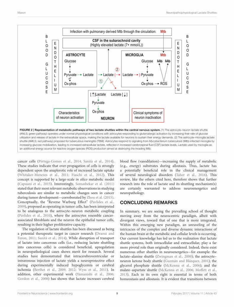
In the remainder of this review an adaptation of the ANLSmodel—the AMLS model (Mason et al., 2015)—is discussed interms of TBM; a chronic, infectious neuroinflammatory disease.
Pathogen-Induced ChronicNeuroinflammationIn the case of a chronic infection in the brain by a persistentpathogen, such asMycobacterium tuberculosis (Mtb), the bacillusresponsible for TBM, there is sustained allostatic overload. Here,the microglia strive to eradicate the scourge but unwittinglybecome the habitat of the persisting pathogen. Similar to theANLS hypothesis, the AMLSmodel proposes that when the brainis in crisis due to infection, energy flow in brain metabolism isshifted away from the neurons, and shunted toward the microglia(see Figure 2). The initial mechanics of the AMLS remain thesame as the ANLS, the only difference being the directionalityof the lactate upon exiting the astrocyte. The AMLS hypothesispostulates that in neuroinflammatory infectious diseases, suchas TBM, lactate produced through glycolysis in astrocytesparticipates in the activated immune response and, in associationwith ketones and gluconeogenic amino acids, is collectivelydirected from the neurons preferentially into microglia. Withinthe microglia, lactate is expected to enter the mitochondrialKrebs cycle, contributing to oxidative phosphorylation and henceproducing high levels of ATP and forms of ROS, such ashydrogen peroxide, required for degradation of the invadingpathogen. Thus, the AMLS hypothesis uses the same line of logicof the ANLS model, but instead the microglia are the primefocus under conditions of neuroinflammatory infectious diseases;increased astrocytic lactate is directed toward the microglia.
Using the same CSF samples from the study by Mason et al.(2015), it was shown in a targeted analysis (Mason et al., 2016a)that the highly elevated amounts of lactate observed in the CSFof confirmed TBM cases was only in the L-form (L-lactate isproduced by the host, whereas D-lactate is of bacterial origin).Thus, elevated lactate found in the CSF of TBM cases is solelya response by the host to the infection. These results from thisfollow-up study by Mason et al. (2016a) provide experimentalevidence to support the proposed AMLS model and highlight thefact that lactate plays an important role in neuroinflammatorydiseases, such as TBM. Activation of microglia, as required in
the AMLS hypothesis, does not, however, present a uniformprocess and involves intricate interactions and feedback loopsbetween the microglia, astrocytes and neurons that hamperattempts to construct basic and linear cascades of cause andeffect; TBM involves a complex integration of the responses fromthe various cell types present within the CNS, with microglia andthe astrocytes as main players.
During the course of a neuroinflammatory infectious disease,such as TBM, the ANLS is not only inadequate but, indeed,a liability as activated neurons will only become vulnerableto neurodestructive components. The bypassing of metabolicenergy intermediates from the neurons effectively inactivatesthem to protect them fromneurodestructive agents in response tochronic neuroinflammation. This neuron inactivation is evidentfrom a clinical viewpoint as the progression of TBM is reflectedby reduced scoring in the Glasgow Coma Score—specifically,decreased consciousness and awareness. The AMLS hypothesiscan thus be viewed as invoking a persistent allostatic overloadstate that becomes activated when allostatic mechanisms fail andthe body enters into a diseased state. The regulation of dynamicsystems that function within the brain, in terms of homeostasis,allostasis and disease states, is delicate. Chronic activation ofmicroglia can lead to neuropathological sequelae (Streit et al.,2004), and is also linked to neuropathic pain (Raghavendra et al.,2003). The persistent activation of microglia can be viewed asthe proverbial double-edged sword—simultaneously exhibitingneuroprotective and neurodestructive properties in an attempt tosave the whole at the expense of the part (Hall et al., 1998; Giaumeet al., 2007).
Using TBM as an example of an infectious neuroinflammatorydisease—an extreme scenario of allostatic overload—it can bespeculated that the Mtb-induced host and microbe markers canreflect: (1) the disease state on admission to hospital, and (2)differentiation in the restoration toward a new condition ofhomeostasis following treatment. Upon admission to hospital,most TBM patients present with moderate to severe symptomsof ketosis. Metabolomics analysis of the urine (Mason et al.,2016b) substantiates this clinical picture through a ketosisurinary biosignature—cases admitted to hospital with very highketosis biomarkers tended to have a poor prognosis, leadingto severe neurological complications irrespective of treatment.A similar profile was also reflected in the CSF biochemistry(Mason et al., 2016a); as the TBM disease progressed intoits later stages there was a deterioration in patient prognosis.Hence, in more advanced TBM stages there is less CSF lactateand therefore fewer energy substrates available for microgliaactivity, and decreased protection of neurons. During this period,irreversible neurological damage can begin to occur—illustratingthe importance of lactate levels during periods of chronicneuroinflammation.
The concept of lactate shuttle systems extends to other typesof neuronal cells, such as the oligodendrocyte–axon lactateshuttle (Baltan, 2015) and the Bergmann glia (BG) Purkinjecell (PC) lactate shuttle (Sawada et al., 2016). Lactate shuttlesextend beyond the brain—they are of particular interest incancer research (Pizzuto et al., 2012). Several cancer studies havesuggested a tumor-to-stroma coupling via a lactate shuttle in
Frontiers in Neuroscience | www.frontiersin.org 8 February 2017 | Volume 11 | Article 43
Mason Neuro(patho)physiological Lactate Shuttles
FIGURE 2 | Representation of metabolic pathways of two lactate shuttles within the central nervous system. (1) The astrocyte–neuron lactate shuttle
(ANLS; green pathway) operates under normal physiological conditions with astrocytes responding to glutamatergic activation by increasing their rate of glucose
utilization and release of lactate in the extracellular space, making the lactate available for neurons to sustain their energy demands. (2) The astrocyte–microglia lactate
shuttle (AMLS; red pathway) proposed for tuberculous meningitis (TBM). Astrocytes respond to signaling from Mycobacterium tuberculosis (Mtb)-infected microglia by
increasing glucose mobilization, leading to increased extracellular lactate, reflected in increased cerebrospinal fluid (CSF) lactate levels. Lactate used by microglia as
an additional energy source for reactive oxygen species (ROS) production aimed at destroying the invading Mtb.
cancer cells (Pértega-Gomes et al., 2014; Sanità et al., 2014).These studies indicate that over-propagation of cells is stronglydependent upon the anaplerotic role of increased lactate uptake(Whitaker-Menezes et al., 2011; Fiaschi et al., 2012). Thisconcept is supported by a large-scale in silico metabolic model(Capuani et al., 2015). Interestingly, Somashekar et al. (2011)stated that their most relevant metabolic observations in studyingtuberculosis are similar to metabolic changes seen in cancerduring tumor development—corroborated by Zhou et al. (2015).Conceptually, the “Reverse Warburg Effect” (Pavlides et al.,2009), proposed as operating in tumor cells, has been interpretedto be analogous to the astrocyte–neuron metabolic coupling(Pavlides et al., 2010), where the astrocytes resemble cancer-associated fibroblasts and the neuron the epithelial tumor cells,resulting in their higher proliferative capacity.
The regulation of lactate shuttles has been discussed as beinga potential therapeutic target in cancer research (Draoui andFeron, 2011; Sanità et al., 2014). While disruption of transportof lactate into cancerous cells (i.e., reducing lactate shuttlinginto cancerous cells) is considered beneficial, upregulationin neuropathological cases shows merit for research. Severalstudies have demonstrated that intracerebroventricular orintravenous injection of lactate yields a neuroprotective effectduring experimentally induced hypoglycemia or cerebralischemia (Berthet et al., 2009, 2012; Wyss et al., 2011). Inaddition, other experimental work (Yamanishi et al., 2006;Gordon et al., 2008) has shown that lactate increases cerebral
blood flow (vasodilation)—increasing the supply of metabolic(e.g., energy) substrates during allostasis. Thus, lactate hasa potentially beneficial role in the clinical managementof several neurological disorders (Taher et al., 2016). Thisreview, like the others cited here, therefore shows that furtherresearch into the role of lactate and its shuttling mechanism(s)are certainly warranted to address neuroenergetics andneuropathology.
CONCLUDING REMARKS
In summary, we are seeing the prevailing school of thoughtmoving away from the neurocentric paradigm, albeit withdivergent views, toward that of one that is more integrated.Within this emerging new paradigm, an unraveling of theintricacies of the complex and diverse dynamic interactions ofthe human brain at the metabolic and cellular levels is occurring.Our current knowledge has led us to the realization that lactateshuttle systems, both intracellular and extracellular, play a farmore pivotal role than originally considered. Indeed, there existnumerous other shuttles in neuroenergetics—for example: thelactate–alanine shuttle (Zwingman et al., 2000); the astrocyte–neuron ketone body shuttle (Guzmán and Blázquez, 2001); theglycerol phosphate shuttle (McKenna et al., 2006); and themalate–aspartate shuttle (McKenna et al., 2006; Moffett et al.,2013). Each in its own right is essential in terms of bothhomeostasis and allostasis. It is evident that transitions between
Frontiers in Neuroscience | www.frontiersin.org 9 February 2017 | Volume 11 | Article 43
Mason Neuro(patho)physiological Lactate Shuttles
different states of homeostasis and allostasis, and disease, aredynamic—they are often difficult to distinguish and/or control.However, the role of neuroenergetics is undeniable in thebrain’s constant objective of protecting itself and trying tomaintain an optimum state. Here, we have discussed, in termsof neuroenergetics, the role of lactate in a shuttle system duringhomeostasis (ANLS) and in the infected state (AMLS). Otherstudies on neurodegenerative diseases have described complexpathophysiological changes that highlighted the importance oflactate (Newington et al., 2013; Finsterwald et al., 2015). Thereis thus overwhelming evidence that astrocytes, lactate and theassociated shuttle systems so far recognized in combination are ofcrucial importance in neuroenergetics—in both the healthy anddiseased state. Perhaps not surprisingly, the latest technology hasrevealed that neuro(patho)physiological metabolism is far morecomplex than originally thought and that further knowledge
of the lactate shuttle systems apparently operating, as outlinedin this article, is key to our understanding of the mechanismsinvolved.
AUTHOR CONTRIBUTIONS
The author confirms being the sole contributor of this work andapproved it for publication.
FUNDING
Research funding was provided by the Technological InnovationAgency (TIA) of the Department of Science and Technology ofSouth Africa. Opinions expressed and conclusions arrived at, arethose of the author and are not necessarily to be attributed to thefunding body TIA.
REFERENCES
Albanese, M., Zagaglia, S., Landi, D., Boffa, L., Nicoletti, C. G., Marciani, M. G.,
et al. (2016). Cerebrospinal fluid lactate is associated with multiple sclerosis
disease progression. J. Neuroinflamm. 13:36. doi: 10.1186/s12974-016-0502-1
Angamo, E. A., Röesner, J., Liotta, A., Kovács, R., and Heinemann, U.
(2016). A neuronal lactate uptake inhibitor slows recovery of extracellular
ion concentration changes in the hippocampal CA3 region by affecting
energy metabolism. J. Neurophysiol. 116, 2420–2430. doi: 10.1152/jn.003
27.2016
Attwell, D., and Laughlin, S. B. (2001). An energy budget for signaling in
the grey matter of the brain. J. Cereb. Blood Flow Metab. 21, 1133–1145.
doi: 10.1097/00004647-200110000-00001
Aubert, A., Costalat, R., Magistretti, P. J., and Pellerin, L. (2005). Brain lactate
kinetics: modeling evidence for neuronal lactate uptake upon activation. Proc.
Natl. Acad. Sci. U.S.A. 102, 16448–16453. doi: 10.1073/pnas.0505427102
Aubert, A., and Costalat, R. (2005). Interaction between astrocytes and
neurons studied using a mathematical model of compartmentalized energy
metabolism. J. Cereb. Blood Flow Metab. 25, 1476–1490. doi: 10.1038/sj.jcbfm.
9600144
Aubert, A., Pellerin, L., Magistretti, P. J., and Costalat, R. (2007). A coherent
neurobiological framework for functional neuroimaging provided by a model
integrating compartmentalized energymetabolism. Proc. Natl. Acad. Sci. U.S.A.
104, 4188–4193. doi: 10.1073/pnas.0605864104
Bak, L. K., Schousboe, A., Sonnewald, U., andWaagepetersen, H. S. (2006). Glucose
is necessary to maintain neurotransmitter homeostasis during synaptic activity
in cultured glutamatergic neurons. J. Cereb. Blood Flow Metab. 26, 1285–1297.
doi: 10.1038/sj.jcbfm.9600281
Baltan, S. (2015). Can lactate serve as an energy substrate for axons in good
times and in bad, in sickness and in health? Metab. Brain Dis. 30, 25–30.
doi: 10.1007/s11011-014-9595-3
Beal, M. F. (2000). Energetics in the pathogenesis of neurodegenerative diseases.
Trends Neurosci. 23, 298–304. doi: 10.1016/S0166-2236(00)01584-8
Bélanger, M., Allaman, I., and Magistretti, P. J. (2011). Brain energy metabolism:
focus on astrocyte-neuron metabolic cooperation. Cell Metab. 14, 724–738.
doi: 10.1016/j.cmet.2011.08.016
Bender, A. S., Young, L. P., and Norenberg, M. D. (1997). Effect of lactic acid on L-
glutamate uptake in cultured astrocytes: mechanistic considerations. Brain Res.
750, 59–66. doi: 10.1016/S0006-8993(96)01331-5
Bergersen, L. H. (2007). Is lactate food for neurons? Comparison of
monocarboxylate transporter subtypes in brain and muscle. Neuroscience
145, 11–19. doi: 10.1016/j.neuroscience.2006.11.062
Berthet, C., Lei, H., Thevenet, J., Gruetter, R., Magistretti, P. J., and Hirt, L. (2009).
Neuroprotective role of lactate after cerebral ischemia. J. Cereb. Blood Flow
Metab. 29, 1780–1789. doi: 10.1038/jcbfm.2009.97
Berthet, C., Castillo, X., Magistretti, P. J., and Hirt, L. (2012). New evidence
of neuroprotection by lactate after transient focal cerebral ischaemia:
extended benefit after intracerebroventricular injection and efficacy of
intravenous administration. Cerebrovasc. Dis. 34, 329–335. doi: 10.1159/0003
43657
Bittar, P. G., Charnay, Y., Pellerin, L., Bouras, C., and Magistretti, P. J. (1996).
Selective distribution of lactate dehydrogenase isoenzymes in neurons and
astrocytes of human brain. J. Cereb. Blood Flow Metab. 16, 1079–1089.
doi: 10.1097/00004647-199611000-00001
Blair, C., Raver, C. C., Granger, D., Mills-Koonce, R., and Hibel, L., (2011).
Allostasis and allostatic load in the context of poverty in early childhood. Dev.
Psychopathol. 23, 845–857. doi: 10.1017/S0954579411000344
Bliss, T. M., Ip, M., Cheng, E., Minami, M., Pellerin, L., Magistretti, P.,
et al. (2004). Dual-gene, dual-cell type therapy against an excitotoxic
insult by bolstering neuroenergetics. J. Neurosci. 4, 6202–6208.
doi: 10.1523/JNEUROSCI.0805-04.2004
Boumezbeur, F., Petersen, K. F., Cline, G. W., Mason, G. F., Behar, K.
L., Shulman, G. I., et al. (2010). The contribution of blood lactate
to brain energy metabolism in humans measured by dynamic 13C
nuclear magnetic resonance spectroscopy. J. Neurosci. 30, 13983–13991.
doi: 10.1523/JNEUROSCI.2040-10.2010
Bourgeois, N. M., Van Herck, S. L., Vancamp, P., Delbaere, J., Zevenbergen, C.,
Kersseboom, S., et al. (2016). Characterization of chicken thyroid hormone
transporters. Endocrinology 157, 2560–2574. doi: 10.1210/en.2015-2025
Bouzat, P., and Oddo, M. (2014). Lactate and the injured brain: friend or foe? Curr.
Opin. Crit. Care 20, 133–140. doi: 10.1097/MCC.0000000000000072
Bouzier-Sore, A. K., Merle, M., Magistretti, P. J., and Pellerin, L. (2002). Feeding
active neurons: (re) emergence of a nursing role for astrocytes. J. Physiol. Paris
96, 273–282. doi: 10.1016/S0928-4257(02)00016-5
Bouzier-Sore, A. K., Serres, S., Canioni, P., and Merle, M. (2003). Lactate
involvement in neuron–glia metabolic interaction: 13C-NMR spectroscopy
contribution. Biochimie 85, 841–848. doi: 10.1016/j.biochi.2003.08.003
Brodersen, P., and Jorgensen, E. O. (1974). Cerebral blood flow and oxygen uptake,
and cerebrospinal fluid biochemistry in severe coma. J. Neurol. Neurosurg.
Psychiatr. 37, 384–391. doi: 10.1136/jnnp.37.4.384
Brooks, G. A. (1985). “Lactate: glycolytic end product and oxidative substrate
during sustained exercise in mammals – the ‘lactate shuttle’,” in Circulation,
Respiration and Metabolism: Current Comparative Approaches, ed R. Gilles
(Berlin: Springer–Verlag), 208–218.
Brooks, G. A. (1986a). The lactate shuttle during exercise and recovery. Med. Sci.
Sports Exerc. 18, 360–368.
Brooks, G. A. (1986b). Lactate production under fully aerobic conditions: the
lactate shuttle during rest and exercise. Fed. Proc. 45, 2924–2929.
Brooks, G. A. (2000). Intra-and extra-cellular lactate shuttles. Med. Sci. Sports
Exerc. 32, 790–799. doi: 10.1097/00005768-200004000-00011
Frontiers in Neuroscience | www.frontiersin.org 10 February 2017 | Volume 11 | Article 43
Mason Neuro(patho)physiological Lactate Shuttles
Brooks, G. A. (2002). Lactate shuttles in nature. Biochem. Soc. Trans. 30, 258–264.
doi: 10.1042/bst0300258
Brooks, G. A. (2009). Cell–cell and intracellular lactate shuttles. J. Physiol. 587,
5591–5600. doi: 10.1113/jphysiol.2009.178350
Brooks, G. A., and Martin, N. A. (2015). Cerebral metabolism following traumatic
brain injury: new discoveries with implications for treatment. Front. Neurosci.
8:408. doi: 10.3389/fnins.2014.0040
Cambron, M., D’haeseleer, M., Laureys, G., Clinckers, R., Debruyne, J., and De
Keyser, J. (2012). White-matter astrocytes, axonal energy metabolism, and
axonal degeneration in multiple sclerosis. J. Cereb. Blood Flow Metab. 32,
413–424. doi: 10.1038/jcbfm.2011.193
Campbell, G. R., Worrall, J. T., and Mahad, D. J. (2014). The central role of
mitochondria in axonal degeneration in multiple sclerosis. Mult. Scler. J. 20,
1806–1813. doi: 10.1177/1352458514544537
Capuani, F., De Martino, D., Marinari, E., and DeMartino, A. (2015). Quantitative
constraint-based computational model of tumor-to-stroma coupling via lactate
shuttle. Sci. Rep. 5:11880. doi: 10.1038/srep11880
Carpenter, K. L., Jalloh, I., Gallagher, C. N., Grice, P., Howe, D. J., Mason, A.,
et al. (2014). 13C-labelled microdialysis studies of cerebral metabolism in TBI
patients. Eur. J. Pharm. Sci. 57, 87–97. doi: 10.1016/j.ejps.2013.12.012
Carpenter, K. L., Jalloh, I., and Hutchinson, P. J. (2015). Glycolysis and the
significance of lactate in traumatic brain injury. Front. Neurosci. 9:112.
doi: 10.3389/fnins.2015.00112
Cater, H. L., Benham, C. D., and Sundstrom, L. E. (2001). Neuroprotective role of
monocarboxylate transport during glucose deprivation in slice cultures of rat
hippocampus. J. Physiol. 531, 459–466. doi: 10.1111/j.1469-7793.2001.0459i.x
Cater, H. L., Chandratheva, A., Benham, C. D., Morrison, B. III., and Sundstrom,
L. E. (2003). Lactate and glucose as energy substrates during, and after, oxygen
deprivation in rat hippocampal acute and cultured slices. J. Neurochem. 87,
1381–1390. doi: 10.1046/j.1471-4159.2003.02100.x
Chaumeil, M. M., Radoul, M., Najac, C., Eriksson, P., Viswanath, P., Blough, M.
D., et al. (2016). Hyperpolarized 13CMR imaging detects no lactate production
in mutant IDH1 gliomas: implications for diagnosis and response monitoring.
Neuroimage Clin. 12, 180–189. doi: 10.1016/j.nicl.2016.06.018
Chen, T., Qian, Y. Z., Di, X., Rice, A., Zhu, J. P., and Bullock, R.
(2000). Lactate/glucose dynamics after rat fluid percussion brain injury. J.
Neurotrauma 17, 135–142. doi: 10.1089/neu.2000.17.135
Chen, Y., and Swanson, R. A. (2003). Astrocytes and brain injury. J. Cereb. Blood
Flow Metab. 23, 137–149. doi: 10.1097/00004647-200302000-00001
Chih, C. P., Lipton, P., and Roberts, E. L. Jr. (2001). Do active cerebral
neurons really use lactate rather than glucose? Trends Neurosci. 24, 573–578.
doi: 10.1016/S0166-2236(00)01920-2
Chih, C. P., and Roberts, E. L. Jr. (2003). Energy substrates for neurons
during neural activity: a critical review of the astrocyte-neuron
lactate shuttle hypothesis. J. Cereb. Blood Flow Metab. 23, 1263–1281.
doi: 10.1097/01.WCB.0000081369.51727.6F
Chiry, O., Fishbein, W. N., Merezhinskaya, N., Clarke, S., Galuske, R., Magistretti,
P. J., et al. (2008). Distribution of the monocarboxylate transporter MCT2 in
human cerebral cortex: an immunohistochemical study. Brain Res. 1226, 61–69.
doi: 10.1016/j.brainres.2008.06.025
Covarrubias-Pinto, A., Moll, P., Solís-Maldonado, M., Acuña, A. I., Riveros,
A., Miró, M. P., et al. (2015). Beyond the redox imbalance: oxidative stress
contributes to an impaired GLUT3 modulation in Huntington’s disease.
Free Radic. Biol. Med. 89, 1085–1096. doi: 10.1016/j.freeradbiomed.2015.
09.024
Cruz, R. S., de Aguiar, R. A., Turnes, T., Penteado Dos Santos, R., Fernandes
Mendes de Oliveira, M., and Caputo, F. (2012). Intracellular shuttle: the lactate
aerobic metabolism. Scientific World J. 2012:420984. doi: 10.1100/2012/420984
Danese, A., and McEwen, B. S. (2012). Adverse childhood experiences,
allostasis, allostatic load, and age-related disease. Physiol. Behav. 106, 29–39.
doi: 10.1016/j.physbeh.2011.08.019
Debernardi, R., Magistretti, P. J., and Pellerin, L. (1999). Trans-inhibition of
glutamate transport prevents excitatory amino acid-induced glycolysis in
astrocytes. Brain Res. 850, 39–46. doi: 10.1016/S0006-8993(99)02022-3
DeSalles, A. A., Kontos, H. A., Becker, D. P., Yang, M. S., Ward, J. D., Moulton, R.,
et al. (1986). Prognostic significance of ventricular CSF lactic acidosis in severe
head injury. J. Neurosurg. 65, 615–624. doi: 10.3171/jns.1986.65.5.0615
De Salles, A. A., Muizelaar, J. P., and Young, H. F. (1987).
Hyperglycemia, cerebrospinal fluid lactic acidosis, and cerebral blood
flow in severely head-injured patients. Neurosurgery 21, 45–50.
doi: 10.1227/00006123-198707000-00009
Dienel, G. A., and Hertz, L. (2001). Glucose and lactate metabolism during brain
activation. J. Neurosci. Res. 66, 824–838. doi: 10.1002/jnr.10079
Dienel, G. A., and Cruz, N. F. (2003). Neighborly interactions of
metabolically-activated astrocytes in vivo. Neurochem. Int. 43, 339–354.
doi: 10.1016/S0197-0186(03)00021-4
Dienel, G. A., and Cruz, N. F. (2004). Nutrition during brain activation: does
cell-to-cell lactate shuttling contribute significantly to sweet and sour food for
thought? Neurochem. Int. 45, 321–351. doi: 10.1016/j.neuint.2003.10.011
Dienel, G. A. (2014). Lactate shuttling and lactate use as fuel after traumatic brain
injury: metabolic considerations. J. Cereb. Blood Flow Metab. 34, 1736–1748.
doi: 10.1038/jcbfm.2014.153
Dienel, G. A., and Cruz, N. F. (2015). Contributions of glycogen to
astrocytic energetics during brain activation. Metab. Brain Dis. 30, 281–298.
doi: 10.1007/s11011-014-9493-8
DiNuzzo, M., Mangia, S., Maraviglia, B., and Giove, F. (2010a). Changes in
glucose uptake rather than lactate shuttle take center stage in subserving
neuroenergetics: evidence from mathematical modeling. J. Cereb. Blood Flow
Metab. 30, 586–602. doi: 10.1038/jcbfm.2009.232
DiNuzzo, M., Mangia, S., Maraviglia, B., and Giove, F. (2010b). Glycogenolysis
in astrocytes supports blood-borne glucose channeling not glycogen-derived
lactate shuttling to neurons: evidence from mathematical modeling. J. Cereb.
Blood Flow Metab. 30, 1895–1904. doi: 10.1038/jcbfm.2010.151
Draoui, N., and Feron, O. (2011). Lactate shuttles at a glance: from physiological
paradigms to anti-cancer treatments. Dis. Model. Mech. 4, 727–732.
doi: 10.1242/dmm.007724
Dutta, R., McDonough, J., Yin, X., Peterson, J., Chang, A., Torres, T., et al. (2006).
Mitochondrial dysfunction as a cause of axonal degeneration in multiple
sclerosis patients. Ann. Neurol. 59, 478–489. doi: 10.1002/ana.20736
Eder, C. (1998). Ion channels in microglia (brain macrophages).Am. J. Physiol. Cell
Physiol. 275, C327–C342.
Erlichman, J. S., Hewitt, A., Damon, T. L., Hart, M., Kurascz, J., Li, A., et al. (2008).
Inhibition of monocarboxylate transporter 2 in the retrotrapezoid nucleus in
rats: a test of the astrocyte–neuron lactate-shuttle hypothesis. J. Neurosci. 28,
4888–4896. doi: 10.1523/JNEUROSCI.5430-07.2008
Fiaschi, T., Marini, A., Giannoni, E., Taddei, M. L., Gandellini, P., De Donatis,
A., et al. (2012). Reciprocal metabolic reprogramming through lactate shuttle
coordinately influences tumor-stroma interplay. Cancer Res. 72, 5130–5140.
doi: 10.1158/0008-5472.CAN-12-1949
Finsterwald, C., Magistretti, P. J., and Lengacher, S. (2015). Astrocytes: new
targets for the treatment of neurodegenerative diseases. Curr. Pharm. Des. 21,
3570–3581. doi: 10.2174/1381612821666150710144502
Fox, P. T., Raichle, M. E., Mintun, M. A., and Dence, C. (1988). Nonoxidative
glucose consumption during focal physiologic neural activity. Science 241,
462–464. doi: 10.1126/science.3260686
Gallagher, C. N., Carpenter, K. L., Grice, P., Howe, D. J., Mason, A., Timofeev, I.,
et al. (2009). The human brain utilizes lactate via the tricarboxylic acid cycle:
a 13C-labelled microdialysis and high-resolution nuclear magnetic resonance
study. Brain 132, 2839–2849. doi: 10.1093/brain/awp202
Gårseth, M., Sonnewald, U., White, L. R., Rød, M., Zwart, J. A., Nygaard,
Ø., et al. (2000). Proton magnetic resonance spectroscopy of cerebrospinal
fluid in neurodegenerative disease: indication of glial energy impairment in
Huntington chorea, but not Parkinson disease. J. Neurosci. Res. 60, 779–782.
doi: 10.1002/1097-4547(20000615)60:6<779::AID-JNR10>3.0.CO;2-M
Genc, S., Kurnaz, I. A., and Ozilgen, M. (2011). Astrocyte-neuron lactate shuttle
may boost more ATP supply to the neuron under hypoxic conditions-in
silico study supported by in vitro expression data. BMC Syst. Biol. 5:162.
doi: 10.1186/1752-0509-5-162
Giaume, C., Kirchhoff, F., Matute, C., Reichenbach, A., and Verkhratsky, A.
(2007). Glia: the fulcrum of brain diseases. Cell Death Differ. 14, 1324–1335.
doi: 10.1038/sj.cdd.4402144
Giaume, C., Koulakoff, A., Roux, L., Holcman, D., and Rouach, N. (2010).
Astroglial networks: a step further in neuroglial and gliovascular interactions.
Nature Rev. Neurosci. 11, 87–99. doi: 10.1038/nrn2757
Frontiers in Neuroscience | www.frontiersin.org 11 February 2017 | Volume 11 | Article 43
Mason Neuro(patho)physiological Lactate Shuttles
Gjedde, A., Marrett, S., and Vafaee, M. (2002). Oxidative and nonoxidative
metabolism of excited neurons and astrocytes. J. Cereb. Blood Flow Metab. 22,
1–14. doi: 10.1097/00004647-200201000-00001
Gjedde, A., and Marrett, S. (2001). Glycolysis in neurons, not astrocytes,
delays oxidative metabolism of human visual cortex during sustained
checkerboard stimulation in vivo. J. Cereb. Blood Flow Metab. 21, 1384–1392.
doi: 10.1097/00004647-200112000-00002
Gladden, L. B. (2004). Lactate metabolism: a new paradigm for the
third millennium. J. Physiol. 558, 5–30. doi: 10.1113/jphysiol.2003.
058701
Glenn, T. C., Martin, N. A., Horning, M. A., McArthur, D. L., Hovda, D. A.,
Vespa, P., et al. (2015a). Lactate: brain fuel in human traumatic brain injury: a
comparison with normal healthy control subjects. J. Neurotrauma 32, 820–832.
doi: 10.1089/neu.2014.3483
Glenn, T. C., Martin, N. A., McArthur, D. L., Hovda, D. A., Vespa, P., Johnson,
M. L., et al. (2015b). Endogenous nutritive support after traumatic brain
injury: peripheral lactate production for glucose supply via gluconeogenesis.
J. Neurotrauma 32, 811–819. doi: 10.1089/neu.2014.3482
Gordon, G. R., Choi, H. B., Rungta, R. L., Ellis-Davies, G. C., and MacVicar, B.
A. (2008). Brain metabolism dictates the polarity of astrocyte control over
arterioles. Nature 456, 745–749. doi: 10.1038/nature07525
Gouarné, C., Tardif, G., Tracz, J., Latyszenok, V., Michaud, M., Clemens,
L. E., et al. (2013). Early deficits in glycolysis are specific to striatal
neurons from a rat model of huntington disease. PLoS ONE 8:e81528.
doi: 10.1371/journal.pone.0081528
Guzmán, M., and Blázquez, C. (2001). Is there an astrocyte–neuron
ketone body shuttle? Trends Endocrinol. Metab. 12, 169–173.
doi: 10.1016/S1043-2760(00)00370-2
Hall, E. D., Oostveen, J. A., and Gurney, M. E. (1998). Relationship
of microglial and astrocytic activation to disease onset and
progression in a transgenic model of familial ALS. Glia 23, 249–256.
doi: 10.1002/(SICI)1098-1136(199807)23:3<249::AID-GLIA7>3.0.CO;2-#
Hashimoto, T., Hussien, R., Cho, H. S., Kaufer, D., and Brooks, G. A. (2008).
Evidence for the mitochondrial lactate oxidation complex in rat neurons:
demonstration of an essential component of brain lactate shuttles. PLoS ONE
3:e2915. doi: 10.1371/journal.pone.0002915
Haydon, P. G., and Carmignoto, G. (2006). Astrocyte control of synaptic
transmission and neurovascular coupling. Physiol. Rev. 86, 1009–1031.
doi: 10.1152/physrev.00049.2005
Hertz, L., Dringen, R., Schousboe, A., and Robinson, S. R. (1999).
Astrocytes: glutamate producers for neurons. J. Neurosci. Res. 57, 417–428.
doi: 10.1002/(SICI)1097-4547(19990815)57:4<417::AID-JNR1>3.0.CO;2-N
Herrero-Mendez, A., Almeida, A., Fernández, E., Maestre, C., Moncada, S., and
Bolaños, J. P. (2009). The bioenergetic and antioxidant status of neurons is
controlled by continuous degradation of a key glycolytic enzyme by APC/C–
Cdh1. Nat. Cell Biol. 11, 747–752. doi: 10.1038/ncb1881
Itoh, Y., Esaki, T., Shimoji, K., Cook, M., Law, M. J., Kaufman, E., et al. (2003).
Dichloroacetate effects on glucose and lactate oxidation by neurons and
astroglia in vitro and on glucose utilization by brain in vivo. Proc. Natl. Acad.
Sci. U.S.A. 100, 4879–4884. doi: 10.1073/pnas.0831078100
Jolivet, R., Allaman, I., Pellerin, L., Magistretti, P. J., and Weber, B.
(2010). Comment on recent modeling studies of astrocyte–neuron
metabolic interactions. J. Cereb. Blood Flow Metab. 30, 1982–1986.
doi: 10.1038/jcbfm.2010.132
Jolivet, R., Coggan, J. S., Allaman, I., and Magistretti, P. J. (2015). Multi-
timescale modeling of activity-dependent metabolic coupling in the
neuron-glia-vasculature ensemble. PLoS Comput. Biol. 11:e1004036.
doi: 10.1371/journal.pcbi.1004036
Jueptner, M., and Weiller, C. (1995). Review: does measurement of regional
cerebral blood flow reflect synaptic activity?—Implications for PET and fMRI.
Neuroimage 2, 148–156. doi: 10.1006/nimg.1995.1017
Karagiannis, A., Sylantyev, S., Hadjihambi, A., Hosford, P. S., Kasparov, S., and
Gourine, A. V. (2016). Hemichannel-mediated release of lactate. J. Cereb. Blood
Flow Metab. 36, 1202–1211. doi: 10.1177/0271678X15611912
Kimelberg, H. K., and Norenberg, M. D. (1989). Astrocytes. Sci. Am. 260, 66–72.
doi: 10.1038/scientificamerican0489-66
Kolko, M., Vosborg, F., Henriksen, U. L., Hasan-Olive, M. M., Diget, E. H.,
Vohra, R., et al. (2016). Lactate transport and receptor actions in retina:
potential roles in retinal function and disease. Neurochem. Res. 41, 1229–1236.
doi: 10.1007/s11064-015-1792-x
Korogod, N., Petersen, C. C., and Knott, G. W. (2015). Ultrastructural analysis of
adult mouse neocortex comparing aldehyde perfusion with cryo fixation. Elife
4:e05793. doi: 10.7554/eLife.05793
Kuchiiwa, T., Nio-Kobayashi, J., Takahashi-Iwanaga, H., Yajima, T., and Iwanaga,
T. (2011). Cellular expression of monocarboxylate transporters in the female
reproductive organ of mice: implications for the genital lactate shuttle.
Histochem. Cell Biol. 135, 351–360. doi: 10.1007/s00418-011-0794-2
Kuhr, W. G., and Korf, J. (1988). Extracellular lactic acid as an indicator
of brain metabolism: continuous on-line measurement in conscious, freely
moving rats with intrastriatal dialysis. J. Cereb. Blood Flow Metab. 8, 130–137.
doi: 10.1038/jcbfm.1988.17
Laskas, J. M. (2009). Game Brain. Gentlemen’s Quarterly. New York, NY: Condé
Nast.
Laskas, J. M. (2015). Concussion. New York, NY: Random House.
Laughton, J. D., Charnay, Y., Belloir, B., Pellerin, L., Magistretti, P. J., and Bouras,
C. (2000). Differential messenger RNA distribution of lactate dehydrogenase
LDH-1 and LDH-5 isoforms in the rat brain. Neuroscience 96, 619–625.
doi: 10.1016/S0306-4522(99)00580-1
Laureys, G., Valentino, M., Demol, F., Zammit, C., Muscat, R., Cambron, M.,
et al. (2014). β 2-Adrenergic receptors protect axons during energetic stress
but do not influence basal glio-axonal lactate shuttling in mouse white matter.
Neuroscience 277, 367–374. doi: 10.1016/j.neuroscience.2014.07.022
Lee, N. Y., and Kang, Y. S. (2016). In vivo and in vitro evidence for brain uptake of
4-phenylbutyrate by the monocarboxylate transporter 1 (MCT1). Pharm. Res.
33, 1711–1722. doi: 10.1007/s11095-016-1912-6
Leen, W. G., Willemsen, M. A., Wevers, R. A., and Verbeek, M. M.
(2012). Cerebrospinal fluid glucose and lactate: age-specific reference
values and implications for clinical practice. PLoS ONE 7:e42745.
doi: 10.1371/journal.pone.004274
Li, C., Zhao, R., Gao, K., Wei, Z., Yin, Y. M., Lau, T. L., et al. (2011). Astrocytes:
implications for neuroinflammatory pathogenesis of Alzheimer’s disease. Curr.
Alzheimer Res. 8, 67–80. doi: 10.2174/156720511794604543
Logan, J. G., and Barksdale, D. J. (2008). Allostasis and allostatic load: expanding
the discourse on stress and cardiovascular disease. J. Clin. Nurs. 17, 201–208.
doi: 10.1111/j.1365-2702.2008.02347.x
Lutz, N. W., Viola, A., Malikova, I., Confort-Gouny, S., Audoin, B., Ranjeva,
J. P., et al. (2007). Inflammatory multiple-sclerosis plaques generate
characteristic metabolic profiles in cerebrospinal fluid. PLoS ONE 2:e595.
doi: 10.1371/journal.pone.0000595
Magistretti, P. J., and Pellerin, L. (1996). Cellular bases of brain energy metabolism
and their relevance to functional brain imaging: evidence for a prominent role
of astrocytes. Cereb. Cortex 6, 50–61. doi: 10.1093/cercor/6.1.50
Magistretti, P. J., and Pellerin, L. (1999). Astrocytes couple synaptic activity to
glucose utilization in the brain. Physiology 14, 177–182.
Magistretti, P. J. (2000). Cellular bases of functional brain imaging:
insights from neuron-glia metabolic coupling. Brain Res. 886, 108–112.
doi: 10.1016/S0006-8993(00)02945-0
Magistretti, P. J. (2006). Neuron–glia metabolic coupling and plasticity. J. Exp. Biol.
209, 2304–2311. doi: 10.1242/jeb.02208
Magistretti, P. J. (2009). Role of glutamate in neuron-glia metabolic coupling. Am.
J. Clin. Nutr. 90, 875S–880S. doi: 10.3945/ajcn.2009.27462CC
Mangia, S., Giove, F., Bianciardi, M., Di Salle, F., Garreffa, G., and Maraviglia, B.
(2003a). Issues concerning the construction of a metabolic model for neuronal
activation. J. Neurosci. Res. 71, 463–467. doi: 10.1002/jnr.10531
Mangia, S., Garreffa, G., Bianciardi, M., Giove, F., Di Salle, F., and Maraviglia,
B. (2003b). The aerobic brain: lactate decrease at the onset of neural activity.
Neuroscience 118, 7–10. doi: 10.1016/S0306-4522(02)00792-3
Mangia, S., Simpson, I. A., Vannucci, S. J., and Carruthers, A. (2009). The in vivo
neuron-to-astrocyte lactate shuttle in human brain: evidence from modeling
of measured lactate levels during visual stimulation. J. Neurochem. 109, 55–62.
doi: 10.1111/j.1471-4159.2009.06003.x
Mason, S., van Furth, A. M., Mienie, L. J., Engelke, U. F., Wevers, R. A., Solomons,
R., et al. (2015). A hypothetical astrocyte–microglia lactate shuttle derived from
a 1H NMR metabolomics analysis of cerebrospinal fluid from a cohort of
South African children with tuberculousmeningitis.Metabolomics 11, 822–837.
doi: 10.1007/s11306-014-0741-z
Frontiers in Neuroscience | www.frontiersin.org 12 February 2017 | Volume 11 | Article 43
Mason Neuro(patho)physiological Lactate Shuttles
Mason, S., Reinecke, C. J., Kulik, W., van Cruchten, A., Solomons, R., and
van Furth, A. M. (2016a). Cerebrospinal fluid in tuberculous meningitis
exhibits only the L-enantiomer of lactic acid. BMC Infect. Dis. 16:251.
doi: 10.1186/s12879-016-1597-9
Mason, S., van Furth, A. M. T., Solomons, R., Wevers, R. A., van Reenen,
M., and Reinecke, C. J. (2016b). A putative urinary biosignature for
diagnosis and follow-up of tuberculous meningitis in children: outcome of
a metabolomics study disclosing host–pathogen responses. Metabolomics 12,
1–16. doi: 10.1007/s11306-016-1053-2
McEwen, B. S. (2006). Sleep deprivation as a neurobiologic and physiologic
stressor: allostasis and allostatic load. Metab. Clin. Exp. 55, S20–S23.
doi: 10.1016/j.metabol.2006.07.008
McEwen, B. S. (2008). Central effects of stress hormones in health and disease:
understanding the protective and damaging effects of stress and stress
mediators. Eur. J. Pharmacol. 583, 174–185. doi: 10.1016/j.ejphar.2007.11.071
McEwen, B. S., and Gianaros, P. J. (2011). Stress- and allostasis-
induced brain plasticity. Annu. Rev. Med. 62, 431–445.
doi: 10.1146/annurev-med-052209-100430
McKenna, M. C., Waagepetersen, H. S., Schousboe, A., and Sonnewald, U. (2006).
Neuronal and astrocytic shuttle mechanisms for cytosolic-mitochondrial
transfer of reducing equivalents: current evidence and pharmacological tools.
Biochem. Pharmacol. 71, 399–407. doi: 10.1016/j.bcp.2005.10.011
Moffett, J. R., Arun, P., Ariyannur, P. S., and Namboodiri, A. M. (2013).
N-Acetylaspartate reductions in brain injury: impact on post-injury
neuroenergetics, lipid synthesis, and protein acetylation. Front.Neuroenergetics
5:11. doi: 10.3389/fnene.2013.00011
Moro, N., Ghavim, S., Harris, N. G., Hovda, D. A., and Sutton, R. L.
(2013). Glucose administration after traumatic brain injury improves cerebral
metabolism and reduces secondary neuronal injury. Brain Res. 1535, 124–136.
doi: 10.1016/j.brainres.2013.08.044
Newington, J. T., Harris, R. A., and Cumming, R. C. (2013). Reevaluating
metabolism in Alzheimer’s disease from the perspective of the
astrocyte-neuron lactate shuttle model. J. Neurodegen. Dis. 2013:234572.
doi: 10.1155/2013/234572
Nijland, P. G., Molenaar, R. J., van der Pol, S. M., van der Valk, P., van Noorden, C.
J., de Vries, H. E., et al. (2015). Differential expression of glucose-metabolizing
enzymes in multiple sclerosis lesions. Acta Neuropathol. Commun. 3:79.
doi: 10.1186/s40478-015-0261-8
Norenberg, M. D., Mozes, L. W., Gregorios, J. B., and Norenberg, L. O. (1987).
Effects of lactic acid on astrocytes in primary culture. J. Neuropathol. Exp.
Neurol. 46, 154–166. doi: 10.1097/00005072-198703000-00004
Olson, J. K., Girvin, A. M., and Miller, S. D. (2001). Direct activation of innate
and antigen-presenting functions ofmicroglia following infection with Theiler’s
virus. J. Virol. 75, 9780–9789. doi: 10.1128/JVI.75.20.9780-9789.2001
Olson, J. K., and Miller, S. D. (2004). Microglia initiate central nervous system
innate and adaptive immune responses through multiple TLRs. J. Immunol.
173, 3916–3924. doi: 10.4049/jimmunol.173.6.3916
Oresic, M., Hänninen, V. A., and Vidal-Puig, A. (2008). Lipidomics: a
new window to biomedical frontiers. Trends Biotechnol. 26, 647–652.
doi: 10.1016/j.tibtech.2008.09.001
Ovanesov, M. V., Ayhan, Y., Wolbert, C., Moldovan, K., Sauder, C., and
Pletnikov, M. V. (2008). Astrocytes play a key role in activation of
microglia by persistent Borna disease virus infection. J. Neuroinflamm. 5:50.
doi: 10.1186/1742-2094-5-50
Patet, C., Suys, T., Carteron, L., and Oddo, M. (2016). Cerebral lactate
metabolism after traumatic brain injury. Curr. Neurol. Neurosci. Rep. 16, 1–7.
doi: 10.1007/s11910-016-0638-5
Pavlides, S., Whitaker-Menezes, D., Castello-Cros, R., Flomenberg, N., Witkiewicz,
A. K., Frank, P. G., et al. (2009). The reverse Warburg effect—aerobic glycolysis
in cancer associated fibroblasts and the tumor stroma. Cell Cycle 8, 3984–4001.
doi: 10.4161/cc.8.23.10238
Pavlides, S., Tsirigos, A., Vera, I., Flomenberg, N., Frank, P. G., Casimiro,
M. C., et al. (2010). Transcriptional evidence for the “Reverse Warburg
Effect” in human breast cancer tumor stroma and metastasis: similarities
with oxidative stress, inflammation, Alzheimer’s disease, and “Neuron-Glia
Metabolic Coupling”. Aging 2, 185–199. doi: 10.18632/aging.100134
Pellerin, L., Bonvento, G., Voutsinos-Porche, B., Tanaka, K., Chatton, J. Y., Brunet,
J. F., et al. (2002). Role of astrocytes in coupling synaptic activity to glucose
utilization. Int. Congr. Ser. 1235, 189–196. doi: 10.1016/S0531-5131(02)00186-3
Pellerin, L. (2003). Lactate as a pivotal element in neuron–
glia metabolic cooperation. Neurochem. Int. 43, 331–338.
doi: 10.1016/S0197-0186(03)00020-2
Pellerin, L., and Magistretti, P. J. (2004). Neuroenergetics: calling
upon astrocytes to satisfy hungry neurons. Neuroscientist 10, 53–62.
doi: 10.1177/1073858403260159
Pellerin, L., Bouzier-Sore, A. K., Aubert, A., Serres, S., Merle, M., Costalat, R., et al.
(2007). Activity-dependent regulation of energy metabolism by astrocytes: an
update. Glia 55, 1251–1262. doi: 10.1002/glia.20528
Pellerin, L. (2010). Food for thought: the importance of glucose and other
energy substrates for sustaining brain function under varying levels of activity.
Diabetes Metab. 36, S59–S63. doi: 10.1016/S1262-3636(10)70469-9
Pellerin, L., and Magistretti, P. J. (2011). Sweet sixteen for ANLS. J. Cereb. Blood
Flow Metab. 32, 1152–1166. doi: 10.1038/jcbfm.2011.149
Pérez-Escuredo, J., Van Hée, V. F., Sboarina, M., Falces, J., Payen, V. L., Pellerin, L.,
et al. (2016). Monocarboxylate transporters in the brain and in cancer. Biochim.
Biophys. Acta 1863, 2481–2497. doi: 10.1016/j.bbamcr.2016.03.013
Pértega-Gomes, N., Vizcaíno, J. R., Attig, J., Jurmeister, S., Lopes, C., and
Baltazar, F. (2014). A lactate shuttle system between tumour and stromal cells
is associated with poor prognosis in prostate cancer. BMC Cancer 14:352.
doi: 10.1186/1471-2407-14-352
Peters, A., and McEwen, B. S. (2015). Stress habituation, body shape
and cardiovascular mortality. Neurosci. Biobehav. Rev. 56, 139–150.
doi: 10.1016/j.neubiorev.2015.07.001
Pierre, K., Pellerin, L., Debernardi, R., Riederer, B. M., and Magistretti, P.
J. (2000). Cell-specific localization of monocarboxylate transporters,
MCT1 and MCT2, in the adult mouse brain revealed by double
immunohistochemical labeling and confocal microscopy. Neuroscience
100, 617–627. doi: 10.1016/S0306-4522(00)00294-3
Pizzuto, R., Paventi, G., Porcile, C., Sarnataro, D., Daniele, A., and Passarella, S.
(2012). L-Lactate metabolism in HEP G2 cell mitochondria due to the L-lactate
dehydrogenase determines the occurrence of the lactate/pyruvate shuttle and
the appearance of oxaloacetate, malate and citrate outside mitochondria.
Biochim. Biophys. Acta 1817, 1679–1690. doi: 10.1016/j.bbabio.2012.
05.010
Porras, O. H., Loaiza, A., and Barros, L. F. (2004). Glutamate mediates
acute glucose transport inhibition in hippocampal neurons. J. Neurosci. 24,
9669–9673. doi: 10.1523/JNEUROSCI.1882-04.2004
Prichard, J., Rothman, D., Novotny, E., Petroff, O., Kuwabara, T., Avison, M.,
et al. (1991). Lactate rise detected by 1H NMR in human visual cortex
during physiologic stimulation. Proc. Natl. Acad. Sci. U.S.A. 88, 5829–5831.
doi: 10.1073/pnas.88.13.5829
Psychogios, N., Hau, D. D., Peng, J., Guo, A. C., Mandal, R., Bouatra,
S., et al. (2011). The human serum metabolome. PLoS ONE 6:e16957.
doi: 10.1371/journal.pone.0016957
Raghavendra, V., Tanga, F., and DeLeo, J. A. (2003). Inhibition of microglial
activation attenuates the development but not existing hypersensitivity
in a rat model of neuropathy. J. Pharm. Exp. Ther. 306, 624–630.
doi: 10.1124/jpet.103.052407
Rake, A. J., Srinivasan, V., Nadkarni, V., Kaptan, R., and Newth, C. J. L. (2010).
Glucose variability and survival in critically ill children: allostasis or harm?
Pediatr. Crit. Care Med. 11, 707–712. doi: 10.1097/PCC.0b013e3181e88b1f
Rakus, D., Gizak, A., and Wisniewski, J. R. (2016). Proteomics unveils fibroblast-
cardiomyocyte lactate shuttle and hexokinase paradox in mouse muscles. J.
Proteome Res. 15, 2479–2490. doi: 10.1021/acs.jproteome.5b01149
Ramautar, R., Berger, R., van der Greef, J., and Hankemeier, T. (2013). Human
metabolomics: strategies to understand biology. Curr. Opin. Chem. Biol. 17,
841–846. doi: 10.1016/j.cbpa.2013.06.015
Ranjbar, M., and Amiri, M. (2015). On the role of astrocyte analog
circuit in neural frequency adaptation. Neural Comput. Appl. 26, 1–13.
doi: 10.1007/s00521-015-2112-8
Regenold, W. T., Phatak, P., Makley, M. J., Stone, R. D., and Kling, M. A.
(2008). Cerebrospinal fluid evidence of increased extra-mitochondrial glucose
metabolism implicates mitochondrial dysfunction in multiple sclerosis disease
progression. J. Neurol. Sci. 275, 106–112. doi: 10.1016/j.jns.2008.07.032
Robinet, C., and Pellerin, L. (2011). Brain-derived neurotrophic factor enhances
the hippocampal expression of key postsynaptic proteins in vivo including
the monocarboxylate transporter MCT2. Neuroscience 192, 155–163.
doi: 10.1016/j.neuroscience.2011.06.059
Frontiers in Neuroscience | www.frontiersin.org 13 February 2017 | Volume 11 | Article 43
Mason Neuro(patho)physiological Lactate Shuttles
Rock, R. B., Gekker, G., Hu, S., Sheng, W. S., Cheeran, M., Lokensgard, J. R., et al.
(2004). Role of microglia in central nervous system infections. Clin. Microbiol.
Rev. 17, 942–964. doi: 10.1128/CMR.17.4.942-964.2004
Rosafio, K., Castillo, X., Hirt, L., and Pellerin, L. (2016). Cell-specific modulation
of monocarboxylate transporter expression contributes to the metabolic
reprograming taking place following cerebral ischemia. Neuroscience 317,
108–120. doi: 10.1016/j.neuroscience.2015.12.052
Rossi, D. J., Brady, J. D., and Mohr, C. (2007). Astrocyte metabolism and signaling
during brain ischemia. Nat. Neurosci. 10, 1377–1386. doi: 10.1038/nn2004
Rothman, D. L., Behar, K. L., Hyder, F., and Shulman, R. G. (2003). In vivo
NMR studies of the glutamate neurotransmitter flux and neuroenergetics:
implications for brain function. Annu. Rev. Physiol. 65, 401–427.
doi: 10.1146/annurev.physiol.65.092101.142131
Rouach, N., Koulakoff, A., Abudara, V., Willecke, K., and Giaume, C. (2008).
Astroglial metabolic networks sustain hippocampal synaptic transmission.
Science 322, 1551–1555. doi: 10.1126/science.1164022
Sack, G. H. Jr. (2010). Mitochondrial matters in Huntington disease. J. Bioenerg.
Biomembr. 42, 189–191. doi: 10.1007/s10863-010-9291-x
Sahlin, K., Fernström, M., Svensson, M., and Tonkonogi, M. (2002). No evidence
of an intracellular lactate shuttle in rat skeletal muscle. J. Physiol. 541, 569–574.
doi: 10.1113/jphysiol.2002.016683
Sampol, D., Ostrofet, E., Jobin, M. L., Raffard, G., Sanchez, S., Bouchaud, V.,
et al. (2013). Glucose and lactate metabolism in the awake and stimulated
rat: a 13C-NMR study. Front. Neuroenergetics 5:5. doi: 10.3389/fnene.2013.
00005
Sanità, P., Capulli, M., Teti, A., Galatioto, G. P., Vicentini, C., Chiarugi,
P., et al. (2014). Tumor-stroma metabolic relationship based on lactate
shuttle can sustain prostate cancer progression. BMC Cancer 14:154.
doi: 10.1186/1471-2407-14-154
Sappey-Marinier, D., Calabrese, G., Fein, G., Hugg, J. W., Biggins, C., and Weiner,
M. W. (1992). Effect of photic stimulation on human visual cortex lactate and
phosphates using 1H and 31Pmagnetic resonance spectroscopy. J. Cereb. Blood
Flow Metab. 12, 584–592. doi: 10.1038/jcbfm.1992.82
Sawada, Y., Konno, A., Nagaoka, J., and Hirai, H. (2016). Inflammation-induced
reversible switch of the neuron-specific enolase promoter from Purkinje
neurons to Bergmann glia. Sci. Rep. 6:27758. doi: 10.1038/srep27758
Schurr, A., West, C. A., and Rigor, B. M. (1988). Lactate-supported synaptic
function in the rat hippocampal slice preparation. Science 240, 1326–1328.
doi: 10.1126/science.3375817
Schurr, A., Payne, R. S., Miller, J. J., and Rigor, B. M. (1997a). Glia are the main
source of lactate utilized by neurons for recovery of function posthypoxia. Brain
Res. 774, 221–224.
Schurr, A., Payne, R. S., Miller, J. J., and Rigor, B. M. (1997b). Brain lactate is
an obligatory aerobic energy substrate for functional recovery after hypoxia:
further in vitro validation. J. Neurochem. 69, 423–426.
Schurr, A., Payne, R. S., Miller, J. J., and Rigor, B. M. (1997c). Brain lactate,
not glucose, fuels the recovery of synaptic function from hypoxia upon
reoxygenation: an in vitro study. Brain Res. 744, 105–111.
Schurr, A., Payne, R. S., Tseng, M. T., Miller, J. J., and Rigor, B. M. (1999a). The
glucose paradox in cerebral ischemia: new insights. Ann. N.Y. Acad. Sci. 893,
386–390.
Schurr, A., Miller, J. J., Payne, R. S., and Rigor, B. M. (1999b). An increase in lactate
output by brain tissue serves to meet the energy needs of glutamate-activated
neurons. J. Neurosci. 19, 34–39.
Schurr, A. (2006). Lactate: the ultimate cerebral oxidative energy substrate? J.
Cereb. Blood Flow Metab. 26, 142–152. doi: 10.1038/sj.jcbfm.9600174
Shannon, M., King, T. L., and Kennedy, H. P. (2007). Allostasis: a
theoretical framework for understanding and evaluating perinatal
health outcomes. J. Obstet. Gynecol. Neonatal Nurs. 36, 125–134.
doi: 10.1111/j.1552-6909.2007.00126.x
Shulman, R. G., Rothman, D. L., Behar, K. L., and Hyder, F. (2004). Energetic basis
of brain activity: implications for neuroimaging. Trends Neurosci. 27, 489–495.
doi: 10.1016/j.tins.2004.06.005
Siesjö, B. K. (1988). Acidosis and ischemic brain damage. Neurochem. Pathol. 9,
31–88.
Simpson, I. A., Carruthers, A., and Vannucci, S. J. (2007). Supply and demand in
cerebral energy metabolism: the role of nutrient transporters. J. Cereb. Blood
Flow Metab. 27, 1766–1791. doi: 10.1038/sj.jcbfm.9600521
Smith, D., Pernet, A., Hallett, W. A., Bingham, E., Marsden, P. K., and Amiel, S. A.
(2003). Lactate: a preferred fuel for human brain metabolism in vivo. J. Cereb.
Blood Flow Metab. 23, 658–664. doi: 10.1097/01.WCB.0000063991.19746.11
Sokoloff, L. (1989). Circulation and energy metabolism of the brain. Basic
Neurochemistry 2, 338–413.
Somashekar, B. S., Amin, A. G., Rithner, C. D., Troudt, J., Basaraba, R., Izzo,
A., et al. (2011). Metabolic profiling of lung granuloma in Mycobacterium
tuberculosis infected guinea pigs: ex vivo 1H magic angle spinning NMR
studies. J. Proteome Res. 10, 4186–4195. doi: 10.1021/pr2003352
Sone, J. Y., Kondziolka, D., Huang, J. H., and Samadani, U. (2016). Helmet efficacy
against concussion and traumatic brain injury: a review. J. Neurosurg. 124,
1–14. doi: 10.3171/2016.2.jns151972
Steele, M. L., and Robinson, S. R. (2012). Reactive astrocytes give neurons less
support: implications for Alzheimer’s disease. Neurobiol. Aging 33, 423–e1.
doi: 10.1016/j.neurobiolaging.2010.09.018
Sterling, P., and Eyer, J. (1988). “Allostasis: a new paradigm to explain arousal
pathology,” in Handbook of Life Stress, Cognition and Health, eds S. Fisher and
J. Reason (Oxford: John Wiley & Sons), 629–649.
Stewart, J. A. (2006). The detrimental effects of allostasis: allostatic load
as a measure of cumulative stress. J. Physiol. Anthropol. 25, 133–145.
doi: 10.2114/jpa2.25.133
Streit, W. J., Mrak, R. E., and Griffin, W. S. (2004). Microglia and
neuroinflammation: a pathological perspective. J. Neuroinflamm. 1:14.
doi: 10.1186/1742-2094-1-14
Stumvoll, M., Tataranni, P. A., Stefan, N., Vozarova, B., and Bogardus, C.
(2003). Perspective in diabetes: glucose allostasis. Diabetes 52, 903–909.
doi: 10.2337/diabetes.52.4.903
Stumvoll, M., Tataranni, P. A., and Bogardus, C. (2004). The role of glucose
allostasis in type 2 diabetes. Rev. Endocr. Metab. Disord. 5, 99–103.
doi: 10.1023/B:REMD.0000021430.56457.2c
Sussman, E. S., Ho, A. L., Pendharkar, A. V., and Ghajar, J. (2016). Clinical
evaluation of concussion: the evolving role of oculomotor assessments.
Neurosurg. Focus 40, E7. doi: 10.3171/2016.1.FOCUS15610
Suzuki, A., Stern, S. A., Bozdagi, O., Huntley, G. W., Walker, R. H., Magistretti,
P. J., et al. (2011). Astrocyte-neuron lactate transport is required for
long-term memory formation. Cell 144, 810–823. doi: 10.1016/j.cell.2011.
02.018
Tabernero, A., Vicario, C., and Medina, J. M. (1996). Lactate spares glucose as a
metabolic fuel in neurons and astrocytes from primary culture. Neurosci. Res.
26, 369–376. doi: 10.1016/S0168-0102(96)01121-2
Taher, M., Leen, W. G., Wevers, R. A., and Willemsen, M. A. (2016). Lactate
and its many faces. Eur. J. Paediatr. Neurol. 20, 3–10. doi: 10.1016/j.ejpn.2015.
09.008
Tannenbaum, B., Tannenbaum, G. S., and Anisman, S. H. (2002). Neurochemical
and behavioural alterations elicited by a chronic intermittent stressor
regimen: implications for allostatic load. Brain Res. 963, 82–92.
doi: 10.1016/S0006-8993(02)03273-0
Tomiyama, A. J., O’Donovan, A., Lin, J., Puterman, E., Lazaro, A., Chan, J., et al.
(2012). Does cellular aging relate to patterns of allostasis? An examination of
basal and stress reactive HPA axis activity and telomere length. Physiol. Behav.
106, 40–45. doi: 10.1016/j.physbeh.2011.11.016
van Hall, G., Strømstad, M., Rasmussen, P., Jans, O., Zaar, M., Gam, C., et al.
(2009). Blood lactate is an important energy source for the human brain. J.
Cereb. Blood Flow Metab. 29, 1121–1129. doi: 10.1038/jcbfm.2009.35
Vannucci, S. J., Maher, F., and Simpson, I. A. (1997). Glucose transporter
proteins in brain: delivery of glucose to neurons and glia. Glia 21, 2–21.
doi: 10.1002/(SICI)1098-1136(199709)21:1<2::AID-GLIA2>3.0.CO;2-C
Verwaest, K. A., Vu, T. N., Laukens, K., Clemens, L. E., Nguyen, H. P., Van Gasse,
B., et al. (2011). 1H NMR based metabolomics of CSF and blood serum: a
metabolic profile for a transgenic rat model of Huntington disease. Biochim.
Biophys. Acta 1812, 1371–1379.
Voutsinos-Porche, B., Bonvento, G., Tanaka, K., Steiner, P., Welker, E., Chatton, J.
Y., et al. (2003). Glial glutamate transporters mediate a functional metabolic
crosstalk between neurons and astrocytes in the mouse developing cortex.
Neuron 37, 275–286. doi: 10.1016/S0896-6273(02)01170-4
Wender, R., Brown, A. M., Fern, R., Swanson, R. A., Farrell, K., and Ransom, B.
R. (2000). Astrocytic glycogen influences axon function and survival during
glucose deprivation in central white matter. J. Neurosci. 20, 6804–6810.
Frontiers in Neuroscience | www.frontiersin.org 14 February 2017 | Volume 11 | Article 43
Mason Neuro(patho)physiological Lactate Shuttles
Whitaker-Menezes, D., Martinez-Outschoorn, U. E., Lin, Z., Ertel, A., Flomenberg,
N., Witkiewicz, A. K., et al. (2011). Evidence for a stromal-epithelial “lactate
shuttle” in human tumors: MCT4 is a marker of oxidative stress in cancer-
associated fibroblasts. Cell Cycle 10, 1772–1783. doi: 10.4161/cc.10.11.15659
Wishart, D. S., Lewis, M. J., Morrissey, J. A., Flegel, M. D., Jeroncic, K.,
Xiong, Y., et al. (2008). The human cerebrospinal fluid metabolome.
J. Chromatogr. B Analyt. Technol. Biomed. Life Sci. 871, 164–173.
doi: 10.1016/j.jchromb.2008.05.001
Wyss, M. T., Jolivet, R., Buck, A., Magistretti, P. J., and Weber, B. (2011). In vivo
evidence for lactate as a neuronal energy source. J. Neurosci. 31, 7477–7485.
doi: 10.1523/JNEUROSCI.0415-11.2011
Yamanishi, S., Katsumura, K., Kobayashi, T., and Puro, D. G. (2006). Extracellular
lactate as a dynamic vasoactive signal in the rat retinal microvasculature. Am. J.
Physiol. Heart Circ. Physiol. 290, H925–H934. doi: 10.1152/ajpheart.01012.2005
Zeis, T., Allaman, I., Gentner, M., Schroder, K., Tschopp, J., Magistretti, P. J., et al.
(2015). Metabolic gene expression changes in astrocytes in Multiple Sclerosis
cerebral cortex are indicative of immune-mediated signaling. Brain Behav.
Immun. 48, 313–325. doi: 10.1016/j.bbi.2015.04.013
Zhou, A., Ni, J., Xu, Z., Wang, Y., Zhang, W., Wu, W., et al. (2015). Metabolomics
specificity of tuberculosis plasma revealed by 1H NMR spectroscopy.
Tuberculosis 95, 294–302. doi: 10.1016/j.tube.2015.02.038
Zuo, R. J., Gu, X. W., Qi, Q. R., Wang, T. S., Zhao, X. Y., Liu, J. L., et al.
(2015). Warburg-like glycolysis and lactate shuttle in mouse decidua during
early pregnancy. J. Biol. Chem. 290, 21280–21291. doi: 10.1074/jbc.M115.6
56629
Zwingman, C., Richter-Landsberg, C., Brand, A., and Leibfritz, D.
(2000). NMR spectroscopic study on the metabolic fate of [3-(13)C]
alanine in astrocytes, neurons, and cocultures: implications for
glia-neuron interactions in neurotransmitter metabolism. Glia 32,
286–303. doi: 10.1002/1098-1136(200012)32:3<286::AID-GLIA80>3.0.
CO;2-P
Conflict of Interest Statement: The author declares that the research was
conducted in the absence of any commercial or financial relationships that could
be construed as a potential conflict of interest.
Copyright © 2017 Mason. This is an open-access article distributed under the terms
of the Creative Commons Attribution License (CC BY). The use, distribution or
reproduction in other forums is permitted, provided the original author(s) or licensor
are credited and that the original publication in this journal is cited, in accordance
with accepted academic practice. No use, distribution or reproduction is permitted
which does not comply with these terms.
Frontiers in Neuroscience | www.frontiersin.org 15 February 2017 | Volume 11 | Article 43