Lacrimal Ductus
8
- Abstrak Twnor retrobulbar nterupakan salah satu penyakit orbita. Tunor terlelakdi belakang bola nata, terkukuttg di antara tulang orbita dan berada di jaringan vital. Ganbaran klinisnya berupa eksoftaluos, dapat disertai gangguan gerak bola nnta dan penurunanr yiszs. Penbedahan cara orbitototni lateral lebih ditujukan pada tutnor orbita yang berada di daerah tenporal, apeks atau sedikit di daerah superior. Tujûan orbitototni lnteral terutama untuk nengangkat tuttror secara en toto, nrennnipulasi organ vital di orbitasedikit tnungkin untuk nenghindarkan komplikasi gatrgguan penglihatan.Peubedahan dengan cara ini nungkin nencapai tujuannya, tetapi tatnpaloya ada pengaruh jenis tunor terhadap keberhasilan pentbedahan arau prognosis penderita buruk Bahan penelitian sejunlah 3I pasien 1'ang dilakukan penbedahan orbitototui lateral di Rwnah sakit Dr. Cipto Mangunkusunto, selanw periode Januari 1985 - 1992. Kriteria prognosis yang dittilai dalan keberhasilan pengangkatan tuilror disertai dengan atau tanpa kontplikasi pada berbagai jenis twnor. Dari keberhasilan operasi, ternlata prognosis penderita terbagi dalan 3 kelonpolç yaitu :1. Hasil operasi tercapai baik dengan prognosis penderita baih 2.Hasil operasi tercapai bailç tetapi prognosis penderiia burulç dan 3.Hasil operasi tidak tercapai dan prognosis penderita buruk. Hasil menggaubarknn bahwa lokasi, t+,alauputt apeks sekaliputr tidak nenpengaruhi operasi orbirolottti lateral. Ketidak berhasilan operasi tanqtaknl,a dipengaruhi oleh jerus tunor. Abstract Retrobulbar tu,,tors are orbital disease processes situared withitt vital tissue behind the eyeball and confined v,ithin the orbital space by the orbital bones. Surgery of the retrobulbar luntors is stil! dfficult to perfonn, due to the conplex anatonl, of the orbit u,hich can deuand variatiotts in surgical approach. In the case oforbital surgerr, a surgeon will do his ut,n(rst to reach the apical area. One of the techniques v,ell knov,n in orbital surgerf is lateral orbitotoms,. This surgery is prinarill' ained at renovitrg zygo,naric bone cotlstituring the rin of the lateral orbital bone. The pritrciple ain of ktteral orbitotortrf is to rennve the twnor en toto by uanipulating vital organs in the orbit as slightll,as possible in order to at'oid the conplications of visiott disorder. Thirtl'-one patients utuleru,ent lateral orbitotonD' at the Dr. Cipto Mangunkusuttto Hospital and other hospitals during the period betv,een January, I 985 to J u11' 1982. The criteria for successful surgery cotrsist of the evaluatiott of su.ccessful tu,,nr re,iloval v,ith or v,ithout cotttplicatiotts. Based on the saccess ofthe surgery and prognosis, the patients are divided into three groups, they 67" :1. Successfu.I su;gery', patient prognosis favorable,2.Successful surgeD:, patient prognosis dubious and 3.Utrsuccessful surgery, palienl prognosis unfavorable. Howe,ter, larcral orbitotottty did not result itt conplicatiorc in several types of tunors although they were located in the apical. This situatiott shrtu,s that even tu,nors in the apical area do not always affect the.srcce.ss of the lateral orbitotony, and thefailure of the surgery tends to be altributed nore to tuilor ry*pe. Keywords : Orbital tutnors, Itteral orbitototttl', T1'pes of tunors 248 Moeloek et al The Results of Lateral Orbitotomy on Orbital Tumors Nila F. Moeloek, Ria Mekarwangi, Tetty A Usman, Widiarti P Riono INTRODUCTION Retrobulbar tumors are orbital diseases situated within vital tissue behind the eyeball and confined within the orbit by the orbital bones. Their clinical appearance is often denronstrated by exophthalmos that can be accompanied by deterioration of vision and limitation of ocular ntove- ments. Med J Univ Indon The major difficulties facing patients with orbital tumors for the most part lie in the attempts to perform diagnosis, primarily because retrobulbar tumors are frequently difficult to palpate. This problent was over- conre by the invention of Conrputerized \xial Tomog- raphy Scanning (CT-scan) as a diagnostic aid in 1972. Utilizing this equipnrent,all the delicate tissues of the orbit surrounding and bones can be clearly scanned. This equipment is highly valuable in establislring diag-
description
lacrimal
Transcript of Lacrimal Ductus
-
Abstrak
Twnor retrobulbar nterupakan salah satu penyakit orbita. Tunor terlelakdi belakang bola nata, terkukuttg di antara tulang orbitadan berada di jaringan vital. Ganbaran klinisnya berupa eksoftaluos, dapat disertai gangguan gerak bola nnta dan penurunanr yiszs.
Penbedahan cara orbitototni lateral lebih ditujukan pada tutnor orbita yang berada di daerah tenporal, apeks atau sedikit di daerahsuperior. Tujûan orbitototni lnteral terutama untuk nengangkat tuttror secara en toto, nrennnipulasi organ vital di orbitasedikit tnungkinuntuk nenghindarkan komplikasi gatrgguan penglihatan.Peubedahan dengan cara ini nungkin nencapai tujuannya, tetapi tatnpaloyaada pengaruh jenis tunor terhadap keberhasilan pentbedahan arau prognosis penderita buruk Bahan penelitian sejunlah 3I pasien
1'ang dilakukan penbedahan orbitototui lateral di Rwnah sakit Dr. Cipto Mangunkusunto, selanw periode Januari 1985 - 1992. Kriteriaprognosis yang dittilai dalan keberhasilan pengangkatan tuilror disertai dengan atau tanpa kontplikasi pada berbagai jenis twnor.Dari keberhasilan operasi, ternlata prognosis penderita terbagi dalan 3 kelonpolç yaitu :1. Hasil operasi tercapai baik denganprognosis penderita baih 2.Hasil operasi tercapai bailç tetapi prognosis penderiia burulç dan 3.Hasil operasi tidak tercapai danprognosis penderita buruk. Hasil menggaubarknn bahwa lokasi, t+,alauputt apeks sekaliputr tidak nenpengaruhi operasi orbirolotttilateral. Ketidak berhasilan operasi tanqtaknl,a dipengaruhi oleh jerus tunor.
Abstract
Retrobulbar tu,,tors are orbital disease processes situared withitt vital tissue behind the eyeball and confined v,ithin the orbitalspace by the orbital bones. Surgery of the retrobulbar luntors is stil! dfficult to perfonn, due to the conplex anatonl, of the orbit u,hichcan deuand variatiotts in surgical approach. In the case oforbital surgerr, a surgeon will do his ut,n(rst to reach the apical area. Oneof the techniques v,ell knov,n in orbital surgerf is lateral orbitotoms,. This surgery is prinarill' ained at renovitrg zygo,naric bonecotlstituring the rin of the lateral orbital bone. The pritrciple ain of ktteral orbitotortrf is to rennve the twnor en toto by uanipulatingvital organs in the orbit as slightll,as possible in order to at'oid the conplications of visiott disorder. Thirtl'-one patients utuleru,entlateral orbitotonD' at the Dr. Cipto Mangunkusuttto Hospital and other hospitals during the period betv,een January, I 985 to J u11' 1982.The criteria for successful surgery cotrsist of the evaluatiott of su.ccessful tu,,nr re,iloval v,ith or v,ithout cotttplicatiotts. Based on thesaccess ofthe surgery and prognosis, the patients are divided into three groups, they 67" :1. Successfu.I su;gery', patient prognosis
favorable,2.Successful surgeD:, patient prognosis dubious and 3.Utrsuccessful surgery, palienl prognosis unfavorable. Howe,ter, larcralorbitotottty did not result itt conplicatiorc in several types of tunors although they were located in the apical. This situatiott shrtu,s thateven tu,nors in the apical area do not always affect the.srcce.ss of the lateral orbitotony, and thefailure of the surgery tends to bealtributed nore to tuilor ry*pe.
Keywords : Orbital tutnors, Itteral orbitototttl', T1'pes of tunors
248 Moeloek et al
The Results of Lateral Orbitotomy on Orbital TumorsNila F. Moeloek, Ria Mekarwangi, Tetty A Usman, Widiarti P Riono
INTRODUCTION
Retrobulbar tumors are orbital diseases situated withinvital tissue behind the eyeball and confined within theorbit by the orbital bones.Their clinical appearance is often denronstratedby exophthalmos that can be accompanied bydeterioration of vision and limitation of ocular ntove-ments.
Med J Univ Indon
The major difficulties facing patients with orbitaltumors for the most part lie in the attempts to performdiagnosis, primarily because retrobulbar tumors arefrequently difficult to palpate. This problent was over-conre by the invention of Conrputerized \xial Tomog-raphy Scanning (CT-scan) as a diagnostic aid in 1972.Utilizing this equipnrent,all the delicate tissues of theorbit surrounding and bones can be clearly scanned.This equipment is highly valuable in establislring diag-
Vol 3, No 4, October-Decenber i,994
noses, and its use can even be expanded to serve as aguidance in planning surgeries.
Surgery of the retrobulbar tumors is still difficultto perform, due to the complex anatomy of the orbitwhich can demand variations in surgical approach. Theprinciples of surgery, including orbital surgery,presuppose a sufficient space for this undertaking inorder to facilitate the removal of the tumor and todiminish the manipulation of other vital tissues. In thecase of orbital surgery, a surgeon will do his utmost toreach the apical area. One of the techniques wellknown in orbital surgery is lateral orbitotomy. Thissurgery is primarily aimed at removing zygomaticbone constituting the rim of the lateral orbital bÀne. l'2'3
Lateral orbitotomy is primarily directed at orbitaltumors situated at the temporal and apical spaces andto sonre extent at the superior area. In the case oftunlors situated nasally it would be advisable to reachlhe tumor through a transcranial approach. But iflateral orbitotomy is to yield the desired results for thistunror,^it should be slighty modified to meet specificneeds.3
The principle aim of lateral orbitotomy is torenlove the tumor en toto by mani al organsin the orbit as possible in orcler to omplica-tion of vision disorders. Surgery d is alsodone for biopsy in which tlre tumor mass can be neitherpalpated nor clinically diagnosed with suppcrtingdiagnostic tools.
Though surgery of this kind nright accomplish itsaim, a strong mutual relationship exists between thetype of tumor and the success of surgery,as well as,thepatients' prognosis.
The objective of this study is to perform a re-as-sessment on the usefulness of lateral orbitotomy basedon the success of tumor rentoval, postoperative com_plications, and patient prognosis.
MATERIAL AND MBTHOD
This study includes data taken from the medicalrecords of patients who had lateral or_bitotonry at Dr. Cipto Man Hospital,January 1985 to July 1992. T of patier,rrswere eye tunror patie undergone treatntentat the Ocular Tumor ment at the Dr. CiptoMangunkusunto Ho study covers a treat_ment period.
Diagnosis of retrobulbar tumor was established,on the basis of clinical examination and supportingexanrination, of which one was CT-scan examination.Histopathological examination still plays a nrajor rolein establishing diagnosis in these patients. The
ktteral Orbitotonl, on Orbital Tunrors 249
majority of patien\s underwent lateral orbitotomy witha Wright incision,3 while the remainder underwent thisorbitotomy with a slight modification, also called theosteoplasty orbitotomy tyW 2 of Nakamura.4The criteria for successful surgery consist of rheevaluation of successful tumor removal with orwithout complications.Patient prognosis was determinedon the basis of tumortype and the necessity further treatment.The data taken into account are :
- tumor removal : tumor can be removed eithcr entoto or in part (excision)
- surgical complications : is there any paresis of eyemuscle, orbital hematoma or ptosis
- visual conditions prior to and post surgery- tumor types based on the histopathologic exanrina_
tlon- further treatment on the basis of tunror types and
the patient's prognosis
Based on the success of the surgery and prog_nosis, the patients are divided into three groups, theyare :
l. successful surgery, patient prognosis favorable2. successfLrl surgery, patient prognosis dubious3. unsuccessful surgery, patient prognosis un_
favorable
The purpose of this three-group division is to reassessthe extent to which lateral orbitotonty could best serveits purpose in order that the surgery could be conductedmore effectively on certain tuntor types.
RESULTS
Thirty-one patientDr. Cipto Mangunduring the periodThey consist of(58.1%); the youngesr parienr being 4,5 years old, andthe oldest 70 years old, the nrode being 3l years old.
The data collected fronr those who underwentlateral orbitotonry include, the results of tunrorremoval; surgical conrplications; resuIting visuaI con_ditions; the histopathologic results; and the patient.sprognosis. These results are evident in the Tables1,2,3,4.
DISCUSSION
During the period of Janr.rary 1985 to July 1992, lateralorbitotonry was perfornted on 3l patients with orbitaltuntors, contprising l3 ntales and l8 fenrales with tlremode age of 3l years.
25O Moeloek et al
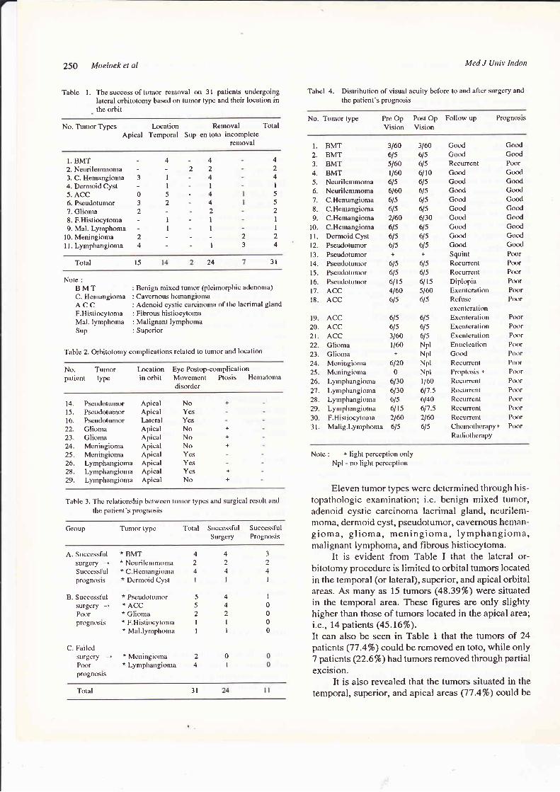
Table l. Thesuccessof lumor rcnoval olt 3l palients undcrgoinglateral orbitotomy based on tumor tyfrc and thcir location inthe orbit
No. Tumor Types Location Removal To(alApical Tenrporal Sup en toto incomplote
temoval
I. BMT2. Neurilemmoma3. C. Henrangionra4. Dermoid Cyst5. ACC6. Pseudotumor7. Glioma8. F.Histiocytoma9. Mal. Lynrphoma
10. MeningiomaI l. Lymphangionra
Tolal
Med J Utriv Indon
Tabcl 4. Distribution of visual acuity bcfore to and aftcr surgcry and
thc paticnt's prognosis
No. Tumor type Pre Op Post Op Follow upVision Vision
Prognosis
-4-422
3l-4-l-l05-432-4^a-l-l-l-l2-4--l
l..,
3.
4.5.6.
7.
8.
9.
10.
I l.t2.13.
14.
424I55
2II24
3t
;3
BMTBMTBMTBMTNeurilemmomaNcurilcnrmomaC.HcmangionraC.HcnrangiomaC.HcmangionraC.HcmangiomaDcrmoid CystPseudolumorPscudotumorPscudotunror
3160 3160
6ls 6lss/60 6lsr/60 6/106ls 6ls6160 6ls6ls 6ls6ls 6ls2160 6/306ls 6ls6ls 6ls6ls 6ls++
GotxlGoodRccurrcntGotxlGoodGoodGoqlGoodGotxlGoodGoodGoodSquintRccurrcnl
GoodGoodPtx;rGocxl
GoodGoodGoodGoodGoodGoodGotxlGcndPtxrrPtxrr
Notè :
BMTC. HenrangiomaACCF.HisticrcytonraMal. lynrphomaStrp
6ls6ls6/lss/606ls
6ls6ls6/ r54l606ls
24l5
: Bcnign mixcd lunror (plcinrorphic ad<:nonra)
: Cavcrnous lrcnrangionra: Adcnoid cystic carcinonra of thc lacrimal gland: Fibrous histiocytonra: Malignant lymphonra: Supcrior
15. Pscudolunror16. Pscudotunror17. ACC18. ACC
Rccurrcrrt Ptxrr
Diplopia PtxrrExcnlcralion PtxrrRcllsc Ptxrr
ACC 6lsACC 6lsACC 3160
Glionra l/60Glioma +
Moningionra 6120
Mcningioma 0
Lyrnphangionra 6130
Lynrphangioma 6/30Lyu.rphangionra 615
Lynrplrangioura 6l15F.Histiocytoma 2160
Malig.Lymphoma 6/5
exclltclallon615 Exonlcration Ptxrr
615 Exr:nlcration Poor
615 Excntotation Ptxrr
Npl Enuclcation PttorNpl Gotxl Poor
Npl Rccurrcnt PtrrrNpl Proptosis + Ptxrr
l/60 Rccurrcnt Pttor
6l'1.5 Rccurrcnt Ptxtr6140 Rccurrcnt Ptxrr617.5 Rccurrcnt Ptxtr
2160 Rccurrctrt Ptxrr615 Chcnrolhcrapy+ Poor
Railiothr:rapy
No(e : + light pcrccption onlyNpl - no light pcrccption
Eleven tumor types were determined through his-topathologic examination; i.e. benign mixed tumor,adenoid cystic carcinoma lacrimal gland, neurilem-moma, dermoid cyst, pseudotumor, cavernous heman-gioma, glioma, meningioma, lymphangioma,malignant lymphoma, and fibrous histiocytoma.
It is evident from Table I that the lateral or-bitotomy procedure is limited to orbital tumors locatedin the temporal (or lateral), superior, and apical orbitalareas. As many as 15 tumors (48.39%) were situatedin the temporal area. These figures are only slightyhigher than those of tumors located in the apical area;
i.e., 14 patients (45.167o).It can also be seen in Table I that the tumors of 24patients (77.4%) could be removed en toto, while only7 patients (22.6%) had tumors removed through partialexcision.
It is also revealed that the tumors situated in thetemporal, superior, and apical areas (77.4%) could be
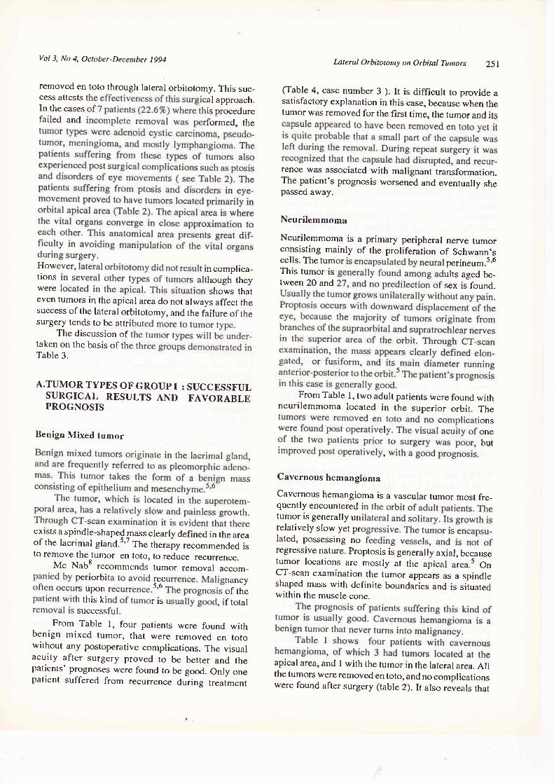
Tablc 2. Orbitotomy complications rclated to tuntor and location
19.
20.2t.22.
23.24.
25.26.
27.28.29.30.
31.
No. Tumor Locationpaticnt lypc in orbit
Eye Poslop-conrplicationMovcmcnt Ptosis Hcnratonradisordcr
14- Pscudolunor15. Pscudolunror16. Pscudotumor22. Glioma23. Glionra24. Mcningionra25. Moningioma26. Lymphangiorna28. Lynrplrangioma29. Lynrphangiona
ApicalApicalLalcralApicalApicalApicalApicalApicalApicalApical
NoYcsYcsNoNoNoYcsYcsYesNo
+
+
+
+
+
Tablc 3. Thc rolationship botwccn tunror typos and surgical rcsult and
lhc palicnl's prognosis
Gtoup Tunror lypc Total Succr:ssl'ul Succcssl'ul
Surgcry Pnrgnosis
A. Succcssl'ulsurgory +Strcccssl ul
ProSnosls
B. Succcssfulsurgcry +Poorprogn()sls
C. Failc<lsurgcry +Ptxtr
Prognosls
* BMT* Ncurilr:nrnroma* C.Hcnrangionra* Dcrnroid Cysl
* Pscudoltttnor* ACC* Glioma* F.Histitrcytonra* Mal.lynrplronra
* Moningioma* Lynrphangionra
44.,)44tl
3
2
4I
5
5
2I
I
442
I
I
0I
00o0
2
4
3tTotal
Vol 3, No 4, October-Decenber 1994
removed en toto through lateral orbitotomy. This suc_cess attests al approach.In the cases is procedurefailed and formed, the
How I ncomplica_tions hough theywere shows thateven tumors in the apical area do not always affect thesuccess of the lateral orbitotomy, and the failure of thesurgery tends to be attr
The discussion oftaken on the basis of thTable 3.
A.TUMOR TYPES OF GROUP I : SUCCESSFULSURGICAL RESULTS AND FAVORABLEPROGNOSIS
Benign Mixed tumor
exists a spindle-shaped mass clearly defined in the areaof the lacrimal gland.s'7 The therapy recommended isto remove the tumor en toto, to reduce recurrence.
Mc NabS recommends tumor ,"aouul accom_periorbi recrs upon .i'e :iï:h this ki r is f totalsuccess
From Table l, four patients were found withbenign mixed tumor, that were removed en totowithout any postoperative compli.cations. The visualacuity after surgery proved to be better and thepatients' prognoses were found to be good. Only onepatient suffered front recurrence during treatment
Lateral Orbitotony on Orbital Tutnors 2Sl
(Table 4, case number 3 ). It is difficult to provide asatisfactory explanation in this case, because when thetumor was removed for the first time, the tumor and its
rence was associated with malignant transformation.The patient's prognosis worsened and eventually shepasseci away.
Neurilemmoma
Neurilemmoma is a primary peripheral nerve tumorconsisting mainly of the proliferation of Schwann.s;iï:,ii".Ti ffJ'ï:i*:tween 20 and sex is found.
From Table l, two adult patients were found withneurilemmoma located in the superior orbit. The
tonsI on.
Cavernous hemangioma
Cavernous hemangioma is a vascular tumor most fre_quently encounteredtumor is generally unrelatively slow yet prlated, possessing noregressive nature. Proptosis is generally axial, becausetumor locations are mostly at the apical area.s OnCT-scan examination the tumor upp"u., as a spindleshaped mass wirh definite boundaiies and is situatedwithin the muscle cone.
apical area, and I with the tumor in the lateral area. Allthe tumors were re moved en toto, and no complicationswere found after surgery (table 2). It also reveals that
252 Moeloek et al
the vision after surgery was good, and even becameincreasingly better (Table 4, cases number 9).
Dermoid Cyst
The cyst walls consist of squamous epithelium and
dermis and the lumen contains keratin and hair. Der-moid cysts are usually in the temporal or superonasalareas of the orbit. Their clinical symptoms varydepending on the location and the size of the cyst. Theappearances of dermoid cysts on CT-scan are quitecharacteristic; i.e, lesions appear to be clearly definedwith thin walls and the lumen is of low density."Dermoid cysts located anteriorly can cause defects ofthe bone to where they are attached, usually in the
zygomatic frontal region.Table I shows a patient with a temporally situated
dermoid cyst which was removed en toto without anypost-operative colnplications (Table 2). Visual acuityafter surgery remained good (Table 4), and thepatient's prognosis was equally good.
It is evident from the data collected that the lateral
orbitotomy performed in the first group was success-
ful; i.e, tumors were removed en toto without post
surgery complications. Even visual acuity showeddefinite improvement in those cases and in all thepatient. Prognoses were favorable except for onepatient whose benign mixed tumor recurred. In orderto avoid recurrence, Mc Nab emphasizes the necessityof removing tumor along with the periorbital fascia.o
It is sufficient to say that lateral orbitotomy performedin the first group of tumor types succeeded in ac-
conrplishing the purposes of the treatment.
B. THE SECOND GROUP OFTUMOR TYPBS :SUCCESSFUL SURGERY WITII DUBIOUSPROGNOSIS
Adenoid Cystic Carcinoma
Adenoid cystic carcinoma is a frequently encounteredepithelial carcinonra of the lacrimal gland. This kindof tumor is one of the malignant tumors of lacrima!gland that can cause death.e Generally this tumor isfound in young adults with symptoms of unilateralproptosis with downward and medial displacement ofthe eye and is accompanied by pain. The pain indicatesthe spread of the tumor to the periorbit and netves.Through CT-scan, it is revealed that the mass isspindle-shaped with irregular boundaries. The boneerosion at the area of the superotemporal orbit is not
always distinct except when the mass is relativelylarge. The prognosis of the patient with malignanttunror of lacrinral gland is usually poor. Recurrence
Med J Uttiv Indon
rate is reasonably high, and it frequently invades orbi-tal bones.
As seen in Table 1, five patients were diagnosedhistopathologically as having adenoid cystic car-cinoma. All the tumors were removed en toto withoutany post surgery complication (Table 2). The patientsvisual acuity both prior to and after the surgeryremained good.However, because of the poor histologic charac-teristics of the tumor, and its further growth to stage IIIand IV, to prolong the patients lives further surgery inthe form of orbital exenteration coupled with radio-therapy was necessary. Visual acuity of the patientswas lost.
Pseudotunror
Pseudotumor is not a true neoplasm, but consists ofinflammatory cells forming u rnur. in the orbit.s'6This tumor usually affects young adults but can also
affect children. There is no sex prevalence, and thetumor can occur unilaterally or bilaterally.The clinical appearance is of palpebral edema, con-junctival chemosis, ocular motility disturbances, prop-tosis, and pain. Occassionally visual reduction ordiplopia may occur.The inflammatory process can involve the whole orbitor certain tissues only, such as the lacrimal gland,extraocular muscles, orbital fat, or vascular tissue.CT-scan appearances are varied. It could appear to be
retrobulbar edema, optic nerve inflammation, muscleenlargement, or an infiltrative *orr.'0
The patient's prognoses is hopeful, yet the preser-vation of visual acuity largely depends on the extentand the severity of the inflanrnatory process and the
effective response to corticosteroids and radiotherapy.
From Table l, it is seen that there wcre fivepatients with pseudolumors locatcd at the temporal andapical areas. Tunrors located at the temporal area
originated from the lacrinral gland and their ren.roval
en toto was relatively easy to perform. On the othcrhand, the en toto removal in the apical area was rela-tively more difficult, and great care hr',J to be taken.
One patient was found to have chronic 'nyositis of thelateral rectus muscle which on CT-scan examinationappeared as a mass. Only an extirpation biopsy was
performed via lateral orbitotomy. Further treatment byradiotherapy proved to have no effect, and the patientstill complained of diplopia (Table 4, case number l6).In four patients whose tumors were removed en toto,post surgical complications occurred consisting oflimited eye movement in one patient and ptosis pal-pebra in another (Table 2, cases number 14,15).
Vol 3, No 4, October-Deceuber 1994
Visual acuity both prior to and after the surgery did notchange significantly overall. During further treatmentit was found that one patient suffered from reccurrenceand another from strabismus, while two others hadcomplaints of diplopia (Table 4, cases number13,14,15,16). It seems that the prognosis for goodvisual acuity is not very encouraging, while the prog-nosis for survival remains favorable.
Glioma
Glioma is a primary tumor of the optic nerve glial cell.sThis tumor is most commonly found among children,and comprises l-27o of all orbital tumors. Nearly 50%of the tumors are diagnosed in children aged under 5.It appears with unilateral proptosis. Bilateral proptosisis a pathognomonic sign of glioma accompained byneurofibroma, and cafe-au-lait spots may be en-countered.5'6Management of the tumor varies according to the sizeand location of the tumor, as well as the visual condi-tion of the patient. It is difficult to assess the patient'sprognosis due to the variation of the disease's history.Generally, the visual prognosis is discouraging, whilethe life prognosis depends on the extension of tumorinvasion intracranially. On CT-scan the optic nerveappears enlarged and intracranial extension of thetunror through the optic foramen can be seen.
Table I reveals two five year old girls withglionra. Their tuntors were renroved in toto, but postsurgical conrplication occurred consisting of limitedeye htovement and ptosis. Visualacuity definitely wor-sened. It was also not possible to maintain the eyes ingood condition, which finally neccessitated enuclea-tion of the eye and repair of the ptosis.
Fibrous Histiocytoma
Fibrous histiocytonra, frequently referred to as fibroxanthonta, is a tuntor of histiocytes usua lly interntixingwith fibroblasts or fibrocytes.) Its clinical appearanceis generally unilateral proptosis, with the tumorsituated at the superior or nasal area of orbit. Thistumor lends to cause reduction of vision on con-junctival chemosis and paralysis of ocular muscles. OnCT-scan it appears as a round-shaped nrass with eitherclear, conjunctival chentosis and paralysis of ocularmuscles definite or irregular boundaries, and it is dif-ficult to differentiate it from neurilemmoma, cavern-ous hemangioma, or hemangiopericytoma. Boneerosion is rarely encountered, except in the advancedstages.)The surest way to deal with this kind of tumor is torenrove all the tumor tissue. Should a recurrence occur,
lnteral Orbitototttl, on Orbital Tunnrs 253
further excision must cover a wider area, and if neces-sary an orbital exenterâtion must be resorted to in thiscase. During the recurrence, the tumor will tend to bemore aggressive and malignant. Generally, radio-therapy carried out on this tumor does not yield goodresults, and its recurrency rate is high. Death couldfollow due to a direct metastases intracranially.
Table I shows one patient who underwent lateralorbitotomy due to fibrous histiocytoma. The tumor wassituated at the apex and was removed in toto withoutpost surgery complications. The visual acuity bothprior to and after surgery remains relatively unchanged(Table 4). In view of the possibility of the recurrenceof this tumor, the patient's prognosis remains dubious.
Malignant Lymphoma
Lymphoid tumors generally originate in lymph glands,yet they can also develop outside lymph glands, suchas those found in the orbit.5'o Clinically lymphoidtumors usually present as progressive proptosis, yetthis kind of tumor is not acconrpanied by pain, disor-ders in ocular motility, reduction in vision, or enlarge-ment of the lacrimal gland.
It is difficult to differentiate malignant lymphomaoriginating in the lacrimal gland-from epithelialprimary tumor of the lacrimal gland.5 On CT-scan thetumor appears oval or elongated and grows along therectus muscle. Most tumor of this kind are confined tosoft tissue of the orbit and do not cause bone erosion.5
Table I reveals one patient who underwent lateralorbitotomy for malignant lymphoma located at thelacrimal gland. The tumor was removed conrpletelywithout post surgery complications. The visual acuityof the patient remained unaffected, yet because thepatient's condition necessitated cytostatic treatmentand radiotherapy, his prognosis depends on the exlentof the disease.
The second group of tunlors exhibit reasonablygood surgical results in which the tumors wererenoved en toto. However on follow-up it was shownthat the patients had poor prognoses. performinglateral orbitotomy on these cases should be limited. Inthe case of adenoid cystic carcinoma, with the tumorat an early stage and of small size, when it is stillpossible to remove the orbital tissue around the tumorwithout resorting to exenteration, a lateral orbitotonrycould be utilized. If the disease has reached and ad-vanced stage, an exenteration is nrore appropriate.Appropriate evaluation would include biopsy to deter-mine diagnosis first. Fine needle aspiration biopsy isrecomnrended in an effort to avoid surgery. Thismethod is also effective in dealing with pseudotumorsin order to ensure a definite diagnosis prior to treatnrent
254 Moeloek et al
with corticosteroid or radiotherapy. Lateral orbitotomycan be utilized if necessary, yet it must be done bytaking into account any possible complications. In thecases of glioma, fibrous histiocytoma, and malignantlymphoma, lateral orbitotomy can be carried out toremove the tumor. It should be borne in mind thatultimately in the patients prognosis may be a functionof the nature of the tumor.
C. THE TIIIRD GROUP OF TUMORS :
THE RESULTS OF SURGERY ARE NOTACCOMPLISHED, AND TIIE PATIENTSPROGNOSIS UNFAVORABLE
Lymphangioma
Lymphangioma is a benign tumor pres-ent since birth.It is slowly progressive, so that clinical signs are onlyapparent after several months or several years afterbirth. Proptosis is found unilaterally. Blood filled mul-tilocular cysts are frequently encountered.Hemorrhage into the orbit results in a mass usuallyknown as a "brown cyst". Pressure on the eye willcause secondary glaucoma, edema of the optic disc,and loss of vision. On CT-scan, a multilocular cysticmass is seen within orbital tissue with undefined ir-regular boundaries, of high density without contrast,and without changes in density after contrast injection,
Four patients with lymphangioma underwentlateral orbitotomy, as can be seen in the Table L Onlyone tumor was completely removed, and complica-tions occurred in three, such as restriction of ocularmovement in two and ptosis in another two. Extirpa-tion of this tumor is diffic'ult, even with gradual,part bypart excision it appears impossible to avoid post opera-tive complications. No improvement of vision occuredafter surgery, and prognosis is doubtful in respect tothe progressive nature of the tumor.
It is difficult to determine the best way to dealwith this type of tumor because treatment will dependon such factors as visual acuity, and size as well as
location of the tumor. If the tumor is confined to theoptic nerve and does not disturb vision, immediatero.g".y is not neces.ury.s On the other hand, if thetumor shows any progressivity and sharp reduction invision occurs, surgery is called for, and the type ofsurgery recommended is lateral orbitotomy. The prog-nosis for visual acuity of the patient with tumor locatedat the apical area is usually discouraging.
Meningioma
Meningioma is a tumor originating fro_m the menin-gothelial cells of the arachnoid layers.rJ The arach-
Med J Univ Indon
noid can be found in the orbit as the outer layers of theoptic nerve. Primary meningioma arises from the opticnerve arachnoidal layer within the orbit of opticforamen. Secondary meningioma arises from the in-tracranial arachnoid layer contiguous to the orbit.
Primary meningioma occurs in the 30 - 50 yearsage group, and secondary meningioma usually presentin the fifth decade. This tumor is most often found inwomen, and usually Caucasion.5'6
Meningioma is a slowly progressive gro_wingtumor of local malignancy,without metastases.t Theclinical characteristic of meningioma is slow butprogressive unilateral loss of vision.
CT-scan examinations are important to evaluatethe extent of the tumor. On CT-scan a high densitylesion can be seen whose density increases upon injec-tion of contrast. The tumors may have regular or ir-regular boundaries.
Table 1 reveals two patient with meningioma whounderwent lateral orbitotomy. The tumors were notremoved en toto, and even excision was difficult be-cause of the hard consistency of the tumor and itstendency to bleed. Complication occuring aftersurgeryin two patients were restricted eye movement andptosis. Visual acuity after surgery progressivelydeteriorated. Recurrences during follow up led tosemi-exenteration of the orbit.
The above data shclw that surgical results in thethird group on lymphangioma and meningioma are notfavorable. Tumors could not be conpletely removedand surgical cornplications frequently occur such as
retricted eye movement and ptosis. Lateral orbitoton-ryon these cases should be carefully considered inrespect to the difficulty of the operation, the surgicalcomplications that may occur, and the poor prognosisof the patient. In the case of lymphangioma and menin-gioma, the lateral orbitotomy is not adequate to yieldthe desired results. There are still numerous difficultiesto be faced in this group if lateral orbitotomy js to beperformed. Recently an alternative method using CO2laser, has been devised.Chardll has already used COz laser therapy on a case
of meningioma of the sphenoid bone by first exposingthe field of operation by means of lateral orbitotonry.The tumor was destroyed by COz laser burning. Themajor advantage of this therapy lies in its capacity toeliminate the possible hemorrhage frequently interfer-ing in the removal of the tumor.
CONCLUSION
In the view of the results of this review of lateralorbitotomy conducted on patients with tumors of theabove mentioned types and the associates patient's
Vol 3, No 4, October-De':entber 1994
prognoses, it is this kind of surgery begiven careful c efore it is attempted inorder to ensure ults,In the light of all these possibilities, it seems that lateralorbitotomy still has a place in dealing with this groupof tumors, yet it would surely be more effective if thiskind of surgery is coupled with other therapy, such asCO2 laser.
REFERENCES
1. Kronlein RU. Zur Pathologie und operativen behandlung derdermoidcysts der Orbita. bruns klin Chir l9g2;4: 149.
2. Henderson JW. orbital turnors. Brian C Decker, a Divisionof Thieme-Stratton Inc. New york, l9g0
3. Bonavolonta G. Evoluzione dell'orbitotomia temporale. Att610 Congr. Societa Oftalrnologica Italiana, Rorne, l9gl.
lnteral Orbitotomy on Orbital Tunors 255
4. Nakamura y. Osteoplastic Orbitotomy for Orbital tumorsurgery, 5th International Symposium on Orbital Disorders,September 1985, Amsterdam. Orbit l9g6;5 :235 _7.
5. Shield JA. Diagnosis and Management of Orbital Ttimors.Philadelphia : WB Saunders, 1989.
6. Henderson fW, Farrow GM, Devine KD, Miller RH. OrbitalTumors. Philadelphia: WB Saunders, 1973.
7. Kincaid MC, Green WR. Diagnosric Merhods in OrbitalDisease.Ophthalmology 1984; 9l : 7 19-25.
8. McNab AA. lacrimal fossa lesions clinical features of over400 consecutive cases. Satellite Symposium on Intraocularand Adnexal Tumorc, and Orbital Diseases, Bali 1990
9. Grove AS. Giant Dermoid Cyst of the Orbit. Ophthal_mologica 1919; 86: 15 13-20.
10. Ensmann D, Donaldson Dermoid SS, Marshall WH, KrissIP. Computed Tomografi in Orbital pseudotumor. Radiol_ogy 1976; l2O:597-û1.
ll. Char ces in orbital Tumors Diagnosis andïrera gress of Asia pasific AcadeÀy of Oph_thalm ,1991.