Lack of Correlation of Plasma HDL With Fecal Cholesterol ... of... · with HDL-C, and (d) existence...
20
ORIGINAL RESEARCH published: 11 September 2018 doi: 10.3389/fphys.2018.01222 Edited by: Luigi Iuliano, Università degli Studi di Roma La Sapienza, Italy Reviewed by: Bernardo Louis Trigatti, McMaster University, Canada John James Mackrill, University College Cork, Ireland *Correspondence: Rai A. K. Srivastava [email protected]; [email protected] Specialty section: This article was submitted to Lipid and Fatty Acid Research, a section of the journal Frontiers in Physiology Received: 27 April 2018 Accepted: 14 August 2018 Published: 11 September 2018 Citation: Srivastava N, Cefalu AB, Averna M and Srivastava RAK (2018) Lack of Correlation of Plasma HDL With Fecal Cholesterol and Plasma Cholesterol Efflux Capacity Suggests Importance of HDL Functionality in Attenuation of Atherosclerosis. Front. Physiol. 9:1222. doi: 10.3389/fphys.2018.01222 Lack of Correlation of Plasma HDL With Fecal Cholesterol and Plasma Cholesterol Efflux Capacity Suggests Importance of HDL Functionality in Attenuation of Atherosclerosis Neelam Srivastava 1 , Angelo B. Cefalu 1 , Maurizio Averna 1 and Rai A. K. Srivastava 2 * 1 Department of Internal Medicine, University of Palermo, Palermo, Italy, 2 Integrated Pharma Solutions, Philadelphia, PA, United States A number of clinical findings suggested HDL-raising as a plausible approach to treat residual risk of CVD. However, lack of CVD risk reduction by elevated HDL cholesterol (HDL-C) through cholesterol ester transfer protein (CETP) inhibition and enhanced risk reduction in apolipoprotein A-I Milano (apoAI-M) individuals with low HDL-C shifted the focus from HDL-C level to HDL function. In the present study, we investigated correlations between HDL-C, HDL function, fecal cholesterol excretion, and ex vivo plasma cholesterol efflux capacity (CEC) in animal models using two HDL modulators, LXR and PPAR-α agonists. In C57Bl mice, LXR agonist, T1317, raised HDL-C by 30%, while PPAR-α agonist, fenofibrate, reduced HDL-C by 30%, but fecal cholesterol showed twofold increase in both cases. CEC showed a 30–40% increase. Combination of LXR and PPAR-α agonists showed no changes in HDL-C, but, interestingly, fecal cholesterol increased by 4.5-fold, and CEC by 40%, suggesting existence of additional pathway for fecal cholesterol excretion. Regression analysis showed a lack of correlation between HDL-C and fecal cholesterol and CEC, while fecal cholesterol showed significant correlation with CEC, a measure of HDL function. ABCA1 and G1, the two important players in RCT showed greater induction with LXR agonist than PPAR-α agonist. HDL-C increased by 40 and 80% in LXR and PPAR-α treated apoA-I transgenic mice, respectively, with 80% increase in fecal cholesterol. A fivefold increase in fecal cholesterol with no correlation with either plasma HDL-C or CEC following co-treatment with LXR and PPAR-α agonists suggested existence of an HDL-independent pathway for body cholesterol elimination. In hyperlipidemic diabetic ob/ob mice also combination of LXR and PPAR-α agonists showed marked increases in fecal cholesterol content (10–20-fold), while HDL-C rise was only 40%, further suggesting HDL-independent elimination of body cholesterol in mice treated with combination of LXR and PPAR-α agonists. Atherosclerosis attenuation by LXR and PPAR-α agonists in LDLr-deficient mice was associated with increased fecal cholesterol, but not HDL-C. However, fecal cholesterol counts showed inverse correlation with aortic cholesteryl ester content. These data suggest: (a) lack of correlation between HDL-C and fecal or aortic cholesterol Frontiers in Physiology | www.frontiersin.org 1 September 2018 | Volume 9 | Article 1222
Transcript of Lack of Correlation of Plasma HDL With Fecal Cholesterol ... of... · with HDL-C, and (d) existence...

fphys-09-01222 September 8, 2018 Time: 13:43 # 1
ORIGINAL RESEARCHpublished: 11 September 2018
doi: 10.3389/fphys.2018.01222
Edited by:Luigi Iuliano,
Università degli Studi di Roma LaSapienza, Italy
Reviewed by:Bernardo Louis Trigatti,
McMaster University, CanadaJohn James Mackrill,
University College Cork, Ireland
*Correspondence:Rai A. K. Srivastava
[email protected];[email protected]
Specialty section:This article was submitted to
Lipid and Fatty Acid Research,a section of the journalFrontiers in Physiology
Received: 27 April 2018Accepted: 14 August 2018
Published: 11 September 2018
Citation:Srivastava N, Cefalu AB, Averna M
and Srivastava RAK (2018) Lackof Correlation of Plasma HDL With
Fecal Cholesterol and PlasmaCholesterol Efflux Capacity Suggests
Importance of HDL Functionalityin Attenuation of Atherosclerosis.
Front. Physiol. 9:1222.doi: 10.3389/fphys.2018.01222
Lack of Correlation of Plasma HDLWith Fecal Cholesterol and PlasmaCholesterol Efflux Capacity SuggestsImportance of HDL Functionality inAttenuation of AtherosclerosisNeelam Srivastava1, Angelo B. Cefalu1, Maurizio Averna1 and Rai A. K. Srivastava2*
1 Department of Internal Medicine, University of Palermo, Palermo, Italy, 2 Integrated Pharma Solutions, Philadelphia, PA,United States
A number of clinical findings suggested HDL-raising as a plausible approach to treatresidual risk of CVD. However, lack of CVD risk reduction by elevated HDL cholesterol(HDL-C) through cholesterol ester transfer protein (CETP) inhibition and enhanced riskreduction in apolipoprotein A-I Milano (apoAI-M) individuals with low HDL-C shiftedthe focus from HDL-C level to HDL function. In the present study, we investigatedcorrelations between HDL-C, HDL function, fecal cholesterol excretion, and ex vivoplasma cholesterol efflux capacity (CEC) in animal models using two HDL modulators,LXR and PPAR-α agonists. In C57Bl mice, LXR agonist, T1317, raised HDL-C by30%, while PPAR-α agonist, fenofibrate, reduced HDL-C by 30%, but fecal cholesterolshowed twofold increase in both cases. CEC showed a 30–40% increase. Combinationof LXR and PPAR-α agonists showed no changes in HDL-C, but, interestingly, fecalcholesterol increased by 4.5-fold, and CEC by 40%, suggesting existence of additionalpathway for fecal cholesterol excretion. Regression analysis showed a lack of correlationbetween HDL-C and fecal cholesterol and CEC, while fecal cholesterol showedsignificant correlation with CEC, a measure of HDL function. ABCA1 and G1, the twoimportant players in RCT showed greater induction with LXR agonist than PPAR-αagonist. HDL-C increased by 40 and 80% in LXR and PPAR-α treated apoA-I transgenicmice, respectively, with 80% increase in fecal cholesterol. A fivefold increase in fecalcholesterol with no correlation with either plasma HDL-C or CEC following co-treatmentwith LXR and PPAR-α agonists suggested existence of an HDL-independent pathwayfor body cholesterol elimination. In hyperlipidemic diabetic ob/ob mice also combinationof LXR and PPAR-α agonists showed marked increases in fecal cholesterol content(10–20-fold), while HDL-C rise was only 40%, further suggesting HDL-independentelimination of body cholesterol in mice treated with combination of LXR and PPAR-αagonists. Atherosclerosis attenuation by LXR and PPAR-α agonists in LDLr-deficientmice was associated with increased fecal cholesterol, but not HDL-C. However, fecalcholesterol counts showed inverse correlation with aortic cholesteryl ester content.These data suggest: (a) lack of correlation between HDL-C and fecal or aortic cholesterol
Frontiers in Physiology | www.frontiersin.org 1 September 2018 | Volume 9 | Article 1222

fphys-09-01222 September 8, 2018 Time: 13:43 # 2
Srivastava et al. HDL Functionality and Atherosclerosis
content; (b) HDL function (CEC) correlated with fecal cholesterol content; (c) associationof reduced aortic lipids in LDLr−/− mice with increased fecal cholesterol, but notwith HDL-C, and (d) existence of an HDL-independent pathway for fecal cholesterolexcretion following co-treatment with LXR and PPAR-α agonists.
Keywords: HDL, mouse, PPAR-α, LXR, reverse cholesterol transport, cholesterol efflux, ABCA1, atherosclerosis
INTRODUCTION
Coronary artery disease remains the leading cause of death in theUnited States and other developed countries (American HeartAssociation HDass-u, 2007). While elevated levels of LDL-Cand triglycerides are established risk factors for developingCAD (Cannon et al., 2004), low levels of HDL is suggestedto be a risk factor for developing premature atherosclerosis(Linsel-Nitschke and Tall, 2005), and may represent the residualrisk factors not covered by statins (Gordon et al., 1986; DiabetesAtherosclerosis Intervention Study Investigators [DAIS], 2001).Although an inverse correlation between HDL level andthe risk of CADs has been suggested (Frick et al., 1987;Rubins et al., 1999), recent efforts to raise HDL through CETPinhibition were disappointing (Barter et al., 2007; Schwartzet al., 2012). Low HDL-C levels are the most common lipidabnormalities observed in men with CAD (Genest et al., 1991).Metabolic Syndrome (MetS) is characterized by a clustering ofrisk factors leading to developing CVDs (Reaven, 1995; Srivastavaand Srivastava, 2004; Moller and Kaufman, 2005). Low HDL-C is identified as one of the features of MetS. The mostimportant atheroprotective function of HDL, however, is theHDL-mediated enhancement of RCT, a process in which HDLreceives excess cholesterol from the peripheral tissues, includingmacrophages in the arterial wall, and is subsequently deliveredto the liver for biliary excretion (Srivastava and Srivastava,2000).
Since apoA-I has been shown to specifically bind to ATPbinding cassette transporter protein A1 (ABCA1) (Oramet al., 2000), the lipid-poor apoA1 (preβ-HDL) functions asan acceptor of cholesterol and phospholipid in an ABCA1-dependent manner resulting the formation of maturecholesterol ester rich spherical α-HDL particles followingthe action of lecithin cholesterol acyl transferase (LCAT).Indeed, overexpression of ABCA1 in mouse macrophagesenhanced cholesterol efflux (Wang et al., 2000) furthersupporting the key role of ABCA1 in apoA-I-dependentcholesterol efflux from cells. The importance of macrophageABCA1 expression in atherosclerosis has been furtherdemonstrated in bone marrow transplantation of ABCA1
Abbreviations: ABCA, ATP binding cassette transporter; ACAT, Aacetyl-CoAacetyltransferase; CAD, coronary artery disease; CVD, cardiovascular disease;Cyp7A1, cholesterol 7-alpha hydroxylase; ELSD, evaporative light scatteringdetector; FAS, fatty acid synthase; HDL-C, high density lipoprotein cholesterol;LDL-C, low-density lipoprotein cholesterol; LRP1, LDL receptor related protein 1;LXR, liver x receptor; NPC1L1, Niemann-Pick C1-Like 1; PPAR, peroxisomeproliferator activated receptor; RCT, reverse cholesterol transport; RXR, Rretinoidx receptor; SCD1, steroyl coA desaturase; SR-BI, scavenger receptor class B type 1;SREBP1, sterol response element binding protein 1; T1317, T0901317; TICE,transintestinal cholesterol efflux.
null macrophages in hyperlipidemic mice (Aiello et al., 2002).Conversely, the over-expression of macrophage ABCA1 inmice reduces atherosclerotic lesion development in low-density lipoprotein receptor deficient mice (Van Eck et al.,2006).
Many factors influence the ability of HDL to efflux cellularcholesterol to acceptor apoproteins, including modification ofthe acceptor proteins under conditions of oxidative stress(Kresanov et al., 2013) and inflammation (Morgantini et al.,2011). Additionally, players in the cells that promote lipidefflux get influenced under certain pathophysiological states,including diabetes (Klein, 1995) and inflammation (Morgantiniet al., 2011). In this study, we tested the hypothesis thatcirculating HDL may not always correlate with the HDLfunctionality and fecal cholesterol excretion. We used severalanimal models, including WT C57Bl and hyperlipidemic LDLr-deficient mice treated with two HDL modulating agents, LXRand PPAR-α agonists, either individually or combined. LXR-selective agonists are known to induce ABCA1 expression incultured cells (Zanotti et al., 2008) and in animal models(Naik et al., 2006; Hazra et al., 2012). LXR-α agonists (Repaet al., 2000) and PPAR-α agonists induce the transcriptionof ABCA1 (Chawla et al., 2001; Chinetti et al., 2001).Addition of PPAR and LXR-α agonists showed additiveeffects on ABCA1 upregulation, suggesting that these agonistsinfluence ABCA1 transcription via independent mechanism.Since LXRα was also induced by PPARα agonists, andsince ABCA1 promoter harbors LXR element, it suggeststhat PPARα agonists induce ABCA1 gene expression viaLXR-mediated pathway (Chinetti et al., 2001). Thus, LXRagonists increase HDL through upregulation of ABCA1. Sincediabetic individuals are at greater risk of CVD, we alsoexamined correlations between HDL concentrations and fecalcholesterol in diabetic ob/ob mice. Our findings suggest thatthere was lack of correlation between circulating HDL levelsand cholesterol excretion, and that atherosclerosis burdenwas inversely associated with fecal cholesterol excretion thancirculating HDL concentrations.
MATERIALS AND METHODS
Animal ModelsC57Bl (WT), apoA-I transgenic, LDLr−/−, ob/ob, and LDLr−/−
mice were used in this study. All animal studies were approvedby the IACUC and all guidance were followed. Animals wereobtained either from the Jackson Laboratories or bred in-house.Details of study procedures with each animal models aredescribed below.
Frontiers in Physiology | www.frontiersin.org 2 September 2018 | Volume 9 | Article 1222

fphys-09-01222 September 8, 2018 Time: 13:43 # 3
Srivastava et al. HDL Functionality and Atherosclerosis
Comparison of Isotopic andNon-isotopic Methods for CholesterolExcretion in the FecesC57Bl male mice (10 weeks old, weighing 25–30 g) were usedto carry out both radioisotopic and non-radioisotopic studies tocompare fecal cholesterol excretion. A potent pan LXR agonist,T0901317 (T1317) (Crestani et al., 2004), was used as a referenceagent for promoting ABCA1-mediated RCT (Zanotti et al., 2008)and using fecal cholesterol as a measure of RCT efficiency(Larrede et al., 2009). As shown in Figure 1A, for non-isotopicstudy, C57Bl mice were divided into two groups, one group(n = 8) was untreated control and the other group (n = 8) wasadministered potent pan LXR agonist, T1317, for 7 days by oralgavage once daily at a dose of 20 mg/kg/day. Mice were fedrodent chow from Research Diets (5001) ad libitum with freeaccess to water. On the eighth day mice from both groups weresacrificed and plasma, liver and feces were isolated to measureplasma lipid profile, liver gene expression, and fecal cholesterolby HPLC (Homan and Anderson, 1998). Feces were collectedonly for the final 24 h during the treatment period. In terms ofgene expression, blood monocytic ABCA1, G1 and hepatic SCD1,SREBP1, FAS mRNA were quantified.
In a second experiment relating to the validation of HDLconcentration and RCT using two methods described aboveinvolved a dose–response experiment with a potent pan LXRagonist, T1317. In this experiment C57Bl mice were administered
three doses of T1317 (0.3, 1.0, and 10 mg/kg/day) for7 days. This was done using isotopic as well as non-isotopicmethod. Feces from both studies were collected 24 h afterinjection of radiolabeled cholesterol loaded macrophages. In thenon-radioactive method, feces were collected after the sixth dayof dosing over 24 h time period. In both studies biliary cholesterolwas also measured by HPLC quantitation (non-radioactive study)or by counting radioisotope (radioisotopic study).
Radioisotopic Method of RCTIn vivo radioisotopic macrophage-specific RCT was measuredin J774 cell as described (Zhang et al., 2003; Naik et al., 2006).As illustrated in Figure 1B, J774 cells were grown, treated withacetylated LDL and then 3[H]-cholesterol was loaded (Zhanget al., 2003; Naik et al., 2006). First, mice were treated withthe test agent for indicated time period followed by injection ofradioisotopic cholesterol loaded J774 cells intraperitonially. After24 and 48 h, feces, plasma, and liver, and in some studies bile,were isolated. Radioisotope was counted in all the fractions asdescribed. LXR agonists are frequently used as a reference agentto study RCT in animal models (Naik et al., 2006; Hazra et al.,2012). We used a potent pan LXR agonist, T1317 (Zanotti et al.,2008). Groups of male C57Bl mice were treated with T1317 for7 days. At the final day of dosing, plasma, liver, and feces wereisolated and analyzed. Feces were collected over 24 h time periodafter the peritoneal injection of J774 cells treated with acetylatedLDL and then loaded with 3[H]-cholesterol.
FIGURE 1 | (A) Measurement of fecal cholesterol in the non-radioisotopic assay. C57Bl male mice were treated with pan LXR agonist, T1317, for 7 days followed bycollection of feces over 24 h after last dosing and cholesterol content measured as described in Section “Materials and Methods.” (B) Quantitation of reversecholesterol transport by measuring of macrophage-derived cholesterol in the feces. As shown, J774 cells were differentiated with acetylated LDL followed by loadingradiolabeled cholesterol. C57Bl male mice (n = 8) were treated with pan LXR agonist, T1317, for 7 days. On day 6, after dosing, mice were administeredintraperitoneally radiolabeled cholesterol loaded J774 cells and after 24 h, plasma, liver, and feces collected for [3H]-cholesterol counts. (C) Effect of LXR andPPAR-α agonists on fecal cholesterol excretion and plasma lipids. C57Bl mice (n = 8) were treated with pan LXR agonist, T1317, and PPAR-α agonist, fenofibrate,either individually or combined for 7 days followed by the measurement of fecal cholesterol and plasma lipids. (D) Effect of LXR and PPAR-α agonists on fecalcholesterol excretion and plasma lipids in male ob/ob mice. ob/ob mice (n = 8) were treated with pan LXR agonists, T1317 and GW3965, and PPAR-α agonist,fenofibrate and GW9578, either individually or combined for 14 days followed by the measurement of glucose, insulin, fecal cholesterol, and plasma lipids.
Frontiers in Physiology | www.frontiersin.org 3 September 2018 | Volume 9 | Article 1222
fphys-09-01222 September 8, 2018 Time: 13:43 # 4
Srivastava et al. HDL Functionality and Atherosclerosis
Studies on HDL-C and RCT in C57BlMiceMale C57Bl mice 9–10 weeks old were procured from JacksonLaboratories, Bar Harbor, Maine and employed to investigate thefunctionality of HDL following treatment with pan LXR agonist,T1317, and PPAR-α agonist, fenofibrate, either individually orcombined. LXR is known to elevate HDL-C (Attie et al., 2001;Hozoji-Inada et al., 2011) and up-regulate players in the RCTlike ABCA1 (Donkin et al., 2010) and ABCG1 (Cumminsand Mangelsdorf, 2006). PPAR-α agonist, fenofibrate, is alsoknown to elevate HDL-C in humans (Arakawa et al., 2005)and increase fecal cholesterol content in mice (Mukherjee et al.,2008). T1317 was used at 10 mg/kg/day and fenofibrate at100 mg/kg/day (Figure 1C). The combination treatment withLXR and PPAR-α agonists was done with 10 mg/kg/day of T1317and 100 mg/kg/day of fenofibrate. The treatment was continuedfor 7 days with n = 8 in each group. On day 8, mice were fastedfor 8 h and sacrificed to isolate plasma and feces for analysis.
Studies on HDL-C and RCT in ApoA-ITransgenic MiceAs described above in Section “Studies on HDL-C and RCTin C57Bl Mice,” LXR and PPAR-α agonists were evaluated fortheir RCT efficacy using apoA-I transgenic mice. It shouldbe noted that PPAR-α agonist, fenofibrate, while increasesHDL-C in humans (Arakawa et al., 2005), it lowers HDL-Cin mice because of differences in the regulation of mouseand human apoA-I gene expression (Vu-Dac et al., 1998;Srivastava et al., 2011). We therefore used apoA-I transgenicmice (Srivastava et al., 2000) expressing human apoA-I geneto investigate whether RCT and fecal cholesterol are driven byHDL-C concentration in mice and whether or not there is acorrelation between HDL-C, HDL functionality as measuredby ex vivo cholesterol efflux and fecal cholesterol content.Male apoA-I transgenic (apoA-I-Tg) mice were obtained fromJackson Laboratories, Bar Harbor, Maine. At the time ofinitiation of study, apoA-I-Tg mice were 9–10 weeks old.All the procedures followed were same as with C57Bl mice(Figure 1C).
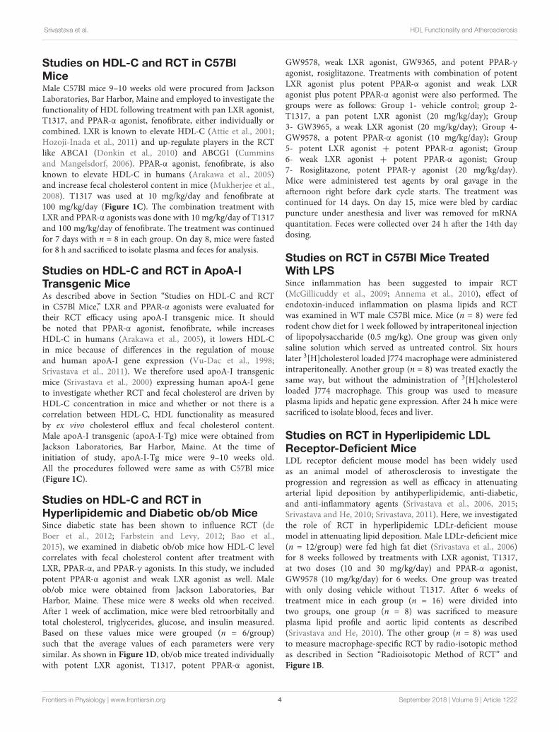
Studies on HDL-C and RCT inHyperlipidemic and Diabetic ob/ob MiceSince diabetic state has been shown to influence RCT (deBoer et al., 2012; Farbstein and Levy, 2012; Bao et al.,2015), we examined in diabetic ob/ob mice how HDL-C levelcorrelates with fecal cholesterol content after treatment withLXR, PPAR-α, and PPAR-γ agonists. In this study, we includedpotent PPAR-α agonist and weak LXR agonist as well. Maleob/ob mice were obtained from Jackson Laboratories, BarHarbor, Maine. These mice were 8 weeks old when received.After 1 week of acclimation, mice were bled retroorbitally andtotal cholesterol, triglycerides, glucose, and insulin measured.Based on these values mice were grouped (n = 6/group)such that the average values of each parameters were verysimilar. As shown in Figure 1D, ob/ob mice treated individuallywith potent LXR agonist, T1317, potent PPAR-α agonist,
GW9578, weak LXR agonist, GW9365, and potent PPAR-γagonist, rosiglitazone. Treatments with combination of potentLXR agonist plus potent PPAR-α agonist and weak LXRagonist plus potent PPAR-α agonist were also performed. Thegroups were as follows: Group 1- vehicle control; group 2-T1317, a pan potent LXR agonist (20 mg/kg/day); Group3- GW3965, a weak LXR agonist (20 mg/kg/day); Group 4-GW9578, a potent PPAR-α agonist (10 mg/kg/day); Group5- potent LXR agonist + potent PPAR-α agonist; Group6- weak LXR agonist + potent PPAR-α agonist; Group7- Rosiglitazone, potent PPAR-γ agonist (20 mg/kg/day).Mice were administered test agents by oral gavage in theafternoon right before dark cycle starts. The treatment wascontinued for 14 days. On day 15, mice were bled by cardiacpuncture under anesthesia and liver was removed for mRNAquantitation. Feces were collected over 24 h after the 14th daydosing.
Studies on RCT in C57Bl Mice TreatedWith LPSSince inflammation has been suggested to impair RCT(McGillicuddy et al., 2009; Annema et al., 2010), effect ofendotoxin-induced inflammation on plasma lipids and RCTwas examined in WT male C57Bl mice. Mice (n = 8) were fedrodent chow diet for 1 week followed by intraperitoneal injectionof lipopolysaccharide (0.5 mg/kg). One group was given onlysaline solution which served as untreated control. Six hourslater 3[H]cholesterol loaded J774 macrophage were administeredintraperitoneally. Another group (n = 8) was treated exactly thesame way, but without the administration of 3[H]cholesterolloaded J774 macrophage. This group was used to measureplasma lipids and hepatic gene expression. After 24 h mice weresacrificed to isolate blood, feces and liver.
Studies on RCT in Hyperlipidemic LDLReceptor-Deficient MiceLDL receptor deficient mouse model has been widely usedas an animal model of atherosclerosis to investigate theprogression and regression as well as efficacy in attenuatingarterial lipid deposition by antihyperlipidemic, anti-diabetic,and anti-inflammatory agents (Srivastava et al., 2006, 2015;Srivastava and He, 2010; Srivastava, 2011). Here, we investigatedthe role of RCT in hyperlipidemic LDLr-deficient mousemodel in attenuating lipid deposition. Male LDLr-deficient mice(n = 12/group) were fed high fat diet (Srivastava et al., 2006)for 8 weeks followed by treatments with LXR agonist, T1317,at two doses (10 and 30 mg/kg/day) and PPAR-α agonist,GW9578 (10 mg/kg/day) for 6 weeks. One group was treatedwith only dosing vehicle without T1317. After 6 weeks oftreatment mice in each group (n = 16) were divided intotwo groups, one group (n = 8) was sacrificed to measureplasma lipid profile and aortic lipid contents as described(Srivastava and He, 2010). The other group (n = 8) was usedto measure macrophage-specific RCT by radio-isotopic methodas described in Section “Radioisotopic Method of RCT” andFigure 1B.
Frontiers in Physiology | www.frontiersin.org 4 September 2018 | Volume 9 | Article 1222

fphys-09-01222 September 8, 2018 Time: 13:43 # 5
Srivastava et al. HDL Functionality and Atherosclerosis
Measurements of Aortic Lipid ContentAortic lipid contents were measured as described (Srivastavaet al., 2006). Briefly, after the removal of blood, the vasculaturewas perfused first with cold PBS containing 5 mM EDTAthrough the left ventricle followed by carefully removing of alladventitious tissues connected to the aorta. The aorta from eachmouse was removed from aortic root to the renal artery andplaced in cold PBS for 15 s and taken out in another dishto remove excess solution by using a paper blot. Aortas wereweighed individually and cut into small pieces followed by theextraction of lipids using chloroform/methanol (2:1) as described(Srivastava et al., 1991, 2006). The measurement of cholesterolester was quantitated in the aortic lipid extract (Srivastava et al.,1991).
Fecal Cholesterol MeasurementsFor non-radioactive fecal cholesterol transport assay, male mice(n = 8/group) were treated with the test agent for specifiedamount of days followed by collection of feces over 24 h period.The feces were extracted with solvent and cholesterol measured.Fecal cholesterol were measured by ELSD using a shortenedmethod of Homan and Anderson (1998). In brief, 100 mg ofeach fecal sample was homogenized in 0.50 ml of 150 mMNaCl/5 mM MOPS/1 mM EDTA/0.01% PMSF followed by theaddition of 0.5 ml of 150 mM NaCl 5 mM MOPS/1 mM EDTAand 200 µl of internal standard (2.0 mg/ml eicosanol dissolvedin 2:1 MeCl2:MeOH). Samples were extracted with 2.0 ml of(2:1 MeCl2:MeOH) and quantified in Agilent Series 1100 HPLCsystem.
Measurement of Plasma CholesterolEfflux CapacityTo measure plasma cholesterol efflux capacity (CEC), firstplasma apoB was depleted. Polyethylene glycol (40 µl), madeby dissolving 20 g in 1000 ml of 200 mM glycine buffer, wasadded to 100 µl mouse plasma followed by incubation for20 min at room temperature. The contents were centrifuged at10,000 × g and 4◦C for 30 min, and supernatant transferred toanother clean Eppendorf tube as described (Rohatgi et al., 2014;Trieb et al., 2016). Cholesterol efflux capacity of apoB-depletedplasma was carried out as described (Rohatgi et al., 2014).In brief, J774 macrophages were grown in DMEM mediumsupplemented with 10% fetal bovine serum. To enhance uptakeof [3H]-cholesterol, the cells were first incubated with 0.3 mM8-(4-chlorophenylthio)-cyclic AMP in serum-free DMEM toinduce ATP-binding cassette transporter protein (ABCA1).Labeling of cells with [3H]-cholesterol was done for 24 h andcells were washed once with DMEM. After washing the cellswith phosphate-buffered saline, cells were incubated at 37◦C withapoB-depleted plasma (2.5%) for 4 h. ApoA-I (20 µg/ml) wasused as a control. After 4 h of incubation, the medium wasremoved and the cells in the well as a monolayer were washedand harvested. Radioactivity was measured in both medium andcells. Cholesterol efflux was expressed as radioactivity in themedium divided by the total radioactivity in the medium and thecells.
Apolipoprotein A-1 MeasurementMouse apolipoprotein A-1 concentrations in plasma sampleswere analyzed by Sandwich-ELISA kit procured fromElab Sciences (Catalog No. E-EL-M0130) according to themanufacturer’s instructions.
Lipoprotein Profile and Plasma LipidMeasurementsLipoprotein profile was measured as described before (Srivastavaet al., 1997b, 2000). In brief, to determine the lipoprotein profile,pooled plasma (400 µl) from each group of mice were loadedonto two Superose 6 columns connected in series; 0.5 ml fractionswere collected using a solution containing 0.15 mM NaCl,0.02% sodium azide and 1 mM EDTA. In each eluted fraction,cholesterol was measured, and the fractions corresponding toVLDL-, LDL-, and HDL-C were pooled to measure respectivelipoprotein concentrations. Plasma levels of total cholesterol andtriglycerides were quantitated using commercial kits as described(Srivastava et al., 2011).
Insulin and Glucose QuantitationIn the ob/ob mice, glucose and insulin were measured asdescribed (Srivastava et al., 2006).
Measurement of RNA by QuantitativePCRHepatic and blood monocytic mRNA were carried out byquantitative polymerase chain reaction following the establishedmethods (Srivastava et al., 1999). Total RNA (50 ng) wasreverse transcribed by reverse transcriptase and the resultantsingle stranded complimentary DNA was amplified using thermalcycler. The sequences of respective genes for mRNA quantitationwere obtained from NCBI for designing primers.
Statistical AnalysisStatistical analyses were performed by one-way ANOVA withthe Dunnett’s multiple comparisons where applicable. Data wereexpressed as mean± SD.
Spearman Non-parametric CorrelationAnalysisSpearman non-parametric correlation analysis was carried outby Prism 6 software to determine correlation and p-values usingregression analysis. Prism 6 calculates exact p-values in regressionanalysis, which are shown in respective figures. Correlationswere determined between apoA-I, HDL, cholesterol efflux, fecalcholesterol content, and aortic cholesterol content.
RESULTS
LXR Agonist, T1317, Show Similar FecalCholesterol Excretion in Radioisotopeand Non-radioisotope AssaysTo examine relationship between circulating HDL-C andHDL functionality measured by fecal cholesterol excretion as
Frontiers in Physiology | www.frontiersin.org 5 September 2018 | Volume 9 | Article 1222

fphys-09-01222 September 8, 2018 Time: 13:43 # 6
Srivastava et al. HDL Functionality and Atherosclerosis
a maker of RCT, first we compared non-radioisotopic andradioisotopic method of RCT. As shown in Figure 1A, innon-radioisotopic method male C57Bl mice were treated withthe reference agent, LXR pan agonist, T1317, known to elevatecirculating HDL (Brunham et al., 2006) and enhances RCT(Zanotti et al., 2008) through induction of players (Delvecchioet al., 2008) in RCT pathway. Following 7 days of treatment,feces were collected, and total cholesterol measured. In theradio-isotopic assay, macrophage-specific RCT was measured. Inthis assay (Figure 1B), macrophages are loaded with radiolabetedcholesterol (3H) and after 7 days treatment with T1317, feceswere collected and 3H counts were measured. The results ofnon-radioisotopic method of RCT measurement together withplasma lipid and hepatic RNA measurements are shown inFigure 2. As expected, the induction of lipogenesis (Schultz et al.,2000) and RCT (Zanotti et al., 2008) by LXR pan agonist, T1317increased both LDL-C and HDL-C (Figure 2A). Compared tovehicle control, HDL-C, LDL-C, and TG increased in the LXRagonist treated mice (Figure 2B). Increased RCT as measuredby fecal cholesterol (>2-fold) was evident in the T1317 treatedmice (Figure 2C). As expected, increases in the fecal cholesterolin the T1317 treated mice paralleled ABCA1 and ABCG1induction in blood monocytes (Figure 2D) and ABCA1, G1, G5,and G8 induction in the liver (Figure 2E). In the radioactivemethod of RCT measurement, T1317 showed increased RCT(Figure 2F) similar to the results in the non-radioisotopicmethod (Figure 2C). These results suggest that T1317 exhibitedexpected efficacy of increased fecal cholesterol in both thenon-radioactive and radioactive methods. Twenty-four hourafter the radiolabeled macrophage administration appears to be
sufficient to show-up labeled cholesterol in the feces (Figure 3A).A dose-dependent increase in the fecal cholesterol was noticedin the T1317 treated mice (Figure 3B). Using both radioactiveand non-radioactive methods, biliary cholesterol measurementsshowed similar dose-dependent increases (Figure 3C). Thesefindings suggest that non-radioactive method provide similarresults in terms of biliary cholesterol excretion as the radioactivemethod. Therefore, all subsequent studies examining HDL-C andRCT were carried out using non-radioisotopic method. However,in order to confirm that fecal cholesterol content indeed reflectsHDL functionality, in a parallel experiment, ex vivo cholesterolefflux was measured in the apoB-depleted plasma and the resultsshown in Figure 3D, suggests that fecal cholesterol concentrationin the T1317 treated mice also increased.
Lack of Correlation Between HDL andRCT in C57Bl MiceCorrelation between HDL-C and RCT was examined usingtwo reference agents, an LXR agonist that increases HDL-Cin mice (Hozoji-Inada et al., 2011) and a PPAR-α agonistthat decreases HDL-C in mice (Berthou et al., 1996; Vu-Dacet al., 1998), but increases in humans (Duez et al., 2005). Asshown in Figures 4A,B, LXR agonist, T1317, known to increaseHDL-C as well as hepatic lipogenesis through induction ofa number of lipogenic genes (Schultz et al., 2000), increasedHDL-C by 30% and fenofibrate decreased HDL-C by 30%(Figures 4A,B). Although fenofibrate reduced plasma levels ofHDL-C, it resulted in the formation of larger HDL particlesas evidenced by the shift of HDL peak toward left. This isconsistent with the earlier findings that fenofibrate increases
FIGURE 2 | Male C57Bl mice (n = 8) were treated with pan LXR agonist, T1317, as described in Figure 1C. (A) lipoprotein profile; (B) Plasma lipids; and (C) fecalcholesterol. ∗ Significantly different (p < 0.01) compared to control; (D) ABCA1 and G1 expression in the blood monocytes; (E) Hepatic gene induction; and (F) fecal[3H]-cholesterol counts. ∗ Significantly different (p < 0.01) compared to control.
Frontiers in Physiology | www.frontiersin.org 6 September 2018 | Volume 9 | Article 1222

fphys-09-01222 September 8, 2018 Time: 13:43 # 7
Srivastava et al. HDL Functionality and Atherosclerosis
FIGURE 3 | Dose–response of pan LXR agonist, T1317, on reverse cholesterol transport. Male C57Bl mice (n = 8) were treated as described in Figure 2, andreverse cholesterol measured as described in Figure 1B. (A) Macrophage-derived Plasma [3H]-cholesterol counts collected during 24 and 48 h after the last dosingof LXR agonist (20 mg/kg/day). (B) Dose–response of LXR agonist, T1317, on fecal [3H]-cholesterol counts. (C) [3H]-cholesterol counts and biliary cholesterol massin bile. (D) Ex vivo cholesterol efflux in apoB-depleted serum. ∗ Significantly different (p < 0.01) compared to control.
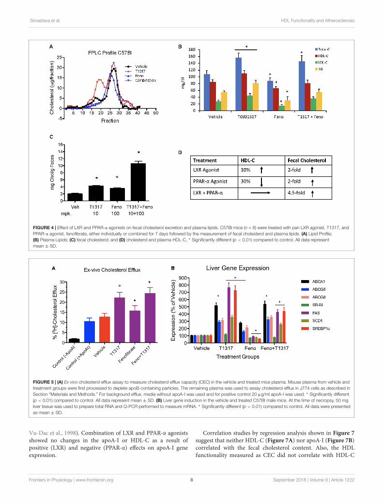
HDL size through induction of phospholipid transfer protein(PLTP) (Srivastava et al., 2011). However, fecal cholesterolexcretion, a measure of RCT, was comparable in both LXRand PPAR-α agonist treated mice (Figure 4C). One groupof mice was also treated with the combination of LXRand PPAR-α agonist, since the former increases lipogenesis,as evidenced by increased TG and LDL-C, and the laterdecreases lipogenesis (Srivastava et al., 2006) as evidencedby decreased TG and LDL-C (Figures 4A,B). Interestingly,while fecal cholesterol excretion increased by approximatelytwofold by LXR and PPAR-α agonists individually, a markedincrease (4.5-fold) was noted in the fecal cholesterol excretionin mice treated with the combination of LXR and PPAR-αagonists (Figures 4C,D). It should be noted that there wasno increase in the total circulating HDL-C in the combinationgroup (Figures 4A,B). This necessitated direct measurementsof the HDL functionality in a cholesterol efflux assay usingthe apoB-depleted plasma samples from all treated groups.As shown in Figure 5A, T1317 increased CEC parallelto the fecal cholesterol content and a somewhat similartrend was evident in the fenofibrate treated mice. However,despite 4.5-fold increase in the fecal cholesterol content, HDLfunction, measured as CEC, was comparable to T1317 treatedgroup.
To further understand the role of players known to influenceRCT and lipogenesis, expression of select genes in the liverwere quantitated. As shown in Figure 5B, as expected, LXRagonist induced ABCA1, G1, G5, and G8 greater than the PPAR-α agonist and induced lipogenic genes, while PPAR-α agonistsuppressed lipogenic genes, consistent with the reported findings(Schultz et al., 2000; Srivastava, 2009). The combination of LXRand PPAR-α agonist did not necessarily show additive effectson the RCT influencing genes, for instance ABCA1 and G1.SR-BI is a significant player in the uptake of HDL by the liver,which may influence fecal cholesterol content, however, in thecurrent study LXR agonist did not significantly influence SR-BI, and fenofibrate suppressed SR-BI expression as reportedearlier (Srivastava et al., 2011) ruling out the possibility of SR-BI in the fecal cholesterol content. Another important playerin the RCT pathway is the apoA-I. We, therefore, measuredplasma levels of apoA-I that functions as the acceptor of cellularcholesterol efflux in the ABCA-1 mediated pathway to findout if apoA-I levels explain some of the unexpected resultson fecal cholesterol content in the combination treatment.LXR agonist does increase plasma apoA-I (Figure 6), whichin turn raises HDL through ABCAI-mediated cholesterol effluxto apoA-I. Fenofibrate, on the other hand, reduces mouseapoA-I and HDL-C as reported earlier (Berthou et al., 1996;
Frontiers in Physiology | www.frontiersin.org 7 September 2018 | Volume 9 | Article 1222
fphys-09-01222 September 8, 2018 Time: 13:43 # 8
Srivastava et al. HDL Functionality and Atherosclerosis
FIGURE 4 | Effect of LXR and PPAR-α agonists on fecal cholesterol excretion and plasma lipids. C57Bl mice (n = 8) were treated with pan LXR agonist, T1317, andPPAR-α agonist, fenofibrate, either individually or combined for 7 days followed by the measurement of fecal cholesterol and plasma lipids. (A) Lipid Profile;(B) Plasma Lipids; (C) fecal cholesterol; and (D) cholesterol and plasma HDL-C. ∗ Significantly different (p < 0.01) compared to control. All data representmean ± SD.
FIGURE 5 | (A) Ex vivo cholesterol efflux assay to measure cholesterol efflux capacity (CEC) in the vehicle and treated mice plasma. Mouse plasma from vehicle andtreatment groups were first processed to deplete apoB-containing particles. The remaining plasma was used to assay cholesterol efflux in J774 cells as described inSection “Materials and Methods.” For background efflux, media without apoA-I was used and for positive control 20 µg/ml apoA-I was used. ∗ Significantly different(p < 0.01) compared to control. All data represent mean ± SD. (B) Liver gene induction in the vehicle and treated C57Bl male mice. At the time of necropsy, 50 mgliver tissue was used to prepare total RNA and Q-PCR performed to measure mRNA. ∗ Significantly different (p < 0.01) compared to control. All data were presentedas mean ± SD.
Vu-Dac et al., 1998). Combination of LXR and PPAR-α agonistsshowed no changes in the apoA-I or HDL-C as a result ofpositive (LXR) and negative (PPAR-α) effects on apoA-I geneexpression.
Correlation studies by regression analysis shown in Figure 7suggest that neither HDL-C (Figure 7A) nor apoA-I (Figure 7B)correlated with the fecal cholesterol content. Also, the HDLfunctionality measured as CEC did not correlate with HDL-C
Frontiers in Physiology | www.frontiersin.org 8 September 2018 | Volume 9 | Article 1222

fphys-09-01222 September 8, 2018 Time: 13:43 # 9
Srivastava et al. HDL Functionality and Atherosclerosis
FIGURE 6 | Mice were treated with vehicle, T1317, fenofibrate, andcombination of T1317 and fenofibrate as described in Figure 4. At the end of7 days treatment, individual mouse plasma was analyzed for apoA-I usingcommercial kit. Data shown are mean ± SD.
(Figure 7C). Interestingly, ex vivo cholesterol efflux (CEC), ameasure of HDL functionality, showed significant correlation
with the fecal cholesterol content (Figure 7D). In this correlationanalysis, all data points were used, including the data oncombination effects where HDL-C did not change, but fecalcholesterol increased by 4.5-fold. These findings confirm thevalidity of fecal cholesterol contents as a measure of RCT.
Plasma Levels of HDL and RCT Does NotShow Correlation in ApoA-I-Tg MiceFenofibrate, an PPAR-α agonist, although increases HDL-C inhumans, it decreases HDL-C in mice as shown in Figures 4A,B.Therefore, we carried out same study with apoA-I Tg mice(Srivastava et al., 2000) following the same protocol as theWT C57Bl mice discussed above (Figure 4). In the apoA-Itransgenic mice expressing human apoA-I, both LXR andPPAR-α agonists increased HDL-C. Although PPAR-α agonistshowed twofold greater increases in HDL-C as compared toLXR agonist, T0901317, the fecal cholesterol increases were same(∼2-fold) when compared to untreated control (Figures 8A,B),suggesting a lack of correlation between HDL-C concentrationand fecal cholesterol excretion. The combination of LXR andPPAR-α agonist treatment increased fecal cholesterol excretionby 5–6-fold (Figure 8C), although increases in HDL-C were sameas the PPAR-α agonist treatment group (Figure 8D), suggesting
FIGURE 7 | Linear regression analysis was carried out by non-parametric (Spearman) correlation using Prism 6 to determine correlations and p-values betweenHDL, apoA-I, fecal cholesterol, and ex vivo cholesterol efflux. (A) HDL vs. fecal cholesterol- no correlation; (B) ApoA-I vs. fecal cholesterol- no correlation; (C) HDLvs. ex vivo cholesterol efflux- no correlation; (D) Fecal cholesterol vs. ex vivo cholesterol efflux- highly significant correlation (p < 0.0001).
Frontiers in Physiology | www.frontiersin.org 9 September 2018 | Volume 9 | Article 1222

fphys-09-01222 September 8, 2018 Time: 13:43 # 10
Srivastava et al. HDL Functionality and Atherosclerosis
FIGURE 8 | Effect of LXR and PPAR-α agonists on fecal cholesterol excretion and plasma lipids in apoA-I transgenic mice. ApoA-I Tg mice (n = 8) were treated withpan LXR agonist, T1317, and PPAR-α agonist, fenofibrate, either individually or combined for 7 days followed by the measurement of fecal cholesterol and plasmalipids. (A) Plasma lipoprotein profile; (B) Plasma lipids; (C) fecal cholesterol; and (D) comparison of fecal cholesterol and plasma HDL-C. ∗ Significantly different(p < 0.01) compared to control. Data presented show mean ± SD.
a clear lack of correlation between HDL-C and fecal cholesterolexcretion. To have mechanistic insights into the RCT pathwayin apoAI-Tg mice, CEC was performed as a measure of HDLfunctionality. As shown in Figure 9, the basal CEC in thevehicle treated mice was twice that of non-transgenic C57Bl mice(Figure 5A).
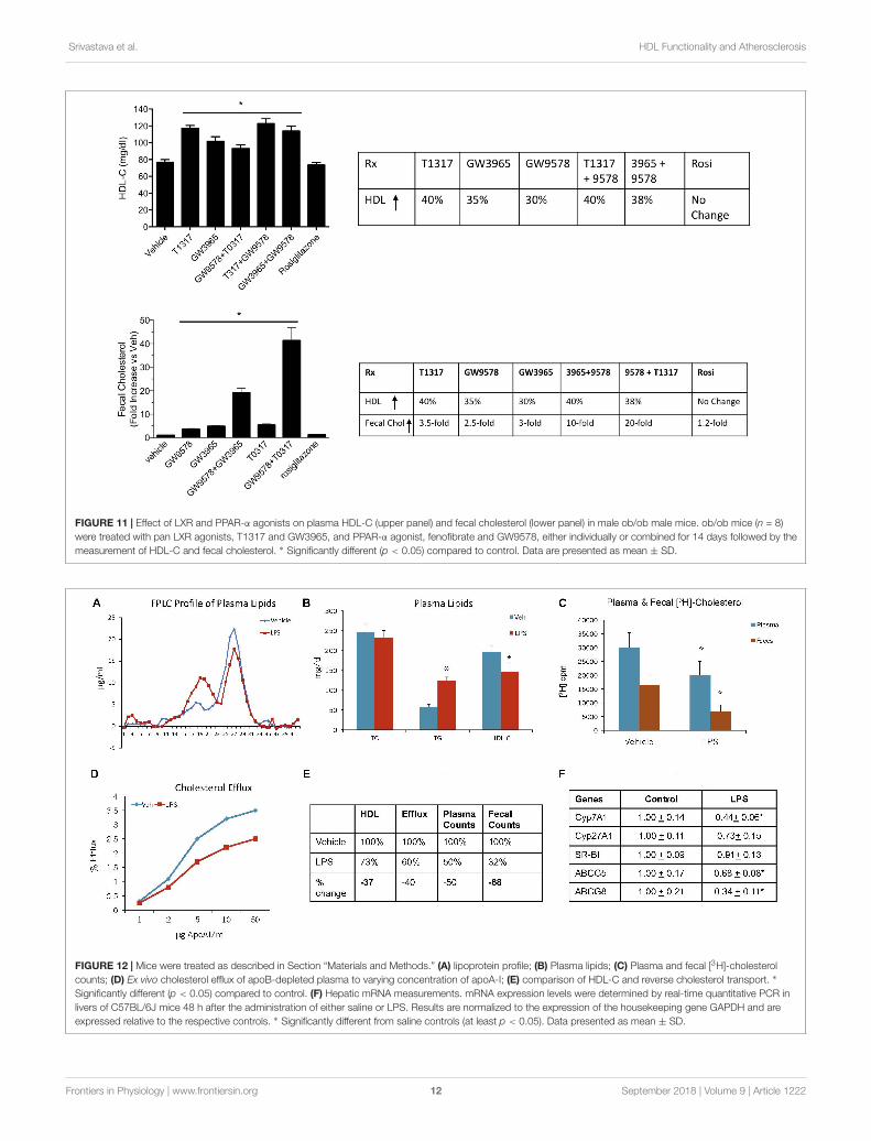
Discordance Between HDLConcentration and Fecal Cholesterol inHyperlipidemic and Diabetic ob/ob MiceSince diabetic conditions are associated with increased risk ofCVD (Srivastava, 2018) and dampening of RCT, we examinedHDL-C levels and fecal cholesterol in ob/ob mice treated withweak (GW3965) and potent (T1317) LXR agonists, respectively,potent PPAR-α agonist (GW9578), and potent PPAR-γ agonist(rosiglitazone). One group of mice was also treated with aweak LXR agonist (GW3965) plus GW9578, and one groupwith T1317 plus GW9578. Since PPAR-α, PPAR-γ, and LXRagonists have antidiabetic efficacy in animal model of diabetes(Srivastava, 2009), we first ascertained that these agonists showantidiabetic efficacy in ob/ob mice. As shown in Figure 10, allthese agonists lowered glucose (left panel) and insulin (rightpanel). LXR agonist as well as combination of LXR and PPAR-αagonists showed maximal antidiabetic efficacy. Except PPAR-γagonist, rosiglitazone, all agonists tested in this study increased
HDL-C with greater HDL-C elevation in groups treated withT1317 and combination of LXR and PPAR-α agonists (Figure 11,upper panel). Fecal cholesterol measurements were done in allgroups and results are shown in Figure 11, lower panel. Similarto the results shown above in WT C57Bl (Figure 4) and apoA-I Tgmice (Figure 8), T1317 showed 40% increase in HDL-C. In thishyperlipidemic diabetic model, potent PPAR-α agonist, GW9578,also showed 30% increase. Combination of LXR and PPAR-αagonist treatment did not further increase circulating HDL-Cin ob/ob mice when compared to the treatment groups withLXR and PPAR-α agonist treatment individually. Most notably,potent PPAR-α agonist, GW9578 increased fecal cholesterol by10-fold despite only 30% increase in HDL-C, when comparedto untreated control. Combination of potent LXR and potentPPAR-α agonist treatment showed a massive 20-fold increase infecal cholesterol, but only 38% increase in circulating HDL-C(Figure 11), suggesting role of HDL-independent pathway inbody cholesterol excretion.
Endotoxin-Induced InflammationReduces RCT in C57Bl WT MiceWhile HDL possesses anti-inflammatory properties (Navab et al.,2005; van der Westhuyzen et al., 2007), in addition to itsprimary function of RCT (Sacks and Jensen, 2018), underinflammatory conditions, HDL function gets compromised
Frontiers in Physiology | www.frontiersin.org 10 September 2018 | Volume 9 | Article 1222

fphys-09-01222 September 8, 2018 Time: 13:43 # 11
Srivastava et al. HDL Functionality and Atherosclerosis
FIGURE 9 | Ex vivo cholesterol efflux assay in apoA-I transgenic mice tomeasure cholesterol efflux capacity (CEC) in the vehicle and treated miceplasma. Mouse plasma from vehicle and treatment groups were firstprocessed to deplete apoB-containing particles. The remaining plasma wasused to assay cholesterol efflux in J774 cells as described in Section“Materials and Methods.” For background efflux, media without apoA-I wasused and for positive control 20 µg/ml apoA-I was used. ∗ Significantlydifferent (p < 0.01) compared to control. All data represent mean ± SD.
(McGillicuddy et al., 2009; Morgantini et al., 2011). Weexamined cholesterol efflux and reverse cholesterol capabilityof C57Bl mice under endotoxin-induced condition. The resultsshown in Figure 12 suggest that pro-inflammatory conditionsraise proatherogenic triglyceride-rich particles as evidenced by
increased TG and VLDL (Figure 12A). Endotoxin-inducedinflammation lowered HDL-C by 27% (Figure 12B). RCTmeasured by plasma and fecal cholesterol counts decreased by50 and 64%, respectively (Figure 12C). Ex vivo cholesterol effluxalso reduced (Figure 12D). Overall, HDL functionality, measuredby fecal cholesterol counts, was compromised more than theHDL-C (Figure 12E). The dampened cholesterol efflux capabilityand RCT was corroborated by decreases in the expressionof cholesterol 7-α hydroxylase A1 (Cyp7A1), ABCG5 and G8(Figure 12F).
HDL Functionality, but Not HDL-C,Correlates With Aortic Lipid Content inLDLr-Deficient MiceTo examine if aortic lipid deposition is influenced by plasmaHDL-C, LDLr-deficient mice were fed-high fat diet and treatedwith HDL raising agents. LDLr-deficient model was used in thisstudy because, with few exceptions, most mouse atherosclerosismodels available are hyperlipidemia-induced models. Whileatherosclerosis progression in this model is apoB-containingproatherogenic lipoprotein driven (Srivastava et al., 2006), weasked the question if RCT and/or HDL-C is responsible forinhibition of progression in this model. As expected, both plasmaand fecal 3H-cholesterol counts increased following treatmentswith PPAR-α and LXR agonists (Figures 13A,B). Fecalcholesterol excretion reached maximal level at 10 mg/kg/daydose of LXR agonist, T1317 (Figure 13B). There was a modestincrease (15%) in circulating HDL-C following treatments withPPAR-α agonist, GW9578 (Figure 13D). Treatment with higherdose of LXR agonist, T1317, showed increased circulating HDL-C by 30%. To examine if fecal cholesterol excretion is anyindication of aortic lipid content, we measured fecal cholesterol
FIGURE 10 | T1317, Potent LXR agonist; GW9578, Potent PPAR-α agonist; GW9365, Weak LXR agonist; Rosi- Rosiglitazone, Potent PPAR-γ agonist. Effect ofLXR and PPAR-α agonists on plasma glucose (left panel) and insulin (right panel) in male ob/ob mice. ob/ob mice (n = 8) were treated with pan LXR agonists (T1317)and GW3965 (PPAR-α agonist), fenofibrate and GW9578, either individually or combined for 14 days followed by the measurement of glucose and insulin.∗ Significantly different (p < 0.05) compared to control. Data presented are mean ± SD.
Frontiers in Physiology | www.frontiersin.org 11 September 2018 | Volume 9 | Article 1222
fphys-09-01222 September 8, 2018 Time: 13:43 # 12
Srivastava et al. HDL Functionality and Atherosclerosis
FIGURE 11 | Effect of LXR and PPAR-α agonists on plasma HDL-C (upper panel) and fecal cholesterol (lower panel) in male ob/ob male mice. ob/ob mice (n = 8)were treated with pan LXR agonists, T1317 and GW3965, and PPAR-α agonist, fenofibrate and GW9578, either individually or combined for 14 days followed by themeasurement of HDL-C and fecal cholesterol. ∗ Significantly different (p < 0.05) compared to control. Data are presented as mean ± SD.
FIGURE 12 | Mice were treated as described in Section “Materials and Methods.” (A) lipoprotein profile; (B) Plasma lipids; (C) Plasma and fecal [3H]-cholesterolcounts; (D) Ex vivo cholesterol efflux of apoB-depleted plasma to varying concentration of apoA-I; (E) comparison of HDL-C and reverse cholesterol transport. ∗
Significantly different (p < 0.05) compared to control. (F) Hepatic mRNA measurements. mRNA expression levels were determined by real-time quantitative PCR inlivers of C57BL/6J mice 48 h after the administration of either saline or LPS. Results are normalized to the expression of the housekeeping gene GAPDH and areexpressed relative to the respective controls. ∗ Significantly different from saline controls (at least p < 0.05). Data presented as mean ± SD.
Frontiers in Physiology | www.frontiersin.org 12 September 2018 | Volume 9 | Article 1222
fphys-09-01222 September 8, 2018 Time: 13:43 # 13
Srivastava et al. HDL Functionality and Atherosclerosis
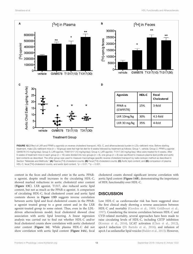
FIGURE 13 | Effect of LXR and PPAR-α agonists on reverse cholesterol transport, HDL-C, and atherosclerosis burden in LDLr-deficient mice. Before startingtreatment, male LDLr-deficient mice (n = 16/group) were fed high fat diet for 8 weeks followed by treatment as follows: Group 1, vehicle; Group 2, PPAR-α agonistGW9578 (10 mg/kg/day); Group 3, LXR agonist, T0901317 (10 mg/kg/day); Group 4, LXR agonist, T1317 (30 mg/kg/day). Mice were treated for 6 weeks. After6 weeks of treatment mice in each group (n = 16) were divided into two groups (n = 8), one group (n = 8) was sacrificed to measure plasma lipid profile and aorticlipid contents as described. The other group was used to measure macrophage-specific reverse cholesterol transport by radio-isotopic method as described inSection “Materials and Methods.” (A) Plasma [3H]-cholesterol counts; (B) Fecal [3H]-cholesterol counts; (C) Aortic lipid content; and (D) comparison of plasmaHDL-C, fecal [3H]-cholesterol counts, and aortic lipid content. ∗p < 0.01; ∗∗p < 0.001.
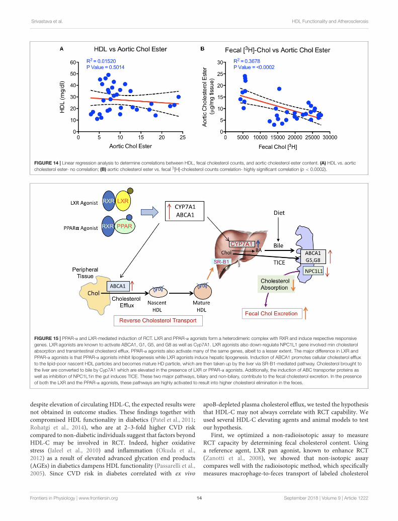
content in the feces and cholesteryl ester in the aorta. PPAR-α agonist, despite small increases in the circulating HDL-C,showed marked reductions in aortic cholesteryl ester content(Figure 13C). LXR agonist, T1317, also reduced aortic lipidcontent, but not as much as the PPAR-α agonist. A comparisonof circulating HDL-C, fecal cholesterol count and aortic lipidcontents shown in Figure 13D suggests inverse correlationbetween aortic lipid and fecal cholesterol counts in the PPAR-α agonist treated group to a great extent and in the LXRagonist-treated group to some extent. Thus, even in the LDL-driven atherosclerosis model, fecal cholesterol showed closeassociation with aortic lipid lowering. A linear regressionanalysis was carried out to find out whether HDL-C and/orfecal cholesterol counts show correlation with aortic cholesterylester content (Figure 14). While plasma HDL-C did notshow correlation with aortic lipid content (Figure 14A), fecal
cholesterol counts showed significant inverse correlation withaortic lipid content (Figure 14B), demonstrating the importanceof HDL functionality over HDL-C.
DISCUSSION
Low HDL-C as cardiovascular risk has been suggested sincethe first clinical study showing a reverse association betweenHDL-C and mortality (Gordon et al., 1989; Goldbourt et al.,1997). Considering the inverse correlation between HDL-C andCVD-related mortality, several approaches have been made toraise circulating levels of HDL-C, including CETP inhibition(Kosmas et al., 2016), LCAT activation (Chen et al., 2012),apoA-I induction (Di Bartolo et al., 2016), and infusion ofapoA-I as unilamellar lipid vesicles (Balder et al., 2013). However,
Frontiers in Physiology | www.frontiersin.org 13 September 2018 | Volume 9 | Article 1222
fphys-09-01222 September 8, 2018 Time: 13:43 # 14
Srivastava et al. HDL Functionality and Atherosclerosis
FIGURE 14 | Linear regression analysis to determine correlations between HDL, fecal cholesterol counts, and aortic cholesterol ester content. (A) HDL vs. aorticcholesterol ester- no correlation; (B) aortic cholesterol ester vs. fecal 3[H]-cholesterol counts correlation- highly significant correlation (p < 0.0002).
FIGURE 15 | PPAR-α and LXR-mediated induction of RCT. LXR and PPAR-α agonists form a heterodimeric complex with RXR and induce respective responsivegenes. LXR agonists are known to activate ABCA1, G1, G5, and G8 as well as Cyp7A1. LXR agonists also down-regulate NPC1L1 gene involved min cholesterolabsorption and transintestinal cholesterol efflux. PPAR-α agonists also activate many of the same genes, albeit to a lesser extent. The major difference in LXR andPPAR-α agonists is that PPAR-α agonists inhibit lipogenesis while LXR agonists induce hepatic lipogenesis. Induction of ABCA1 promotes cellular cholesterol effluxto the lipid-poor nascent HDL particles and becomes mature HD particle, which are then taken up by the liver via SR-B1-mediated pathway. Cholesterol brought tothe liver are converted to bile by Cyp7A1 which are elevated in the presence of LXR or PPAR-α agonists. Additionally, the induction of ABC transporter proteins aswell as inhibition of NPC1L1in the gut induces TICE. These two major pathways, biliary and non-biliary, contribute to the fecal cholesterol excretion. In the presenceof both the LXR and the PPAR-α agonists, these pathways are highly activated to result into higher cholesterol elimination in the feces.
despite elevation of circulating HDL-C, the expected results werenot obtained in outcome studies. These findings together withcompromised HDL functionality in diabetics (Patel et al., 2011;Rohatgi et al., 2014), who are at 2–3-fold higher CVD riskcompared to non-diabetic individuals suggest that factors beyondHDL-C may be involved in RCT. Indeed, higher oxidativestress (Jaleel et al., 2010) and inflammation (Okuda et al.,2012) as a result of elevated advanced glycation end products(AGEs) in diabetics dampens HDL functionality (Passarelli et al.,2005). Since CVD risk in diabetes correlated with ex vivo
apoB-depleted plasma cholesterol efflux, we tested the hypothesisthat HDL-C may not always correlate with RCT capability. Weused several HDL-C elevating agents and animal models to testour hypothesis.
First, we optimized a non-radioisotopic assay to measureRCT capacity by determining fecal cholesterol content. Usinga reference agent, LXR pan agonist, known to enhance RCT(Zanotti et al., 2008), we showed that non-isotopic assaycompares well with the radioisotopic method, which specificallymeasures macrophage-to-feces transport of labeled cholesterol
Frontiers in Physiology | www.frontiersin.org 14 September 2018 | Volume 9 | Article 1222

fphys-09-01222 September 8, 2018 Time: 13:43 # 15
Srivastava et al. HDL Functionality and Atherosclerosis
carried in circulation by macrophages (Figure 1B). As expected,the pan LXR agonist induced players in the RCT includingABCA1, G1, G5, and G8 (Figure 2E). LXR agonists areknown to induce lipogenesis as well (Schultz et al., 2000),and in the present study, we did find induction of lipogenicgenes, SREBP1c, FAS, and SCD1. Therefore, to avoid useof radioactive material, we preferred to use fecal cholesterolconcentration as a measure of RCT in most of the studiesreported here.
Fenofibrate (PPAR-α agonist) and T1317 are widely studiedreference agents to investigate lipid regulation (Schultz et al.,2000; Srivastava et al., 2011) and atherosclerosis (Srivastava,2011). We used these two reference agents to investigate acorrelation between circulating HDL-C and fecal cholesterolexcretion as a measure of RCT. Unlike in humans, fenofibratelowers circulating HDL-C in mice because of the differences inthe human and mouse apoA-I promoter (Vu-Dac et al., 1998).ApoA-I, the main protein component of HDL-C and a goodacceptor in the process of cellular cholesterol efflux determinesthe circulating levels of HDL (Srivastava and Srivastava, 2000). Inthe C57Bl mice, as expected, fenofibrate lowered HDL-C by 30%and LXR agonist increased HDL-C level by 30%. Interestingly,fecal cholesterol increased by twofold in both cases regardless ofwhether HDL-C decreased or increased (Figure 4), suggestingthat circulating HDL-C did not correlate with the fecal cholesterolcontent. To confirm that indeed fecal cholesterol content servesas a measure of RCT, ex vivo cholesterol efflux capacity (CEC)was measured, which paralleled fecal cholesterol content at leastin mice treated with LXR and PPAR-α agonists individually.Combination treatment with PPAR-α and pan LXR agonist didnot change circulating HDL-C, yet increased fecal cholesterolcontent by 4.5-fold, suggesting a lack of correlation betweencirculating levels of HDL-C and fecal cholesterol content. Thesefinding indicates existence of HDL-independent non-biliarycholesterol elimination in the feces (van der Velde et al., 2007).Indeed, similar to present findings, HDL-independent TICEresponsible for fecal cholesterol elimination has been described(Vrins et al., 2012). However, in the present investigation, wedid not focus on the HDL-independent mechanism of fecalcholesterol excretion. To validate the fecal cholesterol contentas a measure of HDL functionality, a correlation analysis wasperformed, and the results clearly demonstrated that HDL-Cdid not correlate with fecal cholesterol content, while ex vivocholesterol efflux capacity (CEC) correlated significantly with thefecal cholesterol content (Figure 7). As shown in Figure 4A,the characteristics of PPAR-α and LXR agonists was evident bythe marked reduction of TG following treatment with PPAR-αagonist (Srivastava et al., 2006) and increased TG by LXR agonist(Schultz et al., 2000). This confirms that the findings on HDL-Cand RCT by these two agonists were mediated via establishedplayers in the RCT pathway (Figure 5B).
To address the opposing effects on HDL-C by PPAR-α inrodent and humans (Vu-Dac et al., 1998) and how it influencesRCT, we utilized transgenic mouse model overexpressing humanapoA-I and carried out exactly the same experiment as withC57Bl mice. In apoA-I transgenic mice overexpressing human
apoA-I, both PPAR-α as well as LXR agonists raised circulatingHDL-C by 80 and 40%, respectively, while fecal cholesterolincreased by 60 and 50%, respectively. Greater HDL-C increasesin the PPAR-α-treated group was due to the upregulation ofhuman apoA-I gene (Srivastava et al., 2011). Combining PPAR-αand LXR agonists did not further increase circulating HDL-C,but showed marked elevation (300%) in the fecal cholesterolcontent, once again suggesting the absence of direct correlationbetween HDL level and fecal cholesterol content. Thus, studiesperformed in wild-type C57Bl and apoA-I transgenic mice showagreement in terms of dissociation between circulating HDL-Cand fecal cholesterol. It is possible that the fecal cholesterolcontents may be the result of a combination of more than onephysiological phenomenon, including LDL receptor-mediateduptake of LDL-C by the liver leading to the conversion intobile acids for excretion (Srivastava and Srivastava, 2000). Sincemice have very low levels of circulating LDL-C on normal rodentchow (Srivastava et al., 1993), it appears unlikely that the majorityof fecal cholesterol are derived from LDL receptor-mediateduptake of LDL-C by the liver. Other prominent players thatinfluence RCT are SR-BI (Van Eck et al., 2005), ABCA1 (Joyceet al., 2002; Srivastava, 2002), and G1 (Kennedy et al., 2005).The composition of HDL may also influence RCT since pre-βHDL particles (Kane and Malloy, 2012) are implicated in theinduction of RCT (Srivastava, 2002). In the present study, wehave not addressed this mechanistic aspect since the goal of thisstudy was to investigate plasma levels of HDL-C and RCT asmeasured by fecal cholesterol or fecal [3H]-cholesterol counts,and ex vivo cholesterol efflux. It is unlikely that RCT is positivelyinfluenced by SR-BI in the PPAR-α agonist treated group,since fenofibrate down-regulates SR-BI in the liver (Mardoneset al., 2003; Srivastava et al., 2011). In addition, fenofibrateinduces SR-BI degradation (Lan and Silver, 2005), ruling outthe role of SR-BI in fecal cholesterol excretion. The PPAR-αagonist-mediated induction of SR-BI in macrophage may partlycontribute to enhance RCT (Chinetti et al., 2000). Since PPAR-αagonist also influences prominent players in RCT via inducingtranscription factor LXR selectively in the macrophages (Nakayaet al., 2011), this may induce ABC transporter proteins. Thesynergistic effects observed in the PPAR-α and LXR agonistcombination treatment appears to occur partly as a result ofinduction of RCT pathway and also via HDL-independentpathway, including TICE (Figure 15), but this appears to belimited to the synergistic effects in the combination treatmentgroup, since hepatic ABC transporter proteins are insufficientto explain massive increases in the fecal cholesterol content.LXR agonists are known to induce intestinal ABCG5 and G8(Repa et al., 2002), which inhibits cholesterol absorption andinduces TICE (van der Veen et al., 2009). It is also suggested thatbiliary cholesterol secretion may not be required for macrophageRCT (Temel et al., 2010). As shown in Figure 15, part ofthe fecal cholesterol in the combination of PPAR-α plus LXRagonists treated group could account from HDL-independentnon-biliary mechanism involving other players like NPC1L1 thatinfluence cholesterol absorption (van der Veen et al., 2005). AcylCoA acetyl transferase-2 (ACAT2) is also known to influence
Frontiers in Physiology | www.frontiersin.org 15 September 2018 | Volume 9 | Article 1222

fphys-09-01222 September 8, 2018 Time: 13:43 # 16
Srivastava et al. HDL Functionality and Atherosclerosis
cholesterol absorption in the gut (Temel et al., 2005). Sinceboth PPAR-α (Valasek et al., 2007) and LXR (Duval et al., 2006)agonists down-regulate NPC1L1 gene, some of the effects seen onfecal cholesterol excretion may be mediated via NPC1L1 in micetreated with combination of PPAR-α and LXR agonists. Indeed,NPC1L1inhibitor, ezetimibe, enhances fecal sterol eliminationthrough TICE-mediated pathway (Jakulj et al., 2016).
Given that the ex vivo cholesterol efflux correlated with CVDmortality in human clinical studies (Patel et al., 2011; Rohatgiet al., 2014; Traldi et al., 2015), one of the questions we askedin this study was to examine association between RCT andaortic lipid deposition. To test this hypothesis, we employedLDLr-deficient mice widely studied to evaluate progression ofatherosclerosis (Srivastava et al., 2006) as well as to understandthe biology of aortic lipid deposition (van Leeuwen et al.,2008). While this animal model of atherosclerosis is heavilydriven by circulating LDL on high fat high cholesterol dietbecause of the absence of LDL receptor, nevertheless, weattempted to investigate the RCT component in the inhibitionof atherosclerosis progression following treatment with LXRand PPAR-α agonists. In the absence of LDL receptor, anotherreceptor, LRP1 carries out hepatic uptake of apoB/E-containinglipoproteins (Gordts et al., 2009). Fecal [3H]-cholesterol countsshowed 3- and 4.5-fold increases in the PPAR-α and LXR treatedanimals, which appeared to correlate with the inhibition ofaortic lipid deposition. However, there were only 15 and 30%increase in HDL-C in the PPAR-α (GW9578) and LXR (T1317)treated groups. Ex vivo cholesterol efflux did show induction inall treatment groups and an inverse correlation between fecalcholesterol counts and aortic cholesteryl ester content. Aorticcholesteryl ester showed a lack of correlation with HDL-C(Figure 14). Thus, present results clearly demonstrate that HDLfunctionality, measured as fecal cholesterol count or contentcorrelated better with atherosclerotic lesion severity compared toHDL-C.
To look further into the details and to understand RCT inhyperlipidemic models, ob/ob mice was employed. All the studiesdiscussed above used two reference compounds, fenofibrate(PPAR-α agonist) and T1317 (pan LXR agonist). Fenofibrateis a mild PPAR-α agonist and T1317 is a potent LXR agonist.To examine HDL-C and RCT correlation in ob/ob mice, wealso used potent PPAR-α agonist (GW9578) and weak LXRagonist (GW3965) either individually or a combination of mild(GW3965) and potent (T1317) LXR agonists with potent PPAR-α(GW9578) agonist. In all the treatment groups, individuallyor combined, HDL-C increased in the range of 30–40%, butfecal cholesterol excretion increased in the range of 2.5- to 20-fold. The increases in the fecal cholesterol were related to thepotency of the respective agonists, suggesting that induction ofthe RCT pathway, in part, played major roles compared to merelyincreasing the circulating HDL-C. One of the limitations of lack
of increases in circulating HDL-C appears to be availability ofthe main protein component, apoA-I. It is possible that despitesmall or no changes in the circulating HDL-C, the RCT pathwaymay still be very efficient in delivering HDL-derived cholesterol tothe liver and recycling apoA-I to serve as an acceptor for cellularcholesterol efflux. In this context macrophage ABCA1 and G1are likely to play greater role in RCT as suggested (Aiello et al.,2002; Van Eck et al., 2006). More than additive effect on fecalcholesterol elimination in the combination treatment group withpotent LXR agonist and potent PPAR-α agonist may have resultedfrom greater induction of ABCA1 and G1 in the macrophage onthe one hand to promote cellular cholesterol efflux to nascentHDL particle that delivers it to liver for excretion, and inductionof ABCG5 and 8 as well as inhibition of NPC1L1 by LXR andPPAR-α agonists (Duval et al., 2006; Valasek et al., 2007) onthe other hand. The later possibly induced HDL-independentpathway to increase fecal cholesterol excretion (Figure 15).
In summary, the data presented using a number of animalmodels demonstrate that measurement of HDL functionality ismore meaningful compared to measuring HDL-C, since even incases where there was no change in HDL, CEC showed markedincreases. These results further support clinical findings shown inindividuals susceptible to CVD (Rohatgi et al., 2014; Traldi et al.,2015).
It should be noted that mice lack CETP and most of thecholesterol in mice are transported as HDL particle with lowlevels of circulating LDL (Srivastava et al., 1991). This resultsinto fivefold lower plasma apoB in mice (Srivastava et al., 1997a)compared to humans (Pepin et al., 1991) and higher apoAI(Srivastava et al., 1992) compared to humans (Ghiselli et al.,1985). Mice have significantly lower biliary cholesterol secretionrelative to biliary salt when compared to humans (van der Wulpet al., 2013). Therefore, transgenic mice expressing apoB andCETP may provide further insights into the physiology of RCT,biliary secretion, and TICE. Despite these limitations with mousemodels, it presents relatively better genetic homogeneity andoffers a variety of genetically modified strains to ask importantbiologic questions. In the present studies with mice, we believethat our findings add further understanding to the roles ofHDL, RCT and fecal cholesterol elimination in attenuatingatherosclerosis progression.
AUTHOR CONTRIBUTIONS
RS evaluated data from the studies and has contributed to theinterpretation and analyses of data as well as writing of themanuscript. NS and AC designed the studies, carried out someof the experiments, and wrote part of the manuscript. MAcontributed to the review and interpretation of data and wrotepart of the manuscript.
REFERENCESAiello, R. J., Brees, D., Bourassa, P. A., Royer, L., Lindsey, S., Coskran, T., et al.
(2002). Increased atherosclerosis in hyperlipidemic mice with inactivation
of ABCA1 in macrophages. Arterioscler. Thromb. Vasc. Biol. 22, 630–637.doi: 10.1161/01.ATV.0000014804.35824.DA
American Heart Association HDass-u (2007). A report from the americanheart association statistics committee and stroke statistics subcommittee.
Frontiers in Physiology | www.frontiersin.org 16 September 2018 | Volume 9 | Article 1222

fphys-09-01222 September 8, 2018 Time: 13:43 # 17
Srivastava et al. HDL Functionality and Atherosclerosis
Circulation 115, e69–e71. doi: 10.1161/CIRCULATIONAHA.106.179918
Annema, W., Nijstad, N., Tolle, M., de Boer, J. F., Buijs, R. V., Heeringa, P., et al.(2010). Myeloperoxidase and serum amyloid A contribute to impaired in vivoreverse cholesterol transport during the acute phase response but not groupIIA secretory phospholipase A(2). J. Lipid Res. 51, 743–754. doi: 10.1194/jlr.M000323
Arakawa, R., Tamehiro, N., Nishimaki-Mogami, T., Ueda, K., and Yokoyama, S.(2005). Fenofibric acid, an active form of fenofibrate, increases apolipoproteinA-I-mediated high-density lipoprotein biogenesis by enhancing transcriptionof ATP-binding cassette transporter A1 gene in a liver X receptor-dependentmanner. Arterioscler. Thromb. Vasc. Biol. 25, 1193–1197. doi: 10.1161/01.ATV.0000163844.07815.c4
Attie, A. D., Kastelein, J. P., and Hayden, M. R. (2001). Pivotal role of ABCA1in reverse cholesterol transport influencing HDL levels and susceptibility toatherosclerosis. J. Lipid Res. 42, 1717–1726.
Balder, J. W., Staels, B., and Kuivenhoven, J. A. (2013). Pharmacologicalinterventions in human HDL metabolism. Curr. Opin. Lipidol. 24, 500–509.doi: 10.1097/MOL.0000000000000018
Bao, L. D., Li, C. Q., Peng, R., Ren, X. H., Ma, R. L., Wang, Y.,et al. (2015). Correlation between the decrease of cholesterol efflux frommacrophages in patients with type II diabetes mellitus and down-regulatedCYP7A1 expression. Genet. Mol. Res. 14, 8716–8724. doi: 10.4238/2015.July.31.20
Barter, P. J., Caulfield, M., Eriksson, M., Grundy, S. M., Kastelein, J. J.,Komajda, M., et al. (2007). Effects of torcetrapib in patients at high risk forcoronary events. N. Engl. J. Med. 357, 2109–2122. doi: 10.1056/NEJMoa0706628
Berthou, L., Duverger, N., Emmanuel, F., Langouet, S., Auwerx, J., Guillouzo, A.,et al. (1996). Opposite regulation of human versus mouse apolipoprotein A-Iby fibrates in human apolipoprotein A-I transgenic mice. J. Clin. Invest. 97,2408–2416. doi: 10.1172/JCI118687
Brunham, L. R., Kruit, J. K., Pape, T. D., Parks, J. S., Kuipers, F., and Hayden, M. R.(2006). Tissue-specific induction of intestinal ABCA1 expression with a liver Xreceptor agonist raises plasma HDL cholesterol levels. Circ. Res. 99, 672–674.doi: 10.1161/01.RES.0000244014.19589.8e
Cannon, C. P., Braunwald, E., McCabe, C. H., Rader, D. J., Rouleau, J. L.,Belder, R., et al. (2004). Intensive versus moderate lipid lowering with statinsafter acute coronary syndromes. N. Engl. J. Med. 350, 1495–1504. doi: 10.1056/NEJMoa040583
Chawla, A., Boisvert, W. A., Lee, C. H., Laffitte, B. A., Barak, Y., Joseph, S. B., et al.(2001). A PPAR gamma-LXR-ABCA1 pathway in macrophages is involved incholesterol efflux and atherogenesis. Mol. Cell. 7, 161–171. doi: 10.1016/S1097-2765(01)00164-2
Chen, Z., Wang, S. P., Krsmanovic, M. L., Castro-Perez, J., Gagen, K., Mendoza, V.,et al. (2012). Small molecule activation of lecithin cholesterol acyltransferasemodulates lipoprotein metabolism in mice and hamsters. Metab. Clin. Exp. 61,470–481. doi: 10.1016/j.metabol.2011.08.006
Chinetti, G., Gbaguidi, F. G., Griglio, S., Mallat, Z., Antonucci, M.,Poulain, P., et al. (2000). CLA-1/SR-BI is expressed in atheroscleroticlesion macrophages and regulated by activators of peroxisome proliferator-activated receptors. Circulation 101, 2411–2417. doi: 10.1161/01.CIR.101.20.2411
Chinetti, G., Lestavel, S., Bocher, V., Remaley, A. T., Neve, B., Torra,I. P., et al. (2001). PPAR-alpha and PPAR-gamma activators inducecholesterol removal from human macrophage foam cells throughstimulation of the ABCA1 pathway. Nat. Med. 7, 53–58. doi: 10.1038/83348
Crestani, M., De Fabiani, E., Caruso, D., Mitro, N., Gilardi, F., Vigil Chacon, A. B.,et al. (2004). LXR (liver X receptor) and HNF-4 (hepatocyte nuclear factor-4):key regulators in reverse cholesterol transport. Biochem. Soc. Trans. 32, 92–96.doi: 10.1042/bst0320092
Cummins, C. L., and Mangelsdorf, D. J. (2006). Liver X receptors and cholesterolhomoeostasis: spotlight on the adrenal gland. Biochem. Soc. Trans. 34,1110–1113. doi: 10.1042/BST0341110
de Boer, J. F., Annema, W., Schreurs, M., van der Veen, J. N., van derGiet, M., Nijstad, N., et al. (2012). Type I diabetes mellitus decreases in vivomacrophage-to-feces reverse cholesterol transport despite increased biliary
sterol secretion in mice. J. Lipid Res. 53, 348–357. doi: 10.1194/jlr.M018671
Delvecchio, C. J., Bilan, P., Nair, P., and Capone, J. P. (2008). LXR-induced reversecholesterol transport in human airway smooth muscle is mediated exclusivelyby ABCA1. Am. J. Physiol. Lung Cell. Mol. Physiol. 295, L949–L957. doi: 10.1152/ajplung.90394.2008
Di Bartolo, B. A., Scherer, D. J., and Nicholls, S. J. (2016). Inducingapolipoprotein A-I synthesis to reduce cardiovascular risk: from assert tosustain and beyond. Arch. Med. Sci. 12, 1302–1307. doi: 10.5114/aoms.2016.62906
Diabetes Atherosclerosis Intervention Study Investigators [DAIS] (2001).Effect of fenofibrate on progression of coronary-artery disease intype 2 diabetes: the diabetes atherosclerosis intervention study, arandomised study. Lancet 357, 905–910. doi: 10.1016/S0140-6736(00)04209-4
Donkin, J. J., Stukas, S., Hirsch-Reinshagen, V., Namjoshi, D., Wilkinson, A.,May, S., et al. (2010). ATP-binding cassette transporter A1 mediates thebeneficial effects of the liver X receptor agonist GW3965 on object recognitionmemory and amyloid burden in amyloid precursor protein/presenilin1 mice. J. Biol. Chem. 285, 34144–34154. doi: 10.1074/jbc.M110.108100
Duez, H., Lefebvre, B., Poulain, P., Torra, I. P., Percevault, F., Luc, G.,et al. (2005). Regulation of human apoA-I by gemfibrozil and fenofibratethrough selective peroxisome proliferator-activated receptor alpha modulation.Arterioscler. Thromb. Vasc. Biol. 25, 585–591. doi: 10.1161/01.ATV.0000154140.73570.00
Duval, C., Touche, V., Tailleux, A., Fruchart, J. C., Fievet, C., Clavey, V., et al.(2006). Niemann-pick C1 like 1 gene expression is down-regulated by LXRactivators in the intestine. Biochem. Biophys. Res. Commun. 340, 1259–1263.doi: 10.1016/j.bbrc.2005.12.137
Farbstein, D., and Levy, A. P. (2012). HDL dysfunction in diabetes: causes andpossible treatments. Expert Rev. Cardiovasc. Ther. 10, 353–361. doi: 10.1586/erc.11.182
Frick, M. H., Elo, O., Haapa, K., Heinonen, O. P., Heinsalmi, P., Helo, P., et al.(1987). Helsinki heart study: primary-prevention trial with gemfibrozil inmiddle-aged men with dyslipidemia. safety of treatment, changes in risk factors,and incidence of coronary heart disease. N. Engl. J. Med. 317, 1237–1245.doi: 10.1056/NEJM198711123172001
Genest, J. J., McNamara, J. R., Salem, D. N., and Schaefer, E. J. (1991). Prevalenceof risk factors in men with premature coronary artery disease. Am. J. Cardiol.67, 1185–1189. doi: 10.1016/0002-9149(91)90924-A
Ghiselli, G., Gotto, A. M. Jr., Tanenbaum, S., and Sherrill, B. C. (1985).Proapolipoprotein A-I conversion kinetics in vivo in human and in rat. Proc.Natl. Acad. Sci. U.S.A. 82, 874–878. doi: 10.1073/pnas.82.3.874
Goldbourt, U., Yaari, S., and Medalie, J. H. (1997). Isolated low HDL cholesterol asa risk factor for coronary heart disease mortality. A 21-year follow-up of 8000men. Arterioscler. Thromb. Vasc. Biol. 17, 107–113. doi: 10.1161/01.ATV.17.1.107
Gordon, D. J., Knoke, J., Probstfield, J. L., Superko, R., and Tyroler,H. A. (1986). High-density lipoprotein cholesterol and coronary heartdisease in hypercholesterolemic men: the lipid research clinics coronaryprimary prevention trial. Circulation 74, 1217–1225. doi: 10.1161/01.CIR.74.6.1217
Gordon, D. J., Probstfield, J. L., Garrison, R. J., Neaton, J. D., Castelli, W. P., Knoke,J. D., et al. (1989). High-density lipoprotein cholesterol and cardiovasculardisease. four prospective american studies. Circulation 79, 8–15. doi: 10.1161/01.CIR.79.1.8
Gordts, P. L., Reekmans, S., Lauwers, A., Van Dongen, A., Verbeek, L., andRoebroek, A. J. (2009). Inactivation of the LRP1 intracellular NPxYxxL motif inLDLR-deficient mice enhances postprandial dyslipidemia and atherosclerosis.Arterioscler. Thromb. Vasc. Biol. 29, 1258–1264. doi: 10.1161/ATVBAHA.109.192211
Hazra, S., Rasheed, A., Bhatwadekar, A., Wang, X., Shaw, L. C., Patel, M.,et al. (2012). Liver X receptor modulates diabetic retinopathy outcome in amouse model of streptozotocin-induced diabetes. Diabetes Metab. Res. Rev. 61,3270–3279. doi: 10.2337/db11-1596
Homan, R., and Anderson, M. K. (1998). Rapid separation and quantitationof combined neutral and polar lipid classes by high-performance
Frontiers in Physiology | www.frontiersin.org 17 September 2018 | Volume 9 | Article 1222
fphys-09-01222 September 8, 2018 Time: 13:43 # 18
Srivastava et al. HDL Functionality and Atherosclerosis
liquid chromatography and evaporative light-scattering mass detection.J. Chromatogr. B Biomed. Sci. Appl. 708, 21–26. doi: 10.1016/S0378-4347(97)00651-8
Hozoji-Inada, M., Munehira, Y., Nagao, K., Kioka, N., and Ueda, K. (2011).Liver X receptor beta (LXRbeta) interacts directly with ATP-binding cassetteA1 (ABCA1) to promote high density lipoprotein formation during acutecholesterol accumulation. J. Biol. Chem. 286, 20117–20124. doi: 10.1074/jbc.M111.235846
Jakulj, L., van Dijk, T. H., de Boer, J. F., Kootte, R. S., Schonewille, M., Paalvast, Y.,et al. (2016). Transintestinal cholesterol transport is active in mice and humansand controls ezetimibe-induced fecal neutral sterol excretion. Cell Metab. 24,783–794. doi: 10.1016/j.cmet.2016.10.001
Jaleel, A., Henderson, G. C., Madden, B. J., Klaus, K. A., Morse, D. M., Gopala, S.,et al. (2010). Identification of de novo synthesized and relatively older proteins:accelerated oxidative damage to de novo synthesized apolipoprotein A-1 intype 1 diabetes. Diabetes Metab. Res. Rev. 59, 2366–2374. doi: 10.2337/db10-0371
Joyce, C. W., Amar, M. J., Lambert, G., Vaisman, B. L., Paigen, B., Najib-Fruchart, J.,et al. (2002). The ATP binding cassette transporter A1 (ABCA1) modulatesthe development of aortic atherosclerosis in C57BL/6 and apoE-knockoutmice. Proc. Natl. Acad. Sci. U.S.A. 99, 407–412. doi: 10.1073/pnas.012587699
Kane, J. P., and Malloy, M. J. (2012). Prebeta-1 HDL and coronary heartdisease. Curr. Opin. Lipidol. 23, 367–371. doi: 10.1097/MOL.0b013e328353eef1
Kennedy, M. A., Barrera, G. C., Nakamura, K., Baldan, A., Tarr, P., Fishbein,M. C., et al. (2005). ABCG1 has a critical role in mediating cholesterol effluxto HDL and preventing cellular lipid accumulation. Cell Metab. 1, 121–131.doi: 10.1016/j.cmet.2005.01.002
Klein, R. (1995). Hyperglycemia and microvascular and macrovascular disease indiabetes. Diabetes Care 18, 258–268. doi: 10.2337/diacare.18.2.258
Kosmas, C. E., DeJesus, E., Rosario, D., and Vittorio, T. J. (2016). CETPinhibition: past failures and future hopes. Clin. Med. Insights Cardiol. 10, 37–42.doi: 10.4137/CMC.S32667
Kresanov, P., Ahotupa, M., Vasankari, T., Kaikkonen, J., Kahonen, M.,Lehtimaki, T., et al. (2013). The associations of oxidized high-densitylipoprotein lipids with risk factors for atherosclerosis: the Cardiovascular Riskin Young Finns Study. Free Radic. Biol. Med. 65, 1284–1290. doi: 10.1016/j.freeradbiomed.2013.09.023
Lan, D., and Silver, D. L. (2005). Fenofibrate induces a novel degradationpathway for scavenger receptor B-I independent of PDZK1. J. Biol. Chem. 280,23390–23396. doi: 10.1074/jbc.M502777200
Larrede, S., Quinn, C. M., Jessup, W., Frisdal, E., Olivier, M., Hsieh, V., et al.(2009). Stimulation of cholesterol efflux by LXR agonists in cholesterol-loaded human macrophages is ABCA1-dependent but ABCG1-independent.Arterioscler. Thromb. Vasc. Biol. 29, 1930–1936. doi: 10.1161/ATVBAHA.109.194548
Linsel-Nitschke, P., and Tall, A. R. (2005). HDL as a target in the treatmentof atherosclerotic cardiovascular disease. Nat. Rev. Drug Discov. 4, 193–205.doi: 10.1038/nrd1658
Mardones, P., Pilon, A., Bouly, M., Duran, D., Nishimoto, T., Arai, H., et al.(2003). Fibrates down-regulate hepatic scavenger receptor class B type I proteinexpression in mice. J. Biol. Chem. 278, 7884–7890. doi: 10.1074/jbc.M211627200
McGillicuddy, F. C., de la Llera Moya, M., Hinkle, C. C., Joshi, M. R.,Chiquoine, E. H., Billheimer, J. T., et al. (2009). Inflammation impairsreverse cholesterol transport in vivo. Circulation 119, 1135–1145. doi: 10.1161/CIRCULATIONAHA.108.810721
Moller, D. E., and Kaufman, K. D. (2005). Metabolic syndrome: a clinical andmolecular perspective. Annu. Rev. Med. 56, 45–62. doi: 10.1146/annurev.med.56.082103.104751
Morgantini, C., Natali, A., Boldrini, B., Imaizumi, S., Navab, M., Fogelman,A. M., et al. (2011). Anti-inflammatory and antioxidant properties of HDLsare impaired in type 2 diabetes. Diabetes Metab. Res. Rev. 60, 2617–2623.doi: 10.2337/db11-0378
Mukherjee, R., Locke, K. T., Miao, B., Meyers, D., Monshizadegan, H.,Zhang, R., et al. (2008). Novel peroxisome proliferator-activated receptor alpha
agonists lower low-density lipoprotein and triglycerides, raise high-densitylipoprotein, and synergistically increase cholesterol excretion with a liver Xreceptor agonist. J. Pharmacol. Exp. Ther. 327, 716–726. doi: 10.1124/jpet.108.143271
Naik, S. U., Wang, X., Da Silva, J. S., Jaye, M., Macphee, C. H., Reilly,M. P., et al. (2006). Pharmacological activation of liver X receptors promotesreverse cholesterol transport in vivo. Circulation 113, 90–97. doi: 10.1161/CIRCULATIONAHA.105.560177
Nakaya, K., Tohyama, J., Naik, S. U., Tanigawa, H., MacPhee, C., Billheimer,J. T., et al. (2011). Peroxisome proliferator-activated receptor-alpha activationpromotes macrophage reverse cholesterol transport through a liver X receptor-dependent pathway. Arterioscler. Thromb. Vasc. Biol. 31, 1276–1282. doi: 10.1161/ATVBAHA.111.225383
Navab, M., Anantharamaiah, G. M., and Fogelman, A. M. (2005). The role of high-density lipoprotein in inflammation. Trends Cardiovasc. Med. 15, 158–161.doi: 10.1016/j.tcm.2005.05.008
Okuda, L. S., Castilho, G., Rocco, D. D., Nakandakare, E. R., Catanozi, S.,and Passarelli, M. (2012). Advanced glycated albumin impairs HDL anti-inflammatory activity and primes macrophages for inflammatory response thatreduces reverse cholesterol transport. Biochim. Biophys. Acta 1821, 1485–1492.doi: 10.1016/j.bbalip.2012.08.011
Oram, J. F., Lawn, R. M., Garvin, M. R., and Wade, D. P. (2000). ABCA1is the cAMP-inducible apolipoprotein receptor that mediates cholesterolsecretion from macrophages. J. Biol. Chem. 275, 34508–34511. doi: 10.1074/jbc.M006738200
Passarelli, M., Tang, C., McDonald, T. O., O’Brien, K. D., Gerrity, R. G.,Heinecke, J. W., et al. (2005). Advanced glycation end productprecursors impair ABCA1-dependent cholesterol removal from cells.Diabetes Metab. Res. Rev. 54, 2198–2205. doi: 10.2337/diabetes.54.7.2198
Patel, D. C., Albrecht, C., Pavitt, D., Paul, V., Pourreyron, C., Newman, S. P.,et al. (2011). Type 2 diabetes is associated with reduced ATP-binding cassettetransporter A1 gene expression, protein and function. PLoS One 6:e22142.doi: 10.1371/journal.pone.0022142
Pepin, J. M., O’Neil, J. A., and Hoff, H. F. (1991). Quantification ofapo[a] and apoB in human atherosclerotic lesions. J. Lipid Res. 32,317–327.
Reaven, G. M. (1995). Pathophysiology of insulin resistance in humandisease. Physiol. Rev. 75, 473–486. doi: 10.1152/physrev.1995.75.3.473
Repa, J. J., Berge, K. E., Pomajzl, C., Richardson, J. A., Hobbs, H., and Mangelsdorf,D. J. (2002). Regulation of ATP-binding cassette sterol transporters ABCG5 andABCG8 by the liver X receptors alpha and beta. J. Biol. Chem. 277, 18793–18800.doi: 10.1074/jbc.M109927200
Repa, J. J., Turley, S. D., Lobaccaro, J. A., Medina, J., Li, L., Lustig, K., et al.(2000). Regulation of absorption and ABC1-mediated efflux of cholesterolby RXR heterodimers. Science 289, 1524–1529. doi: 10.1126/science.289.5484.1524
Rohatgi, A., Khera, A., Berry, J. D., Givens, E. G., Ayers, C. R., Wedin, K. E.,et al. (2014). HDL cholesterol efflux capacity and incident cardiovascularevents. N. Engl. J. Med. 371, 2383–2393. doi: 10.1056/NEJMoa1409065
Rubins, H. B., Robins, S. J., Collins, D., Fye, C. L., Anderson, J. W., Elam,M. B., et al. (1999). Gemfibrozil for the secondary prevention of coronaryheart disease in men with low levels of high-density lipoprotein cholesterol.veterans affairs high-density lipoprotein cholesterol intervention trial studygroup. N. Engl. J. Med. 341, 410–418. doi: 10.1056/NEJM199908053410604
Sacks, F. M., and Jensen, M. K. (2018). From high-density lipoproteincholesterol to measurements of function: prospects for the developmentof tests for high-density lipoprotein functionality in cardiovascular disease.Arterioscler. Thromb. Vasc. Biol. 38, 487–499. doi: 10.1161/ATVBAHA.117.307025
Schultz, J. R., Tu, H., Luk, A., Repa, J. J., Medina, J. C., Li, L., et al. (2000). Roleof LXRs in control of lipogenesis. Genes Dev. 14, 2831–2838. doi: 10.1101/gad.850400
Schwartz, G. G., Olsson, A. G., Abt, M., Ballantyne, C. M., Barter, P. J.,Brumm, J., et al. (2012). Effects of dalcetrapib in patients with a recent acute
Frontiers in Physiology | www.frontiersin.org 18 September 2018 | Volume 9 | Article 1222
fphys-09-01222 September 8, 2018 Time: 13:43 # 19
Srivastava et al. HDL Functionality and Atherosclerosis
coronary syndrome. N. Engl. J. Med. 367, 2089–2099. doi: 10.1056/NEJMoa1206797
Srivastava, N. (2002). ATP binding cassette transporter A1–key roles in cellularlipid transport and atherosclerosis. Mol. Cell. Biochem. 237, 155–164. doi: 10.1023/A:1016506221047
Srivastava, R. A. (2009). Fenofibrate ameliorates diabetic and dyslipidemicprofiles in KKAy mice partly via down-regulation of 11beta-HSD1, PEPCKand DGAT2. Comparison of PPARalpha, PPARgamma, and liver x receptoragonists. Eur. J. Pharmacol. 607, 258–263. doi: 10.1016/j.ejphar.2009.02.024
Srivastava, R. A. (2011). Evaluation of anti-atherosclerotic activities of PPAR-alpha,PPAR-gamma, and LXR agonists in hyperlipidemic atherosclerosis-susceptibleF(1)B hamsters. Atherosclerosis 214, 86–93. doi: 10.1016/j.atherosclerosis.2010.10.033
Srivastava, R. A., Baumann, D., and Schonfeld, G. (1993). In vivo regulation of low-density lipoprotein receptors by estrogen differs at the post-transcriptional levelin rat and mouse. Eur. J. Biochem. 216, 527–538. doi: 10.1111/j.1432-1033.1993.tb18171.x
Srivastava, R. A., and He, S. (2010). Anti-hyperlipidemic and insulin sensitizingactivities of fenofibrate reduces aortic lipid deposition in hyperlipidemic GoldenSyrian hamster. Mol. Cell. Biochem. 345, 197–206. doi: 10.1007/s11010-010-0573-8
Srivastava, R. A., He, S., and Newton, R. S. (2011). Differential regulation of humanapolipoprotein AI and high-density lipoprotein by fenofibrate in hapoAIand hapoAI-CIII-AIV transgenic mice. Biochim. Biophys. Acta 1811, 76–83.doi: 10.1016/j.bbalip.2010.11.004
Srivastava, R. A., Jahagirdar, R., Azhar, S., Sharma, S., and Bisgaier, C. L. (2006).Peroxisome proliferator-activated receptor-alpha selective ligand reducesadiposity, improves insulin sensitivity and inhibits atherosclerosis in LDLreceptor-deficient mice. Mol. Cell. Biochem. 285, 35–50. doi: 10.1007/s11010-005-9053-y
Srivastava, R. A., Jiao, S., Tang, J. J., Pfleger, B. A., Kitchens, R. T., andSchonfeld, G. (1991). In vivo regulation of low-density lipoprotein receptorand apolipoprotein B gene expressions by dietary fat and cholesterol in inbredstrains of mice. Biochim. Biophys. Acta 1086, 29–43. doi: 10.1016/0005-2760(91)90151-7
Srivastava, R. A., Krul, E. S., Lin, R. C., and Schonfeld, G. (1997a). Regulationof lipoprotein metabolism by estrogen in inbred strains of mice occursprimarily by posttranscriptional mechanisms. Mol. Cell. Biochem. 173,161–168.
Srivastava, R. A., Srivastava, N., Averna, M., Lin, R. C., Korach, K. S.,Lubahn, D. B., et al. (1997b). Estrogen up-regulates apolipoprotein E(ApoE) gene expression by increasing ApoE mRNA in the translatingpool via the estrogen receptor alpha-mediated pathway. J. Biol. Chem. 272,33360–33366.
Srivastava, R. A., Mistry, S., and Sharma, S. (2015). A novel anti-inflammatory natural product from Sphaeranthus indicus inhibitsexpression of VCAM1 and ICAM1, and slows atherosclerosis progressionindependent of lipid changes. Nutr. Metab. 12:20. doi: 10.1186/s12986-015-0018-1
Srivastava, R. A., and Srivastava, N. (2000). High density lipoprotein,apolipoprotein A-I, and coronary artery disease. Mol. Cell. Biochem. 209,131–144. doi: 10.1023/A:1007111830472
Srivastava, R. A., and Srivastava, N. (2004). Search for obesity drugs:targetingcentral and peripheral pathways. Curr. Med. Chem. Immun. Endoc. Metab.Agents 4, 75–90. doi: 10.2174/1568013043357806
Srivastava, R. A., Srivastava, N., and Averna, M. (2000). Dietary cholic acidlowers plasma levels of mouse and human apolipoprotein A-I primarily viaa transcriptional mechanism. Eur. J. Biochem. 267, 4272–4280. doi: 10.1046/j.1432-1033.2000.01473.x
Srivastava, R. A., Srivastava, N., Averna, M., Cefalu, A. B., and Schonfeld, G.(1999). Molecular bases of low production rates of apolipoprotein B-100 and truncated apoB-82 in a mutant HepG2 cell line generated bytargeted modification of the apolipoprotein B gene. J. Lipid Res. 40,901–912.
Srivastava, R. A., Tang, J., Krul, E. S., Pfleger, B., Kitchens, R. T., and Schonfeld, G.(1992). Dietary fatty acids and dietary cholesterol differ in their effect on thein vivo regulation of apolipoprotein A-I and A-II gene expression in inbred
strains of mice. Biochim. Biophys. Acta 1125, 251–261. doi: 10.1016/0005-2760(92)90053-X
Srivastava, R. A. K. (2018). Dysfunctional HDL in diabetes mellitus and its role inthe pathogenesis of cardiovascular disease. Mol. Cell. Biochem. 440, 167–187.doi: 10.1007/s11010-017-3165-z
Temel, R. E., Lee, R. G., Kelley, K. L., Davis, M. A., Shah, R., Sawyer, J. K.,et al. (2005). Intestinal cholesterol absorption is substantially reduced in micedeficient in both ABCA1 and ACAT2. J. Lipid Res. 46, 2423–2431. doi: 10.1194/jlr.M500232-JLR200
Temel, R. E., Sawyer, J. K., Yu, L., Lord, C., Degirolamo, C., McDaniel, A.,et al. (2010). Biliary sterol secretion is not required for macrophage reversecholesterol transport. Cell Metab. 12, 96–102. doi: 10.1016/j.cmet.2010.05.011
Traldi, P., Castilho, G., Sartori, C. H., Machado-Lima, A., Nakandakare, E. R.,Correa-Giannella, M. L., et al. (2015). Glycated human serum albuminisolated from poorly controlled diabetic patients impairs cholesterol efflux frommacrophages: an investigation by mass spectrometry. Eur. J. Mass Spectrom. 21,233–244. doi: 10.1255/ejms.1322
Trieb, M., Horvath, A., Birner-Gruenberger, R., Spindelboeck, W., Stadlbauer, V.,Taschler, U., et al. (2016). Liver disease alters high-density lipoproteincomposition, meolism, and function. Biochim. Biophys. Acta 1861, 630–638.doi: 10.1016/j.bbalip.2016.04.013
Valasek, M. A., Clarke, S. L., and Repa, J. J. (2007). Fenofibrate reduces intestinalcholesterol absorption via PPARalpha-dependent modulation of NPC1L1expression in mouse. J. Lipid Res. 48, 2725–2735. doi: 10.1194/jlr.M700345-JLR200
van der Veen, J. N., Kruit, J. K., Havinga, R., Baller, J. F., Chimini, G.,Lestavel, S., et al. (2005). Reduced cholesterol absorption uponPPARdelta activation coincides with decreased intestinal expressionof NPC1L1. J. Lipid Res. 46, 526–534. doi: 10.1194/jlr.M400400-JLR200
van der Veen, J. N., van Dijk, T. H., Vrins, C. L., van Meer, H., Havinga, R.,Bijsterveld, K., et al. (2009). Activation of the liver X receptor stimulates trans-intestinal excretion of plasma cholesterol. J. Biol. Chem. 284, 19211–19219.doi: 10.1074/jbc.M109.014860
van der Velde, A. E., Vrins, C. L., van den Oever, K., Kunne, C., OudeElferink, R. P., Kuipers, F., et al. (2007). Direct intestinal cholesterolsecretion contributes significantly to total fecal neutral sterol excretionin mice. Gastroenterology 133, 967–975. doi: 10.1053/j.gastro.2007.06.019
van der Westhuyzen, D. R., de Beer, F. C., and Webb, N. R. (2007). HDL cholesteroltransport during inflammation. Curr. Opin. Lipidol. 18, 147–151. doi: 10.1097/MOL.0b013e328051b4fe
van der Wulp, M. Y., Verkade, H. J., and Groen, A. K. (2013). Regulation ofcholesterol homeostasis. Mol. Cell. Endocrinol. 368, 1–16. doi: 10.1016/j.mce.2012.06.007
Van Eck, M., Pennings, M., Hoekstra, M., Out, R., and Van Berkel,T. J. (2005). Scavenger receptor BI and ATP-binding cassettetransporter A1 in reverse cholesterol transport and atherosclerosis.Curr. Opin. Lipidol. 16, 307–315. doi: 10.1097/01.mol.0000169351.28019.04
Van Eck, M., Singaraja, R. R., Ye, D., Hildebrand, R. B., James, E. R., Hayden, M. R.,et al. (2006). Macrophage ATP-binding cassette transporter A1 overexpressioninhibits atherosclerotic lesion progression in low-density lipoprotein receptorknockout mice. Arterioscler. Thromb. Vasc. Biol. 26, 929–934. doi: 10.1161/01.ATV.0000208364.22732.16
van Leeuwen, M., Gijbels, M. J., Duijvestijn, A., Smook, M., van de Gaar,M. J., Heeringa, P., et al. (2008). Accumulation of myeloperoxidase-positive neutrophils in atherosclerotic lesions in LDLR-/- mice.Arterioscler. Thromb. Vasc. Biol. 28, 84–89. doi: 10.1161/ATVBAHA.107.154807
Vrins, C. L., Ottenhoff, R., van den Oever, K., de Waart, D. R., Kruyt, J. K., Zhao, Y.,et al. (2012). Trans-intestinal cholesterol efflux is not mediated throughhigh density lipoprotein. J. Lipid Res. 53, 2017–2023. doi: 10.1194/jlr.M022194
Vu-Dac, N., Chopin-Delannoy, S., Gervois, P., Bonnelye, E., Martin, G., Fruchart,J. C., et al. (1998). The nuclear receptors peroxisome proliferator-activatedreceptor alpha and Rev-erbalpha mediate the species-specific regulation of
Frontiers in Physiology | www.frontiersin.org 19 September 2018 | Volume 9 | Article 1222
fphys-09-01222 September 8, 2018 Time: 13:43 # 20
Srivastava et al. HDL Functionality and Atherosclerosis
apolipoprotein A-I expression by fibrates. J. Biol. Chem. 273, 25713–25720.doi: 10.1074/jbc.273.40.25713
Wang, N., Silver, D. L., Costet, P., and Tall, A. R. (2000). Specific binding of ApoA-I, enhanced cholesterol efflux, and altered plasma membrane morphologyin cells expressing ABC1. J. Biol. Chem. 275, 33053–33058. doi: 10.1074/jbc.M005438200
Zanotti, I., Poti, F., Pedrelli, M., Favari, E., Moleri, E., Franceschini, G., et al. (2008).The LXR agonist T0901317 promotes the reverse cholesterol transport frommacrophages by increasing plasma efflux potential. J. Lipid Res. 49, 954–960.doi: 10.1194/jlr.M700254-JLR200
Zhang, Y., Zanotti, I., Reilly, M. P., Glick, J. M., Rothblat, G. H., and Rader, D. J.(2003). Overexpression of apolipoprotein A-I promotes reverse transport ofcholesterol from macrophages to feces in vivo. Circulation 108, 661–663. doi:10.1161/01.CIR.0000086981.09834.E0
Conflict of Interest Statement: RS worked at Integrated Pharma Solutions andcurrently employed at Gemphire Therapeutics Inc.
The remaining authors declare that the research was conducted in the absence ofany commercial or financial relationships that could be construed as a potentialconflict of interest.
Copyright © 2018 Srivastava, Cefalu, Averna and Srivastava. This is an open-accessarticle distributed under the terms of the Creative Commons Attribution License(CC BY). The use, distribution or reproduction in other forums is permitted, providedthe original author(s) and the copyright owner(s) are credited and that the originalpublication in this journal is cited, in accordance with accepted academic practice.No use, distribution or reproduction is permitted which does not comply with theseterms.
Frontiers in Physiology | www.frontiersin.org 20 September 2018 | Volume 9 | Article 1222