Lab 3 Cell Biology 2015, Kristina Ruuth Characterisation of novel anticancer drugs PART 2 Most...
14
3 Cell Biology 2015, Kristina Ruuth racterisation of novel anticancer drugs PART 2 • Most currently used anticancer drugs work by targeting the cytoskeleton, cell cycle progression or the apoptotic machinery. • Tumor resistance to anti- cancer drugs Ref Per Holmfeldt/Mikael Sellin, Umeå Unive
-
Upload
tori-hartgrove -
Category
Documents
-
view
227 -
download
4
Transcript of Lab 3 Cell Biology 2015, Kristina Ruuth Characterisation of novel anticancer drugs PART 2 Most...

Lab 3 Cell Biology 2015, Kristina RuuthCharacterisation of novel anticancer drugs PART 2
• Most currently used anticancer drugs work by targeting the cytoskeleton, cell cycle progression or the apoptotic machinery.
• Tumor resistance to anti-cancer drugs
Ref Per Holmfeldt/Mikael Sellin, Umeå University

Cell viabilty:
1. Tryptan blue exclusion after 24 and 48 h. 2. Metabolic activity: Cell mediated reduction of resazurin after 24 h.
Giemsa stain.
FACS analysis: Cells double stained with immuno fluorescence technique for micro tubules and PI for cell cycle status. Dipak M et al
Nature Comm 2013
Lab3: Each group will test 7 substanses from a chemical library plus 1 solvent control on Jurkat cells
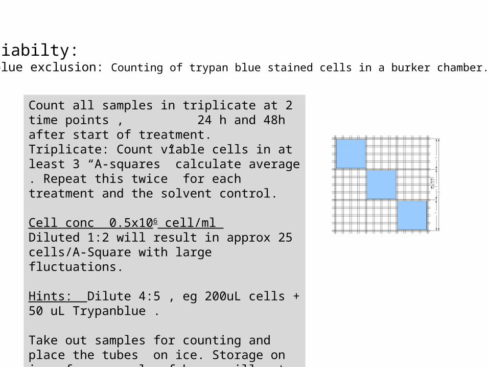
Cell viabilty:Trypan blue exclusion: Counting of trypan blue stained cells in a burker chamber.
Count all samples in triplicate at 2 time points , 24 h and 48h after start of treatment.Triplicate: Count viable cells in at least 3 “A-squares” calculate average . Repeat this twice for each treatment and the solvent control.
Cell conc 0.5x10⁶ cell/ml Diluted 1:2 will result in approx 25 cells/A-Square with large fluctuations.
Hints: Dilute 4:5 , eg 200uL cells + 50 uL Trypanblue .
Take out samples for counting and place the tubes on ice. Storage on ice for a couple of hours will not affect cell viability.
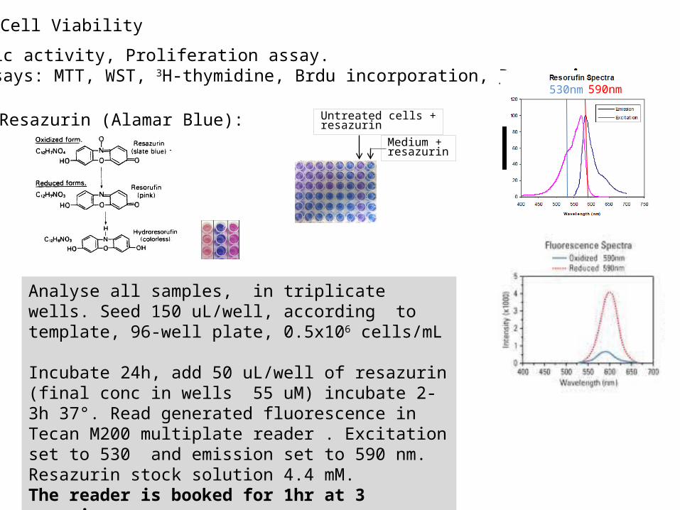
Metabolic activity, Proliferation assay.Many assays: MTT, WST, 3H-thymidine, Brdu incorporation, Resazurin.
Analyse all samples, in triplicate wells. Seed 150 uL/well, according to template, 96-well plate, 0.5x10⁶ cells/mL
Incubate 24h, add 50 uL/well of resazurin (final conc in wells 55 uM) incubate 2-3h 37°. Read generated fluorescence in Tecan M200 multiplate reader . Excitation set to 530 and emission set to 590 nm. Resazurin stock solution 4.4 mM.The reader is booked for 1hr at 3 occasions on Tuesday 10/3: 12:00-13:00, 14:00-15:00, 16:00-17:00
Resazurin (Alamar Blue): Medium + resazurin
Untreated cells +resazurin
590nm 530nm
Cell Viability

Consider results from the two viability assays “trypan blue exclusion” and “resazurin assay.” Which samples from the chemical library affected growth?
Choose 4 samples together with the solvent control and spin out cells on slides and stain cells with May Grunewald/Giemsa according to instructions given in Lab 1. After staining look through the slides and search for a phenotype for that specific drug. Estimate ratio “phenotypic cells”/normal cells among 100-150 cells.
3. Giemsa/May Grunewald stain.
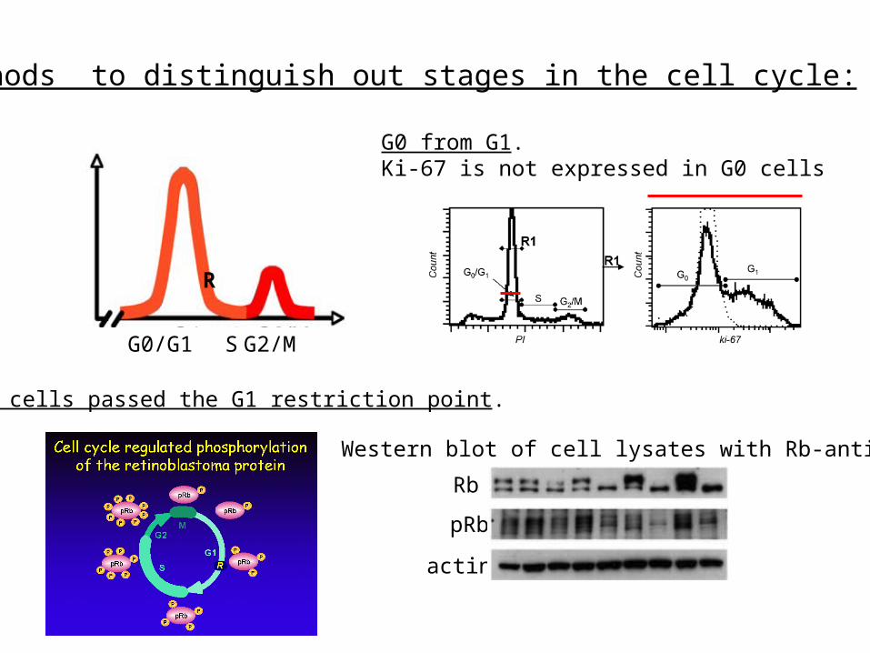
Methods to distinguish out stages in the cell cycle:
G0/G1 S G2/M
R
G0 from G1.Ki-67 is not expressed in G0 cells
Has cells passed the G1 restriction point.
Rb
pRb
actin
Western blot of cell lysates with Rb-antibodies
G0/G1 S G2/M
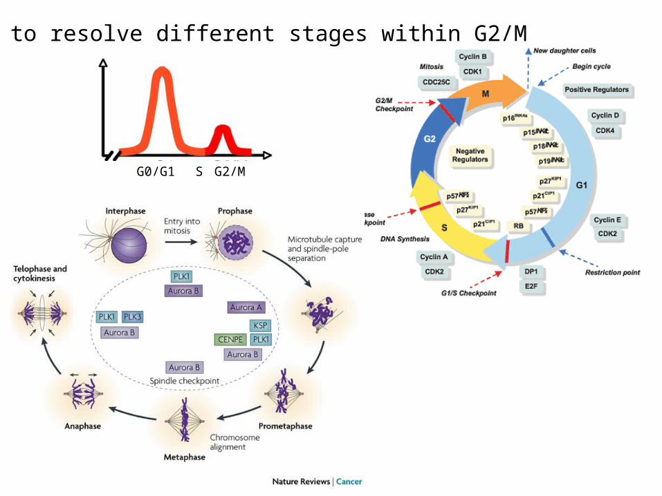
How to resolve different stages within G2/M

μ-tubules
Control Drug
Ref Dipak M et al Nature Comm 2013
Jurkat cells
Intact cells
Perforation of plasma membrane in presence of saponin and taxol; Unpolymerised tubulins will diffuse out of the cell
Ref: Per Holmfeldt, Umeå university
After fixation incubate cells with anti-tubulin antibodies followed by incubation with FITC-labelled secondary antibodies.
FACS analysis of micro-tubules

Solutions: MT-STABPEM buffer, pH 6.90.1 % Saponin (stock 10%)10 µg/ml RNase (stock 5mg/ml)50 nM Taxol (stock 1,1mM) MT-FIX ~50 % PEM buffer, pH 6.9~50 % PFA in H2O (from 8% stock)0.1 % Saponin (add 1/100 of a 10 % stock, which may be stored ~1 week) BLOCK90 % PBSA / 10 % FCS / 0,1 % Saponin WASH PBSA / 0,1 % Saponin
PI SOL.PBSA10µg/ml Propidium Iodide0,1% Triton X-100 (add 1/100 of a 10 % stock)10µg/ml RNase
Staining of microtubules for FACS(copied from “-MG lab methods 2011-“)
1. Spin down 500.000 cells per FACS tubes for 2 min, 1100rpm (250xg).
2. Aspirate the supernatant and resuspend the pelleted cells gently using the shaking table.
3. Extraction of cells; add 400µl 37 °C MT-STAB and incubate at 37 °C for 4 minutes.
4. Fixation of cells; add 400 µl 37°C MT-FIX and incubate at 5. 37 °C for 12 minutes.6. Spin down the cells, 3 min, 1100rpm.7. Aspirate the supernatant and resuspend the cells by shaking.8. Add 1 ml BLOCK and incubate at 37 °C for 10 minutes.9. Spin down the cells, 5 min, 1100rpm.10. Aspirate the supernatant and resuspend the cells by shaking.11. Add 150 µl primary antibody (anti--tubulin T5168 B-5-1-2 dil
1:250 in BLOCK). NOTE! Include a tube with no addition of primary antibody to define the background.
12. Incubate at 37 °C for 60 minutes while shaking.13. Add 1 ml WASH.14. Spin down the cells, 5 min, 1100rpm.15. Aspirate the supernatant and resuspend the cells by shaking.16. Add 100 µl secondary antibody (Rabbit-anti-mouse (FITC)
DAKO F0313 dilute 1:20 in WASH).17. Incubate at 37 °C for 30 minutes while shaking.18. Add 1 ml WASH.19. Spin down the cells, 5 min, 1100rpm.20. Aspirate the supernatant and resuspend the cells by shaking.21. Add 500µl PI SOL and keep samples in refrigerator until
analysis.

500 μL cells
Aspirate medium
Centrifuge 1100 rpm 2min
Loosen pellet by gently shaking on shakerboard
Add 150 μL Mouse anti-α–tubulin Antibody and inc 60min 37° in shakingwaterbath.
Extraction of “free” tubulin Add 400 μL MT-STAB 37° inc 37° 4 min.
Fixation of cellsAdd 400 μL MT-FIX 37° inc 37° 12 min
Aspirate supand shake gently
Blocking of unspecific protein binding sites.Add 1ml BLOCK and inc 10min 37°
Aspirate supand shake gently
Aspirate sup and shake gently
Add 100 μL Secondary ab: FITC Rabbit α mouse IgG, inc 30min 37° in shaking waterbath
Aspirate supand shake gently
Add 1 mL WASH
Centrifuge 1100 rpm 5 min
Centrifuge 1100 rpm 3 min
Centrifuge 1100 rpm 5 min
Add 1 mL WASH
Centrifuge 1100 rpm 5 min
micro-tubules är very britle
Add 0.5ml PI-soution.Store cells in fridge until FACS analysis
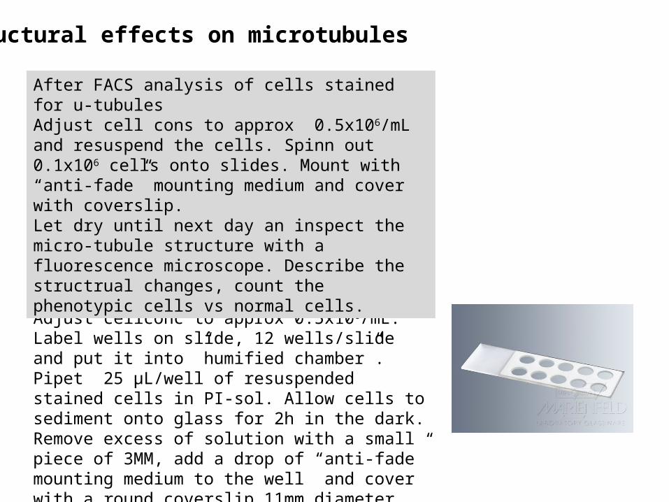
Microscope slides with wellsan alternative to cytospinningAdjust cellconc to approx 0.5x10⁶/mL.Label wells on slide, 12 wells/slide and put it into ”humified chamber”. Pipet 25 μL/well of resuspended stained cells in PI-sol. Allow cells to sediment onto glass for 2h in the dark. Remove excess of solution with a small piece of 3MM, add a drop of “anti-fade” mounting medium to the well and cover with a round coverslip 11mm diameter.
After FACS analysis of cells stained for u-tubulesAdjust cell cons to approx 0.5x10⁶/mL and resuspend the cells. Spinn out 0.1x10⁶ cells onto slides. Mount with “anti-fade” mounting medium and cover with coverslip.Let dry until next day an inspect the micro-tubule structure with a fluorescence microscope. Describe the structrual changes, count the phenotypic cells vs normal cells.
Structural effects on microtubules

Safety issues:
• PFA (paraformaldehyde) is skin and airway irritant and could cause allergic reactions! Wash exposed skin areas with water and soap.
• May Grunewald staining solution is flammable and skin irritant. Rinse with ethanol followed by water in large amounts!
• Giemsa is skin irritant. Rinse with ethanol followed by water in large amounts!
• PI (propidium iodide) binds to DNA and should be handle with care. Rinse with ethanol followed by water in large amounts!
• All drugs are cytotoxic drugs. Rinse with ethanol followed by washing of exposed areas with water and soap.

Group wise presentation: Fully planned experiments
Reagents and supplies and amount needed as well as a time scheduleDrugs: 200x final conc. Jurkat cells: Amount of cells and RPMI1640 10% FCS.Reagents : MT-STAB, MT-FIX, PI-solution, trypan blue solution, resazurin 4.4 mM, saponin 10%, antibodies, buffers……………………………………………..Other suppliesTubes Epp-tubes, multiwell plates, slides, cover slips ……………………………………………. Equipment:
You do not need to include pipett tips , disposable pipets, May Grunewald and Giemsa staining solutions and mounting media.
Carefully planned time schedule for Monday 10/3 Tuesday 11/3.Calculations how to obtain original cell conc when cells are diluted 200ul cells + 50 ul trypanblue.

Time Slots for group discussions:
Monday 10/3group
8:00 15, 16, 178:30 12,13, 149:00 9, 10, 11
12:00 6, 7, 812:30 3, 4, 513:00 1, 2 C Solvent control
S1 Cells treated with drug #1
S2 Cells treated with drug #2 and so onB culture medium without cells
CCC
BB
B
S1
S1S1
S2
S3
S2S4
S2
S6
S7S3S3
S4S4
S5
S6S6
S5S5
S7S7
CC
C
Format for seeding cells into 96-well plate for resazurin