Lab 1 Microscopy Parts & use of microscope. Lab 2 Cells & Tissues Cell theory Cell structure, review...
67
Lab 1 Microscopy • Parts & use of microscope
-
date post
19-Dec-2015 -
Category
Documents
-
view
220 -
download
2
Transcript of Lab 1 Microscopy Parts & use of microscope. Lab 2 Cells & Tissues Cell theory Cell structure, review...

Lab 1 Microscopy
• Parts & use of microscope

Lab 2 Cells & Tissues
• Cell theory• Cell structure, review pp. 17 & 18• Histology: epithelial, connective, muscular &
nervous• Slides– List on pp. 16 (plus frog skin)

Cardiac muscle

Motor end plate

Lab 4 Development
• 5 phases• 3 germ layers• Cleavage: radial v. spiral• Development in sea star (slide) & frog (model)– Zygote, morula, blastula, gastrula, neurula (frog),
larva
• Development in chick (slides)• Extraembronic membranes (4)

Sea star development

Frog development

Lab 5 Morphology
• Symmetry• Body cavity• Metamerism• Cephalization• Protostomes v. Deuterostomes

Lab 6 Protozoa
• Single celled, eukaryotic, animal-like protists• Flagellated protozoa– Euglena (live & prepared)– Trypanosoma
• Amoeboid– Amoeba (live & prepared)– Foraminiferans (slide)– Radiolarians (slide)

Euglena

Amoeba
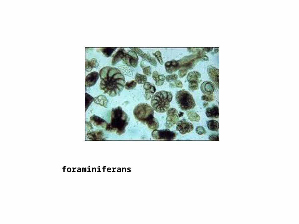
foraminiferans

radiolarians

Protozoa
• Alveolates– Paramecium caudatum

Lab 7 Porifera
• Sponges– Spicules of calcium carbonate or silica– Fibers of collagen or spongin– Outer pinacocytes– Inner choanocytes– Mesohyl in the middle
• Canal systems: asconoid, syconoid or leuconoid

Sponges
• Class Demospongiae: horny sponges• Class Calcispongiae: calcareous sponges• Class Hexactinellida: glass sponges
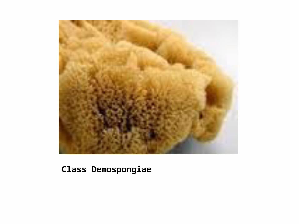
Class Demospongiae

Clathrina
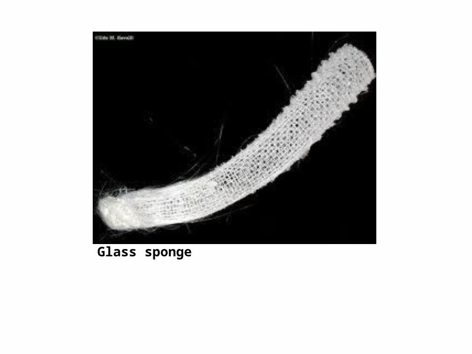
Glass sponge

Phylum Cnidaria
• Radial symmetry• Diploblastic• Outer epidermis• Inner gastrodermis• Cnidocytes—nematocysts• Polyp & medusa stage

Cnidaria
• Class Hydrozoa– Obelia (prepared slide)– Hydra (live & prepared)– Physalia (preserved)
• Class Scyphozoa– Aurelia
• Class Anthozoa– Sea anemones & corals

Obelia

Hydra

Physalia

Aurelia

Anthozoans

Lab 9 Platyhelminthes
• Flatworms• Triploblastic & bilateral• Class Turbellaria– Planaria (live & prepared slides)
• Class Trematoda– Complex life cycle—2+ hosts– Clonorchis sinensis—human liver fluke (slide)– Fasciola hepatica—sheep liver fluke (slide)– Cercaria—tadpole like stage (slide)
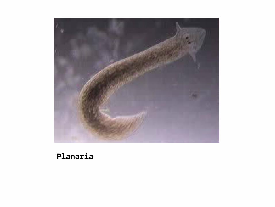
Planaria

Clonorchis sinensis

Fasciola hepatica
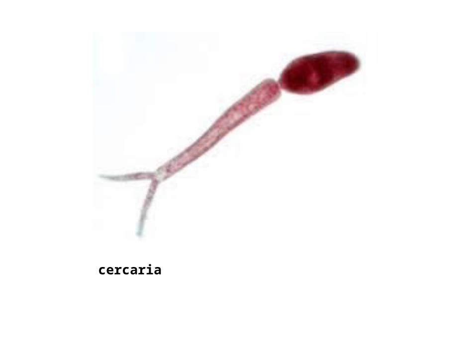
cercaria
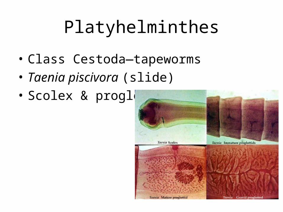
Platyhelminthes
• Class Cestoda—tapeworms• Taenia piscivora (slide)• Scolex & proglottids
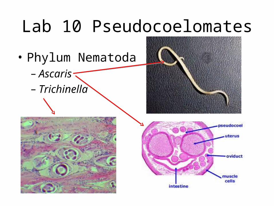
Lab 10 Pseudocoelomates
• Phylum Nematoda– Ascaris– Trichinella
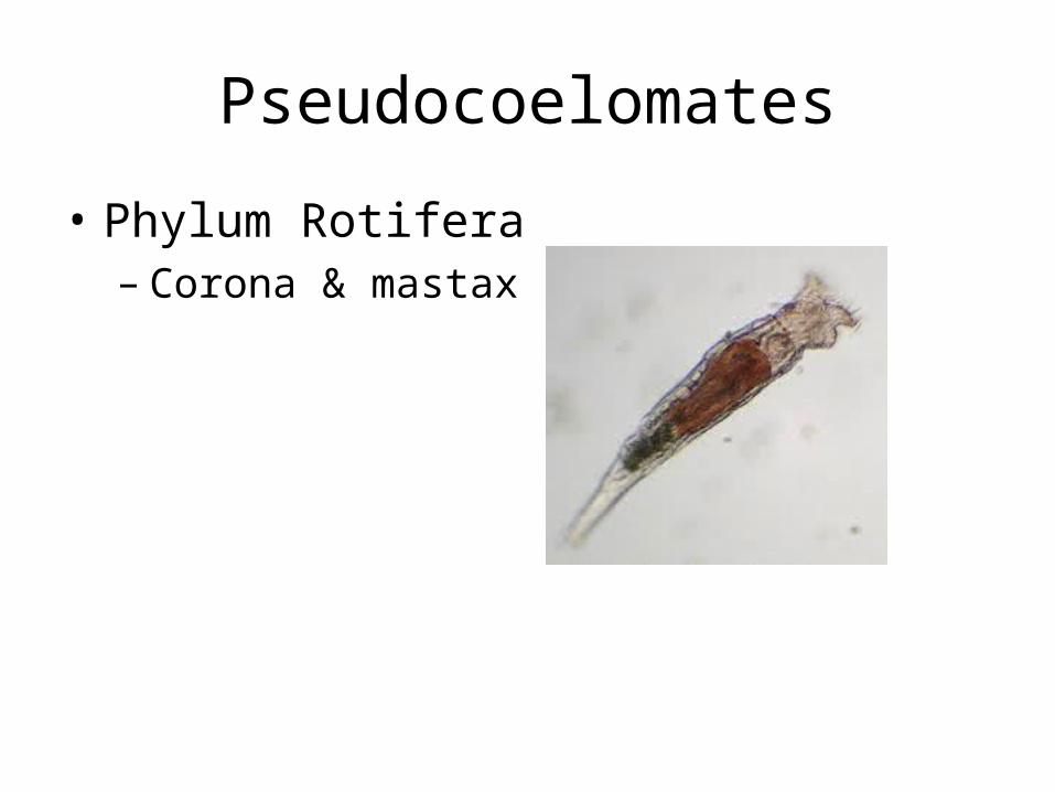
Pseudocoelomates
• Phylum Rotifera– Corona & mastax

Lab 11 Phylum Mollusca
• Soft body• Mantle secretes shell• Triploblastic & coelomate• Radula

Class Polyplacophora

Class Bivalvia

Class Gastropods

Class Cephalopoda


Lab 12 Phylum Annelida
• Metamerism• setae
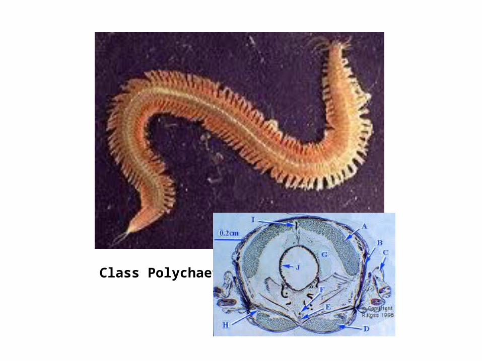
Class Polychaeta

Earthworm dissection

Class Oligochaeta

Class Hirudinea

Lab 13 Phylum Arthropoda
• Hardened exoskeleton• Jointed appendages• Molting

Subphylum Trilobita

Class MerostomataSubphylym Chelicerata

Subphylum Chelicerata

Subphylum MyriapodaClass DiplopodaClass Chilopoda

Class BranchiopodaSubphylum Crustacea

Class MaxillopodaSubphylum Crustacea

Class MalacostracaSubphylum Crustracea


Class InsectaSubphylum Hexapoda