
l enta l i n ic lp Journal of Clinical & Experimental ht f ... · Orbital Plasmablastic...
4
Orbital Plasmablastic Plasmacytoma in Multiple Myeloma Soyeon Jung, Suk Jin Choi and Sungmo Kang * Department of Ophthalmology, Inha University College of Medicine, Incheon, Korea * Corresponding author: Sungmo Kang, Department of Ophthalmology, Inha University College of Medicine, Incheon, Korea, Tel: +82-32-890-2400; Fax: +82-32-890-2417; E-mail: [email protected] Received date: June 17, 2015; Accepted date: August 21, 2015; Published date: August 31, 2015 Copyright: © 2015 Jung S, et al. This is an open-access article distributed under the terms of the Creative Commons Attribution License, which permits unrestricted use, distribution, and reproduction in any medium, provided the original author and source are credited. Abstract Multiple myeloma is a malignant disease characterized by the proliferation of clonal plasma cells in bone marrow, extramedullary involvement with multiple myeloma is generally a manifestation of advanced disease. Orbital involvement by multiple myeloma is uncommon and intraocular involvement is rare. The authors present an uncommon case of orbital plasmablastic plasmacytoma. Keywords: Orbital plasmacytoma; Plasmacytoma; Orbital plasmablastic plasmacytoma; Multiple myeloma Introduction Multiple myeloma is a malignant disease characterized by the proliferation of clonal plasma cells in bone marrow [1], extramedullary involvement with multiple myeloma is generally a manifestation of advanced disease [2]. Orbital involvement by multiple myeloma is uncommon and intraocular involvement is rare [3,4]. Case Report A 73-year-old man was referred to the department of ophthalmology for swelling and ptosis of left eyelid and right lateral and left superior orbital wall masses by brain MRI. He first visited the department of neurology for headache and paresthesia of the left chin and orbit areas. The neurologist recommended to undergo brain MRI for evaluation purposes, and subsequently, multiple variably sized osteolytic lesions were observed in skull, a T1 and T2 iso signal intensity mass in clivus, expansile masses in the right lateral and left superior orbital walls involving adjacent soft tissue, and T1 low signal intensity of C2 vertebra (Figure 1). The radiologist propounded the diagnostic possibilities of multiple myeloma and bone metastasis. The patient was referred to the department of hemato-oncology, where he underwent a systemic evaluation for multiple myeloma. Visual acuity of right and left eyes were 0.9 and 0.8, respectively. Intraocular pressure of the right eye was 11mmHg and of the left was 15mmHg by NCT. Through an eye examination, mild swelling with injection and dermatochalasis of both upper eyelids and ptosis of the left eye (Figure 2) were reported. On examination of extraocular muscle movement, no limitation of extraocular muscle movement or diplopia was observed. His Hertel exophthalmometry reading was 14 mm OD, 13 mm OS and baseline was 115 mm. MRD1 was 5 mm OD and 3 mm OS. LFT of right and left eyes were 15 and 10 mm, respectively. Subsequent, orbital CT revealed an enhancing bone destructive mass in the left superior orbital wall (Figure 3), extending into orbit and upper eyelid, and another enhancing mass in sphenoid sinus and sphenoid bone involving both vidian canals. Based on orbital CT and MRI, the diagnostic possibilities of multiple myeloma and bone metastasis were considered. Orbitotomy and excisional biopsy of the left superior orbital mass was performed and a multiseptated 2.5 × 1.0 cm mass was excised (Figure 4). Post-operative orbital CT (Figure 5) confirmed removal of the left upper eyelid portion of the mass, but a residual enhancing bony destructive mass was detected in the left orbital superior wall bulging into the extraconal area of the orbit. Histopathologic evaluation confirmed the mass as plasmablastic plasmacytoma (Figure 6), and immunohistochemical staining showed positivity in for MUM1 and a 60-70% Ki67 labeling index and negativity in for CD3, CD20, PAX5, CD138, CD30, Bcl2, and ALK. Immunostain of Bcl2 showed weak- positive in about 20~40% of cells. Immunohitochemical staining of CD79a or CD19 was not performed. Tumor cells were immunopositive for lambda light chain and negative for kappa light chain. Figure 1: Brain MR images (a) Multiple variable sized osteolytic lesions in skull. (b) T1 iso signal intensity mass in clivus. (c) Left superior orbital wall mass involving adjacent soft tissue. (d) T1 low signal intensity of C2 vertebra. Jung et al., J Clin Exp Ophthalmol 2015, 6:4 DOI: 10.4172/2155-9570.1000462 Case Report Open Access J Clin Exp Ophthalmol ISSN:2155-9570 JCEO, an open access journal Volume 6 • Issue 4 • 1000462 Journal of Clinical & Experimental Ophthalmology J o ur n a l o f C l i n ic a l & E x pe r i m e n t a l O p h t h a l m o lo g y ISSN: 2155-9570
Transcript of l enta l i n ic lp Journal of Clinical & Experimental ht f ... · Orbital Plasmablastic...
Orbital Plasmablastic Plasmacytoma in Multiple MyelomaSoyeon Jung, Suk Jin Choi and Sungmo Kang*
Department of Ophthalmology, Inha University College of Medicine, Incheon, Korea*Corresponding author: Sungmo Kang, Department of Ophthalmology, Inha University College of Medicine, Incheon, Korea, Tel: +82-32-890-2400; Fax:+82-32-890-2417; E-mail: [email protected]
Received date: June 17, 2015; Accepted date: August 21, 2015; Published date: August 31, 2015
Copyright: © 2015 Jung S, et al. This is an open-access article distributed under the terms of the Creative Commons Attribution License, which permits unrestricteduse, distribution, and reproduction in any medium, provided the original author and source are credited.
Abstract
Multiple myeloma is a malignant disease characterized by the proliferation of clonal plasma cells in bone marrow,extramedullary involvement with multiple myeloma is generally a manifestation of advanced disease. Orbitalinvolvement by multiple myeloma is uncommon and intraocular involvement is rare. The authors present anuncommon case of orbital plasmablastic plasmacytoma.
Keywords: Orbital plasmacytoma; Plasmacytoma; Orbitalplasmablastic plasmacytoma; Multiple myeloma
IntroductionMultiple myeloma is a malignant disease characterized by the
proliferation of clonal plasma cells in bone marrow [1], extramedullaryinvolvement with multiple myeloma is generally a manifestation ofadvanced disease [2]. Orbital involvement by multiple myeloma isuncommon and intraocular involvement is rare [3,4].
Case ReportA 73-year-old man was referred to the department of
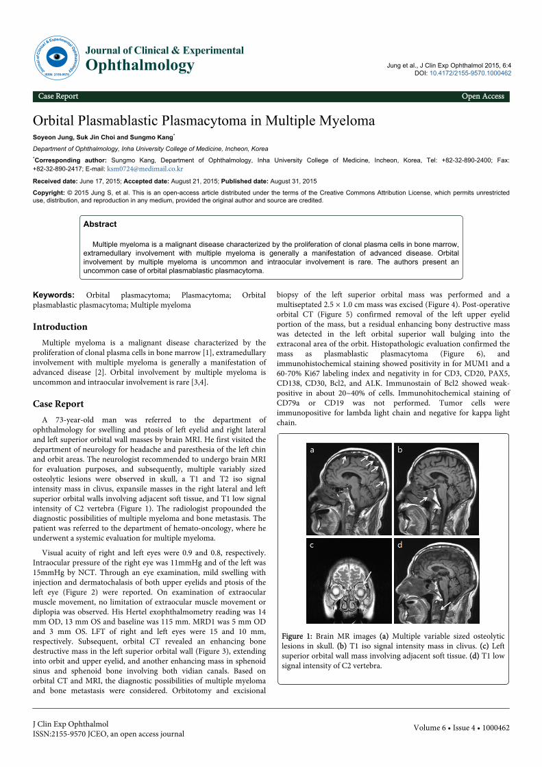
ophthalmology for swelling and ptosis of left eyelid and right lateraland left superior orbital wall masses by brain MRI. He first visited thedepartment of neurology for headache and paresthesia of the left chinand orbit areas. The neurologist recommended to undergo brain MRIfor evaluation purposes, and subsequently, multiple variably sizedosteolytic lesions were observed in skull, a T1 and T2 iso signalintensity mass in clivus, expansile masses in the right lateral and leftsuperior orbital walls involving adjacent soft tissue, and T1 low signalintensity of C2 vertebra (Figure 1). The radiologist propounded thediagnostic possibilities of multiple myeloma and bone metastasis. Thepatient was referred to the department of hemato-oncology, where heunderwent a systemic evaluation for multiple myeloma.
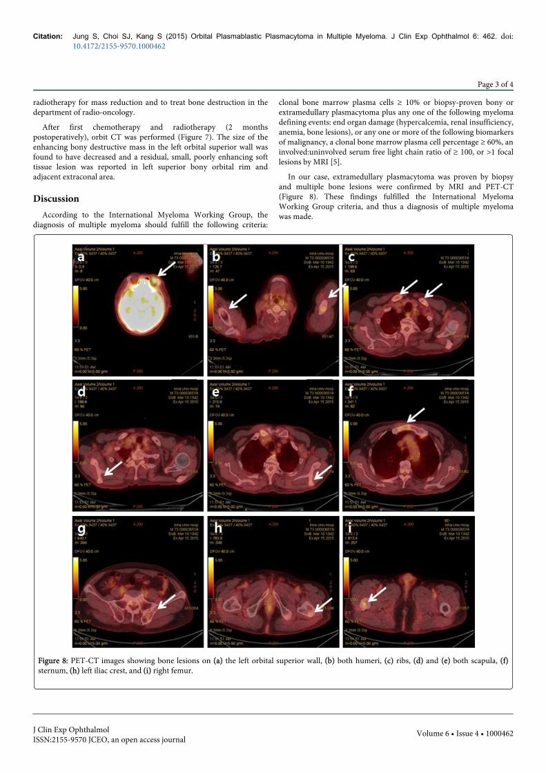
Visual acuity of right and left eyes were 0.9 and 0.8, respectively.Intraocular pressure of the right eye was 11mmHg and of the left was15mmHg by NCT. Through an eye examination, mild swelling withinjection and dermatochalasis of both upper eyelids and ptosis of theleft eye (Figure 2) were reported. On examination of extraocularmuscle movement, no limitation of extraocular muscle movement ordiplopia was observed. His Hertel exophthalmometry reading was 14mm OD, 13 mm OS and baseline was 115 mm. MRD1 was 5 mm ODand 3 mm OS. LFT of right and left eyes were 15 and 10 mm,respectively. Subsequent, orbital CT revealed an enhancing bonedestructive mass in the left superior orbital wall (Figure 3), extendinginto orbit and upper eyelid, and another enhancing mass in sphenoidsinus and sphenoid bone involving both vidian canals. Based onorbital CT and MRI, the diagnostic possibilities of multiple myelomaand bone metastasis were considered. Orbitotomy and excisional
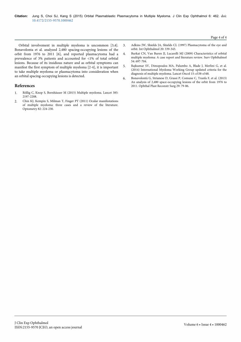
biopsy of the left superior orbital mass was performed and amultiseptated 2.5 × 1.0 cm mass was excised (Figure 4). Post-operativeorbital CT (Figure 5) confirmed removal of the left upper eyelidportion of the mass, but a residual enhancing bony destructive masswas detected in the left orbital superior wall bulging into theextraconal area of the orbit. Histopathologic evaluation confirmed themass as plasmablastic plasmacytoma (Figure 6), andimmunohistochemical staining showed positivity in for MUM1 and a60-70% Ki67 labeling index and negativity in for CD3, CD20, PAX5,CD138, CD30, Bcl2, and ALK. Immunostain of Bcl2 showed weak-positive in about 20~40% of cells. Immunohitochemical staining ofCD79a or CD19 was not performed. Tumor cells wereimmunopositive for lambda light chain and negative for kappa lightchain.
Figure 1: Brain MR images (a) Multiple variable sized osteolyticlesions in skull. (b) T1 iso signal intensity mass in clivus. (c) Leftsuperior orbital wall mass involving adjacent soft tissue. (d) T1 lowsignal intensity of C2 vertebra.
Jung et al., J Clin Exp Ophthalmol 2015, 6:4 DOI: 10.4172/2155-9570.1000462
Case Report Open Access
J Clin Exp OphthalmolISSN:2155-9570 JCEO, an open access journal
Volume 6 • Issue 4 • 1000462
Journal of Clinical & Experimental OphthalmologyJo
urna
l of C
linica
l & Experimental Ophthalmology
ISSN: 2155-9570
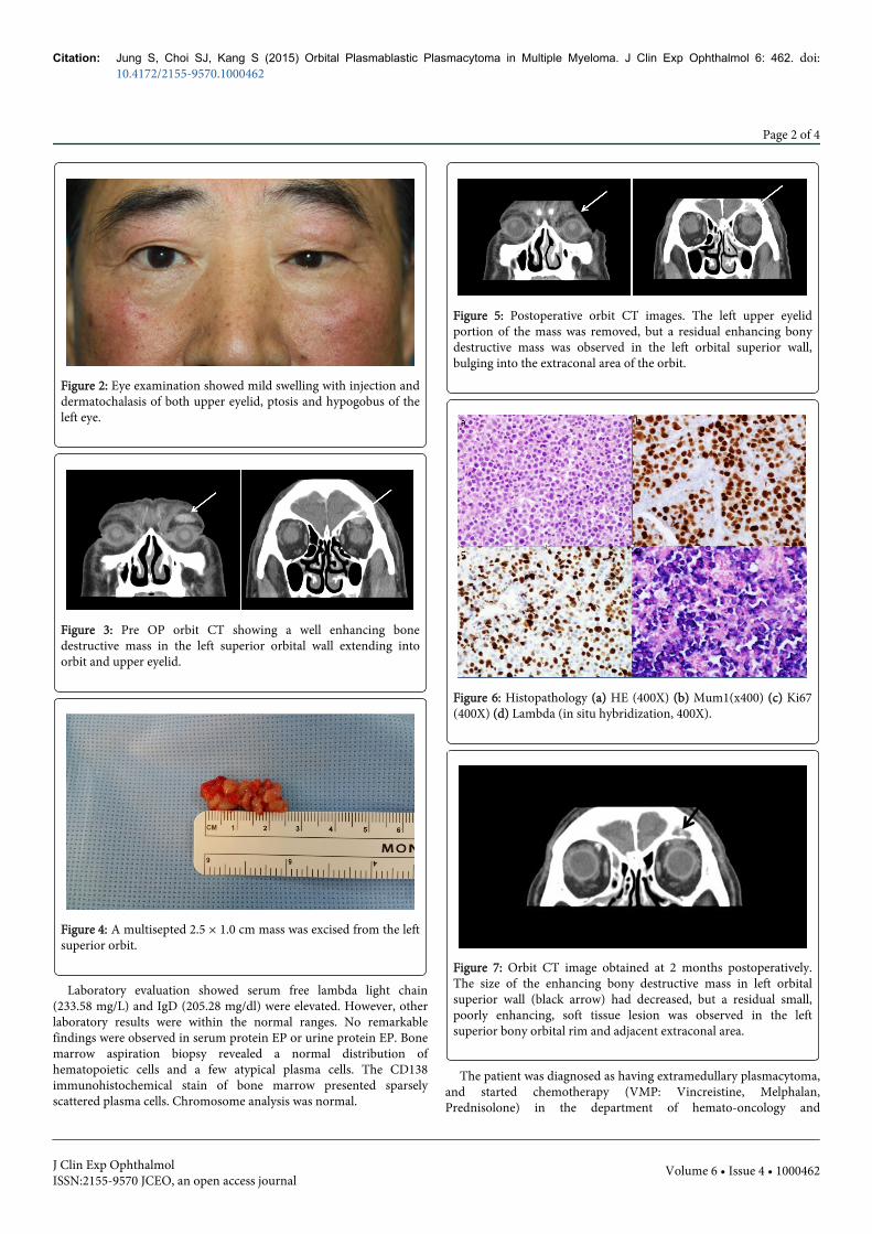
Figure 2: Eye examination showed mild swelling with injection anddermatochalasis of both upper eyelid, ptosis and hypogobus of theleft eye.
Figure 3: Pre OP orbit CT showing a well enhancing bonedestructive mass in the left superior orbital wall extending intoorbit and upper eyelid.
Figure 4: A multisepted 2.5 × 1.0 cm mass was excised from the leftsuperior orbit.
Laboratory evaluation showed serum free lambda light chain(233.58 mg/L) and IgD (205.28 mg/dl) were elevated. However, otherlaboratory results were within the normal ranges. No remarkablefindings were observed in serum protein EP or urine protein EP. Bonemarrow aspiration biopsy revealed a normal distribution ofhematopoietic cells and a few atypical plasma cells. The CD138immunohistochemical stain of bone marrow presented sparselyscattered plasma cells. Chromosome analysis was normal.
Figure 5: Postoperative orbit CT images. The left upper eyelidportion of the mass was removed, but a residual enhancing bonydestructive mass was observed in the left orbital superior wall,bulging into the extraconal area of the orbit.
Figure 6: Histopathology (a) HE (400X) (b) Mum1(x400) (c) Ki67(400X) (d) Lambda (in situ hybridization, 400X).
Figure 7: Orbit CT image obtained at 2 months postoperatively.The size of the enhancing bony destructive mass in left orbitalsuperior wall (black arrow) had decreased, but a residual small,poorly enhancing, soft tissue lesion was observed in the leftsuperior bony orbital rim and adjacent extraconal area.
The patient was diagnosed as having extramedullary plasmacytoma,and started chemotherapy (VMP: Vincreistine, Melphalan,Prednisolone) in the department of hemato-oncology and
Citation: Jung S, Choi SJ, Kang S (2015) Orbital Plasmablastic Plasmacytoma in Multiple Myeloma. J Clin Exp Ophthalmol 6: 462. doi:10.4172/2155-9570.1000462
Page 2 of 4
J Clin Exp OphthalmolISSN:2155-9570 JCEO, an open access journal
Volume 6 • Issue 4 • 1000462
radiotherapy for mass reduction and to treat bone destruction in thedepartment of radio-oncology.
After first chemotherapy and radiotherapy (2 monthspostoperatively), orbit CT was performed (Figure 7). The size of theenhancing bony destructive mass in the left orbital superior wall wasfound to have decreased and a residual, small, poorly enhancing softtissue lesion was reported in left superior bony orbital rim andadjacent extraconal area.
DiscussionAccording to the International Myeloma Working Group, the
diagnosis of multiple myeloma should fulfill the following criteria:
clonal bone marrow plasma cells ≥ 10% or biopsy-proven bony orextramedullary plasmacytoma plus any one of the following myelomadefining events: end organ damage (hypercalcemia, renal insufficiency,anemia, bone lesions), or any one or more of the following biomarkersof malignancy, a clonal bone marrow plasma cell percentage ≥ 60%, aninvolved:uninvolved serum free light chain ratio of ≥ 100, or >1 focallesions by MRI [5].
In our case, extramedullary plasmacytoma was proven by biopsyand multiple bone lesions were confirmed by MRI and PET-CT(Figure 8). These findings fulfilled the International MyelomaWorking Group criteria, and thus a diagnosis of multiple myelomawas made.
Figure 8: PET-CT images showing bone lesions on (a) the left orbital superior wall, (b) both humeri, (c) ribs, (d) and (e) both scapula, (f)sternum, (h) left iliac crest, and (i) right femur.
Citation: Jung S, Choi SJ, Kang S (2015) Orbital Plasmablastic Plasmacytoma in Multiple Myeloma. J Clin Exp Ophthalmol 6: 462. doi:10.4172/2155-9570.1000462
Page 3 of 4
J Clin Exp OphthalmolISSN:2155-9570 JCEO, an open access journal
Volume 6 • Issue 4 • 1000462
Orbital involvement in multiple myeloma is uncommon [3,4].Bonavolonta et al. analyzed 2,480 spacing-occupying lesions of theorbit from 1976 to 2011 [6], and reported plasmacytoma had aprevealence of 3% patients and accounted for <1% of total orbitallesions. Because of its insidious nature and as orbital symptoms canmanifest the first symptom of multiple myeloma [2-4], it is importantto take multiple myeloma or plasmacytoma into consideration whenan orbital spacing-occupying lesions is detected.
References1. Röllig C, Knop S, Bornhäuser M (2015) Multiple myeloma. Lancet 385:
2197-2208.2. Chin KJ, Kempin S, Milman T, Finger PT (2011) Ocular manifestations
of multiple myeloma: three cases and a review of the literature.Optometry 82: 224-230.
3. Adkins JW, Shields JA, Shields CL (1997) Plasmacytoma of the eye andorbit. Int Ophthalmol 20: 339-343.
4. Burkat CN, Van Buren JJ, Lucarelli MJ (2009) Characteristics of orbitalmultiple myeloma: A case report and literature review. Surv Ophthalmol54: 697-704.
5. Rajkumar SV, Dimopoulos MA, Palumbo A, Blade J, Merlini G, et al.(2014) International Myeloma Working Group updated criteria for thediagnosis of multiple myeloma. Lancet Oncol 15: e538-e548.
6. Bonavolontà G, Strianese D, Grassi P, Comune C, Tranfa F, et al. (2013)An analysis of 2,480 space-occupying lesions of the orbit from 1976 to2011. Ophthal Plast Reconstr Surg 29: 79-86.
Citation: Jung S, Choi SJ, Kang S (2015) Orbital Plasmablastic Plasmacytoma in Multiple Myeloma. J Clin Exp Ophthalmol 6: 462. doi:10.4172/2155-9570.1000462
Page 4 of 4
J Clin Exp OphthalmolISSN:2155-9570 JCEO, an open access journal
Volume 6 • Issue 4 • 1000462