L-Amino Acid Oxidase from Venoms - link.springer.com · L-Amino Acid Oxidase from Venoms Payel...
21
L-Amino Acid Oxidase from Venoms Payel Bhattacharjee, Jyotirmoy Mitra and Debasish Bhattacharyya* Division of Structural Biology and Bioinformatics, CSIR-Indian Institute of Chemical Biology, Jadavpur, Kolkata, West Bengal, India Abstract L-Amino acid oxidase (LAAO), a flavoenzyme, is a component of many animal venoms of which the enzyme from snake venom is particularly well characterized. Since the oxidation of L-amino acids produces hydrogen peroxide, the enzyme induces toxicity. Presumably, a glycan part of the enzyme anchors the molecule to cell surface and thereby generates high concentration of hydrogen peroxide locally on cell surface leading to cytotoxicity. Depending on localization of LAAO on normal cells or unwanted cells, the enzyme may be toxic or possess therapeutic potential. The mechanism of action of the enzyme is known, and the crystal structure of snake venom LAAOs has been solved. They are structurally well-conserved enzymes. Therefore, inhibitors of the enzyme may be synthesized using the template of the substrate and features of its catalytic site. Though snake venom LAAO is viewed as toxic, several beneficial activities of the enzyme are known. The physiological actions of the enzyme are not clearly understood, e.g., some contradictory results exist on its platelet aggregation activity. This article updates present knowledge on the enzyme covering various aspects of its toxicity along with its therapeutic potential. Keywords L-Amino acid oxidase; Venoms; Toxicity; Enzyme inhibition; Therapeutic applications Introduction L-Amino acid oxidase (LAAO, E.C. 1.4.3.2) is a flavoenzyme that catalyzes stereospecific oxidative deamination of an L-amino acid to form corresponding a-keto acid along with ammonia and hydrogen peroxide (H 2 O 2 ) (Fig. 1). This enzyme is involved in the metabolic pathways of eight amino acids, e.g., alanine, aspartate, methionine, valine, leucine, tyrosine, phenylalanine, and tryptophan; degradation of isoleucine and phenylalanine; and biosynthesis of tyrosine, tryptophan, and some alkaloids (Mason et al. 2004). LAAO is potentially useful for quantification of amino acids because a product of the enzyme-catalyzed reaction is H 2 O 2 , which can be easily quantified by colorimetric assays (Kameya et al. 2013). LAAOs are widely distributed in several taxa. In bacteria, fungi, and green algae, LAAO appears to use amino acids for nitrogen fixation (Vallon et al. 1993). LAAOs found in the venoms of snakes, insects, and marine animals act as an effective deterrent against enemies and/or predators. LAAO activity has been reported in peroxisomes and mitochondria of rat kidney and liver (Mason et al. 2004). The antibacterial activity of mammalian milk is owing to the presence of LAAO (Nagaoka et al. 2009). Mason et al. first *Email: [email protected] *Email: [email protected] Toxins and Drug Discovery DOI 10.1007/978-94-007-6726-3_11-1 # Springer Science+Business Media Dordrecht 2015 Page 1 of 21
Transcript of L-Amino Acid Oxidase from Venoms - link.springer.com · L-Amino Acid Oxidase from Venoms Payel...
L-Amino Acid Oxidase from Venoms
Payel Bhattacharjee, Jyotirmoy Mitra and Debasish Bhattacharyya*Division of Structural Biology and Bioinformatics, CSIR-Indian Institute of Chemical Biology, Jadavpur, Kolkata,West Bengal, India
Abstract
L-Amino acid oxidase (LAAO), a flavoenzyme, is a component of many animal venoms of which theenzyme from snake venom is particularly well characterized. Since the oxidation of L-amino acidsproduces hydrogen peroxide, the enzyme induces toxicity. Presumably, a glycan part of the enzymeanchors the molecule to cell surface and thereby generates high concentration of hydrogen peroxidelocally on cell surface leading to cytotoxicity. Depending on localization of LAAO on normal cells orunwanted cells, the enzyme may be toxic or possess therapeutic potential. The mechanism of action of theenzyme is known, and the crystal structure of snake venom LAAOs has been solved. They are structurallywell-conserved enzymes. Therefore, inhibitors of the enzyme may be synthesized using the template ofthe substrate and features of its catalytic site. Though snake venom LAAO is viewed as toxic, severalbeneficial activities of the enzyme are known. The physiological actions of the enzyme are not clearlyunderstood, e.g., some contradictory results exist on its platelet aggregation activity. This article updatespresent knowledge on the enzyme covering various aspects of its toxicity along with its therapeuticpotential.
Keywords
L-Amino acid oxidase; Venoms; Toxicity; Enzyme inhibition; Therapeutic applications
Introduction
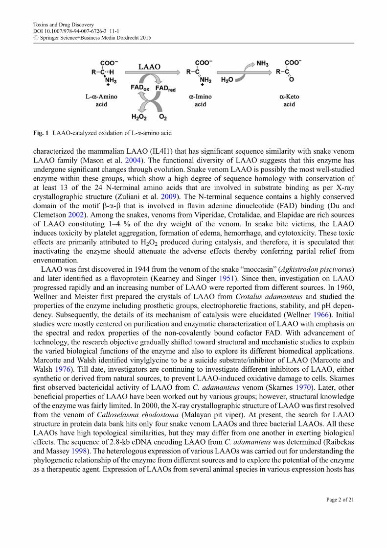
L-Amino acid oxidase (LAAO, E.C. 1.4.3.2) is a flavoenzyme that catalyzes stereospecific oxidativedeamination of an L-amino acid to form corresponding a-keto acid along with ammonia and hydrogenperoxide (H2O2) (Fig. 1). This enzyme is involved in the metabolic pathways of eight amino acids, e.g.,alanine, aspartate, methionine, valine, leucine, tyrosine, phenylalanine, and tryptophan; degradation ofisoleucine and phenylalanine; and biosynthesis of tyrosine, tryptophan, and some alkaloids (Masonet al. 2004). LAAO is potentially useful for quantification of amino acids because a product of theenzyme-catalyzed reaction is H2O2, which can be easily quantified by colorimetric assays (Kameyaet al. 2013).
LAAOs are widely distributed in several taxa. In bacteria, fungi, and green algae, LAAO appears to useamino acids for nitrogen fixation (Vallon et al. 1993). LAAOs found in the venoms of snakes, insects, andmarine animals act as an effective deterrent against enemies and/or predators. LAAO activity has beenreported in peroxisomes and mitochondria of rat kidney and liver (Mason et al. 2004). The antibacterialactivity of mammalian milk is owing to the presence of LAAO (Nagaoka et al. 2009). Mason et al. first
*Email: [email protected]
*Email: [email protected]
Toxins and Drug DiscoveryDOI 10.1007/978-94-007-6726-3_11-1# Springer Science+Business Media Dordrecht 2015
Page 1 of 21
characterized the mammalian LAAO (IL4I1) that has significant sequence similarity with snake venomLAAO family (Mason et al. 2004). The functional diversity of LAAO suggests that this enzyme hasundergone significant changes through evolution. Snake venom LAAO is possibly the most well-studiedenzyme within these groups, which show a high degree of sequence homology with conservation ofat least 13 of the 24 N-terminal amino acids that are involved in substrate binding as per X-raycrystallographic structure (Zuliani et al. 2009). The N-terminal sequence contains a highly conserveddomain of the motif b-a-b that is involved in flavin adenine dinucleotide (FAD) binding (Du andClemetson 2002). Among the snakes, venoms from Viperidae, Crotalidae, and Elapidae are rich sourcesof LAAO constituting 1–4 % of the dry weight of the venom. In snake bite victims, the LAAOinduces toxicity by platelet aggregation, formation of edema, hemorrhage, and cytotoxicity. These toxiceffects are primarily attributed to H2O2 produced during catalysis, and therefore, it is speculated thatinactivating the enzyme should attenuate the adverse effects thereby conferring partial relief fromenvenomation.
LAAO was first discovered in 1944 from the venom of the snake “moccasin” (Agkistrodon piscivorus)and later identified as a flavoprotein (Kearney and Singer 1951). Since then, investigation on LAAOprogressed rapidly and an increasing number of LAAO were reported from different sources. In 1960,Wellner and Meister first prepared the crystals of LAAO from Crotalus adamanteus and studied theproperties of the enzyme including prosthetic groups, electrophoretic fractions, stability, and pH depen-dency. Subsequently, the details of its mechanism of catalysis were elucidated (Wellner 1966). Initialstudies were mostly centered on purification and enzymatic characterization of LAAO with emphasis onthe spectral and redox properties of the non-covalently bound cofactor FAD. With advancement oftechnology, the research objective gradually shifted toward structural and mechanistic studies to explainthe varied biological functions of the enzyme and also to explore its different biomedical applications.Marcotte and Walsh identified vinylglycine to be a suicide substrate/inhibitor of LAAO (Marcotte andWalsh 1976). Till date, investigators are continuing to investigate different inhibitors of LAAO, eithersynthetic or derived from natural sources, to prevent LAAO-induced oxidative damage to cells. Skarnesfirst observed bactericidal activity of LAAO from C. adamanteus venom (Skarnes 1970). Later, otherbeneficial properties of LAAO have been worked out by various groups; however, structural knowledgeof the enzyme was fairly limited. In 2000, the X-ray crystallographic structure of LAAOwas first resolvedfrom the venom of Calloselasma rhodostoma (Malayan pit viper). At present, the search for LAAOstructure in protein data bank hits only four snake venom LAAOs and three bacterial LAAOs. All theseLAAOs have high topological similarities, but they may differ from one another in exerting biologicaleffects. The sequence of 2.8-kb cDNA encoding LAAO from C. adamanteus was determined (Raibekasand Massey 1998). The heterologous expression of various LAAOs was carried out for understanding thephylogenetic relationship of the enzyme from different sources and to explore the potential of the enzymeas a therapeutic agent. Expression of LAAOs from several animal species in various expression hosts has
Fig. 1 LAAO-catalyzed oxidation of L-a-amino acid
Toxins and Drug DiscoveryDOI 10.1007/978-94-007-6726-3_11-1# Springer Science+Business Media Dordrecht 2015
Page 2 of 21
been attempted, but the levels of expression were usually very low. A major limitation in producingrecombinant LAAO is that the enzyme itself produces H2O2, which in turn generates harmful reactiveoxygen species and induces cytotoxicity. Further, the expression of a protein in the absence of secondarymodifications could lead to an insoluble variety of protein, e.g., LAAO carries glycan moieties thatpresumably help in its solubilization. Therefore, recombinant LAAOs without these moieties becomeinsoluble after purification, once the level of expression is fairly improved. As the active site of LAAOmaintains complex configuration containing FAD, alteration in pH, temperature, presence of specific ions,and repeated freeze-thaw cycles lead to its inactivation (Tan and Fung 2009).
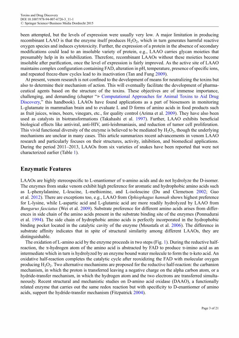
At present, venom research is not confined to the development of means for neutralizing the toxins butalso to determine their mechanism of action. This will eventually facilitate the development of pharma-ceutical agents based on the structure of the toxins. These objectives are of immense importance,challenging, and demanding (chapter “▶Computational Approaches for Animal Toxins to Aid DrugDiscovery,” this handbook). LAAOs have found applications as a part of biosensors in monitoringL-glutamate in mammalian brain and to evaluate L and D forms of amino acids in food products suchas fruit juices, wines, beers, vinegars, etc., for quality control (Arima et al. 2009). They have also beenused as catalysts in biotransformations (Takahashi et al. 1997). Further, LAAO exhibits beneficialbiological effects like antiviral, anti-HIV, anti-leishmaniasis, and reduction of tumor cell proliferation.This vivid functional diversity of the enzyme is believed to be mediated by H2O2, though the underlyingmechanisms are unclear in many cases. This article summarizes recent advancements in venom LAAOresearch and particularly focuses on their structures, activity, inhibition, and biomedical applications.During the period 2011–2013, LAAOs from six varieties of snakes have been reported that were notcharacterized earlier (Table 1).
Enzymatic Features
LAAOs are highly stereospecific to L-enantiomer of a-amino acids and do not hydrolyze the D-isomer.The enzymes from snake venom exhibit high preference for aromatic and hydrophobic amino acids suchas L-phenylalanine, L-leucine, L-methionine, and L-isoleucine (Du and Clemetson 2002; Guoet al. 2012). There are exceptions too, e.g., LAAO from Ophiophagus hannah shows highest preferencefor L-lysine, while L-aspartic acid and L-glutamic acid are more readily hydrolyzed by LAAO fromBungarus fasciatus (Wei et al. 2009). Substrate preference for different amino acids arises from differ-ences in side chain of the amino acids present in the substrate binding site of the enzymes (Ponnuduraiet al. 1994). The side chain of hydrophobic amino acids is perfectly incorporated in the hydrophobicbinding pocket located in the catalytic cavity of the enzyme (Moustafa et al. 2006). The difference insubstrate affinity indicates that in spite of structural similarity among different LAAOs, they aredistinguishable.
The oxidation of L-amino acid by the enzyme proceeds in two steps (Fig. 1). During the reductive half-reaction, the a-hydrogen atom of the amino acid is abstracted by FAD to produce a-imino acid as anintermediate which in turn is hydrolyzed by an enzyme bound water molecule to form the a-keto acid. Anoxidative half-reaction completes the catalytic cycle after reoxidizing the FAD with molecular oxygenproducing H2O2. Two alternative mechanisms are proposed for the reductive half-reaction: the carbanionmechanism, in which the proton is transferred leaving a negative charge on the alpha carbon atom, or ahydride-transfer mechanism, in which the hydrogen atom and the two electrons are transferred simulta-neously. Recent structural and mechanistic studies on D-amino acid oxidase (DAAO), a functionallyrelated enzyme that carries out the same redox reaction but with specificity to D-enantiomer of aminoacids, support the hydride-transfer mechanism (Fitzpatrick 2004).
Toxins and Drug DiscoveryDOI 10.1007/978-94-007-6726-3_11-1# Springer Science+Business Media Dordrecht 2015
Page 3 of 21
Tab
le1
Sum
marized
profile
ofsnakevenom
LAAOas
referred
inrecent
works
a
Species
Physical
properties
Substrate
specificity
Platelets
aggregation
Apoptosis
Anti-
leishm
aniasis
Antim
icrobial
Other
biologicalactiv
ityReferences
Bothrops
pirajai
Hom
odim
erof
66kD
amonom
eric
entity,pI
4.9
L-Phe,L
-Leu,
andL-Ile
Induce
Yes
Yes
Yes
Cytotoxic,p
otentiateseffectof
imatinib
mesylatedrug
onleukem
iccells,antiparasitic
Izidoro
etal.2
011;
Burin
etal.2
013
Bothrops
leucurus
60kD
amonom
er,p
I5.8–
6.1
L-M
et,
L-N
orleu,
L-Leu,L
-Phe,
andL-Trp
Inhibit
Yes
Yes
ND
Cytotoxic
Naumann
etal.2
011
Crotalus
durissus
cumanensis
55kD
a,monom
er,p
I8.0
L-Leu
ND
No
ND
Yes
Non-cytotoxic
Vargas
etal.2
013
Daboia
russelii
russelii
62kD
a,monom
er,p
I6.8–
8.8
L-Phe,L
-Tyr,
L-M
et,L
-Leu,
L-Trp,and
L-Ile
Induce
ND
ND
ND
ND
Chen
etal.2
012
Lachesis
muta
Hom
odim
erof
60kD
a,monom
eric
entity,pI
5.1
L-Leu
ND
No
Yes
ND
Cytotoxic,m
yotoxic,
antitum
orBregge-Silv
aetal.2
012
Najanaja
(Indian
cobra)
62kD
a,monom
er,p
I8.12
L-Leu
ND
ND
ND
ND
ND
Dineshkum
arand
Muthuvelan
2012
a Previousreportson
snakevenom
LAAOswhich
werealreadybeen
review
edhave
notb
eenincluded
inthistable
Toxins and Drug DiscoveryDOI 10.1007/978-94-007-6726-3_11-1# Springer Science+Business Media Dordrecht 2015
Page 4 of 21
Earlier studies focusing on characterization of the redox activity of snake venom LAAO showed thatthe enzyme forms a ternary complex constituting the enzyme, substrate, and oxygen and that reduction ofthe flavin involves formation of a semiquinone (Massey and Curti 1967). The presence of FAD isresponsible for the yellow color of snake venom and the typical absorption maxima of LAAO at380 and 465 nm. LAAOs particularly from snake venom have unique properties of inactivation andreactivation upon temperature and pH variations. Inactivation is accompanied by changes in spectralproperties of the enzyme, which get restored upon reactivation. This suggests that the inactivation isreversible in nature and is due to conformational changes in protein structure, particularly in the vicinity ofthe FAD cofactor. Heat-mediated inactivation of the enzyme is pH dependent where inactivation increaseswith increase in pH (Kearney and Singer 1951; Wellner 1966). Curti et al. (1968) showed that the enzymeis inactivated when stored under frozen conditions with maximal inactivation at �20 �C. However, thefrozen enzyme maintained at �60 �C did not undergo any inactivation. Heat- and freeze-inactivatedLAAO could be reactivated by reheating the enzyme after adjusting the pH to 5 (Kearney and Singer1951;Wellner 1966; Curti et al. 1968). This illustrates conformational flexibility and stability of the nativestate of the enzyme.
Activity of LAAOs is usually estimated by coupled assay using horseradish peroxidase (HRP). In thisprotocol, H2O2 generated by LAAO oxidizes o-dianisidine to color products in the presence of HRPwhich is monitored at 436 nm (Bergmeyer 1983). Based on the same principle, a spectrophotometricmicroplate and an in-gel assay method have been developed for processing large number of samples(Kishimoto and Takahashi 2001; Rau and Fischer 2011). Prussian blue agar assay coupled withSDS-PAGE has been acclaimed to be a simple, rapid, sensitive, and cost-effective method for quantitativein-gel determination of LAAO (Yu et al. 2013).
Structural Features
Basic StructureLAAOs are usually homodimeric FAD-binding glycoproteins of molecular mass 110–150 kDa whenmeasured by gel filtration under non-denaturing conditions, while the monomeric subunits are of50–70 kDa as detected by mass spectrometry and SDS-PAGE under reducing and nonreducing conditions(Du and Clemetson 2002; Bregge-Silva et al. 2012). However, LAAOs from Bungarus fasciatus andBothrops leucurus are monomeric (Wei et al. 2009; Naumann et al. 2011). LAAO has a wide range ofisoelectric points (pI) from 4.4 to 8.8 (Zhong et al. 2009). Interestingly, like snake venom phospholipaseA2 (PLA2), there are acidic, neutral, and basic LAAOs and these too exist in the same venom. Thepresence of LAAO isoforms in venoms of Bothrops alternatus, Pseudechis australis, Vipera berus berus,Daboia russelii russelii, Daboia russelii siamensis, Bothrops jararaca, and Agkistrodon blomhoffiiussurensis has been reported, and such phenomenon is a consequence of protein glycosylation or proteinsynthesis from different genes (Stabeli et al. 2004; Samel et al. 2008; Mandal and Bhattacharyya 2008;Ciscotto et al. 2009; Sun et al. 2010). These LAAO isoforms are thought to possess different pharmaco-logical properties similar to those of PLA2 isoforms, which originate from different substrate bindingsites. This prediction appears to be true for two LAAO isoforms from D. russelii venom (Mandal andBhattacharyya 2008).
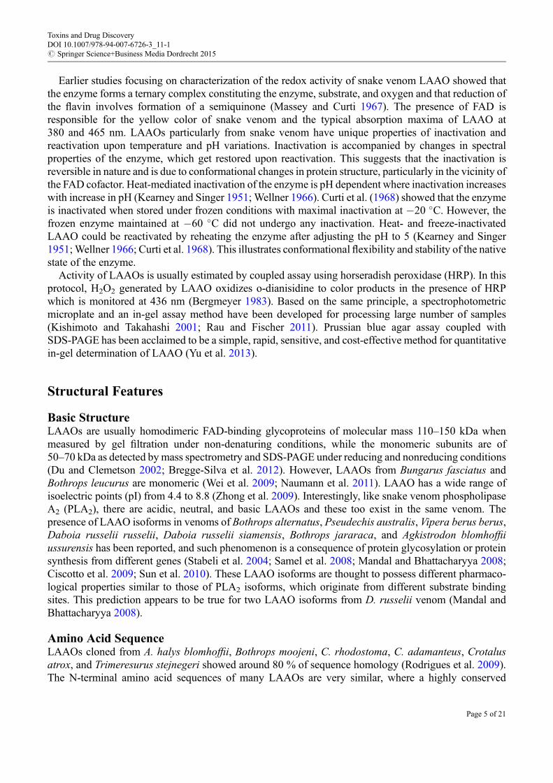
Amino Acid SequenceLAAOs cloned from A. halys blomhoffii, Bothrops moojeni, C. rhodostoma, C. adamanteus, Crotalusatrox, and Trimeresurus stejnegeri showed around 80 % of sequence homology (Rodrigues et al. 2009).The N-terminal amino acid sequences of many LAAOs are very similar, where a highly conserved
Toxins and Drug DiscoveryDOI 10.1007/978-94-007-6726-3_11-1# Springer Science+Business Media Dordrecht 2015
Page 5 of 21
glutamic acid-rich motif is observed. The structure of LAAO from C. rhodostoma revealed that residues5–25 constitute a part of the substrate-binding domain. The conserved amino acids may, therefore, play animportant role in the substrate binding. LAAOs share sequence homology with human monoamineoxidase and also with bacterial, fungal, and mouse interleukin 4-induced Fig1-protein (Raibekas andMassey 1998). Though all these proteins have low sequence homology with LAAOs, they share a similartopological architecture, except the highly preserved catalytic site (Du and Clemetson 2002; Rodrigueset al. 2009).
Three-Dimensional StructureThe X-ray crystallographic structure of the LAAO from C. rhodostoma was the first to be solved thatprovided important insights into its mechanism of substrate binding and catalysis (Pawelek et al. 2000).Recently 3-D structures of LAAOs from the venoms of Agkistrodon halys pallas, Vipera ammodytesammodytes, and Bothrops jararacussu have also been reported (Zhang et al. 2004; Georgieva et al. 2011;Ullah et al. 2012). These enzymes share an average of 85 % of sequence identity with C. rhodostomaalong with high structural similarity (Fig. 2; Zhang et al. 2004; Ullah et al. 2012). The high-resolutionX-ray structure of LAAO from C. rhodostoma indicated that it is a functional dimer where each subunitconsists of 15 a-helices and 22 b-stands that fold into three well-defined domains: an FAD-bindingdomain (residues 35–64, 242–318, and 446–471), a substrate-binding domain (residues 5–25, 73–129,233–236, and 323–420), and a helical domain (residues 130–230). This helical domain contributes to aY-shaped entrance to the enzyme’s active site, which is located at the base of this long funnel extending25 Å from the surface into the interior of the protein. The isoalloxazine ring of the FAD cofactor, essentialfor catalysis, lies at the base of this funnel. In fact, there are two access routes to the active site whichmakes the funnel Y-shaped (Fig. 3). It is proposed that one route is for the entry of the substrate while theother is for the exit of H2O2. The common stem of this Y-shaped funnel is approximately 9 Å in length andextends from the substrate-binding cavity to the bifurcation point. Although L-glutamate oxidase fromStreptomyces sp. and polyamine oxidase (PAO) have high topological similarity with snake venomLAAO, their catalytic funnels differ greatly from that of snake venom LAAO (Arima et al. 2009). Theshape of the funnel and length of L-glutamate oxidase which is ~30 Å resemble PAO. PAO has a U-shapedcatalytic funnel which is narrower than the funnel of snake venom LAAO but broader than that ofL-glutamate oxidase (Arima et al. 2009).
Crystallographic structures of LAAO complexes with the reversible inhibitor o-aminobenzoate andthe substrate L-Phe provided important insights into the mode of substrate binding and the possiblemechanism of catalysis (Moustafa et al. 2006; Pawelek et al. 2000). The substrate L-Phe remains boundto the reface of the cofactor FAD, and the side chain of L-Phe is extended away from the cofactor.It is accommodated in a subpocket which is composed of the side chains of Ile374, His223, and Arg322residues. The carboxylate group of the substrate is involved in a salt-bridge interaction with theguanidinium group of Arg90 and a hydrogen bond with the OH group of Tyr372. The a-C atom of thesubstrate, representing the site of oxidative attack, is positioned over the isoalloxazine ring at a distance of3.1 Å from N5 atom of FAD. The amino group of the substrate forms hydrogen bond with the carbonyloxygen atom of Gly464. Conformational changes of the key components of the active site residues(cf. His223, Arg322, and FAD) can be related to the mechanism of direct transfer of hydride ion. His223alters its side-chain conformation in a fashion to activate the substrate by deprotonating its a-amino group.Thereafter, the lone pair of electrons from the nitrogen atom of the amino group moves to the a-carbonatom followed by hydride transfer to FAD and forms the imine compound. The conserved water moleculeat Lys326 plays a role in the reductive half-reaction, thereby assisting the formation of H2O2. Thus,conformational change of His223 is important for substrate entry as well as for hydride transfer and ishighly conserved in LAAOs. The presence of Gly316 in place of His223 in L-glutamate oxidase indicates
Toxins and Drug DiscoveryDOI 10.1007/978-94-007-6726-3_11-1# Springer Science+Business Media Dordrecht 2015
Page 6 of 21
that catalysis in bacterial LAAO may occur through a different route or may be through the involvementof other residues which need clarification. Structural superposition of several LAAOs showed that the keyresidues for amino acid recognition are fully conserved including Tyr372, Arg90, Trp465, ILe430,His223, and Arg322. This conservation indicates a highly specific function of LAAOs during the processof envenomation. The volume of the active site also influences the substrate specificity. Ullah et al. (2012)reported that in B. jararacussu LAAO, the presence of Ile in place of Trp430 leads to increased volume ofactive-site cavity which increases the specificity of the enzyme toward bulky hydrophobic amino acids. Itwas observed that the key residues involved in dimerization of B. jararacussu LAAO are Lys191,Arg317, His314, Arg317, Ser318, Arg300, Arg301, Tyr436, Asp376, Asp349, Asp210, Asp201, H320,
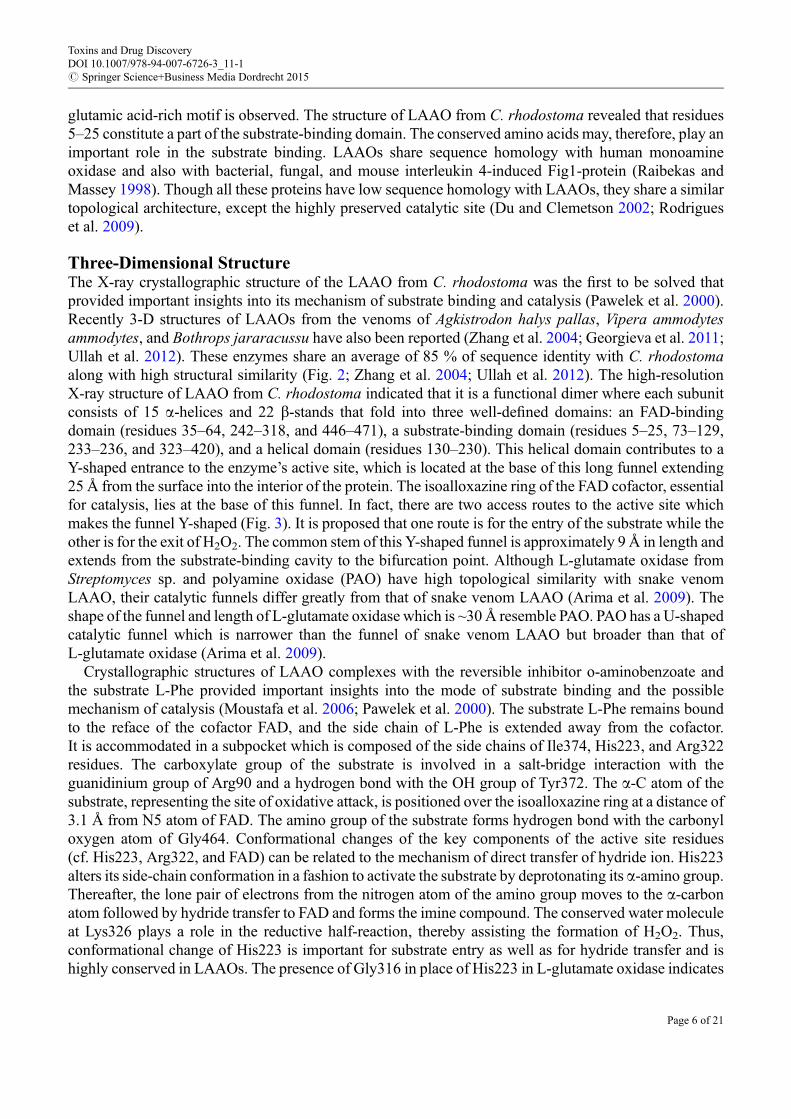
Fig. 2 LAAOs from snake venoms are structurally conserved. The ribbon models derived from X-ray crystallographicstructures of LAAOs from Malayan pit viper (C. rhodostoma) (orange, PDB 1F8S) and cotton mouth viper (A. halys pallas)(green, PDB 1REO) show high extent of overlapping
Fig. 3 Comparison of the shape of the funnels constituting the catalytic site of LAAO from snake venom (C. rhodostoma)(PDB ID: 2IID) (left) and bacteria (Streptomyces sp. X-119-6) (PDB ID: 2E1M) (right). Snake venom LAAO has a Y-shapedfunnel whereas that from bacteria has a U-shaped funnel. Brown dotted lines indicate entry of substrate on the left and exit ofproducts on the right
Toxins and Drug DiscoveryDOI 10.1007/978-94-007-6726-3_11-1# Springer Science+Business Media Dordrecht 2015
Page 7 of 21
Thr182, Asp205, Asp388, Lys186, Arg297, and His440. Electrostatic surface analysis of monomers ofLAAO from V. ammodytes and B. jararacussu indicates that dimerization is mediated by Coulombicforces between negatively charged substrate-binding domains and positively charged FAD-bindingdomain (Fig. 4; Ullah et al. 2012). Furthermore, the entrance of the substrate to the active site for eachprotomer is located on opposite surface of the dimer. Also the adenine dinucleotide part of FADs isextended in opposite directions.
V. ammodytes LAAO is tetrameric with a zinc ion present in the interface of the tetramer coordinatingwith His75 and GLy279 residues. It was observed that the presence or absence of zinc ion did not interferewith the catalytic activity and the absence of zinc in LAAO from C. rhodostoma and B. jararacussustrongly suggest that the zinc ion plays an important role in the stabilization of the tetrameric structure.
Glycan ResiduesThere are good indications that LAAO-induced cytotoxicity is not due to the sole effect of H2O2 that isgenerated during catalysis but to some extent also related to its ability to interact with the cell surfacethrough its glycan moiety (Suhr and Kim 1996; Ande et al. 2006). This glycan moiety consists of only3–5 % of LAAOs by mass. Fluorescence microscopy using FITC (fluorescein isothiocyanate)-labeledLAAO revealed direct attachment of the enzyme to the cell surface which varies depending on the cell line(Suhr and Kim 1996). Deglycosylation of the enzyme isolated from C. atrox (Apoxin1) abolished itscatalytic activity, whereas the activity is reduced by 75 % in case of A. halys pallas LAAO (Toriiet al. 2000). However, removal of the glycan had no effect on enzymatic activities of LAAOs fromBothrops pauloensis, B. jararaca, B. alternatus, and B. moojeni (Stabeli et al. 2004; Ciscotto et al. 2009;Rodrigues et al. 2009). The structure of LAAO from C. rhodostoma reveals two glycosylation sites,Asn172 and Asn361, where the glycan is linked to the amide nitrogen atom of the residues (Paweleket al. 2000). Glycosylation at Asn361 is absent in LAAO from V. ammodytes. The oligosaccharide atAsn172 is exposed on the protein surface irrespective of its multimericity. Isolated glycan from LAAO ofC. rhodostoma venom using N-glycosidase F (PNGase F) and subsequent analysis using 2-D NMR andMALDI mass spectrometry identified the glycan as bis-sialylated, biantennary, core-fucosylateddodecasaccharide (Geyer et al. 2001). The glycan moiety at Asn172 lies near to the proposed H2O2 exit
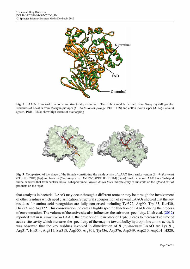
Fig. 4 Simplified two-dimensional representation of LAAO dimer. Functional dimeric form of the protein is stabilized byopposite charges on the surface. This is indicated by two color shades. The catalytic funnel with probable substrate entry andproduct (H2O2) exit paths are marked. FAD is extended in opposite direction in the dimeric form. The glycan attachments atAsn172 are exposed on the surface, are located almost diagonally, and are close to the H2O2 exit path
Toxins and Drug DiscoveryDOI 10.1007/978-94-007-6726-3_11-1# Springer Science+Business Media Dordrecht 2015
Page 8 of 21
channel. It was hypothesized that putative binding of LAAO to cell surface through the glycan moietyresults in production/release of locally high concentration of H2O2 near the binding interface, which inturn may damage the structural elements of the cells through oxidative stress leading to cell apoptosis(Geyer et al. 2001). In contrast to snake venom LAAO, PAO has only one glycosylation site at Asn77,whereas no such glycosylation is observed in LAAO from bacterial sources (Arima et al. 2009). So theprecise role of glycosylation in snake venom LAAOs could not be predicted with certainty.
Toxicology of LAAO
LAAO is an interesting molecule for research because of its hemolytic, hemorrhagic, cytotoxic, antico-agulant, genotoxic, and other physiological effects. All these effects are thought to be mediated by H2O2
generated during catalysis because H2O2 scavenger such as catalase attenuates the effects. However, somestudies imply that H2O2 production is not the only factor responsible for all these biological effects.Several LAAOs were reported to exhibit moderate toxicity having LD50 of 5–9 mg/g in mouse (i.v.). Infact, LD50 of snake venom LAAO is usually higher than that of the corresponding venom (Tan andSaifuddin 1989). The structure and function of LAAOs from different sources have been extensivelyreviewed by several authors (Du and Clemetson 2002; Tan and Fung 2009; Guo et al. 2012). Here, recentprogresses in the toxicological studies of LAAO will be discussed.
Edema-Inducing and Hemorrhagic ActivitiesLAAOs are involved in the pathogenesis of snakebite-induced inflammation. Wei et al (2007) reportedthat LAAO could cause pneumorrhagia, pulmonary interstitial edema, fusion of pulmonary alveoli,cardiac interstitial edema, and liver cell necrosis. It could also stimulate lymphocytes and monocytes torelease IL-6, IL-2, IL-12, and T cells. Later, the same group demonstrated that LAAO from B. fasciatusvenom causes local inflammation by evoking the cytotoxicity and pro-inflammatory activities in mice(Wei et al. 2009). LAAO fromO. hannah exhibited “delayed type” strong edema-inducing activity, whichindicates that the edema formation caused by this enzyme was not mediated through release of amines(Tan and Choy 1993). The edema-inducing activity of the enzyme was not inhibited by the anti-allergicand anti-inflammatory drugs, diphenhydramine or dexamethasone. Izidoro et al. (2006) suggested thatedema formation is due to activation of the inflammatory response by the H2O2 generated, as adminis-tration of glutathione (an antioxidant) to the mouse paw inhibited the edema-inducing activity of theenzyme (Izidoro et al. 2011). All these facts point toward toxicity of LAAOs that may cause seriouspathogenesis in snakebite victims.
Anticoagulant EffectsSakurai et al. (2003) initially reported factor IX-specific anticoagulant activity of LAAO fromAgkistrodon halys blomhoffii venom. But the molecular mechanism of this action is not clear. Later,Fujisawa et al. (2009) purified LAAO from the venom of Gloydius blomhoffii (previously known asA. halys blomhoffii) that contains a 39 kDa fibrinogenolytic metalloproteinase. It induces anticoagulantactivity through the degradation of Aa- chain of fibrinogen, and this activity is independent of theenzymatic activity of LAAO or generation of H2O2.
Genotoxic EffectMarcussi et al. (2013) evaluated the genotoxic potential of venoms from B. jararacussu, B. atrox,Bothrops moojeni, B. alternatus, and B. brazili snakes and some isolated toxins (MjTX-I, BthTX-I andBthTX-II myotoxins, BjussuMP-II metalloprotease, and Batx LAAO) on human lymphocytes. BthTX-I,
Toxins and Drug DiscoveryDOI 10.1007/978-94-007-6726-3_11-1# Springer Science+Business Media Dordrecht 2015
Page 9 of 21
BthTX-II, and LAAO caused significant DNA damage and enhanced the formation of micronuclei(Marcussi et al. 2013).
Strategies for Inhibition of LAAO
Currently, administration of polyvalent antivenom serum (AVS) is the only and effective means oftreatment for snake envenomation. Although monovalent antivenom has often been considered moreefficacious, the production of polyvalent AVS is preferred in many countries as identification of theinvading snake species is generally not possible by the attending physician. Polyvalent AVS that isprepared by raising antibody in horse against four to five different species of snakes often causes allergicreactions in victim’s body due to the presence of nonspecific components. Apart from this, pooravailability, improper maintenance of storage conditions, and high price limit the application of theAVS (Gutierrez et al. 2010; Deshpande et al. 2013). Moreover, the success of AVS treatment varies greatlywith the geographical area as the venom composition and the clinical manifestation of envenomation varywith geographical location (Warrell 1989). The search for an alternative antidote against envenomationthat can be used in case of emergency, when AVS is not readily available or not sufficiently effective, isalways in demand. Such an antidote should allow victims to survive and also to prevent severe localinflammatory effects of envenomation, e.g., muscle necrosis, abnormal bleeding that often lead togangrenous limbs, etc., until established and efficacious treatment is available. The situation may becompared with the application of anticholinesterase drugs such as edrophonium that can partly overcomeblockade by postsynaptic neurotoxins and have shown good results in cobra envenomation (Warrell1999). The discovery of specific inhibitors of snake venom toxins promises great therapeutic applicationin snakebite envenomation.
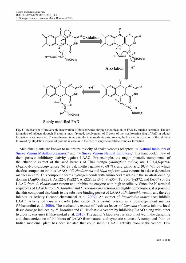
So far, all known LAAOs are flavoenzymes except lysine oxidase fromMarinomonas mediterranea. Inparallel, an increasing number of other flavoenzymes have been inhibited by substrate analogs. However,reports on inhibitors of LAAO are limited (Yu and Qiao 2012). In general, substrate analogs are wellrecognized by the catalytic residues of an enzyme and hence are exploited for both reversible andirreversible inhibitions of enzymes. The binding mode of o-amino benzoic acid, a reversible inhibitorof LAAO, was demonstrated using crystallographic structure of the enzyme-inhibitor complex (Paweleket al. 2000). Moreover, kinetic studies of other reversible inhibitors predicted the presence of differentelectrostatic and hydrophobic patches within the catalytic funnel of two LAAO isoforms of D. russeliivenom (Mandal and Bhattacharyya 2008). Similar to other flavoenzymes, irreversible inactivation ofLAAOs could be achieved by “mechanism-based inhibitor” or “suicide substrate.” These are a specialclass of irreversible inhibitors having structural and chemical features resembling those of the normalsubstrate of an enzyme. In addition, the inhibitors contain a functional group at the enzyme binding sitewhich is converted to a highly reactive moiety within the active site of the enzyme during catalysis. Thisreactive moiety generated at the transition state either attacks an essential amino acid residue or prostheticgroup of the enzyme to form a covalent linkage leading to irreversible inactivation of the enzyme. Hence,the inhibitors are highly specific and suitable for designing drugs based on enzyme inhibition. A notableexample is the inhibition of monoamine oxidase (MAO) by drugs such as clorgiline, selegiline, andrasagiline. These drugs have great potency against Parkinson’s disease (Youdim 2013). Themechanism ofmodification of the flavin residue by this class of drugs is shown in Fig. 5. Vinylglycine has beendescribed as a suicide substrate of C. adamanteus LAAO (Marcotte and Walsh 1976). Later,L-propargylglycine was also shown to inhibit the enzyme from the same venom in a similar fashion,where inactivation of the enzyme occurs through covalent modification of the active site His223 (Mitraand Bhattacharyya 2013). Additional endeavors are likely to yield more potent inhibitors of LAAO.
Toxins and Drug DiscoveryDOI 10.1007/978-94-007-6726-3_11-1# Springer Science+Business Media Dordrecht 2015
Page 10 of 21
Medicinal plants are known to neutralize toxicity of snake venoms (chapters “▶Natural Inhibitors ofSnake Venom Metalloproteinases,” and “▶ Snake Venom Natural Inhibitors,” this handbook). Few ofthem possess inhibitory activity against LAAO. For example, the major phenolic components ofthe ethanolic extract of the seed kernels of Thai mango (Mangifera indica) are 1,2,3,4,6-penta-O-galloyl-b-D-glucopyranose (61.28 %), methyl gallate (0.68 %), and gallic acid (0.44 %), of whichthe first component inhibits LAAO ofC. rhodostoma andNaja naja kaouthia venoms in a dose-dependentmanner in vitro. This compound forms hydrogen bonds with amino acid residues in the substrate-bindingdomain (Arg90, His223, Asp224, Phe227, Ala228, Lys345, Phe354, Tyr356, Tyr372, and Ile374) of theLAAO from C. rhodostoma venom and inhibits the enzyme with high specificity. Since the N-terminalsequences of LAAOs from N. kaouthia and C. rhodostoma venoms are highly homologous, it is possiblethat this compound also binds to the substrate-binding pocket of LAAO ofN. kaouthia venom and therebyinhibits its activity (Leanpolchareanchai et al. 2009). An extract of Tamarindus indica seed inhibitsLAAO activity of Vipera russelii (also called D. russelii) venom in a dose-dependent manner(Ushanandini et al. 2006). The methanolic extract of fresh tea leaves of Camellia sinensis inhibits localtissue damage induced by N. kaouthia and C. rhodostoma venom by inhibiting LAAO along with otherhydrolytic enzymes (Pithayanukul et al. 2010). The author’s laboratory is also involved in the designingand characterization of inhibitors of LAAO from natural and synthetic sources. A compound from anIndian medicinal plant has been isolated that could inhibit LAAO activity from snake venom. Few
Fig. 5 Mechanism of irreversible inactivation of flavoenzymes through modification of FAD by suicide substrate. Thoughformation of adducts through N atom is most favored, involvement of C atom of the isoalloxazine ring of FAD in adductformation is also reported. The mechanism is very similar to normal catalysis process; the first step is oxidation of the inhibitorfollowed by alkylation instead of product release as in the case of enzyme-substrate complex formation
Toxins and Drug DiscoveryDOI 10.1007/978-94-007-6726-3_11-1# Springer Science+Business Media Dordrecht 2015
Page 11 of 21
derivatives of this compound have been synthesized to increase inhibitory potential with reduced toxicityas compared to the original one (Bhattacharjee 2014).
Therapeutic Potentials of LAAO
In the last two decades, LAAO drew the attention of more researchers because of its multifunctionaleffects on different biological systems. This section summarizes information regarding the antiprotozoal,antibacterial, antiviral, and apoptosis-inducing effects of LAAO including its ambiguous action onplatelet aggregation. Through judicious modification of the structure of LAAO, one could reduce itstoxicity on host cells retaining its beneficial function for therapeutic applications.
Antiprotozoal PropertyLeishmaniasis is an endemic tropical disease with human infections ranging from self-healing cutaneousulceration to lethal visceral infection. Approximately 350 million people are at risk from the disease, andan estimated 1.6 million new cases occur annually, with its prevalence in about 88 nations (Stockdale andNewton 2013).
Many reports suggest that LAAO possesses strong antileishmanial activity (Toyama et al. 2006). H2O2
generated by the enzyme induces apoptosis in promastigotes of Leishmania sp. by enhancing oxidativestress, activating heat shock proteins leading to disorganization of its cell membrane and cytoplasmicvesicles. In other words, the H2O2 produced by LAAO breaks the antioxidant system of the promastigotecells to induce apoptosis. LAAO from Bothrops marajoensis could inhibit the growth of promastigotes ofL. chagasi and L. amazonensis in a dose-dependent manner (Costa Torres et al. 2010). LAAO fromB. moojeni significantly inhibits the growth of promastigotes of L. amazonensis, L. chagasi, andL. panamensis exhibiting IC50 of 1 mg/ml. The enzyme also inhibits the growth of Trypanosoma cruziand L. major promastigotes by causing cytoplasmic vacuolization and increasing the volume of mito-chondria. It causes the death of promastigotes by disrupting the respiratory chain of the parasite leading toreduced production of ATP. The antileishmanial activity is negatively correlated with the levels of catalaseand glutathione peroxidase. It has been demonstrated that catalase and glutathione peroxidase are presentthree and 14 times more in amastigotes of L. donovani than in promastigotes, which potentially makes theformer four times more resistant to LAAO than the latter (Toyama et al. 2006). Bregge-Silva et al. (2012)reported that LAAO from Lachesis muta demonstrates antiparasitic activity on Leishmania braziliensis(IC50 2.22 mg/mL) but is ineffective for T. cruzi infection.
At present, only a few drugs are available for the treatment of leishmaniasis, although they are highlytoxic. As LAAOs can produce high concentrations of H2O2 in certain localized areas of the cell, a smallamount of the enzyme can target toward the parasitophorous vacuole, and this would represent a highlyspecific treatment not only for leishmaniasis but also for other intracellular parasites. Insightful expeditioninto the mode of action of LAAO upon parasites, other than the effect exerted by H2O2, may trigger thedesign of new drugs or improved therapeutic approaches for leishmaniasis.
Antibacterial PropertiesThe emergence of drug-resistant strains of bacteria has increased the risk of morbidity and mortalityworldwide. Therefore, the discovery of new powerful therapeutics is necessary for treatment of infectionscaused by the resistant microorganisms. Recently, powerful antibacterial components against gram-positive and gram-negative bacteria from natural sources have drawn attention owing to their fewerside effects (Tõnismägi et al. 2006; Samel et al. 2008; Costa Torres et al. 2010; Lee et al. 2011; Vargaset al. 2013). Venoms are rich sources of antimicrobial compounds such as peptides (chapter
Toxins and Drug DiscoveryDOI 10.1007/978-94-007-6726-3_11-1# Springer Science+Business Media Dordrecht 2015
Page 12 of 21
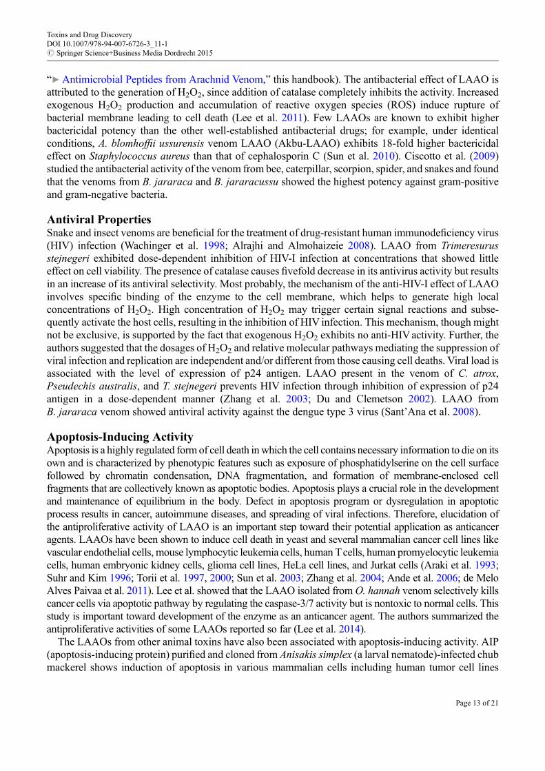
“▶Antimicrobial Peptides from Arachnid Venom,” this handbook). The antibacterial effect of LAAO isattributed to the generation of H2O2, since addition of catalase completely inhibits the activity. Increasedexogenous H2O2 production and accumulation of reactive oxygen species (ROS) induce rupture ofbacterial membrane leading to cell death (Lee et al. 2011). Few LAAOs are known to exhibit higherbactericidal potency than the other well-established antibacterial drugs; for example, under identicalconditions, A. blomhoffii ussurensis venom LAAO (Akbu-LAAO) exhibits 18-fold higher bactericidaleffect on Staphylococcus aureus than that of cephalosporin C (Sun et al. 2010). Ciscotto et al. (2009)studied the antibacterial activity of the venom from bee, caterpillar, scorpion, spider, and snakes and foundthat the venoms from B. jararaca and B. jararacussu showed the highest potency against gram-positiveand gram-negative bacteria.
Antiviral PropertiesSnake and insect venoms are beneficial for the treatment of drug-resistant human immunodeficiency virus(HIV) infection (Wachinger et al. 1998; Alrajhi and Almohaizeie 2008). LAAO from Trimeresurusstejnegeri exhibited dose-dependent inhibition of HIV-I infection at concentrations that showed littleeffect on cell viability. The presence of catalase causes fivefold decrease in its antivirus activity but resultsin an increase of its antiviral selectivity. Most probably, the mechanism of the anti-HIV-I effect of LAAOinvolves specific binding of the enzyme to the cell membrane, which helps to generate high localconcentrations of H2O2. High concentration of H2O2 may trigger certain signal reactions and subse-quently activate the host cells, resulting in the inhibition of HIV infection. This mechanism, though mightnot be exclusive, is supported by the fact that exogenous H2O2 exhibits no anti-HIVactivity. Further, theauthors suggested that the dosages of H2O2 and relative molecular pathways mediating the suppression ofviral infection and replication are independent and/or different from those causing cell deaths. Viral load isassociated with the level of expression of p24 antigen. LAAO present in the venom of C. atrox,Pseudechis australis, and T. stejnegeri prevents HIV infection through inhibition of expression of p24antigen in a dose-dependent manner (Zhang et al. 2003; Du and Clemetson 2002). LAAO fromB. jararaca venom showed antiviral activity against the dengue type 3 virus (Sant’Ana et al. 2008).
Apoptosis-Inducing ActivityApoptosis is a highly regulated form of cell death inwhich the cell contains necessary information to die on itsown and is characterized by phenotypic features such as exposure of phosphatidylserine on the cell surfacefollowed by chromatin condensation, DNA fragmentation, and formation of membrane-enclosed cellfragments that are collectively known as apoptotic bodies. Apoptosis plays a crucial role in the developmentand maintenance of equilibrium in the body. Defect in apoptosis program or dysregulation in apoptoticprocess results in cancer, autoimmune diseases, and spreading of viral infections. Therefore, elucidation ofthe antiproliferative activity of LAAO is an important step toward their potential application as anticanceragents. LAAOs have been shown to induce cell death in yeast and several mammalian cancer cell lines likevascular endothelial cells, mouse lymphocytic leukemia cells, human Tcells, human promyelocytic leukemiacells, human embryonic kidney cells, glioma cell lines, HeLa cell lines, and Jurkat cells (Araki et al. 1993;Suhr and Kim 1996; Torii et al. 1997, 2000; Sun et al. 2003; Zhang et al. 2004; Ande et al. 2006; de MeloAlves Paivaa et al. 2011). Lee et al. showed that the LAAO isolated fromO. hannah venom selectively killscancer cells via apoptotic pathway by regulating the caspase-3/7 activity but is nontoxic to normal cells. Thisstudy is important toward development of the enzyme as an anticancer agent. The authors summarized theantiproliferative activities of some LAAOs reported so far (Lee et al. 2014).
The LAAOs from other animal toxins have also been associated with apoptosis-inducing activity. AIP(apoptosis-inducing protein) purified and cloned from Anisakis simplex (a larval nematode)-infected chubmackerel shows induction of apoptosis in various mammalian cells including human tumor cell lines
Toxins and Drug DiscoveryDOI 10.1007/978-94-007-6726-3_11-1# Springer Science+Business Media Dordrecht 2015
Page 13 of 21
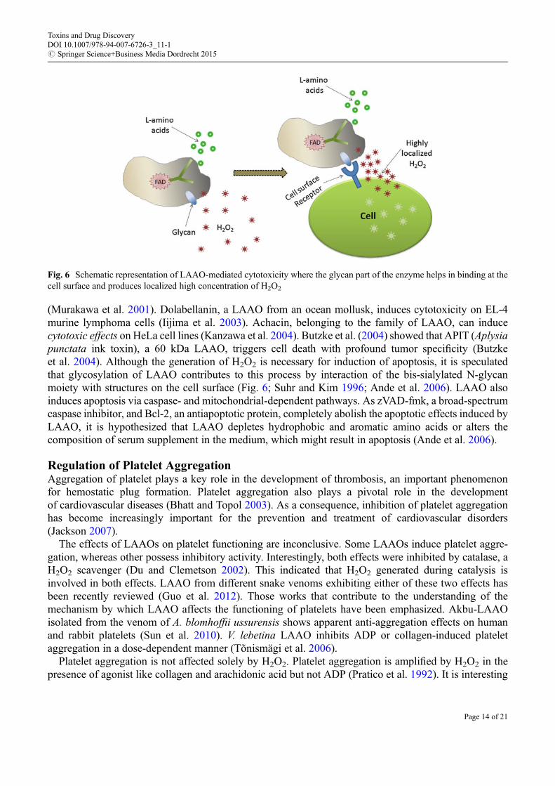
(Murakawa et al. 2001). Dolabellanin, a LAAO from an ocean mollusk, induces cytotoxicity on EL-4murine lymphoma cells (Iijima et al. 2003). Achacin, belonging to the family of LAAO, can inducecytotoxic effects on HeLa cell lines (Kanzawa et al. 2004). Butzke et al. (2004) showed that APIT (Aplysiapunctata ink toxin), a 60 kDa LAAO, triggers cell death with profound tumor specificity (Butzkeet al. 2004). Although the generation of H2O2 is necessary for induction of apoptosis, it is speculatedthat glycosylation of LAAO contributes to this process by interaction of the bis-sialylated N-glycanmoiety with structures on the cell surface (Fig. 6; Suhr and Kim 1996; Ande et al. 2006). LAAO alsoinduces apoptosis via caspase- and mitochondrial-dependent pathways. As zVAD-fmk, a broad-spectrumcaspase inhibitor, and Bcl-2, an antiapoptotic protein, completely abolish the apoptotic effects induced byLAAO, it is hypothesized that LAAO depletes hydrophobic and aromatic amino acids or alters thecomposition of serum supplement in the medium, which might result in apoptosis (Ande et al. 2006).
Regulation of Platelet AggregationAggregation of platelet plays a key role in the development of thrombosis, an important phenomenonfor hemostatic plug formation. Platelet aggregation also plays a pivotal role in the developmentof cardiovascular diseases (Bhatt and Topol 2003). As a consequence, inhibition of platelet aggregationhas become increasingly important for the prevention and treatment of cardiovascular disorders(Jackson 2007).
The effects of LAAOs on platelet functioning are inconclusive. Some LAAOs induce platelet aggre-gation, whereas other possess inhibitory activity. Interestingly, both effects were inhibited by catalase, aH2O2 scavenger (Du and Clemetson 2002). This indicated that H2O2 generated during catalysis isinvolved in both effects. LAAO from different snake venoms exhibiting either of these two effects hasbeen recently reviewed (Guo et al. 2012). Those works that contribute to the understanding of themechanism by which LAAO affects the functioning of platelets have been emphasized. Akbu-LAAOisolated from the venom of A. blomhoffii ussurensis shows apparent anti-aggregation effects on humanand rabbit platelets (Sun et al. 2010). V. lebetina LAAO inhibits ADP or collagen-induced plateletaggregation in a dose-dependent manner (Tõnismägi et al. 2006).
Platelet aggregation is not affected solely by H2O2. Platelet aggregation is amplified by H2O2 in thepresence of agonist like collagen and arachidonic acid but not ADP (Pratico et al. 1992). It is interesting
Fig. 6 Schematic representation of LAAO-mediated cytotoxicity where the glycan part of the enzyme helps in binding at thecell surface and produces localized high concentration of H2O2
Toxins and Drug DiscoveryDOI 10.1007/978-94-007-6726-3_11-1# Springer Science+Business Media Dordrecht 2015
Page 14 of 21
that catalase inhibits platelet aggregation induced by collagen in a dose-dependent manner whereas it doesnot affect the aggregation by other agonists like ADP or thrombin (Pignatelli et al. 1998). Catalase inhibitsthromboxane A2 production and release of arachidonic acid from platelet membrane. This study showedthat collagen-induced platelet aggregation is associated with the production of H2O2 that acts as a secondmessenger by stimulating the arachidonic acid metabolism and phospholipase C pathway. Toyamaet al. (2006) reported that the platelet aggregation-inducing activity of Crotalus durissus cascavellavenom LAAO (Casca LAAO) was inhibited by cyclooxygenase pathway inhibitors such as indomethacinand aspirin. It was suggested that H2O2 generated by LAAO was involved in the production ofinflammatory enzyme thromboxane A2, which consequently resulted in platelet aggregation (Du andClemetson 2002; Toyama et al. 2006). However, thromboxane A2 was not produced by washed humanplatelets when stimulated by H2O2, collagen, or arachidonic acid alone but by H2O2 in the presence ofsub-threshold concentration (non-aggregating concentration) of the collagen or arachidonic acid (Praticoet al. 1992). So it remains unclear how H2O2 produced by LAAO induces platelet aggregation. Naumannet al. (2011) reported that Bothrops leucurus LAAO does not interact with endothelial cells. Therefore, theother possibility that LAAO may activate platelet in a receptor-dependent manner similar to the variedassociation of the enzyme with different cell lines needs further investigation.
The inhibitory potency of LAAO over platelet aggregation may be connected with reduced bindingaffinity for ADP in platelet exposed to H2O2 or the interference of H2O2 in the interaction between theactivated platelet integrin aIIb/b3 and fibrinogen in the platelet-rich plasma (PRP) system (Suhr and Kim1996). It should be considered that physiological conditions of platelet aggregation are quite differentfrom what is measured in an aggregometer. In the instrument, platelet clumping occurs under low,nonlaminar shear conditions, whereas platelets are exposed to a broad range of hemodynamic conditionsranging from relatively low flow situations in venules and large veins (typical wall shear rates<500 s�1)or small arterioles (shear rates up to 5000 s�1) to stenosed arteries with shear rates as high as 40,000 s�1
(Jackson 2007). Moreover, the experimental conditions and procedures for platelet poor preparation(PPP) are vital for subsequent aggregation experiments. So the contradictory results on induction/inhibition of platelet aggregation by LAAO and the underlying mechanism need to be revisited usingthe recent advancement of technology like improved live-cell imaging techniques which enable high-resolution visualization of the platelet aggregation processes operating in vitro and in vivo.
Industrial ApplicationsThe use of LAAOs as biosensors has great potential. Biosensors utilizing bacterial L-glutamate oxidaseare commercially available and are a useful tool for in vitro and in vivo monitoring of mammalian brainL-glutamate (Ryan et al. 1997). LAAO-based biosensors are used in food industries for quality assuranceof edible products through evaluation of L and D enantiomers of amino acids which enter into foodstuffduring fermentation of yeast and bacteria (Lee and Huh 2009; Lata and Pundir 2013). Biotransformationis another application of LAAOs which includes industrial production of a-keto acids and separation ofenantiomers from racemic mixture of amino acids; particularly for the preparation of D-isomers(Takahashi et al. 1997). H2O2 and its precursors are often used as bleaching agent in detergents. LAAOmay be advantageously incorporated, together with a substrate, into detergent compositions for genera-tion of H2O2. The formulation also comprises a peroxidase system to regulate the activity of H2O2;otherwise, the dye may be transferred from dyed fabric to other fabrics during washing (Schneider 1994).LAAO from Rhodococcus sp. is successfully used in the synthesis of aminoadipic derivatives that areprecursors for b-lactam antibiotics (Isobe et al. 2008). All industrial applications of LAAO are limited tomicrobial enzymes as they are easy to produce. Whether venom LAAO is superior remains unknown astheir commercial applications require overproduction in microorganisms.
Toxins and Drug DiscoveryDOI 10.1007/978-94-007-6726-3_11-1# Springer Science+Business Media Dordrecht 2015
Page 15 of 21
Conclusion and Future Direction
LAAOs from snake venoms and other organisms have overall structural similarity. The biologicalfunctions of LAAOs especially from snake venom on platelet aggregation, cell apoptosis, antimicrobialactivity, antileishmanial activity, antitumor, and anti-HIVactivity are mostly induced by H2O2 generatedduring catalysis. Enzyme-cell membrane receptor interaction seems to play certain role in inducing manyof these biological effects, which need to be further established. The exact mechanism of toxicity anddifferent means of neutralization of LAAOs await further studies. The enzyme holds great potentialapplication in cancer therapy. Better understanding of the mode of action of the enzyme upon parasitesmay lead to development of new therapeutic approaches for the treatment of various bacteria, virus, andprotozoa-borne diseases. This multifunctional enzyme has also gained popularity in diagnostics andindustrial applications. Strong antimicrobial activity of LAAO may be utilized for future application infood and cosmetics.
Acknowledgment
PB and JM were supported by UGC-NET and CSIR-NET senior research fellowships, respectively. Wethank Dr. Debashree De for critical reading of the manuscript.
Cross-References
▶Antimicrobial Peptides from Arachnid Venom▶Computational Approaches for Animal Toxins to Aid Drug Discovery▶Natural Inhibitors of Snake Venom Metalloproteinases▶ Snake Venom Natural Inhibitors
References
Alrajhi AA, Almohaizeie A. Snake venom preparation for drug-resistant human immunodeficiency virus.Ann Saudi Med. 2008;28:292–3.
Ande SR, Kommoju PR, Draxl S, Murkovic M, Macheroux P, Ghisla S, Ferrando-May E. Mechanisms ofcell death induction by L-amino acid oxidase, a major component of ophidian venom. Apoptosis.2006;11:1439–51.
Araki S, Ishida T, Yamamoto T, Kaji K, Hayashi H. Induction of apoptosis by hemorrhagic snake venomin vascular endothelial cells. Biochem Biophys Res Commun. 1993;190:148–53.
Arima J, Sasaki C, Sakaguchi C, Mizuno H, Tamura T, Kashima A, Kusakabe H, Sugio S, Inagaki K.Structural characterization of L-glutamate oxidase from Streptomyces sp. X-119-6. FEBSJ. 2009;276:3894–903.
Bergmeyer HV. L-amino acid oxidase. In: Methods in enzymatic analysis, vol. 2. Weinheim: VerlagChimie; 1983. p. 149–50.
Bhatt DL, Topol EJ. Scientific and therapeutic advances in antiplatelet therapy. Nat Rev Drug Discov.2003;2:15–28.
Bhattacharjee P. Evaluation of bioactive components from natural products for prevention of toxicity andneurological disorders. Ph.D. thesis, Jadavpur University, Kolkata. 2014.
Toxins and Drug DiscoveryDOI 10.1007/978-94-007-6726-3_11-1# Springer Science+Business Media Dordrecht 2015
Page 16 of 21
Bregge-Silva C, Nonato MC, de Albuquerque S, Ho PL, Junqueira de Azevedo IL, Vasconcelos DinizMR, Lomonte B, Rucavado A, Díaz C, Gutiérrez JM, Arantes EC. Isolation and biochemical,functional and structural characterization of a novel L-amino acid oxidase from Lachesis muta snakevenom. Toxicon. 2012;60:1263–76.
Burin SM, Ayres LR, Neves RP, Ambrósio L, de Morais FR, Dias-BaruffiM, Sampaio SV, Pereira-CrottLS, de Castro FA. L-amino acid oxidase isolated from Bothrops pirajai induces apoptosis inBCR-ABL-positive cells and potentiates imatinib mesylate effect. Basic Clin Pharmacol Toxicol.2013;113:103–12.
Butzke D,Machuy N, Thiede B, Hurwitz R, Goedert S, Rudel T. Hydrogen peroxide produced by Aplysiaink toxin kills tumor cells independent of apoptosis via peroxiredoxin I sensitive pathways. Cell DeathDiffer. 2004;11:608–17.
Chen HS, Wang YM, Huang WT, Huang KF, Tsai IH. Cloning, characterization and mutagenesis ofRussell’s viper venom L-amino acid oxidase: insights into its catalytic mechanism. Biochimie.2012;94:335–44.
Ciscotto P, Machado de Avila RA, Coelho EA, Oliveira J, Diniz CG, Farías LM, de Carvalho MA, MariaWS, Sanchez EF, Borges A, Chávez-Olórtegui C. Antigenic, microbicidal and antiparasitic propertiesof an L-amino acid oxidase isolated from Bothrops jararaca snake venom. Toxicon. 2009;53:330–41.
Costa Torres AF, Dantas RT, Toyama MH, Diz Filho E, Zara FJ, Rodrigues de Queiroz MG, PintoNogueira NA, Rosa de Oliveira M, de Oliveira Toyama D, Monteiro HS, Martins AM. Antibacterialand antiparasitic effects of Bothrops marajoensis venom and its fractions: phospholipase A2 andL-amino acid oxidase. Toxicon. 2010;55:795–804.
Curti B, Massey V, Zmudka M. Inactivation of snake venom L-amino acid oxidase by freezing. J BiolChem. 1968;243:2306–14.
de Melo Alves Paivaa R, de Freitas Figueiredo R, Antonucci GA, Paiva HH, de Lourdes Pires Bianchi M,Rodrigues KC, Lucarini R, Caetanoa RC, Linhari Rodrigues Pietroc RC, Gomes Martins CH, deAlbuquerquea S, Sampaio SV. Cell cycle arrest evidence, parasiticidal and bactericidal propertiesinduced by L-amino acid oxidase from Bothrops atrox snake venom. Biochimie. 2011;93:941–7.
Deshpande RP, Motghare VM, Padwal SL, Pore RR, Bhamare CG, Deshmukh VS, Pise HN. Adversedrug reaction profile of anti-snake venom in a rural tertiary care teaching hospital. J Young Pharm.2013;5:41–5.
Dineshkumar P, Muthuvelan B. Isolation and purification of L-amino acid oxidase from Indian CobraNaja naja. Curr Res J Biol Sci. 2012;3:6–11.
Du XY, Clemetson KJ. Snake venom L-amino acid oxidases. Toxicon. 2002;40:659–65.Fitzpatrick PF. Carbanion versus hydride transfer mechanisms in flavoprotein-catalyzed dehydrogena-
tions. Bioorg Chem. 2004;32:125–39.Fujisawa D, Yamazaki Y, Morita T. Re-evaluation of M-LAO, L-amino acid oxidase, from the venom of
Gloydius blomhoffii as an anticoagulant protein. J Biochem. 2009;146:43–9.Georgieva D, Murakami M, Perband M, Arni RK, Betzel C. The structure of a native L-amino acid
oxidase, the major component of the Vipera ammodytes ammodytes venomic, reveals dynamic activesite and quaternary structure stabilization by divalent ions. Mol BioSyst. 2011;7:379–84.
Geyer A, Fitzpatrick TB, Pawelek PD, Kitzing K, Vrielink A, Ghisla S, Macheroux P. Structure andcharacterization of glycan moiety of L-amino-acid oxidase from the Malayan pit viper Calloselasmarhodostoma. Eur JBiochem. 2001;268:4044–53.
Guo C, Liu S, Yao Y, Zhang Q, Sun MZ. Past decade study of snake venom L-amino acid oxidase.Toxicon. 2012;60:302–11.
Gutierrez JM, Williams D, Fan HW, Warrell DA. Snakebite envenoming from a global perspective:towards an integrated approach. Toxicon. 2010;56:1223–35.
Toxins and Drug DiscoveryDOI 10.1007/978-94-007-6726-3_11-1# Springer Science+Business Media Dordrecht 2015
Page 17 of 21
Iijima R, Kisugi J, Yamazaki M. L-amino acid oxidase activity of an antineoplastic factor of a marinemollusk and its relationship to cytotoxicity. Dev Comp Immunol. 2003;27:505–12.
Isobe K, Fukuda N, Nagasawa S. Analysis of selective production of Nalpha-benzyloxycarbonyl-L-aminoadipate-delta-semialdehyde and Nalpha-benzyloxycarbonyl-L-aminoadipic acid byRhodococcus sp. AIU Z-35-1. J Biosci Bioeng. 2008;105:152–156.
Izidoro LFM, Ribeiro MC, Souza GR, Sant’Ana CD, Hamaguchi A, Homsi-Brandeburgo MI, GoulartLR, Beleboni RO, Nomizo A, Sampaio SV, Soares AM, Rodrigues VM. Biochemical and functionalcharacterization of an L-amino acid oxidase isolated from Bothrops pirajai snake venom. Bioorg MedChem. 2006;14:7034–43.
Izidoro LFM, Alves LM, Rodrigues VM, Silva DAO, Mineo JR. Bothrops pirajai snake venom L-aminoacid oxidase: in vitro effects on infection of Toxoplasma gondii in human foreskin fibroblasts. BrazJ Pharmacogn. 2011;21:477–85.
Jackson SP. The growing complexity of platelet aggregation. Blood. 2007;109:5087–95.Kameya M, Onakaa H, Asano Y. Selective tryptophan determination using tryptophan oxidases involved
in bis-indole antibiotic biosynthesis. Anal Biochem. 2013;438:124–32.Kanzawa N, Shintani S, Ohta K, Kitajima S, Ehara T, Kobayashi H, Kizaki H, Tsuchiya T. Achacin
induces cell death in HeLa cells through two different mechanisms. Arch Biochem Biophys.2004;422:103–9.
Kearney EB, Singer TP. The L-amino acid oxidases of snake venom. V. Mechanism of the reversibleinactivation. Arch Biochem. 1951;33:414–26.
Kishimoto M, Takahashi T. A spetrophotometric microplate assay for L-amino acid oxidase. AnalBiochem. 2001;298:136–9.
Lata S, Pundir CS. L-amino acid biosensor based on L-amino acid oxidase immobilized onto NiHCNFe/c-MWCNT/PPy/GC electrode. Int J Biol Macromol. 2013;54:250–7.
Leanpolchareanchai J, Pithayanukul P, Bavovada R, Saparpakorn P. Molecular docking studies and anti-enzymatic activities of Thai mango seed kernel extract against snake venoms. Molecules.2009;14:1404–22.
Lee YC, HuhMH. Development of a biosensor with immobilized L-amino acid oxidase for determinationof L-amino acids. J Food Biochem. 2009;23:173–185.
Lee ML, Tan NH, Fung SY, Sekaran SD. Antibacterial action of a heat-stable form of L-amino acidoxidase isolated from King Cobra (Ophiophagus hannah) venom. Comp Biochem Physiol C ToxicolPharmacol. 2011;153:237–42.
Lee ML, Chung I, Fung SY, Kanthimathi MS, Tan NH. Antiproliferative activity of King Cobra(Ophiophagus hannah) venom L-amino acid oxidase. Basic Clin Pharmacol Toxicol.2014;114:336–43.
Mandal S, Bhattacharyya D. Two L-amino acid oxidase isoenzymes from Russell’s viper (Daboia russellirusselli) venom with different mechanisms of inhibition by substrate analogs. FEBS J.2008;275:2078–2095.
Marcotte P, Walsh C. Vinylglycine and propargylglycine: complementary suicide substrates for L-aminoacid oxidase and D-amino acid oxidase. Biochemistry. 1976;15:3070–6.
Marcussi S, Stábeli RG, Santos-Filho NA, Menaldo DL, Silva Pereira LL, Zuliani JP, Calderon LA, daSilva SL, Antunes LM, Soares AM. Genotoxic effect of Bothrops snake venoms and isolated toxins onhuman lymphocyte DNA. Toxicon. 2013;65:9–14.
Mason JM, Naidu MD, Barcia M, Porti D, Chavan SS, Chu CC. IL-4-induced gene-1 is a leukocyteL-amino acid oxidase with an unusual acidic pH preference and lysosomal localization. J Immunol.2004;173:4561–7.
Toxins and Drug DiscoveryDOI 10.1007/978-94-007-6726-3_11-1# Springer Science+Business Media Dordrecht 2015
Page 18 of 21

Massey V, Curti B. On the reaction mechanism of Crotalus adamanteus L-amino acid oxidase. J BiolChem. 1967;242:1259–64.
Mitra J, Bhattacharyya D. Irreversible inactivation of snake venom L-amino acid oxidase by covalentmodification during catalysis of L-propargylglycine. FEBS Open Bio. 2013;3:135–143.
Moustafa IM, Foster S, Lyubimov AY, Vrielink A. Crystal structure of LAAO from Calloselasmarhodostoma with an L-phenylalanine substrate: insights into structure and mechanism. J Mol Biol.2006;364:991–1002.
Murakawa M, Jung S-K, Iijima K, Yonehara S. Apoptosis-inducing protein, AIP, from parasite-infectedfish induces apoptosis in mammalian cells by two different molecular mechanisms. Cell Death Differ.2001;8:298–307.
Nagaoka K, Aoki F, Hayashi M, Muroi Y, Sakurai T, Itoh K, Ikawa M, Okabe M, Imakawa K, Sakai S.L-amino acid oxidase plays a crucial role in host defense in the mammary glands. FASEBJ. 2009;23:2514–20.
Naumann GB, Silva LF, Silva L, Faria G, Richardson M, Evangelista K, Kohlhoff M, Gontijo CMF,Navdaev A, de Rezende FF, Eble JA, Sanchez EF. Cytotoxicity and inhibition of platelet aggregationcaused by an L-amino acid oxidase from Bothrops leucurus venom. Biochim Biophys Acta.2011;1810:683–94.
Pawelek PD, Cheah J, Coulombe R, Macheroux P, Ghisla S, Vrielink A. The structure of L-amino acidoxidase reveals the substrate trajectory into an enantiomerically conserved active site. EMBOJ. 2000;19:4204–15.
Pignatelli P, Pulcinelli FM, Lenti L, Gazzaniga PP, Violi F. Hydrogen peroxide is involved in collagen-induced platelet activation. Blood. 1998;91:484–490.
Pithayanukul P, Leanpolchareanchai J, Bavovada R. Inhibitory effect of tea polyphenols on local tissuedamage induced by snake venoms. Phytother Res. 2010;24:S56–62.
Ponnudurai G, Chung MC, Tan NH. Purification and properties of the L-amino acid oxidase fromMalayan pit viper (Calloselasma rhodostoma) venom. Arch Biochem Biophys. 1994;313:373–8.
Pratico D, Iuliano L, Pulcinelli FM, Bonavita MS, Gazzaniga PP, Violi F. Hydrogen peroxide triggersactivation of human platelets selectively exposed to nonaggregating concentrations of arachidonic acidand collagen. J Lab Clin Med. 1992;119:364–70.
Raibekas AA, Massey V. Primary structure of the snake venom L-amino acid oxidase shows highhomology with the mouse B cell interleukin 4-induced Fig1 protein. Biochem Biophys Res Commun.1998;248:476–8.
Rau JE, Fischer U. In-gel detection of L-amino acid oxidases based on the visualisation of hydrogenperoxide production. J Microbiol Methods. 2011;85:228–9.
Rodrigues RS, da Silva JF, Boldrini Franca J, Fonseca FP, Otaviano AR, Henrique Silva F, Hamaguchi A,Magro AJ, Braz AS, dos Santos JI, Homsi-Brandeburgo MI, Fontes MR, Fuly AL, Soares AM,Rodrigues VM. Structural and functional properties of Bp-LAAO, a new L-amino acid oxidase isolatedfrom Bothrops pauloensis snake venom. Biochimie. 2009;91:490–501.
Ryan MR, Lowry JP, O’Neill RD. Biosensor for neurotransmitter L-glutamic acid designed for efficientuse of L-glutamate oxidase and effective rejection of interference. Analyst. 1997;122:1419–24.
Sakurai Y, Shima M, Matsumoto T, Takatsuka H, Nishiya K, Kasuda S, Fujimura Y, Yoshioka A.Anticoagulant activity of M-LAO, L-amino acid oxidase purified from Agkistrodon halys blomhoffii,through selective inhibition of factor IX. Biochim Biophys Acta. 2003;1649:51–7.
Samel M, Tõnismägi K, RönnholmG, Vija H, Siigur J, Kalkkinen N, Siigur E. L-amino acid oxidase fromNaja naja oxiana venom. Comp Biochem Physiol B Biochem Mol Biol. 2008;149:572–80.
Sant’Ana CD, Menaldo DL, Costa TR, Godoy H, Muller VD, Aquino VH, Albuquerque S, Sampaio SV,MonteiroMC, Stabeli RG, Soares AM. Antiviral and antiparasite properties of an L-amino acid oxidase
Toxins and Drug DiscoveryDOI 10.1007/978-94-007-6726-3_11-1# Springer Science+Business Media Dordrecht 2015
Page 19 of 21
from the snake Bothrops jararaca: cloning and identification of a complete cDNA sequence. BiochemPharmacol. 2008;76:279–88.
Schneider P, Pedersen AH, Hansen SA. L-amino acid oxidase. WO1994025574 A1. November 10, 1994.Skarnes RC. L-amino-acid oxidase, a bactericidal system. Nature. 1970;225:1072–3.Stabeli RG, Marcussi S, Carlos GB, Pietro RC, Selistre-de-Araujo HS, Giglio JR, Oliveira EB, Soares
AM. Platelet aggregation and antibacterial effects of an L-amino acid oxidase purified from Bothropsalternatus snake venom. Bioorg Med Chem. 2004;12:2881–6.
Stockdale L, Newton R. A review of preventative methods against human leishmaniasis infection. PLoSNegl Trop Dis. 2013;7, e2278.
Suhr SM, Kim DS. Identification of the snake venom substance that induces apoptosis. Biochem BiophysRes Commun. 1996;224:134–9.
Sun LK, Yoshii Y, Hyodo A, Tsurushima H, Saito A, Harakuni T, Li YP, Kariya K, Nozaki M, Morine N.Apoptotic effect in the glioma cells induced by specific protein extracted from Okinawa Habu(Trimeresurus flavoviridis) venom in relation to oxidative stress. Toxicol In Vitro. 2003;17:169–77.
Sun MZ, Guo C, Tian Y, Chen D, Greenaway FT, Liu S. Biochemical, functional and structuralcharacterization of Akbu-LAAO: a novel snake venom L-amino acid oxidase from Agkistrodonblomhoffii ussurensis. Biochimie. 2010;92:343–9.
Takahashi E, Furui M, Seko H, Shibatani T. D-methionine preparation from racemic methionines byProteus vulgaris IAM 12003 with asymmetric degrading activity. Appl Microbiol Biotechnol.1997;47:173–9.
Tan NH, Choy SK. The edema-inducing activity of Ophiophagus hannah (King Cobra) venom L-aminoacid oxidase. In: Tan NH, Oo SL, Thambyrajah V, Azila N, editors. Advances in venom and toxinresearch. Kuala Lumpur: Malaysian Society on Toxinology; 1993. p. 268–73.
Tan NH, Fung SY. Snake venom L-amino acid oxidases. In: Mackessy SP, editor. Handbook of venomsand toxins of reptiles. Boka Raton: Taylor and Francis/CRC Press; 2009. p. 221–36.
Tan NH, Saifuddin MN. Isolation and characterization of an unusual form of L-amino acid oxidase fromKing Cobra (Ophiophagus hannah) venom. Biochem Int. 1989;19:937–44.
Tõnismägi K, Samel M, Trummal K, Rönnholm G, Siigur J, Kalkkinen N, Siigur E. L-amino acid oxidasefrom Vipera lebetina venom: isolation, characterization, effects on platelets and bacteria. Toxicon.2006;48:227–37.
Torii S, Naito M, Tsuruo T. Apoxin I, a novel apoptosis-inducing factor with L-amino acid oxidaseactivity purified from Western diamondback rattlesnake venom. J Biol Chem. 1997;272:9539–42.
Torii S, Yamane K, Mashima T, Haga N, Yamamoto K, Fox JW, Naito M, Tsuruo T. Molecular cloningand functional analysis of apoxin I, a snake venom-derived apoptosis-inducing factor with L-aminoacid oxidase activity. Biochemistry. 2000;39:3197–205.
Toyama MH, Toyama DO, Passero LFD, Laurenti MD, Corbett CE, Tomokane TY, Fonseca FV,Antunes E, Joazeiro PP, Beriam LOS, Martins MAC, Monteiro HSA, Fonteles MC. Isolation of anew L-amino acid oxidase from Crotalus durissus cascavella venom. Toxicon. 2006;47:47–57.
Ullah A, Souza TA, Abrego JR, Betzel C, Murakami MT, Arni RK. Structural insights into selectivity andcofactor binding in snake venom L-amino acid oxidases. Biochem Biophys Res Commun.2012;421:124–8.
Ushanandini S, Nagaraju S, Harish Kumar K, Vedavathi M, Machiah DK, Kemparaju K, Vishwanath BS,Gowda TV, Girish KS. The anti-snake venom properties of Tamarindus indica (leguminosae) seedextract. Phytother Res. 2006;20:851–8.
Vallon O, Bulté L, Kuras R, Olive J, Wollman FA. Extensive accumulation of an extracellular L-amino-acid oxidase during gametogenesis of Chlamydomonas reinhardtii. Eur J Biochem. 1993;215:351–60.
Toxins and Drug DiscoveryDOI 10.1007/978-94-007-6726-3_11-1# Springer Science+Business Media Dordrecht 2015
Page 20 of 21
Vargas LJ, Quintana JC, Pereañez JA, Núñez V, Sanz L, Calvete J. Cloning and characterization of anantibacterial L-amino acid oxidase from Crotalus durissus cumanensis venom. Toxicon.2013;64:1–11.
Wachinger M, Kleinschmidt A, Winder D, von Pechmann N, Ludvigsen A, Neumann M, Holle R,Salmons B, Erfle V, Brack-Werner R. Antimicrobial peptides melittin and cecropin inhibit replicationof human immunodeficiency virus 1 by suppressing viral gene expression. J Gen Virol.1998;79:731–40.
Warrell DA. Snake venoms in science and clinical medicine 1. Russell’s viper: biology, venom andtreatment of bites. Trans R Soc Trop Med Hyg. 1989;83:732–40.
Warrell DA. The clinical management of snake bites in the Southeast Asian region. Southeast AsianJ Trop Med Public Health. 1999;30:1.
Wei XL, Wei JF, Li T, Qiao LY, Liu YL, Huang T, He SH. Purification, characterization and potent lunglesion activity of an L-amino acid oxidase from Agkistrodon blomhoffii ussurensis snake venom.Toxicon. 2007;50:1126–39.
Wei JF, Yang HW, Wei XL, Qiao LY, Wang WY, He SH. Purification, characterization and biologicalactivities of the L-amino acid oxidase from Bungarus fasciatus snake venom. Toxicon.2009;54:262–71.
Wellner D. Evidence for conformational changes in L-amino acid oxidase associated with reversibleinactivation. Biochemistry. 1966;5:1585–91.
YoudimMBH.Multi target neuroprotective and neurorestorative anti-Parkinson and anti-alzheimer drugsLadostigil and M30 derived from Rasagiline. Exp Neurobiol. 2013;22:1–10.
Yu Z, Qiao H. Advances in non-snake venom L-amino acid oxidase. Appl Biochem Biotechnol.2012;167:1–13.
Yu Z, Zhou N, Zhao C, Qiu J. In-gel determination of L-amino acid oxidase activity based on thevisualization of Prussian blue-forming reaction. PLoS One. 2013;8, e55548.
Zhang YJ, Wang JH, Lee WH, Wang Q, Liu H, Zheng YT, Zhang Y. Molecular characterization ofTrimeresurus stejnegeri venom L-amino acid oxidase with potential anti-HIV activity. BiochemBiophys Res Commun. 2003;309:598–604.
Zhang HM, TengM, Niu L,Wang Y,Wang Y, Liu Q, Huang Q, Hao Q, Dong Y, Liu P. Purification, partialcharacterization, crystallization and structural determination of AHP-LAAOa novel L-amino-acidoxidase with cell apoptosis-inducing activity from Agkistrodon halys pallas venom. Acta CrystallogrD Biol Crystallogr. 2004;60:974–7.
Zhong SR, Jin Y,Wu JB, Jia YH, Xu GL,Wang GC, Xiong YL, Lu QM. Purification and characterizationof a new L-amino acid oxidase from Daboia russelii siamensis venom. Toxicon. 2009;54:763–71.
Zuliani JP, Kayano AM, Zaqueo KD, Neto AC, Sampaio SV, Soares AM, Stabeli RG. Snake venomL-amino acid oxidases: some consideration about their functional characterization. Protein Pept Lett.2009;16:908–12.
Toxins and Drug DiscoveryDOI 10.1007/978-94-007-6726-3_11-1# Springer Science+Business Media Dordrecht 2015
Page 21 of 21