Kuliah NOSE
20
THE NOSE Topics : 1. NASUS EXTERNUS 2. NASAL CAVITY 3. PARANASAL SINUS Lab. Anatomy Histology FKUB
-
Upload
vardian-mahardika -
Category
Documents
-
view
60 -
download
0
description
Anatomy dll
Transcript of Kuliah NOSE
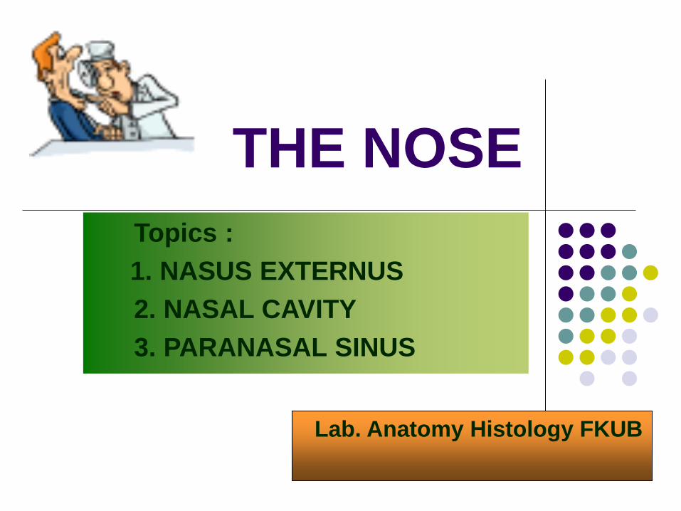
THE NOSE
Topics :
1. NASUS EXTERNUS
2. NASAL CAVITY
3. PARANASAL SINUS
Lab. Anatomy Histology FKUB
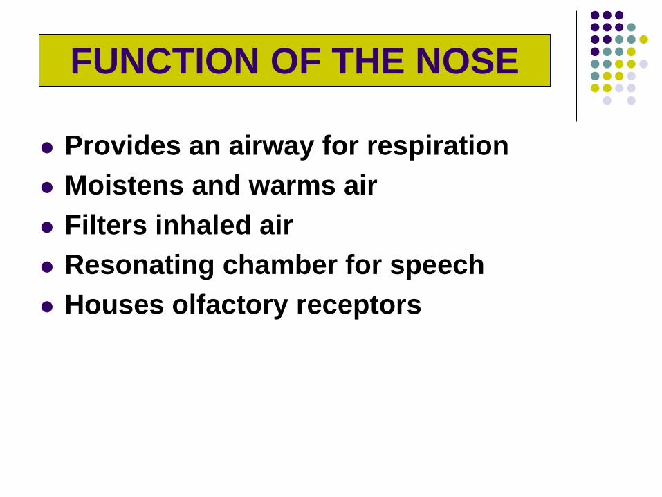
Provides an airway for respiration
Moistens and warms air
Filters inhaled air
Resonating chamber for speech
Houses olfactory receptors
FUNCTION OF THE NOSE
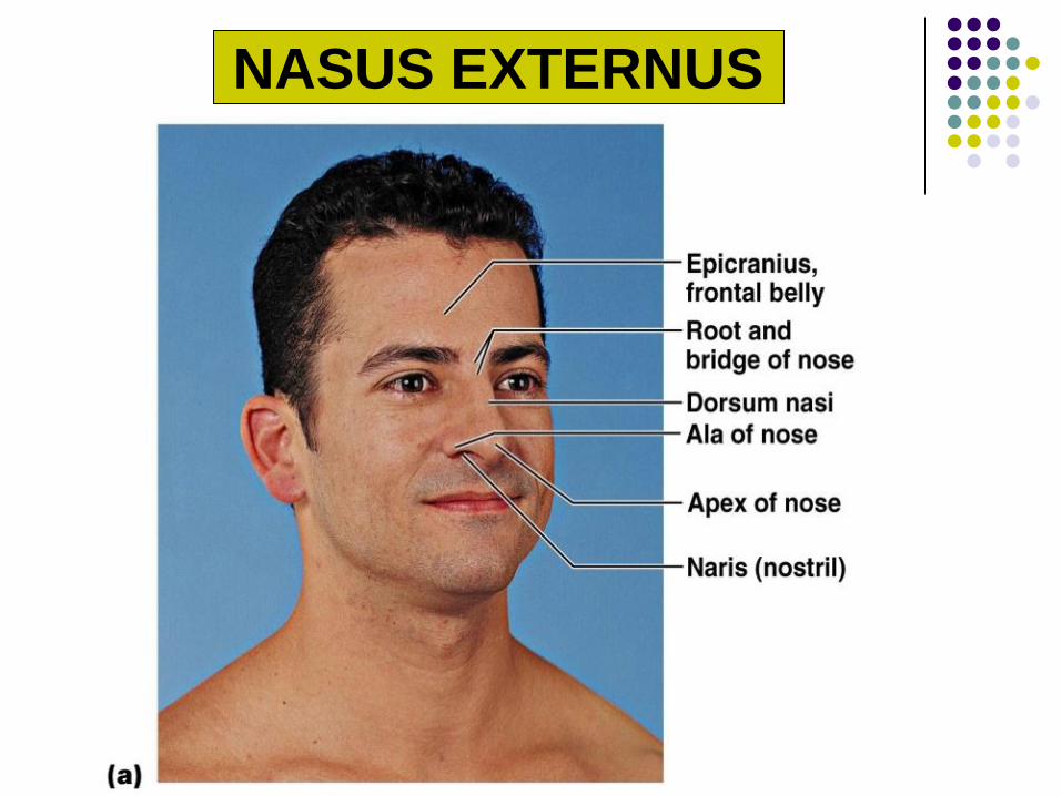
NASUS EXTERNUS
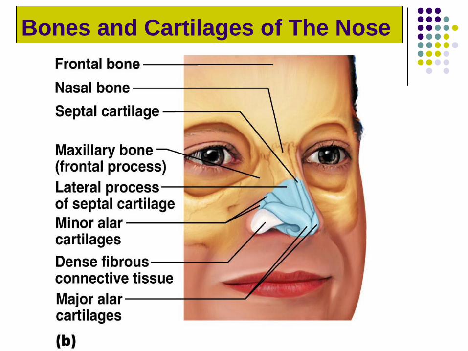
Bones and Cartilages of The Nose
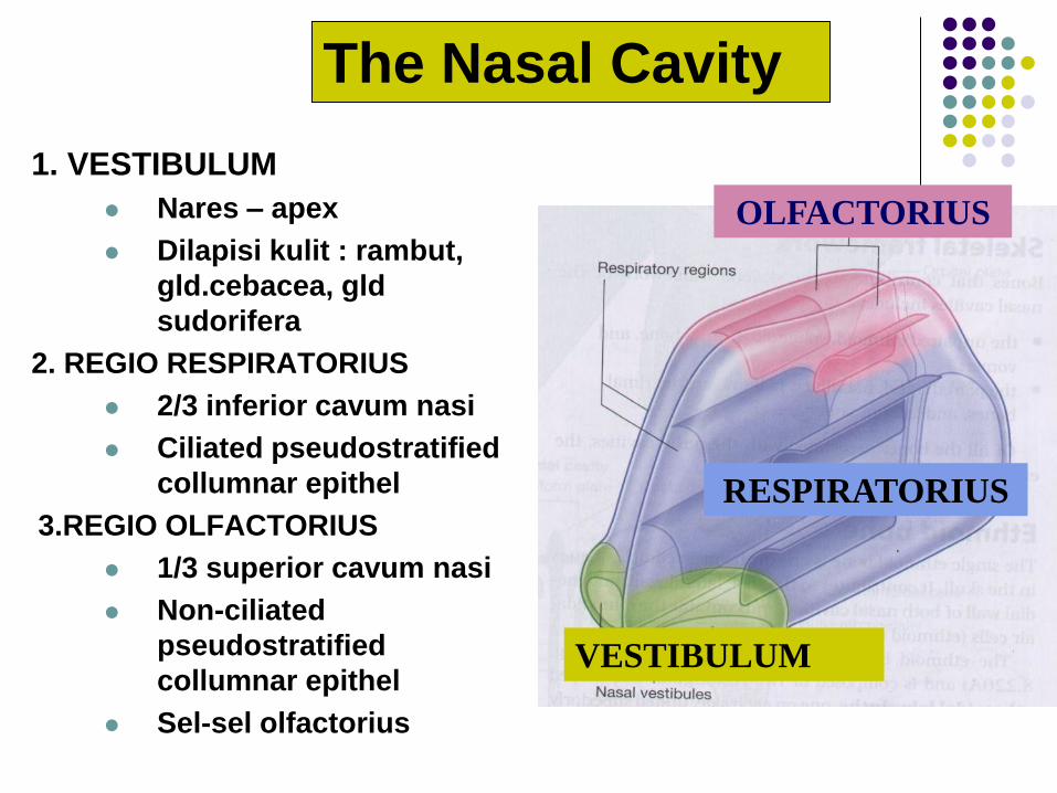
1. VESTIBULUM
Nares – apex
Dilapisi kulit : rambut,
gld.cebacea, gld
sudorifera
2. REGIO RESPIRATORIUS
2/3 inferior cavum nasi
Ciliated pseudostratified
collumnar epithel
3.REGIO OLFACTORIUS
1/3 superior cavum nasi
Non-ciliated
pseudostratified
collumnar epithel
Sel-sel olfactorius
VESTIBULUM
RESPIRATORIUS
OLFACTORIUS
The Nasal Cavity
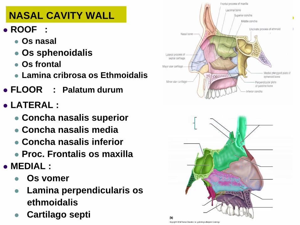
NASAL CAVITY WALL
ROOF : Os nasal
Os sphenoidalis
Os frontal
Lamina cribrosa os Ethmoidalis
FLOOR : Palatum durum
LATERAL : Concha nasalis superior
Concha nasalis media
Concha nasalis inferior
Proc. Frontalis os maxilla
MEDIAL :
Os vomer
Lamina perpendicularis os
ethmoidalis
Cartilago septi
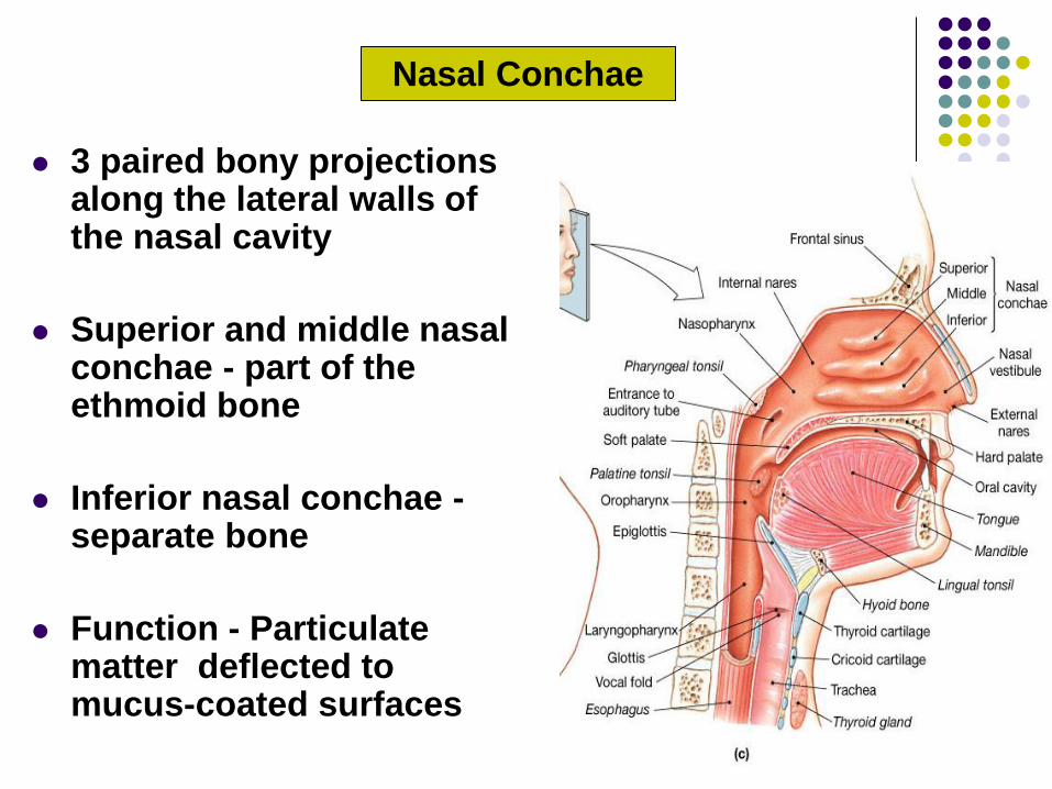
3 paired bony projections along the lateral walls of the nasal cavity
Superior and middle nasal conchae - part of the ethmoid bone
Inferior nasal conchae - separate bone
Function - Particulate matter deflected to mucus-coated surfaces
Nasal Conchae
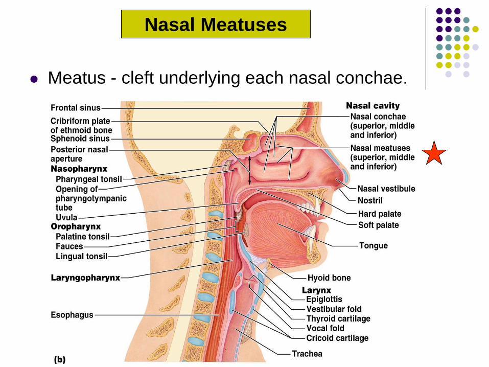
Meatus - cleft underlying each nasal conchae.
Nasal Meatuses
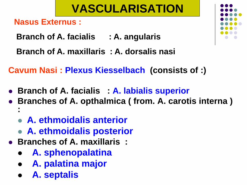
VASCULARISATION
Cavum Nasi : Plexus Kiesselbach (consists of :)
Branch of A. facialis : A. labialis superior
Branches of A. opthalmica ( from. A. carotis interna ) :
A. ethmoidalis anterior
A. ethmoidalis posterior Branches of A. maxillaris :
A. sphenopalatina
A. palatina major
A. septalis
Nasus Externus :
Branch of A. facialis : A. angularis
Branch of A. maxillaris : A. dorsalis nasi
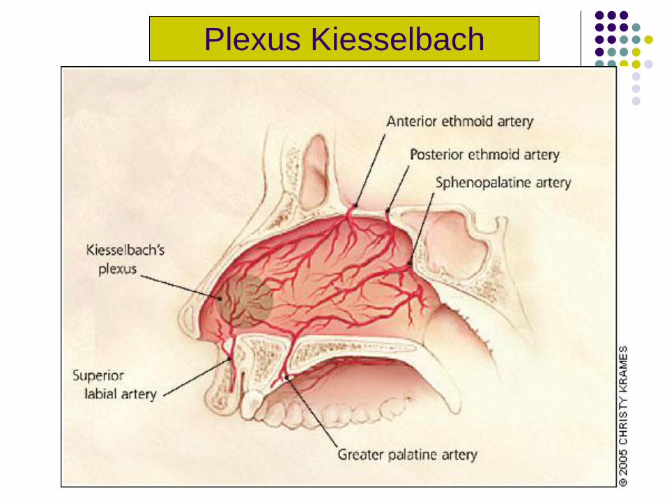
Plexus Kiesselbach
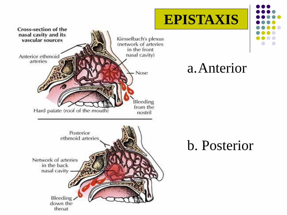
EPISTAXIS
a.Anterior
b. Posterior
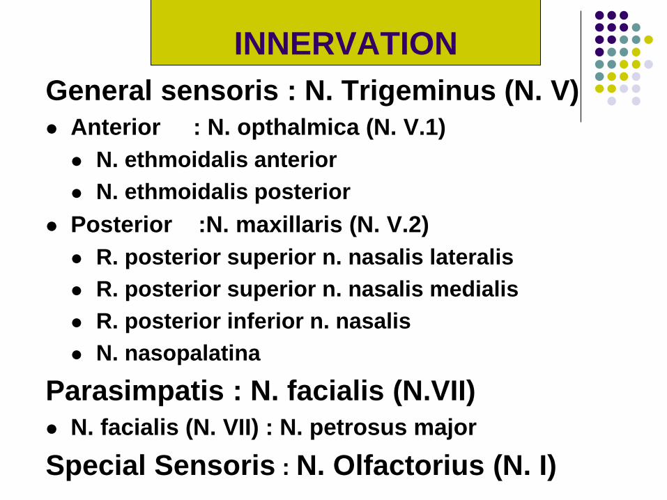
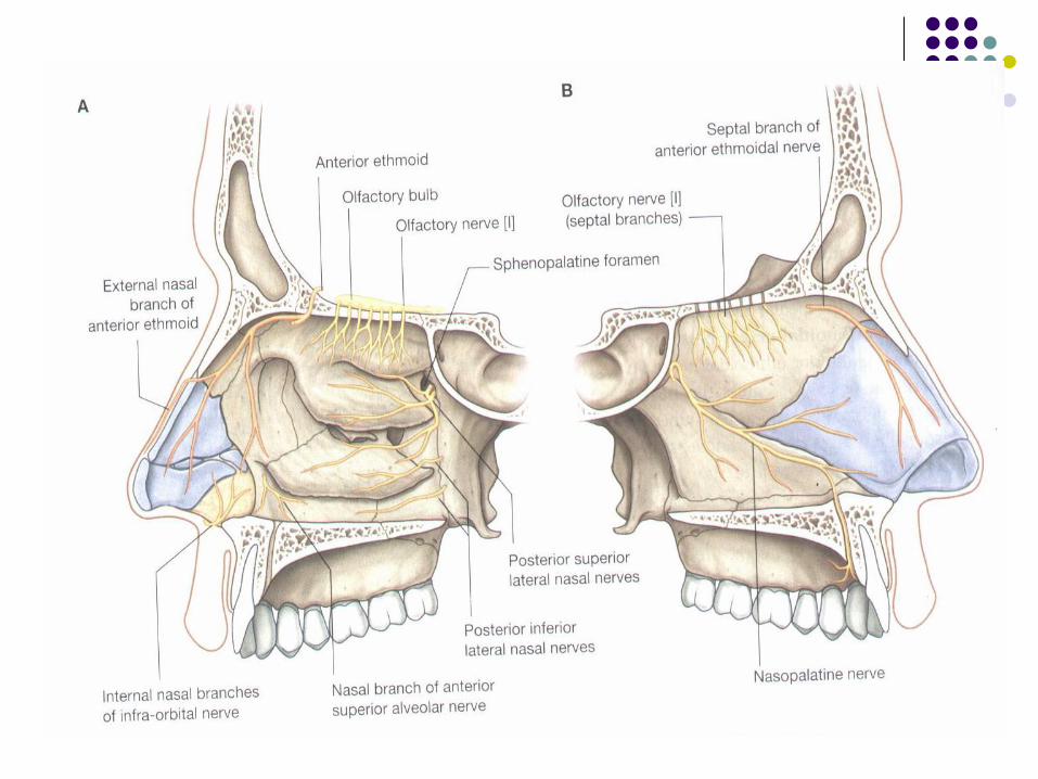
General sensoris : N. Trigeminus (N. V)
Anterior : N. opthalmica (N. V.1)
N. ethmoidalis anterior
N. ethmoidalis posterior
Posterior :N. maxillaris (N. V.2)
R. posterior superior n. nasalis lateralis
R. posterior superior n. nasalis medialis
R. posterior inferior n. nasalis
N. nasopalatina
Parasimpatis : N. facialis (N.VII)
N. facialis (N. VII) : N. petrosus major
Special Sensoris : N. Olfactorius (N. I)
INNERVATION
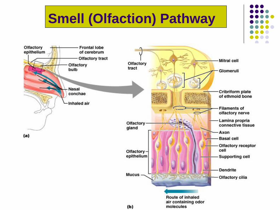
Smell (Olfaction) Pathway
Smell (Olfaction)
Receptors are part of the olfactory epithelium
Olfactory epithelium composed of:
Cell bodies of olfactory receptor cells
Supporting cells – columnar cells
Basal cells – form new olfactory receptor cells
Axons of olfactory epithelium
Gather into bundles – filaments of the olfactory
nerve
Pass through the cribriform plate of the ethmoid
bone
Attach to the olfactory bulbs
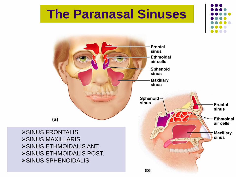
The Paranasal Sinuses
Figure 7.11a, b
SINUS FRONTALIS
SINUS MAXILLARIS
SINUS ETHMOIDALIS ANT.
SINUS ETHMOIDALIS POST.
SINUS SPHENOIDALIS
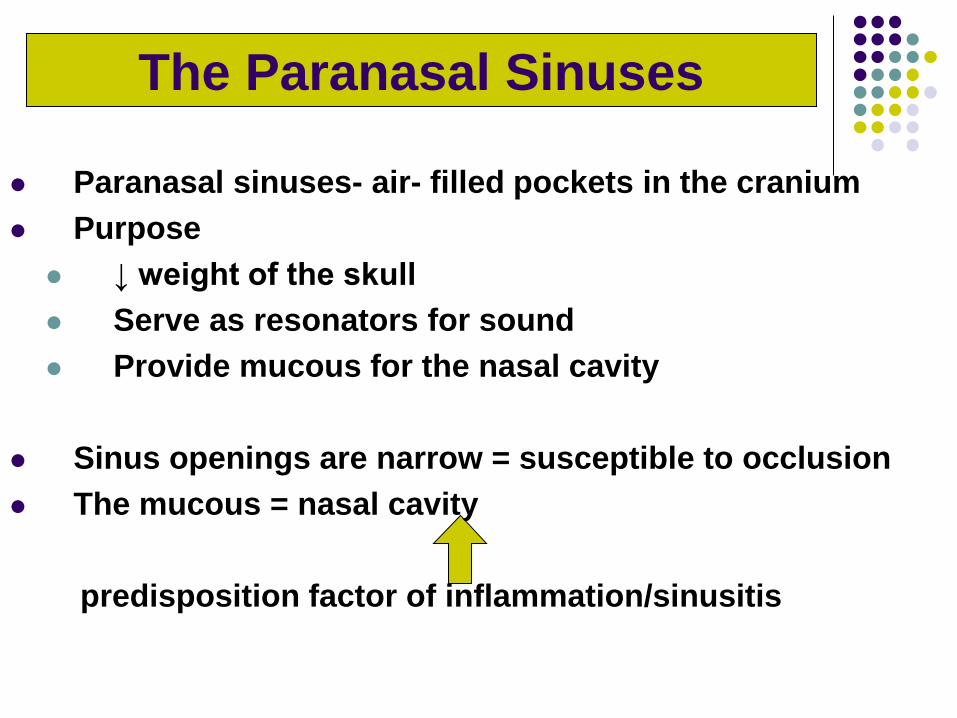
Paranasal sinuses- air- filled pockets in the cranium
Purpose
↓ weight of the skull
Serve as resonators for sound
Provide mucous for the nasal cavity
Sinus openings are narrow = susceptible to occlusion
The mucous = nasal cavity
predisposition factor of inflammation/sinusitis
The Paranasal Sinuses
Sinus
frontalis
Hyatus maxillaris
Sinus sphenoidalis
Sinus
ethmoidalis ant.
Sinus
maxillaris
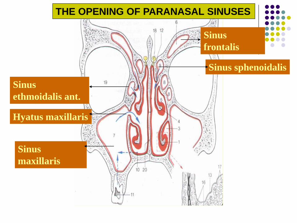
THE OPENING OF PARANASAL SINUSES

Sinus Frontalis
Sinus Maxillaris Meatus Nasi Media
Sinus Ethmoidalis Ant.
Sinus Ethmoidalis Post. Meatus Nasi Superior
Sinus Sphenoidalis Recessus spheno-
ethmoidale
THE OPENING OF PARANASAL SINUSES
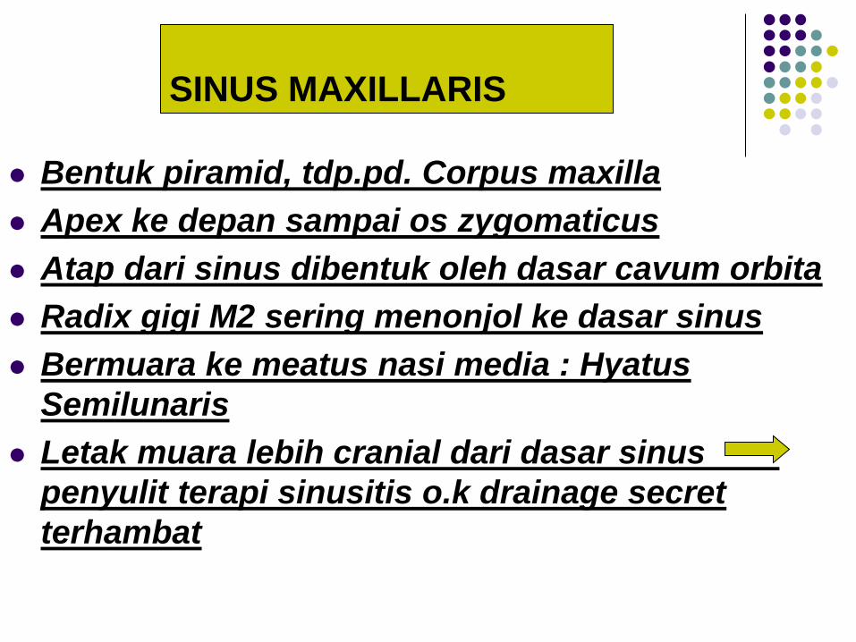
SINUS MAXILLARIS
Bentuk piramid, tdp.pd. Corpus maxilla
Apex ke depan sampai os zygomaticus
Atap dari sinus dibentuk oleh dasar cavum orbita
Radix gigi M2 sering menonjol ke dasar sinus
Bermuara ke meatus nasi media : Hyatus
Semilunaris
Letak muara lebih cranial dari dasar sinus
penyulit terapi sinusitis o.k drainage secret
terhambat