Kshivets O. Cardioesophageal Cancer Surgery
12
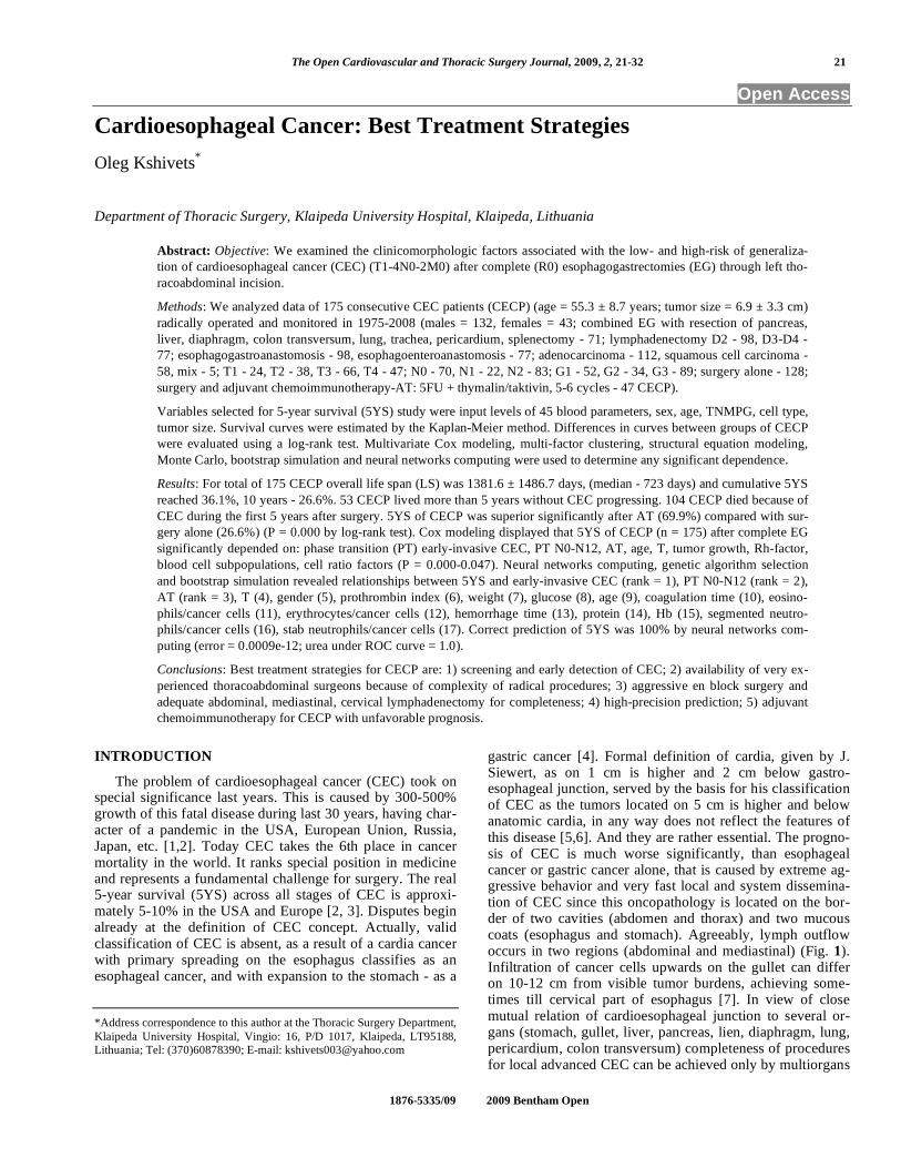
The Open Cardiovascular and Thoracic Surgery Journal, 2009, 2, 21-32 21 1876-5335/09 2009 Bentham Open Open Access Cardioesophageal Cancer: Best Treatment Strategies Oleg Kshivets * Department of Thoracic Surgery, Klaipeda University Hospital, Klaipeda, Lithuania Abstract: Objective: We examined the clinicomorphologic factors associated with the low- and high-risk of generaliza- tion of cardioesophageal cancer (CEC) (T1-4N0-2M0) after complete (R0) esophagogastrectomies (EG) through left tho- racoabdominal incision. Methods: We analyzed data of 175 consecutive CEC patients (CECP) (age = 55.3 ± 8.7 years; tumor size = 6.9 ± 3.3 cm) radically operated and monitored in 1975-2008 (males = 132, females = 43; combined EG with resection of pancreas, liver, diaphragm, colon transversum, lung, trachea, pericardium, splenectomy - 71; lymphadenectomy D2 - 98, D3-D4 - 77; esophagogastroanastomosis - 98, esophagoenteroanastomosis - 77; adenocarcinoma - 112, squamous cell carcinoma - 58, mix - 5; T1 - 24, T2 - 38, T3 - 66, T4 - 47; N0 - 70, N1 - 22, N2 - 83; G1 - 52, G2 - 34, G3 - 89; surgery alone - 128; surgery and adjuvant chemoimmunotherapy-AT: 5FU + thymalin/taktivin, 5-6 cycles - 47 CECP). Variables selected for 5-year survival (5YS) study were input levels of 45 blood parameters, sex, age, TNMPG, cell type, tumor size. Survival curves were estimated by the Kaplan-Meier method. Differences in curves between groups of CECP were evaluated using a log-rank test. Multivariate Cox modeling, multi-factor clustering, structural equation modeling, Monte Carlo, bootstrap simulation and neural networks computing were used to determine any significant dependence. Results: For total of 175 CECP overall life span (LS) was 1381.6 ± 1486.7 days, (median - 723 days) and cumulative 5YS reached 36.1%, 10 years - 26.6%. 53 CECP lived more than 5 years without CEC progressing. 104 CECP died because of CEC during the first 5 years after surgery. 5YS of CECP was superior significantly after AT (69.9%) compared with sur- gery alone (26.6%) (P = 0.000 by log-rank test). Cox modeling displayed that 5YS of CECP (n = 175) after complete EG significantly depended on: phase transition (PT) early-invasive CEC, PT N0-N12, AT, age, T, tumor growth, Rh-factor, blood cell subpopulations, cell ratio factors (P = 0.000-0.047). Neural networks computing, genetic algorithm selection and bootstrap simulation revealed relationships between 5YS and early-invasive CEC (rank = 1), PT N0-N12 (rank = 2), AT (rank = 3), T (4), gender (5), prothrombin index (6), weight (7), glucose (8), age (9), coagulation time (10), eosino- phils/cancer cells (11), erythrocytes/cancer cells (12), hemorrhage time (13), protein (14), Hb (15), segmented neutro- phils/cancer cells (16), stab neutrophils/cancer cells (17). Correct prediction of 5YS was 100% by neural networks com- puting (error = 0.0009e-12; urea under ROC curve = 1.0). Conclusions: Best treatment strategies for CECP are: 1) screening and early detection of CEC; 2) availability of very ex- perienced thoracoabdominal surgeons because of complexity of radical procedures; 3) aggressive en block surgery and adequate abdominal, mediastinal, cervical lymphadenectomy for completeness; 4) high-precision prediction; 5) adjuvant chemoimmunotherapy for CECP with unfavorable prognosis. INTRODUCTION The problem of cardioesophageal cancer (CEC) took on special significance last years. This is caused by 300-500% growth of this fatal disease during last 30 years, having char- acter of a pandemic in the USA, European Union, Russia, Japan, etc. [1,2]. Today CC takes the 6th place in cancer mortality in the world. It ranks special position in medicine and represents a fundamental challenge for surgery. The real 5-year survival (5YS) across all stages of CEC is approxi- mately 5-10% in the USA and Europe [2, 3]. Disputes begin already at the definition of CEC concept. Actually, valid classification of CEC is absent, as a result of a cardia cancer with primary spreading on the esophagus classifies as an esophageal cancer, and with expansion to the stomach - as a *Address correspondence to this author at the Thoracic Surgery Department, Klaipeda University Hospital, Vingio: 16, P/D 1017, Klaipeda, LT95188, Lithuania; Tel: (370)60878390; E-mail: [email protected] gastric cancer [4]. Formal definition of cardia, given by J. Siewert, as on 1 cm is higher and 2 cm below gastro- esophageal junction, served by the basis for his classification of CEC as the tumors located on 5 cm is higher and below anatomic cardia, in any way does not reflect the features of this disease [5,6]. And they are rather essential. The progno- sis of CEC is much worse significantly, than esophageal cancer or gastric cancer alone, that is caused by extreme ag- gressive behavior and very fast local and system dissemina- tion of CEC since this oncopathology is located on the bor- der of two cavities (abdomen and thorax) and two mucous coats (esophagus and stomach). Agreeably, lymph outflow occurs in two regions (abdominal and mediastinal) (Fig. 1). Infiltration of cancer cells upwards on the gullet can differ on 10-12 cm from visible tumor burdens, achieving some- times till cervical part of esophagus [7]. In view of close mutual relation of cardioesophageal junction to several or- gans (stomach, gullet, liver, pancreas, lien, diaphragm, lung, pericardium, colon transversum) completeness of procedures for local advanced CEC can be achieved only by multiorgans
-
Upload
oleg-kshivets -
Category
Health & Medicine
-
view
1.998 -
download
0
Transcript of Kshivets O. Cardioesophageal Cancer Surgery
The Open Cardiovascular and Thoracic Surgery Journal, 2009, 2, 21-32 21
1876-5335/09 2009 Bentham Open
Open Access
Cardioesophageal Cancer: Best Treatment Strategies
Oleg Kshivets*
Department of Thoracic Surgery, Klaipeda University Hospital, Klaipeda, Lithuania
Abstract: Objective: We examined the clinicomorphologic factors associated with the low- and high-risk of generaliza-
tion of cardioesophageal cancer (CEC) (T1-4N0-2M0) after complete (R0) esophagogastrectomies (EG) through left tho-
racoabdominal incision.
Methods: We analyzed data of 175 consecutive CEC patients (CECP) (age = 55.3 ± 8.7 years; tumor size = 6.9 ± 3.3 cm)
radically operated and monitored in 1975-2008 (males = 132, females = 43; combined EG with resection of pancreas,
liver, diaphragm, colon transversum, lung, trachea, pericardium, splenectomy - 71; lymphadenectomy D2 - 98, D3-D4 -
77; esophagogastroanastomosis - 98, esophagoenteroanastomosis - 77; adenocarcinoma - 112, squamous cell carcinoma -
58, mix - 5; T1 - 24, T2 - 38, T3 - 66, T4 - 47; N0 - 70, N1 - 22, N2 - 83; G1 - 52, G2 - 34, G3 - 89; surgery alone - 128;
surgery and adjuvant chemoimmunotherapy-AT: 5FU + thymalin/taktivin, 5-6 cycles - 47 CECP).
Variables selected for 5-year survival (5YS) study were input levels of 45 blood parameters, sex, age, TNMPG, cell type,
tumor size. Survival curves were estimated by the Kaplan-Meier method. Differences in curves between groups of CECP
were evaluated using a log-rank test. Multivariate Cox modeling, multi-factor clustering, structural equation modeling,
Monte Carlo, bootstrap simulation and neural networks computing were used to determine any significant dependence.
Results: For total of 175 CECP overall life span (LS) was 1381.6 ± 1486.7 days, (median - 723 days) and cumulative 5YS
reached 36.1%, 10 years - 26.6%. 53 CECP lived more than 5 years without CEC progressing. 104 CECP died because of
CEC during the first 5 years after surgery. 5YS of CECP was superior significantly after AT (69.9%) compared with sur-
gery alone (26.6%) (P = 0.000 by log-rank test). Cox modeling displayed that 5YS of CECP (n = 175) after complete EG
significantly depended on: phase transition (PT) early-invasive CEC, PT N0-N12, AT, age, T, tumor growth, Rh-factor,
blood cell subpopulations, cell ratio factors (P = 0.000-0.047). Neural networks computing, genetic algorithm selection
and bootstrap simulation revealed relationships between 5YS and early-invasive CEC (rank = 1), PT N0-N12 (rank = 2),
AT (rank = 3), T (4), gender (5), prothrombin index (6), weight (7), glucose (8), age (9), coagulation time (10), eosino-
phils/cancer cells (11), erythrocytes/cancer cells (12), hemorrhage time (13), protein (14), Hb (15), segmented neutro-
phils/cancer cells (16), stab neutrophils/cancer cells (17). Correct prediction of 5YS was 100% by neural networks com-
puting (error = 0.0009e-12; urea under ROC curve = 1.0).
Conclusions: Best treatment strategies for CECP are: 1) screening and early detection of CEC; 2) availability of very ex-
perienced thoracoabdominal surgeons because of complexity of radical procedures; 3) aggressive en block surgery and
adequate abdominal, mediastinal, cervical lymphadenectomy for completeness; 4) high-precision prediction; 5) adjuvant
chemoimmunotherapy for CECP with unfavorable prognosis.
INTRODUCTION
The problem of cardioesophageal cancer (CEC) took on special significance last years. This is caused by 300-500% growth of this fatal disease during last 30 years, having char-acter of a pandemic in the USA, European Union, Russia, Japan, etc. [1,2]. Today C C takes the 6th place in cancer mortality in the world. It ranks special position in medicine and represents a fundamental challenge for surgery. The real 5-year survival (5YS) across all stages of CEC is approxi-mately 5-10% in the USA and Europe [2, 3]. Disputes begin already at the definition of CEC concept. Actually, valid classification of CEC is absent, as a result of a cardia cancer with primary spreading on the esophagus classifies as an esophageal cancer, and with expansion to the stomach - as a
*Address correspondence to this author at the Thoracic Surgery Department,
Klaipeda University Hospital, Vingio: 16, P/D 1017, Klaipeda, LT95188,
Lithuania; Tel: (370)60878390; E-mail: [email protected]
gastric cancer [4]. Formal definition of cardia, given by J. Siewert, as on 1 cm is higher and 2 cm below gastro-esophageal junction, served by the basis for his classification of CEC as the tumors located on 5 cm is higher and below anatomic cardia, in any way does not reflect the features of this disease [5,6]. And they are rather essential. The progno-sis of CEC is much worse significantly, than esophageal cancer or gastric cancer alone, that is caused by extreme ag-gressive behavior and very fast local and system dissemina-tion of CEC since this oncopathology is located on the bor-der of two cavities (abdomen and thorax) and two mucous coats (esophagus and stomach). Agreeably, lymph outflow occurs in two regions (abdominal and mediastinal) (Fig. 1). Infiltration of cancer cells upwards on the gullet can differ on 10-12 cm from visible tumor burdens, achieving some-times till cervical part of esophagus [7]. In view of close mutual relation of cardioesophageal junction to several or-gans (stomach, gullet, liver, pancreas, lien, diaphragm, lung, pericardium, colon transversum) completeness of procedures for local advanced CEC can be achieved only by multiorgans

22 The Open Cardiovascular and Thoracic Surgery Journal, 2009, Volume 2 Oleg Kshivets
resections. Therefore treatment of tumors for this localiza-tion is one of the most difficult and disputable sections of thoracoabdominal surgery [3]. Extensive abdominal (D2-D4), mediastinal, and, in some cases, cervical lymphadenec-tomy (2-3 folds) is the cornerstone of the radical esophagogastrectomy [8]. It has not only medical and diag-nostic, but also prognostic value. All this demands from the surgeon of virtuosous techniques, huge experience, force and endurance. That is why the surgery of CEC always remains the privilege of a narrow circle of the best surgeons of the world, which deficiency accrues every year. And finally, usually the basic attention during analysis of CEC was given to the nearest postoperative results in view of complexity of surgical interventions and features of postoperative monitor-ing of patients, and the remote period remains in a shade. Considering the premises, we analyzed the long time results of treatment of patients with CEC (CECP) after radical (R0) esophagogastrectomies through the left thoracoabdominal incision (Garlock operation). We developed best treatment strategies that incorporate bolus chemotherapy and immuno-therapy after radical, aggressive en-block surgery and ade-quate abdominal, mediastinal and cervical lymph node dis-section.
PATIENTS AND METHODS
We performed a review of prospectively collected data-base of European patients undergoing the complete (R0) esophagogastrectomy for CEC between 1975 and 2008. 175 consecutive CECP (male - 132, female - 43; age = 55.3 ± 8.7 years, tumor size = 6.9 ± 3.3 cm) (mean ± standard devia-tion) entered this trial. Patients were not considered eligible if they had stage IV (nonregional lymph nodes metastases and distant metastases), previous treatment with chemother-apy, immunotherapy or radiotherapy or if there were two primary tumors at the time of diagnosis. CECP after non-radical procedures and patients, who died postoperatively, were excluded to provide a homogeneous patient group. The preoperative staging protocol included clinical history, physical examination, complete blood count with differen-tials, biochemistry and electrolyte panel, chest X-rays, rönt-genoesophagogastroscopy, computed tomography scan of thorax, abdominal ultrasound, fibroesophagogastroscopy, electrocardiogram. Computed tomography scan of abdomen, liver and bone radionuclide scan were performed whenever needed. Mediastinoscopy was not used. All CECP were di-agnosed with histologically confirmed CEC. All had meas-urable tumor and ECOG performance status 0 or 1. Before any treatment each patient was carefully examined by a medical panel composed of thoracic surgeon, chemothera-peutist, radiologist and gastroenterologist to confirm the stage of disease. All patients signed a written informed con-sent form approved by the local Institutional Review Board.
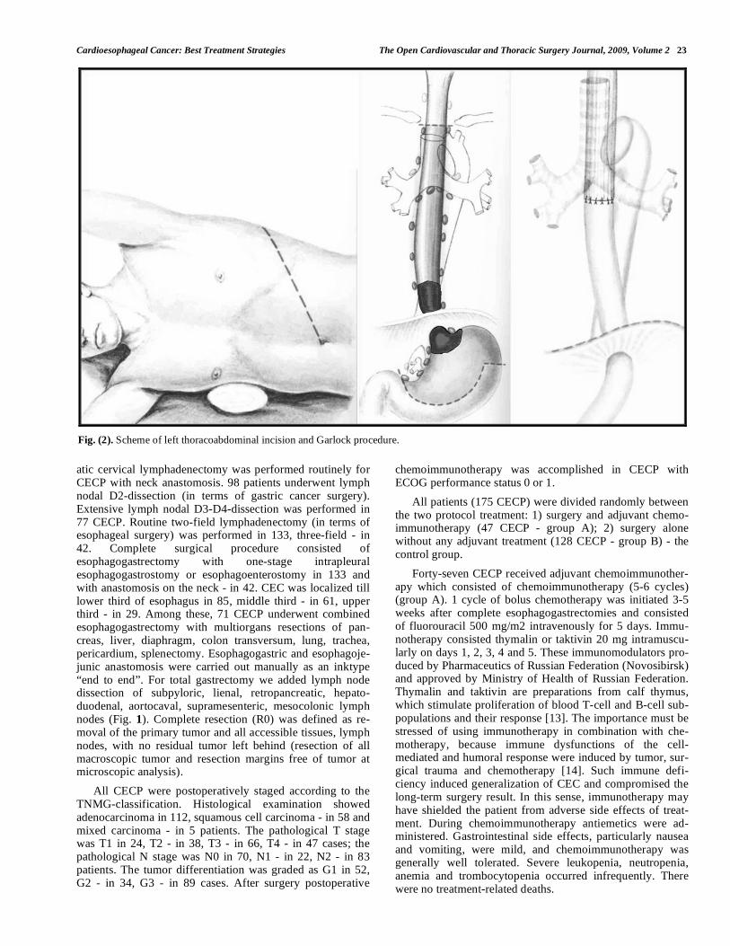
The initial treatment was started with surgery. We are basic supporters of Garlock’s operation through left thoraco-abdominal incision for CEC as more convenient and less traumatic, than Lewis's operation (Figs. 2, 3). Conclusive advantages of left thoracoabdominal approach are unsur-passed conveniences for combined multiorgans resections, extensive lymphadenectomy and exact correlation of length of a gastric tube or jejunum with the level of anastomosis [9-12]. At this procedure we removed the most part of stomach
Fig. (1). Scheme of lymph node metastasis for cardioesophageal
cancer: 1 - right paracardial; 2 - left paracardial; 7 - along left gas-
tric artery; 8 - along common hepatic artery; 9 - along celiac trunk;
10 - lien hilar; 11 - along lienal artery; 16 - aortocaval and paraaor-
tal abdominal; 104a - right supraclavicular; 104b - left supraclavi-
cular; 105 - upper paraesophageal; R106 - right paratracheal; L106
- left paratracheal; 107 - bifurcational; 108 - middle paraesophag-
eal; 109 - lung hilar; 110 - lower paraesophageal; 111 - diaphrag-
matic; 112 - paraaortal thoracic.
with preservation of right gastroepiploic vessels or total stomach with an esophagus on 6-12 m above proximal tu-mor burden with intraoperative express-histology for clear-ance without fail, minor and major omentum, and also lymph nodes on the both sides of a diaphragm (lymph nodes along of celiac trunk, common hepatic artery, splenic artery toward the splenic hilum, paracardial, paraesophageal, sub- and su-pradiaphragmatic, mediastinal, bifurcational, paraaortal and aortocaval lymph nodes) (Figs. 1, 3). The left gastric artery was always transected at its origin and remained with the specimen. The present analysis was restricted to CECP with complete resected tumors with negative surgical resection margin and with N0-2 lymph nodes. Patients with the CEC infiltration till upper third of esophagus had an additional formal extended mediastinal lymphadenectomy comprising all nodes at the tracheal bifurcation along the left and right main stem bronchi, aorta window, the upper mediastinal compartment, and along the left recurrent nerve. A system-
Cardioesophageal Cancer: Best Treatment Strategies The Open Cardiovascular and Thoracic Surgery Journal, 2009, Volume 2 23
atic cervical lymphadenectomy was performed routinely for CECP with neck anastomosis. 98 patients underwent lymph nodal D2-dissection (in terms of gastric cancer surgery). Extensive lymph nodal D3-D4-dissection was performed in 77 CECP. Routine two-field lymphadenectomy (in terms of esophageal surgery) was performed in 133, three-field - in 42. Complete surgical procedure consisted of esophagogastrectomy with one-stage intrapleural esophagogastrostomy or esophagoenterostomy in 133 and with anastomosis on the neck - in 42. CEC was localized till lower third of esophagus in 85, middle third - in 61, upper third - in 29. Among these, 71 CECP underwent combined esophagogastrectomy with multiorgans resections of pan-creas, liver, diaphragm, colon transversum, lung, trachea, pericardium, splenectomy. Esophagogastric and esophagoje-junic anastomosis were carried out manually as an inktype “end to end”. For total gastrectomy we added lymph node dissection of subpyloric, lienal, retropancreatic, hepato-duodenal, aortocaval, supramesenteric, mesocolonic lymph nodes (Fig. 1). Complete resection (R0) was defined as re-moval of the primary tumor and all accessible tissues, lymph nodes, with no residual tumor left behind (resection of all macroscopic tumor and resection margins free of tumor at microscopic analysis).
All CECP were postoperatively staged according to the TNMG-classification. Histological examination showed adenocarcinoma in 112, squamous cell carcinoma - in 58 and mixed carcinoma - in 5 patients. The pathological T stage was T1 in 24, T2 - in 38, T3 - in 66, T4 - in 47 cases; the pathological N stage was N0 in 70, N1 - in 22, N2 - in 83 patients. The tumor differentiation was graded as G1 in 52, G2 - in 34, G3 - in 89 cases. After surgery postoperative
chemoimmunotherapy was accomplished in CECP with ECOG performance status 0 or 1.
All patients (175 CECP) were divided randomly between the two protocol treatment: 1) surgery and adjuvant chemo-immunotherapy (47 CECP - group A); 2) surgery alone without any adjuvant treatment (128 CECP - group B) - the control group.
Forty-seven CECP received adjuvant chemoimmunother-apy which consisted of chemoimmunotherapy (5-6 cycles) (group A). 1 cycle of bolus chemotherapy was initiated 3-5 weeks after complete esophagogastrectomies and consisted of fluorouracil 500 mg/m2 intravenously for 5 days. Immu-notherapy consisted thymalin or taktivin 20 mg intramuscu-larly on days 1, 2, 3, 4 and 5. These immunomodulators pro-duced by Pharmaceutics of Russian Federation (Novosibirsk) and approved by Ministry of Health of Russian Federation. Thymalin and taktivin are preparations from calf thymus, which stimulate proliferation of blood T-cell and B-cell sub-populations and their response [13]. The importance must be stressed of using immunotherapy in combination with che-motherapy, because immune dysfunctions of the cell-mediated and humoral response were induced by tumor, sur-gical trauma and chemotherapy [14]. Such immune defi-ciency induced generalization of CEC and compromised the long-term surgery result. In this sense, immunotherapy may have shielded the patient from adverse side effects of treat-ment. During chemoimmunotherapy antiemetics were ad-ministered. Gastrointestinal side effects, particularly nausea and vomiting, were mild, and chemoimmunotherapy was generally well tolerated. Severe leukopenia, neutropenia, anemia and trombocytopenia occurred infrequently. There were no treatment-related deaths.
Fig. (2). Scheme of left thoracoabdominal incision and Garlock procedure.

24 The Open Cardiovascular and Thoracic Surgery Journal, 2009, Volume 2 Oleg Kshivets
A follow-up examination was, generally, done every 3 month for the first 2 years, every 6 month after that and yearly after 5 years, including a physical examination, a complete blood count, blood chemistry, and chest roent-genography. Fibroesophagogastroscopy was done every 6 months for the first 3 years. Zero time was the data of surgi-cal procedures. No one was lost during the follow-up period and we regarded the outcome as death through personal knowledge, physician's reports, autopsy or death certificates. Survival time (days) was measured from the date of surgery until death or the most-recent date of follow-up for surviving patients.
Variables selected for 5YS and life span (LS) study were the input levels of 45 blood parameters, sex, age, TNMG, cell type, and tumor size. Survival curves were estimated by the Kaplan-Meier method. Differences in curves between groups of CECP were evaluated using a log-rank test. Multi-variate proportional hazard Cox regression, structural equa-tion modeling (SEPATH), Monte Carlo simulation, bootstrap simulation and neural networks computing were used to de-termine any significant dependence [15-21]. Neural networks computing, system, biometric and statistical analyses were conducted using CLASS-MASTER program (Stat Dialog, Inc., Moscow, Russia), SANI program (Stat Dialog, Inc., Moscow, Russia), DEDUCTOR program (BaseGroup Labs, Inc., Riazan, Russia), SPSS (SPSS Inc., Chicago, IL, USA), STATISTICA and STATISTICA Neural Networks program (Stat Soft, Inc., Tulsa, OK, USA), MATHCAD (MathSoft,
Inc., Needham, MA, USA), SIMSTAT (Provalis Research, Inc., Montreal, QC, Canada). All tests were considered sig-nificant if the resulting P value was less than 0.05.
RESULTS
For the entire sample of 175 patients overall LS was 1381.6 ± 1486.7 days (mean ± standard deviation) (95% CI, 1159.8-1603.4 days; median = 723). General cumulative 5YS reached 36.1%, 10-year survival - 26.6%. 61 CECP (34.9%) were alive till now, 53 CECP (30.3%) lived more than 5 years (LS = 3212.7 ± 1512.0 days) without any fea-tures of CEC progressing. 104 CECP (59.4%) died because of generalization during the first 5 years after surgery (LS = 587.3 ± 316.8 days) (Fig. 4).
It is necessary to pay attention to the two very important prognostic phenomenas. First, we found 100% 5YS for CECP with early cancer (T1N0, n = 18) versus 28.4% for other CECP (n = 157) after esophagogastrectomies (P = 0.000 by log-rank test) (Fig. 5). Early CEC was defined, based on the final histopathologic report of the resection specimen, as tumor limited to the mucosa or submucosa and not extending into the muscular wall of the cardioesophageal junction, up to 2 cm in diameter with N0 [12]. All patients with stage T1N0 did not receive adjuvant chemoimmuno-therapy. Correspondingly, the overall 10-year survival for CECP with the early cancer was 92.6% and was significantly better compared to 18.4% for others patients (P = 0.000).
Fig. (3). Our methodology of esophagogastrectomy through left thoracoabdominal approach.

Cardioesophageal Cancer: Best Treatment Strategies The Open Cardiovascular and Thoracic Surgery Journal, 2009, Volume 2 25
Surv iv al Function
5-Y ear Surv iv al of C ardioesophageal Cancer Patients=36.1%; 10-Y ear Surv iv al=26.6%;
n=175
C om plete Censored
-5 0 5 10 15 20 25
Y ears af ter Esophagogas trec tom y
0.1
0.2
0.3
0.4
0.5
0.6
0.7
0.8
0.9
1.0
1.1
1.2
v
Fig. (4). General cumulative survival of CECP with stage T1-4N0-2M0, n = 175 after radical esophagogastrectomies: cumulative 5-year
survival = 36.1%, 10-year survival = 26.6%.
Cum ulat iv e Proport ion Surv iv ing (Kaplan-Meier)
5-Y ear Surv iv al of Early Cardioesophageal Cancer Patients=100%;
5-Y ear Surv iv al of Inv as iv e C ardioesophageal C ancer Patients=28.4%;
P=0.000 by Log-Rank Tes t
C om plete C ensored
0 5 10 15 20 25
Y ears af ter Esophagogas trec tom y
0.0
0.1
0.2
0.3
0.4
0.5
0.6
0.7
0.8
0.9
1.0
u
Inv as iv e Cardioesophageal C ancer Patients , n=157
Early C ardioesophageal Cancer Patients , n=18
Fig. (5). Survival of CECP with early cancer (n = 18) was significantly better compared with invasive cancer (n = 157) (P = 0.000 by log-
rank test).
Cu
mu
lative P
ropo
rtio
n S
urv
ivin
gC
um
ula
tive P
ropo
rtio
n S
urv
ivin
g
26 The Open Cardiovascular and Thoracic Surgery Journal, 2009, Volume 2 Oleg Kshivets
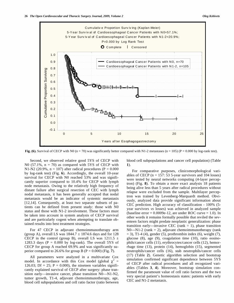
Second, we observed relative good 5YS of CECP with N0 (57.1%, n = 70) as compared with 5YS of CECP with N1-N2 (20.9%, n = 107) after radical procedures (P = 0.000 by log-rank test) (Fig. 6). Accordingly, the overall 10-year survival for CECP with N0 reached 53% and was signifi-cantly superior compared to 10.4% for CECP with lymph node metastasis. Owing to the relatively high frequency of distant failure after surgical resection of CEC with lymph nodal metastasis, it has been generally accepted that nodal metastasis would be an indicator of systemic metastasis [12,14]. Consequently, at least two separate subsets of pa-tients can be defined from present study: those with N0 status and those with N1-2 involvement. These factors must be taken into account in system analysis of CECP survival and are particularly cogent when attempting to translate ob-tained results into best treatment strategies.
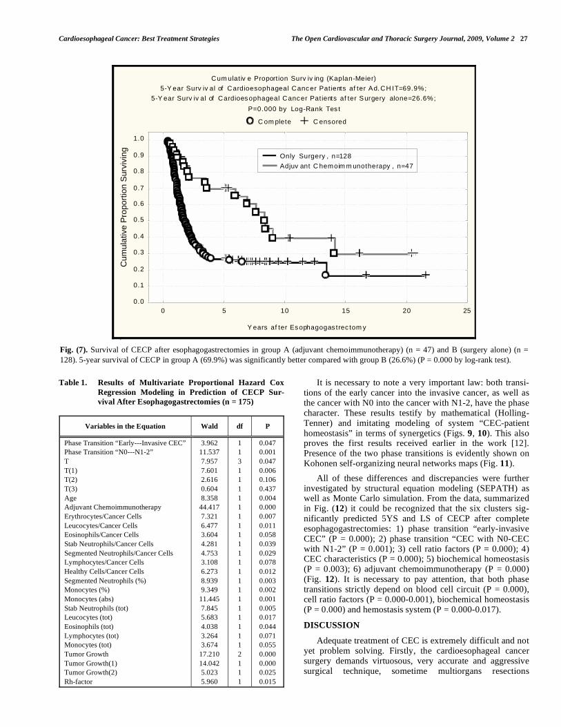
For 47 CECP in adjuvant chemoimmunotherapy arm (group A), overall LS was 1844.7 ± 1874.6 days and for 128 CECP in the control (group B), overall LS was 1211.5 ± 1283.3 days (P = 0.000 by log-rank). The overall 5YS of CECP for group A reached 69.9% and was significantly su-perior compared to 26.6% for group B (P = 0.000) (Fig. 7).
All parameters were analyzed in a multivariate Cox model. In accordance with this Cox model (global
2 =
126.03; Df = 25; P = 0.000), the nineteen variables signifi-cantly explained survival of CECP after surgery: phase tran-sition early---invasive cancer, phase transition N0---N1-N2, tumor growth, T1-4, adjuvant chemoimmunotherapy, age, blood cell subpopulations and cell ratio factor (ratio between
blood cell subpopulations and cancer cell population) (Table 1).
For comparative purposes, clinicomorphological vari-ables of CECP (n = 157: 53 5-year survivors and 104 losses) were tested by neural networks computing (4-layer percep-tron) (Fig. 8). To obtain a more exact analysis 18 patients being alive less than 5 years after radical procedures without relapse were excluded from the sample. Multilayer percep-tron was trained by Levenberg-Marquardt method. Obvi-ously, analyzed data provide significant information about CEC prediction. High accuracy of classification - 100% (5-year survivors vs losses) was achieved in analyzed sample (baseline error = 0.0009e-12, are under ROC curve = 1.0). In other words it remains formally possible that reviled the sev-enteen factors might predate neoplastic generalization: phase transition early---invasive CEC (rank = 1), phase transition N0---N1-2 (rank = 2), adjuvant chemoimmunotherapy (rank = 3), T1-4 (4), gender (5), prothrombin index (6), weight (7), glucose (8), age (9), coagulation time (10), ratio eosino-phils/cancer cells (11), erythrocytes/cancer cells (12), hemor-rhage time (13), protein (14), hemoglobin (15), segmented neutrophils/cancer cells (16), stab neutrophils/cancer cells (17) (Table 2). Genetic algorithm selection and bootstrap simulation confirmed significant dependence between 5YS of CECP after radical procedures and all recognized vari-ables (Tables 3, 4). Moreover, bootstrap simulation con-firmed the paramount value of cell ratio factors and the two very special patient’s homeostasis states: patients with early CEC and N1-2 metastasis.
Cum ulat iv e Proport ion Surv iv ing (Kaplan-Meier)
5-Y ear Surv iv al of Cardioesophageal C ancer Patients with N 0=57.1%;
5-Y ear Surv iv al of C ardioesophageal Cancer Patients with N1-2=20.9%;
P=0.000 by Log Rank Tes t
C om plete C ensored
0 5 10 15 20 25
Y ears af t er Esophagogas trect om y
-0.1
0.0
0.1
0.2
0.3
0.4
0.5
0.6
0.7
0.8
0.9
1.0
u
C ardioesophageal C ancer Patients wit h N 0, n=70
C ardioesophageal C ancer Patients wit h N 1-2, n=105
Fig. (6). Survival of CECP with N0 (n = 70) was significantly better compared with N1-2 metastases (n = 105) (P = 0.000 by log-rank test).
Cum
ulat
ive
Pro
porti
on S
urvi
ving
Cardioesophageal Cancer: Best Treatment Strategies The Open Cardiovascular and Thoracic Surgery Journal, 2009, Volume 2 27
Table 1. Results of Multivariate Proportional Hazard Cox
Regression Modeling in Prediction of CECP Sur-
vival After Esophagogastrectomies (n = 175)
Variables in the Equation Wald df P
Phase Transition “Early---Invasive CEC”
Phase Transition “N0---N1-2”
T
T(1)
T(2)
T(3)
Age
Adjuvant Chemoimmunotherapy
Erythrocytes/Cancer Cells
Leucocytes/Cancer Cells
Eosinophils/Cancer Cells
Stab Neutrophils/Cancer Cells
Segmented Neutrophils/Cancer Cells
Lymphocytes/Cancer Cells
Healthy Cells/Cancer Cells
Segmented Neutrophils (%)
Monocytes (%)
Monocytes (abs)
Stab Neutrophils (tot)
Leucocytes (tot)
Eosinophils (tot)
Lymphocytes (tot)
Monocytes (tot)
Tumor Growth
Tumor Growth(1)
Tumor Growth(2)
Rh-factor
3.962
11.537
7.957
7.601
2.616
0.604
8.358
44.417
7.321
6.477
3.604
4.281
4.753
3.108
6.273
8.939
9.349
11.445
7.845
5.683
4.038
3.264
3.674
17.210
14.042
5.023
5.960
1
1
3
1
1
1
1
1
1
1
1
1
1
1
1
1
1
1
1
1
1
1
1
2
1
1
1
0.047
0.001
0.047
0.006
0.106
0.437
0.004
0.000
0.007
0.011
0.058
0.039
0.029
0.078
0.012
0.003
0.002
0.001
0.005
0.017
0.044
0.071
0.055
0.000
0.000
0.025
0.015
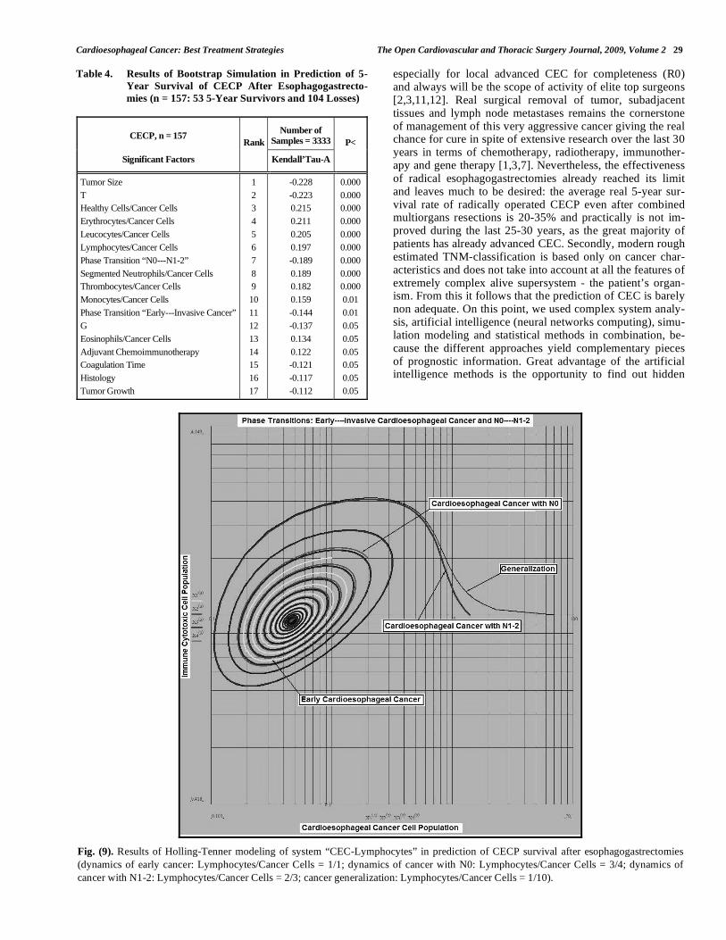
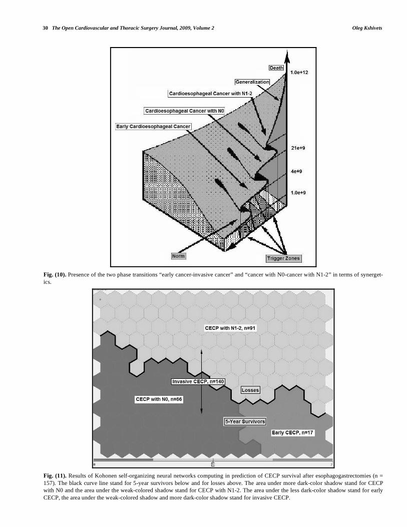
It is necessary to note a very important law: both transi-tions of the early cancer into the invasive cancer, as well as the cancer with N0 into the cancer with N1-2, have the phase character. These results testify by mathematical (Holling-Tenner) and imitating modeling of system “CEC-patient homeostasis” in terms of synergetics (Figs. 9, 10). This also proves the first results received earlier in the work [12]. Presence of the two phase transitions is evidently shown on Kohonen self-organizing neural networks maps (Fig. 11).
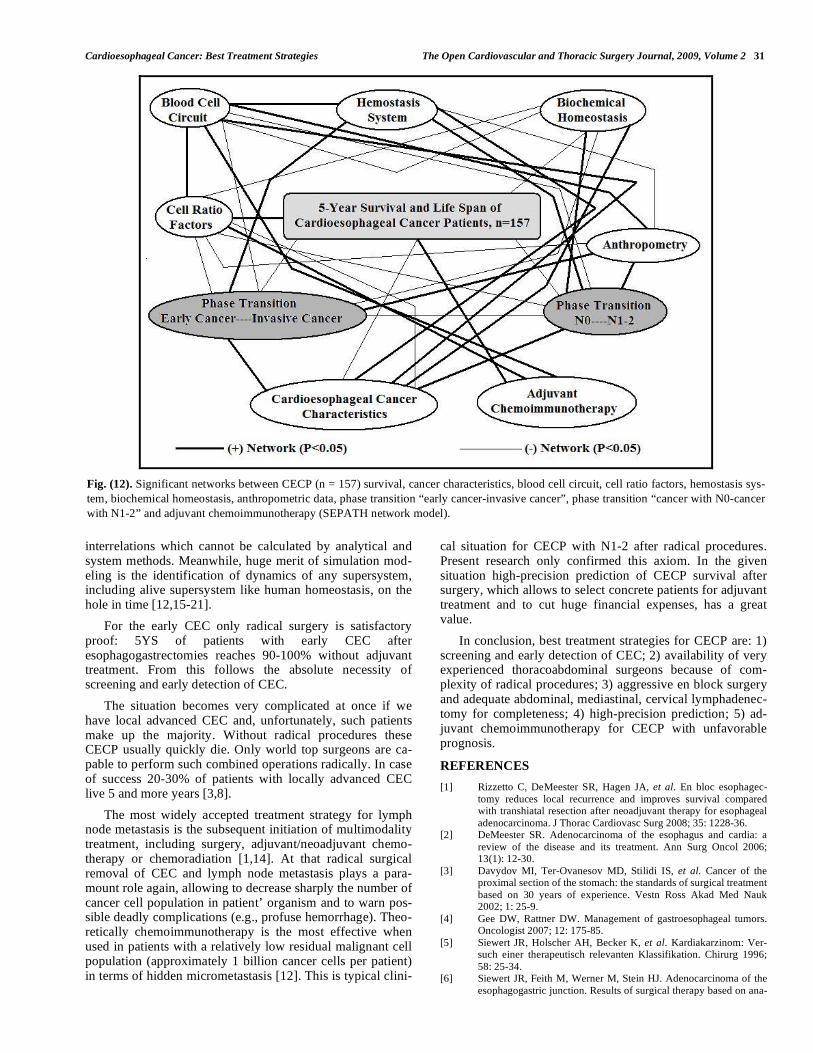
All of these differences and discrepancies were further investigated by structural equation modeling (SEPATH) as well as Monte Carlo simulation. From the data, summarized in Fig. (12) it could be recognized that the six clusters sig-nificantly predicted 5YS and LS of CECP after complete esophagogastrectomies: 1) phase transition “early-invasive CEC” (P = 0.000); 2) phase transition “CEC with N0-CEC with N1-2” (P = 0.001); 3) cell ratio factors (P = 0.000); 4) CEC characteristics (P = 0.000); 5) biochemical homeostasis (P = 0.003); 6) adjuvant chemoimmunotherapy (P = 0.000) (Fig. 12). It is necessary to pay attention, that both phase transitions strictly depend on blood cell circuit (P = 0.000), cell ratio factors (P = 0.000-0.001), biochemical homeostasis (P = 0.000) and hemostasis system (P = 0.000-0.017).
DISCUSSION
Adequate treatment of CEC is extremely difficult and not yet problem solving. Firstly, the cardioesophageal cancer surgery demands virtuosous, very accurate and aggressive surgical technique, sometime multiorgans resections
Cum ulat iv e Proport ion Surv iv ing (Kaplan-Meier)
5-Y ear Surv iv al of Cardioesophageal Cancer Patients af ter Ad. CH IT=69.9%;
5-Y ear Surv iv al of Cardioesophageal Cancer Patients af ter Surgery alone=26.6%;
P=0.000 by Log-Rank Tes t
C om plete C ensored
0 5 10 15 20 25
Y ears af ter Esophagogas trec tom y
0.0
0.1
0.2
0.3
0.4
0.5
0.6
0.7
0.8
0.9
1.0
u
Only Surgery , n=128
Adjuv ant C hemoim m unotherapy , n=47
Fig. (7). Survival of CECP after esophagogastrectomies in group A (adjuvant chemoimmunotherapy) (n = 47) and B (surgery alone) (n =
128). 5-year survival of CECP in group A (69.9%) was significantly better compared with group B (26.6%) (P = 0.000 by log-rank test).
Cu
mu
lative P
ropo
rtio
n S
urv
ivin
g

28 The Open Cardiovascular and Thoracic Surgery Journal, 2009, Volume 2 Oleg Kshivets
Table 2. Results of Neural Networks Computing in Predic-
tion of 5-Year Survival of CECP After
Esophagogastrectomies (n = 157: 53 5-Year Survi-
vors and 104 Losses)
Sample n = 157 NN Factors Rank
Error Ratio
1
2
3
4
5
6
7
8
9
10
11
12
13
14
15
16
17
Phase Transition “Early---Invasive Cancer”
Phase Transition “N0---N1-2”
Adjuvant Chemoimmunotherapy
T
Gender
Prothrombin Index
Weight
Glucose
Age
Coagulation Time
Eosinophils/Cancer Cells
Erythrocytes/Cancer Cells
Hemorrhage Time
Protein
Hemoglobin
Segmented Neutrophils/Cancer Cells
Stab Neutrophils/Cancer Cells
1
2
3
4
5
6
7
8
9
10
11
12
13
14
15
16
17
0.539
0.426
0.329
0.300
0.285
0.176
0.155
0.122
0.080
0.080
0.080
0.080
0.017
0.000
0.000
0.000
0.000
539e+7
426e+7
329e+7
300e+7
285E+7
176e+7
155e+7
122e+7
799e+6
798e+6
798e+6
798e+6
166e+6
155e+2
1852.5
474.43
1.323
Baseline Error
Area under ROC Curve
Correct Classification Rate (%)
0.0009e-12
1.000
100.00
Table 3. Results of Neural Networks Genetic Algorithm Se-
lection in Prediction of 5-Year Survival of CECP Af-
ter Esophagogastrectomies (n = 157: 53 5-Year Sur-
vivors and 104 Losses)
NN CECP, n = 157
Factors Useful for 5-Year
Survival
1
2
3
4
5
6
7
8
9
10
11
12
13
14
15
16
17
18
Phase Transition “N0---N1-2”
Phase Transition “Early---Invasive Cancer”
Adjuvant Chemoimmunotherapy
Erythrocyte/Cancer Cells
Leucocytes/Cancer Cells
Eosinophils/Cancer Cells
Segmented Neutrophils/Cancer Cells
Healthy Cells/Cancer Cells
Tumor Size
T
Hemoglobin
Coagulation Time
Hemorrhage Time
Prothrombin Index
Glucose
Rest Nitrogen
Bilirubin
Gender
Yes
Yes
Yes
Yes
Yes
Yes
Yes
Yes
Yes
Yes
Yes
Yes
Yes
Yes
Yes
Yes
Yes
Yes
Fig. (8). Configuration of neural networks: 4-layer perceptron.
Cardioesophageal Cancer: Best Treatment Strategies The Open Cardiovascular and Thoracic Surgery Journal, 2009, Volume 2 29
Table 4. Results of Bootstrap Simulation in Prediction of 5-
Year Survival of CECP After Esophagogastrecto-
mies (n = 157: 53 5-Year Survivors and 104 Losses)
CECP, n = 157 Number of
Samples = 3333
Significant Factors
Rank
Kendall’Tau-A
P<
Tumor Size
T
Healthy Cells/Cancer Cells
Erythrocytes/Cancer Cells
Leucocytes/Cancer Cells
Lymphocytes/Cancer Cells
Phase Transition “N0---N1-2”
Segmented Neutrophils/Cancer Cells
Thrombocytes/Cancer Cells
Monocytes/Cancer Cells
Phase Transition “Early---Invasive Cancer”
G
Eosinophils/Cancer Cells
Adjuvant Chemoimmunotherapy
Coagulation Time
Histology
Tumor Growth
1
2
3
4
5
6
7
8
9
10
11
12
13
14
15
16
17
-0.228
-0.223
0.215
0.211
0.205
0.197
-0.189
0.189
0.182
0.159
-0.144
-0.137
0.134
0.122
-0.121
-0.117
-0.112
0.000
0.000
0.000
0.000
0.000
0.000
0.000
0.000
0.000
0.01
0.01
0.05
0.05
0.05
0.05
0.05
0.05
especially for local advanced CEC for completeness (R0) and always will be the scope of activity of elite top surgeons [2,3,11,12]. Real surgical removal of tumor, subadjacent tissues and lymph node metastases remains the cornerstone of management of this very aggressive cancer giving the real chance for cure in spite of extensive research over the last 30 years in terms of chemotherapy, radiotherapy, immunother-apy and gene therapy [1,3,7]. Nevertheless, the effectiveness of radical esophagogastrectomies already reached its limit and leaves much to be desired: the average real 5-year sur-vival rate of radically operated CECP even after combined multiorgans resections is 20-35% and practically is not im-proved during the last 25-30 years, as the great majority of patients has already advanced CEC. Secondly, modern rough estimated TNM-classification is based only on cancer char-acteristics and does not take into account at all the features of extremely complex alive supersystem - the patient’s organ-ism. From this it follows that the prediction of CEC is barely non adequate. On this point, we used complex system analy-sis, artificial intelligence (neural networks computing), simu-lation modeling and statistical methods in combination, be-cause the different approaches yield complementary pieces of prognostic information. Great advantage of the artificial intelligence methods is the opportunity to find out hidden
Fig. (9). Results of Holling-Tenner modeling of system “CEC-Lymphocytes” in prediction of CECP survival after esophagogastrectomies
(dynamics of early cancer: Lymphocytes/Cancer Cells = 1/1; dynamics of cancer with N0: Lymphocytes/Cancer Cells = 3/4; dynamics of
cancer with N1-2: Lymphocytes/Cancer Cells = 2/3; cancer generalization: Lymphocytes/Cancer Cells = 1/10).
30 The Open Cardiovascular and Thoracic Surgery Journal, 2009, Volume 2 Oleg Kshivets
Fig. (10). Presence of the two phase transitions “early cancer-invasive cancer” and “cancer with N0-cancer with N1-2” in terms of synerget-
ics.
Fig. (11). Results of Kohonen self-organizing neural networks computing in prediction of CECP survival after esophagogastrectomies (n =
157). The black curve line stand for 5-year survivors below and for losses above. The area under more dark-color shadow stand for CECP
with N0 and the area under the weak-colored shadow stand for CECP with N1-2. The area under the less dark-color shadow stand for early
CECP, the area under the weak-colored shadow and more dark-color shadow stand for invasive CECP.
Cardioesophageal Cancer: Best Treatment Strategies The Open Cardiovascular and Thoracic Surgery Journal, 2009, Volume 2 31
interrelations which cannot be calculated by analytical and system methods. Meanwhile, huge merit of simulation mod-eling is the identification of dynamics of any supersystem, including alive supersystem like human homeostasis, on the hole in time [12,15-21].
For the early CEC only radical surgery is satisfactory proof: 5YS of patients with early CEC after esophagogastrectomies reaches 90-100% without adjuvant treatment. From this follows the absolute necessity of screening and early detection of CEC.
The situation becomes very complicated at once if we have local advanced CEC and, unfortunately, such patients make up the majority. Without radical procedures these CECP usually quickly die. Only world top surgeons are ca-pable to perform such combined operations radically. In case of success 20-30% of patients with locally advanced CEC live 5 and more years [3,8].
The most widely accepted treatment strategy for lymph node metastasis is the subsequent initiation of multimodality treatment, including surgery, adjuvant/neoadjuvant chemo-therapy or chemoradiation [1,14]. At that radical surgical removal of CEC and lymph node metastasis plays a para-mount role again, allowing to decrease sharply the number of cancer cell population in patient’ organism and to warn pos-sible deadly complications (e.g., profuse hemorrhage). Theo-retically chemoimmunotherapy is the most effective when used in patients with a relatively low residual malignant cell population (approximately 1 billion cancer cells per patient) in terms of hidden micrometastasis [12]. This is typical clini-
cal situation for CECP with N1-2 after radical procedures. Present research only confirmed this axiom. In the given situation high-precision prediction of CECP survival after surgery, which allows to select concrete patients for adjuvant treatment and to cut huge financial expenses, has a great value.
In conclusion, best treatment strategies for CECP are: 1) screening and early detection of CEC; 2) availability of very experienced thoracoabdominal surgeons because of com-plexity of radical procedures; 3) aggressive en block surgery and adequate abdominal, mediastinal, cervical lymphadenec-tomy for completeness; 4) high-precision prediction; 5) ad-juvant chemoimmunotherapy for CECP with unfavorable prognosis.
REFERENCES
[1] Rizzetto C, DeMeester SR, Hagen JA, et al. En bloc esophagec-
tomy reduces local recurrence and improves survival compared with transhiatal resection after neoadjuvant therapy for esophageal
adenocarcinoma. J Thorac Cardiovasc Surg 2008; 35: 1228-36. [2] DeMeester SR. Adenocarcinoma of the esophagus and cardia: a
review of the disease and its treatment. Ann Surg Oncol 2006; 13(1): 12-30.
[3] Davydov MI, Ter-Ovanesov MD, Stilidi IS, et al. Cancer of the proximal section of the stomach: the standards of surgical treatment
based on 30 years of experience. Vestn Ross Akad Med Nauk 2002; 1: 25-9.
[4] Gee DW, Rattner DW. Management of gastroesophageal tumors. Oncologist 2007; 12: 175-85.
[5] Siewert JR, Holscher AH, Becker K, et al. Kardiakarzinom: Ver-such einer therapeutisch relevanten Klassifikation. Chirurg 1996;
58: 25-34. [6] Siewert JR, Feith M, Werner M, Stein HJ. Adenocarcinoma of the
esophagogastric junction. Results of surgical therapy based on ana-
Fig. (12). Significant networks between CECP (n = 157) survival, cancer characteristics, blood cell circuit, cell ratio factors, hemostasis sys-
tem, biochemical homeostasis, anthropometric data, phase transition “early cancer-invasive cancer”, phase transition “cancer with N0-cancer
with N1-2” and adjuvant chemoimmunotherapy (SEPATH network model).
32 The Open Cardiovascular and Thoracic Surgery Journal, 2009, Volume 2 Oleg Kshivets
tomical /topographic classification in 1002 consecutive patients.
Ann Surg 2000; 232: 353-61. [7] Barbour AP, Rizk NP, Gonen M, et al. Adenocarcinoma of the
gastroesophageal junction: influence of esophageal resection mar-gin and operative approach on outcome. Ann Surg 2007; 246: 1-8.
[8] Pedrazzani C, de Manzoni G, Marrelli D, et al. Lymph node in-volvement in advanced gastroesophageal junction adenocarcinoma.
J Thorac Cardiovasc Surg 2007; 134: 378-85. [9] Shen KR, Cassiva St D, Deschamps C, et al. Surgical treatment of
tumours of the proximal stomach with involvement of the distal esophagus: A 26-years experience with Siewert type III tumours. J
Thorac Cardiovasc Surg 2006; 132: 755-62. [10] Nakamura T, Oguma H, Sasagawa T, et al. Left thoracoabdominal
approach for adenocarcinoma of the esophagogastric junction. Hepatogastroenterology 2008; 55: 1332-7.
[11] Yonemura Y, Kojima N, Kawamura T, et al. Treatment results of adenocarcinoma of the gastroesophageal junction. Hepatogastroen-
terology 2008; 55: 475-81. [12] Kshivets O. Expert system in diagnosis and prognosis of malignant
neoplasms. Dissertation for Sc.D. Tomsk 1995; p. 486.
[13] Morozow VG, Chavinson VC. Isolation, refinement and identifica-
tion of immunomodulated polypeptide from calf and human thy-mus. Biochemistry 1981; 9: 1652-9.
[14] Kshivets O. Esophageal cancer: Optimization of management. Open Cardiovasc Thorac Surg J 2008; 1: 1-11.
[15] Bazykin AD. Mathematical biophysics of cooperating populations. Science: Moscow 1985; p. 181.
[16] Haken H. Information and self-organization. A macroscopic ap-proach to complex systems. Springer: Berlin 2006; p. 240.
[17] Odom-Maryon T. Biostatistical methods in oncology. Cancer man-agement: A multidisciplinary approach. 1st ed. Huntington, NY:
PRP Inc 1996; 788-802. [18] Mirkin B.G. A sequential fitting procedure for linear data analysis
models. J Classification 1990; 7: 167-96. [19] Joreskog KG, Sorbom D. Recent development in structural equa-
tion modeling. J Mark Res 1982; 19: 404-16. [20] Bostwick DG, Burke HB. Prediction of individual patient outcome
in cancer: comparison of artificial neural networks and Kaplan-Meier methods. Cancer 2001; 91(8): 1643-6.
[21] Husmeier D. The Bayesian evidence scheme for regularizing prob-ability-density estimating neural networks. Neural Comput 2000;
12(11): 2685-717.
Received: February 12, 2009 Revised: February 18, 2009 Accepted: February 18, 2009
© Oleg Kshivets; Licensee Bentham Open.
This is an open access article licensed under the terms of the Creative Commons Attribution Non-Commercial License (http://creativecommons.org/licenses/by-
nc/3.0/) which permits unrestricted, non-commercial use, distribution and reproduction in any medium, provided the work is properly cited.