KS4 Physical Education - | St Christopher's CE High School. joints tendons and... · The different...
37
© Boardworks Ltd 2006 1 of 37 Joints, Tendons and Ligaments © Boardworks Ltd 2006 1 of 37 These icons indicate that teacher’s notes or useful web addresses are available in the Notes Page. This icon indicates that the slide contains activities created in Flash. These activities are not editable. For more detailed instructions, see the Getting Started presentation. KS4 Physical Education
Transcript of KS4 Physical Education - | St Christopher's CE High School. joints tendons and... · The different...
© Boardworks Ltd 20061 of 37
Joints, Tendons and Ligaments
© Boardworks Ltd 20061 of 37
These icons indicate that teacher’s notes or useful web addresses are available in the Notes Page.
This icon indicates that the slide contains activities created in Flash. These activities are not editable.
For more detailed instructions, see the Getting Started presentation.
KS4 Physical
Education
© Boardworks Ltd 20062 of 37
Le
arn
ing
ob
jecti
ves
© Boardworks Ltd 20062 of 37
Learning objectives
What joints are
Classifying joints as fixed, slightly moveable and
freely moveable
The 3 types of connective tissue and their functions
The different types of synovial joint and how they
are used in various sporting movements
The structure of different joints
Analysing joint functions in different movements
How joints and flexibility are effected by physical
activity and age.
What we will learn in this presentation:
© Boardworks Ltd 20063 of 37
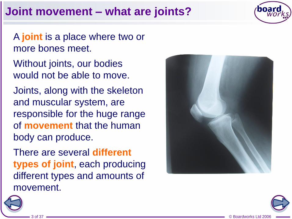
Joint movement – what are joints?
A joint is a place where two or
more bones meet.
Without joints, our bodies
would not be able to move.
Joints, along with the skeleton
and muscular system, are
responsible for the huge range
of movement that the human
body can produce.
There are several different
types of joint, each producing
different types and amounts of
movement.
© Boardworks Ltd 20064 of 37
Different types of joint
There are 3 different types of joint:
1. Immovable (or fixed) joints
3. Movable (or synovial) joints
2. Slightly movable joints
© Boardworks Ltd 20065 of 37
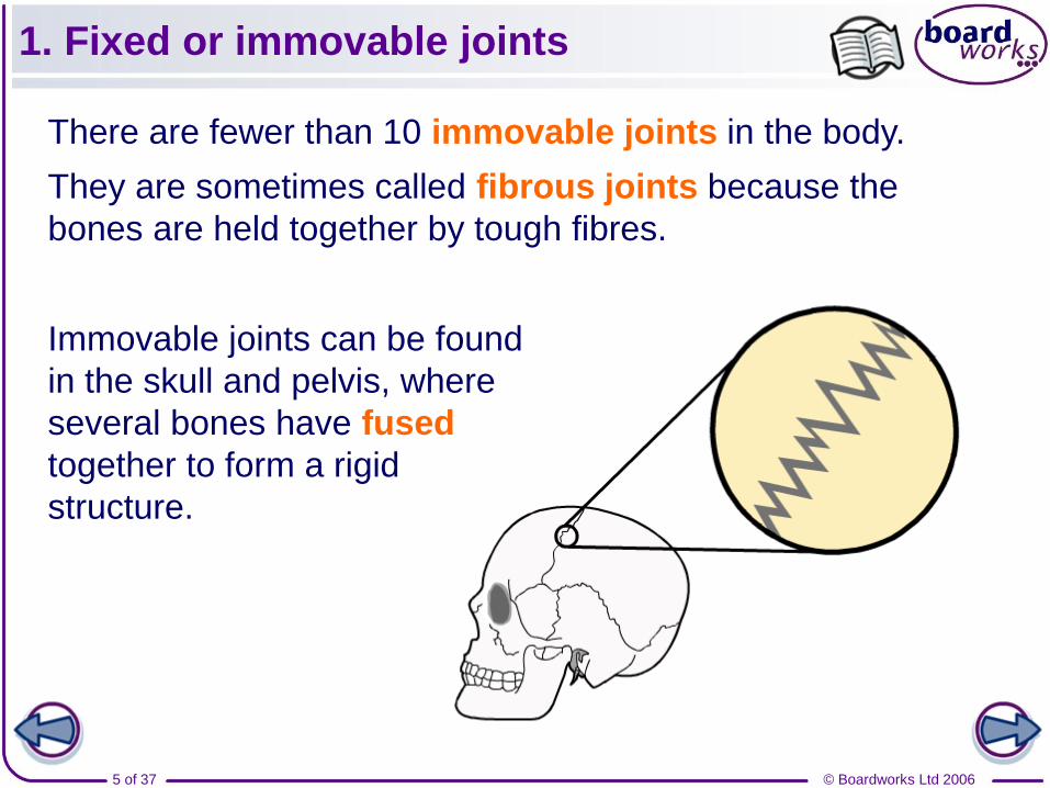
1. Fixed or immovable joints
There are fewer than 10 immovable joints in the body.
They are sometimes called fibrous joints because the
bones are held together by tough fibres.
Immovable joints can be found
in the skull and pelvis, where
several bones have fused
together to form a rigid
structure.
© Boardworks Ltd 20066 of 37
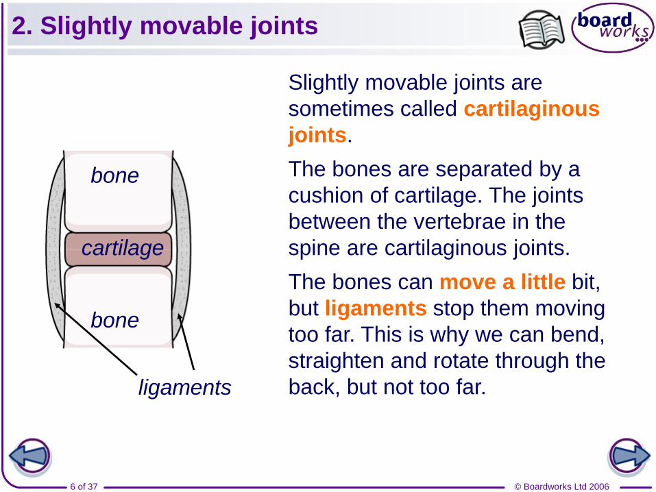
2. Slightly movable joints
Slightly movable joints are
sometimes called cartilaginous
joints.
The bones are separated by a
cushion of cartilage. The joints
between the vertebrae in the
spine are cartilaginous joints.
The bones can move a little bit,
but ligaments stop them moving
too far. This is why we can bend,
straighten and rotate through the
back, but not too far.
bone
ligaments
cartilage
bone
© Boardworks Ltd 20067 of 37
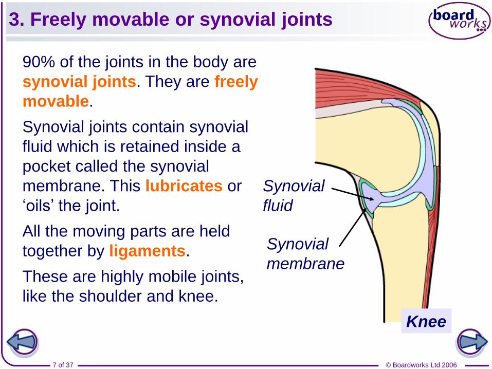
3. Freely movable or synovial joints
90% of the joints in the body are
synovial joints. They are freely
movable.
Synovial joints contain synovial
fluid which is retained inside a
pocket called the synovial
membrane. This lubricates or
‘oils’ the joint.
All the moving parts are held
together by ligaments.
These are highly mobile joints,
like the shoulder and knee.
Synovial
fluid
Knee
Synovial
membrane

© Boardworks Ltd 20068 of 37
Different types of joint
© Boardworks Ltd 20069 of 37
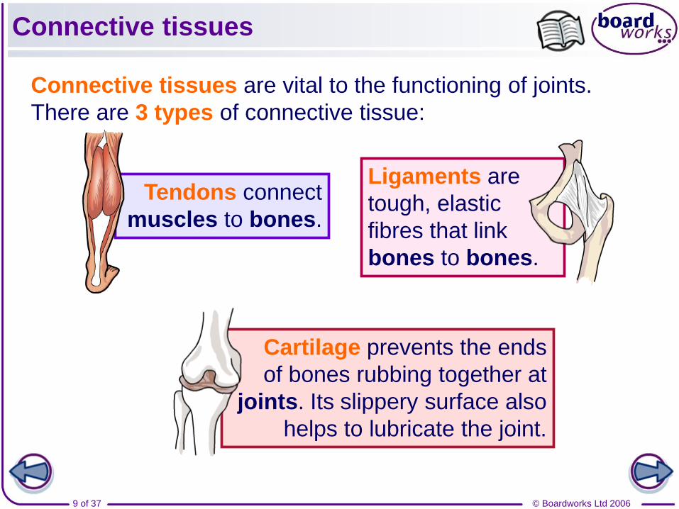
Connective tissues
Connective tissues are vital to the functioning of joints.
There are 3 types of connective tissue:
Ligaments are
tough, elastic
fibres that link
bones to bones.
Tendons connect
muscles to bones.
Cartilage prevents the ends
of bones rubbing together at
joints. Its slippery surface also
helps to lubricate the joint.
© Boardworks Ltd 200610 of 37
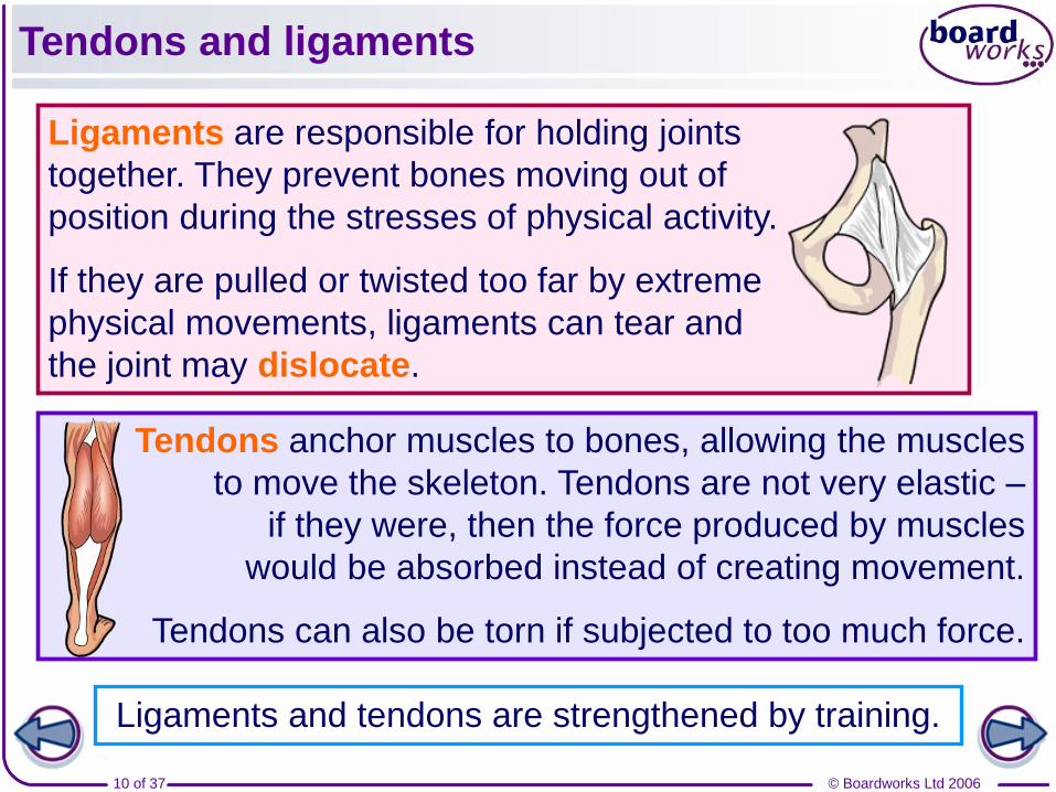
Tendons and ligaments
Ligaments and tendons are strengthened by training.
Ligaments are responsible for holding joints
together. They prevent bones moving out of
position during the stresses of physical activity.
If they are pulled or twisted too far by extreme
physical movements, ligaments can tear and
the joint may dislocate.
Tendons anchor muscles to bones, allowing the muscles
to move the skeleton. Tendons are not very elastic –
if they were, then the force produced by muscles
would be absorbed instead of creating movement.
Tendons can also be torn if subjected to too much force.
© Boardworks Ltd 200611 of 37
Tendons and ligaments
© Boardworks Ltd 200612 of 37
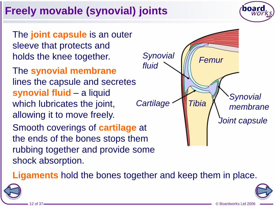
Freely movable (synovial) joints
The joint capsule is an outer
sleeve that protects and
holds the knee together.
The synovial membrane
lines the capsule and secretes
synovial fluid – a liquid
which lubricates the joint,
allowing it to move freely.
Femur
Tibia
Joint capsule
Synovial
membrane
Synovial
fluid
Ligaments hold the bones together and keep them in place.
Cartilage
Smooth coverings of cartilage at
the ends of the bones stops them
rubbing together and provide some
shock absorption.
© Boardworks Ltd 200613 of 37
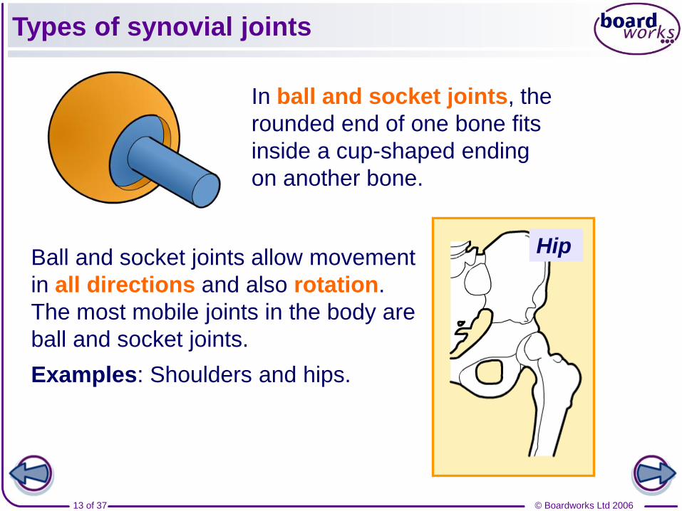
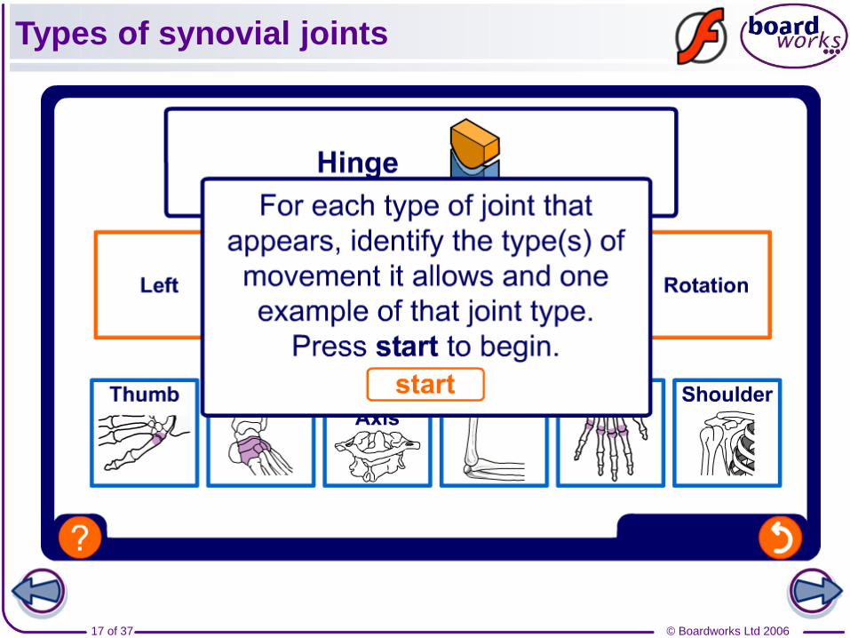
Types of synovial joints
In ball and socket joints, the
rounded end of one bone fits
inside a cup-shaped ending
on another bone.
Ball and socket joints allow movement
in all directions and also rotation.
The most mobile joints in the body are
ball and socket joints.
Examples: Shoulders and hips.
Hip
© Boardworks Ltd 200614 of 37
Types of synovial joints
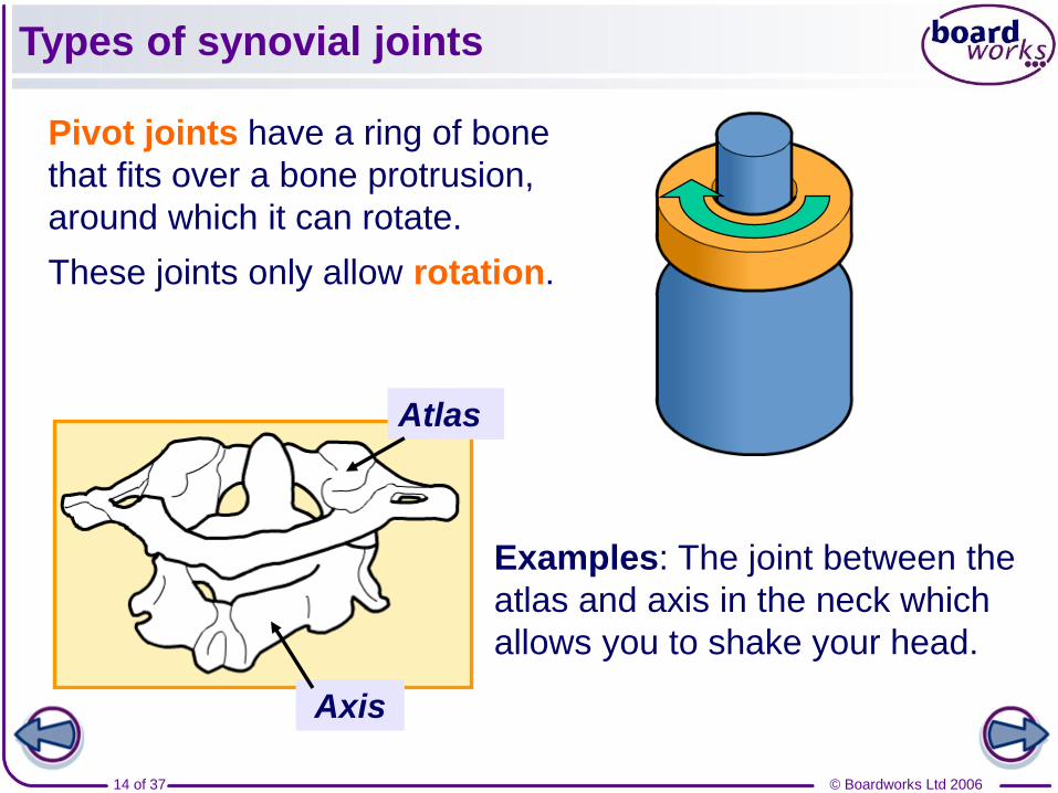
Pivot joints have a ring of bone
that fits over a bone protrusion,
around which it can rotate.
These joints only allow rotation.
Examples: The joint between the
atlas and axis in the neck which
allows you to shake your head.
Axis
Atlas

© Boardworks Ltd 200615 of 37
Types of synovial joints
In saddle joints, the ends of the two
bones fit together in a special way,
allowing movement forwards and
backwards and left to right, but not
rotation.
Examples: The thumb is the only one.
Hinge joints – as their name
suggests – only allow forwards
and backwards movement.
Examples: The knee and elbow. Elbow
© Boardworks Ltd 200616 of 37
Types of synovial joints
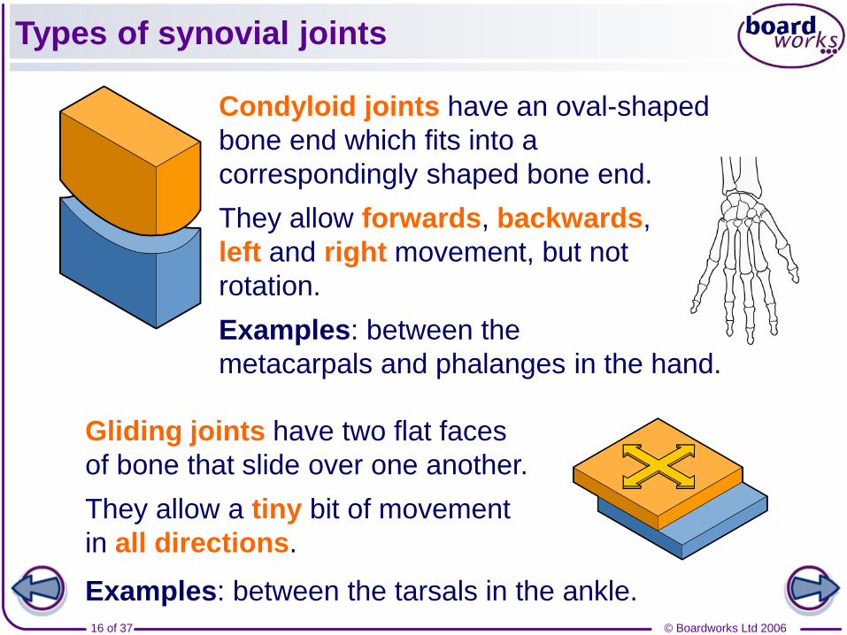
Condyloid joints have an oval-shaped
bone end which fits into a
correspondingly shaped bone end.
They allow forwards, backwards,
left and right movement, but not
rotation.
Examples: between the
metacarpals and phalanges in the hand.
Gliding joints have two flat faces
of bone that slide over one another.
They allow a tiny bit of movement
in all directions.
Examples: between the tarsals in the ankle.
© Boardworks Ltd 200617 of 37
Types of synovial joints
© Boardworks Ltd 200618 of 37
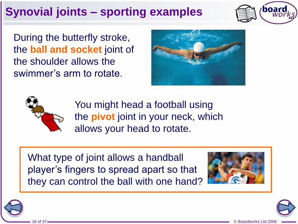
Synovial joints – sporting examples
During the butterfly stroke,
the ball and socket joint of
the shoulder allows the
swimmer’s arm to rotate.
You might head a football using
the pivot joint in your neck, which
allows your head to rotate.
What type of joint allows a handball
player’s fingers to spread apart so that
they can control the ball with one hand?
© Boardworks Ltd 200619 of 37
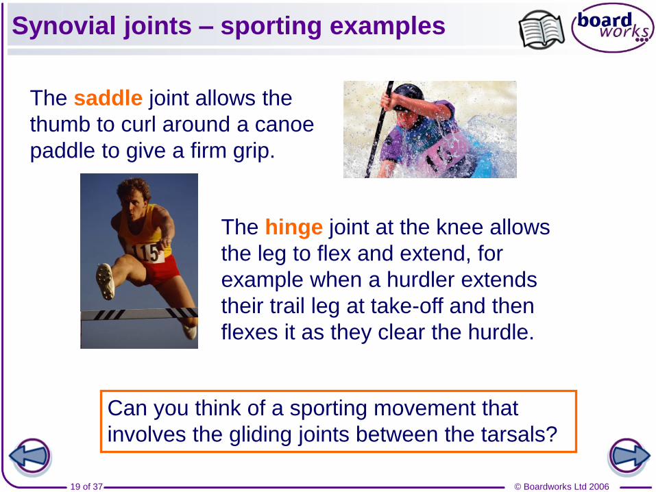
Synovial joints – sporting examples
The saddle joint allows the
thumb to curl around a canoe
paddle to give a firm grip.
Can you think of a sporting movement that
involves the gliding joints between the tarsals?
The hinge joint at the knee allows
the leg to flex and extend, for
example when a hurdler extends
their trail leg at take-off and then
flexes it as they clear the hurdle.
© Boardworks Ltd 200620 of 37
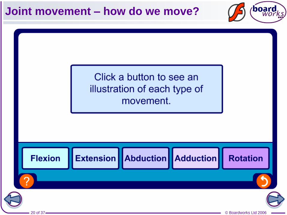
Joint movement – how do we move?
© Boardworks Ltd 200621 of 37
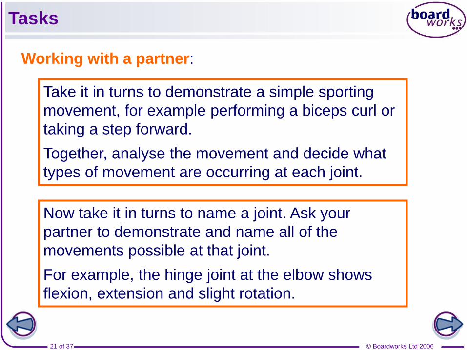
Tasks
Working with a partner:
Take it in turns to demonstrate a simple sporting
movement, for example performing a biceps curl or
taking a step forward.
Together, analyse the movement and decide what
types of movement are occurring at each joint.
Now take it in turns to name a joint. Ask your
partner to demonstrate and name all of the
movements possible at that joint.
For example, the hinge joint at the elbow shows
flexion, extension and slight rotation.
© Boardworks Ltd 200622 of 37
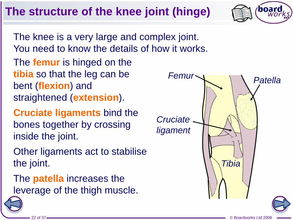
The structure of the knee joint (hinge)
The knee is a very large and complex joint.
You need to know the details of how it works.
The femur is hinged on the
tibia so that the leg can be
bent (flexion) and
straightened (extension).
Cruciate ligaments bind the
bones together by crossing
inside the joint.
Other ligaments act to stabilise
the joint.
The patella increases the
leverage of the thigh muscle.
Femur
Tibia
Patella
Cruciate
ligament
© Boardworks Ltd 200623 of 37
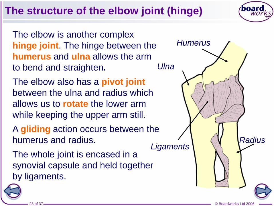
The structure of the elbow joint (hinge)
The elbow is another complex
hinge joint. The hinge between the
humerus and ulna allows the arm
to bend and straighten.
The elbow also has a pivot joint
between the ulna and radius which
allows us to rotate the lower arm
while keeping the upper arm still.
A gliding action occurs between the
humerus and radius.
The whole joint is encased in a
synovial capsule and held together
by ligaments.
Ulna
Humerus
RadiusLigaments
© Boardworks Ltd 200624 of 37
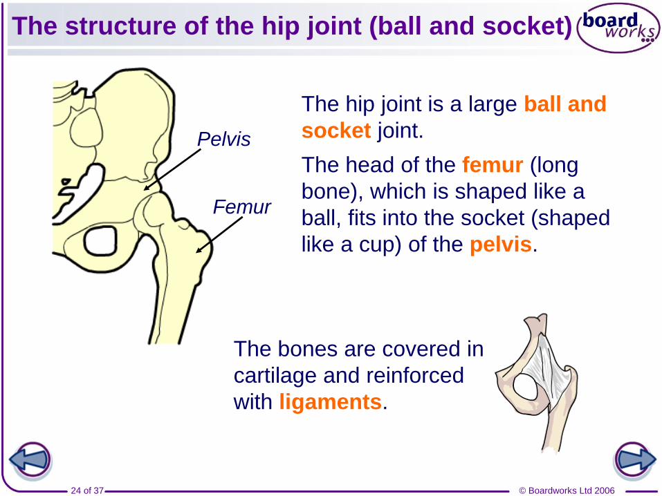
The structure of the hip joint (ball and socket)
The hip joint is a large ball and
socket joint.
The head of the femur (long
bone), which is shaped like a
ball, fits into the socket (shaped
like a cup) of the pelvis.
Femur
Pelvis
The bones are covered in
cartilage and reinforced
with ligaments.
© Boardworks Ltd 200625 of 37
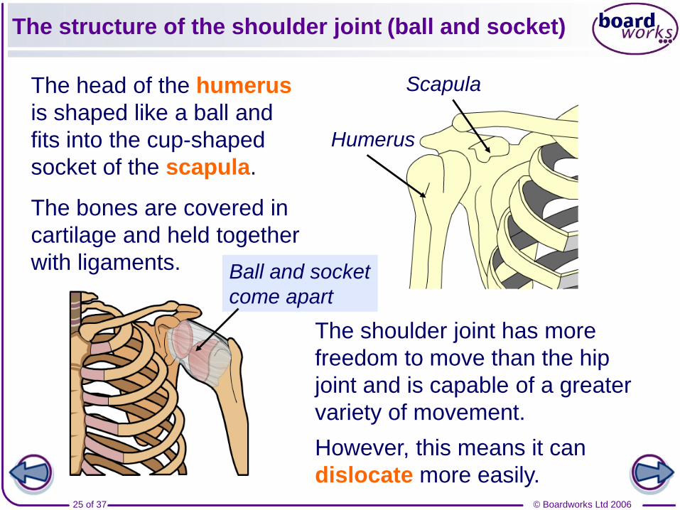
The structure of the shoulder joint (ball and socket)
The head of the humerus
is shaped like a ball and
fits into the cup-shaped
socket of the scapula.
The bones are covered in
cartilage and held together
with ligaments.
Humerus
The shoulder joint has more
freedom to move than the hip
joint and is capable of a greater
variety of movement.
However, this means it can
dislocate more easily.
Scapula
Ball and socket
come apart
© Boardworks Ltd 200626 of 37
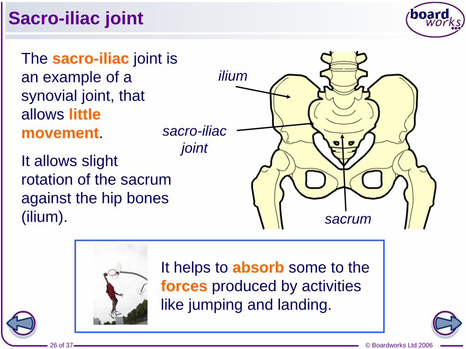
Sacro-iliac joint
The sacro-iliac joint is
an example of a
synovial joint, that
allows little
movement.
It allows slight
rotation of the sacrum
against the hip bones
(ilium).
It helps to absorb some to the
forces produced by activities
like jumping and landing.
ilium
sacrum
sacro-iliac
joint
© Boardworks Ltd 200627 of 37
Name the bones in these joints
© Boardworks Ltd 200628 of 37
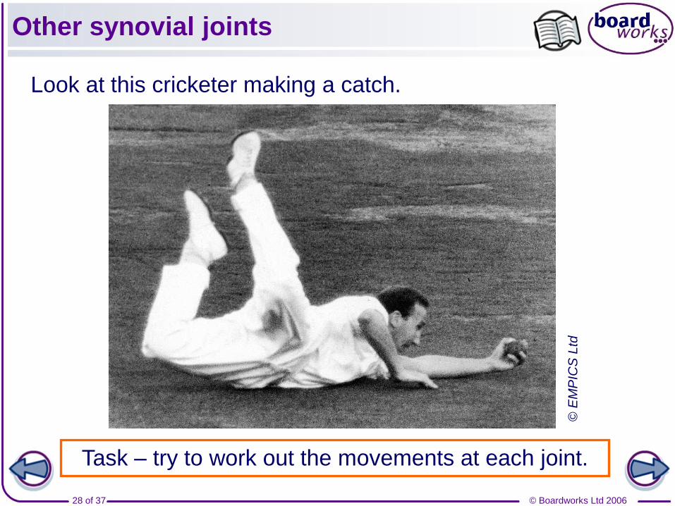
Other synovial joints
Look at this cricketer making a catch.
Task – try to work out the movements at each joint.
© E
MP
ICS
Ltd
© Boardworks Ltd 200629 of 37
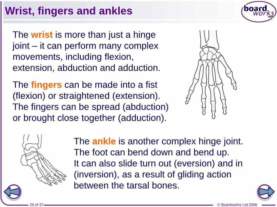
Wrist, fingers and ankles
The wrist is more than just a hinge
joint – it can perform many complex
movements, including flexion,
extension, abduction and adduction.
The fingers can be made into a fist
(flexion) or straightened (extension).
The fingers can be spread (abduction)
or brought close together (adduction).
The ankle is another complex hinge joint.
The foot can bend down and bend up.
It can also slide turn out (eversion) and in
(inversion), as a result of gliding action
between the tarsal bones.
© Boardworks Ltd 200630 of 37
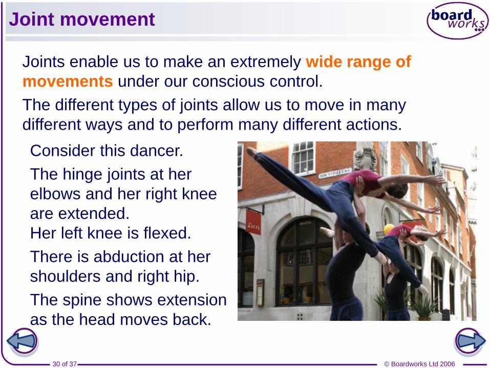
Joint movement
Joints enable us to make an extremely wide range of
movements under our conscious control.
The different types of joints allow us to move in many
different ways and to perform many different actions.
Consider this dancer.
The hinge joints at her
elbows and her right knee
are extended.
Her left knee is flexed.
There is abduction at her
shoulders and right hip.
The spine shows extension
as the head moves back.
© Boardworks Ltd 200631 of 37
Sporting movement
© Boardworks Ltd 200632 of 37
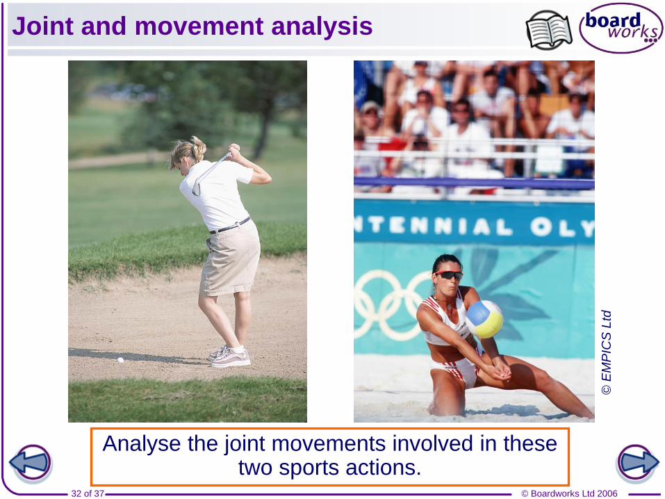
Joint and movement analysis
Analyse the joint movements involved in these two sports actions.
© E
MP
ICS
Ltd
© Boardworks Ltd 200633 of 37
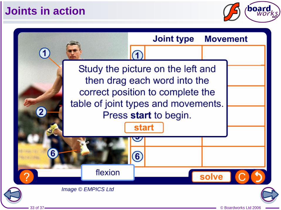
Joints in action
Image © EMPICS Ltd

© Boardworks Ltd 200634 of 37
Joints and sport
Flexibility exercises increase
the range of movement at joints.
This can reduce the risk of
injury and damage as the joints
are more able to absorb forces.
However, overstretching joints
can cause injury to them.
Joint flexibility is important in sport, especially in activities
like gymnastics and diving that require extreme movements.
Participants in all sports however, can benefit from the
greater range of movement that comes with improved
flexibility.
© Boardworks Ltd 200635 of 37
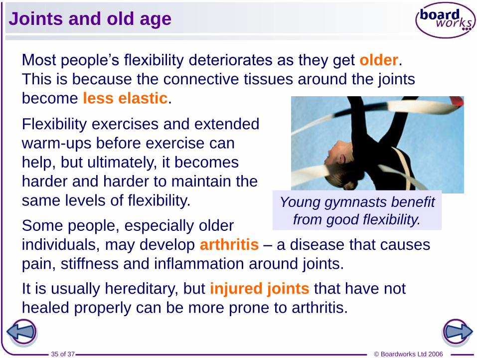
Joints and old age
Some people, especially older
individuals, may develop arthritis – a disease that causes
pain, stiffness and inflammation around joints.
It is usually hereditary, but injured joints that have not
healed properly can be more prone to arthritis.
Most people’s flexibility deteriorates as they get older.
This is because the connective tissues around the joints
become less elastic.
Flexibility exercises and extended
warm-ups before exercise can
help, but ultimately, it becomes
harder and harder to maintain the
same levels of flexibility. Young gymnasts benefit
from good flexibility.
© Boardworks Ltd 200636 of 37
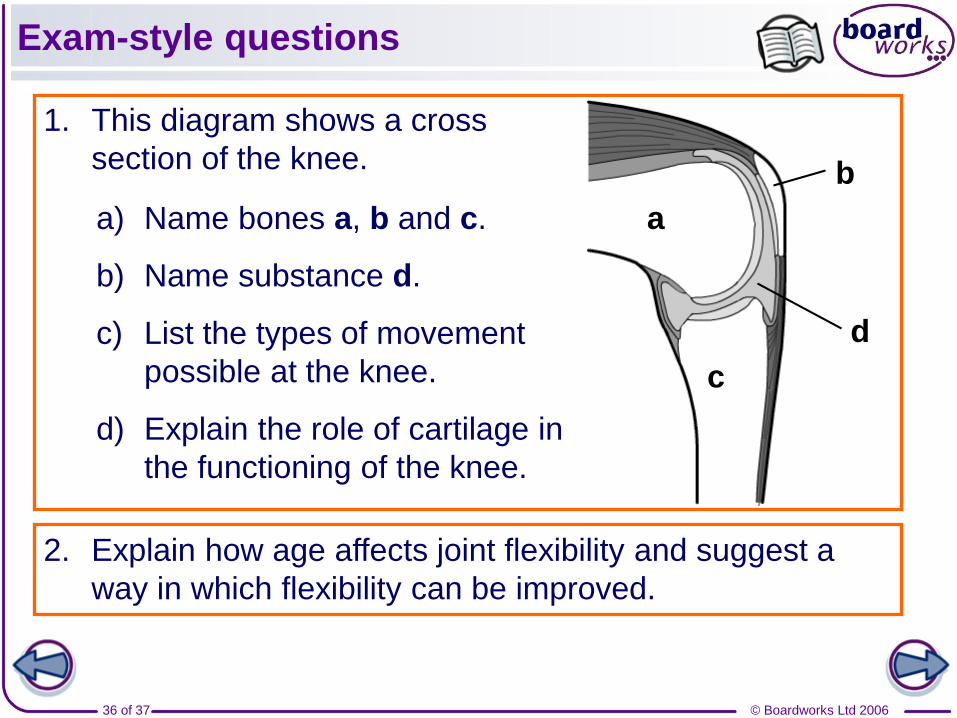
Exam-style questions
1. This diagram shows a cross
section of the knee.
a
b
c
d
a) Name bones a, b and c.
b) Name substance d.
c) List the types of movement
possible at the knee.
d) Explain the role of cartilage in
the functioning of the knee.
2. Explain how age affects joint flexibility and suggest a
way in which flexibility can be improved.
© Boardworks Ltd 200637 of 37
Can you remember all these keywords?
Joint – a place where two or more bones meet.
Flexibility – the range of movement possible at a joint.
Ligaments – strong, elastic fibres that join bones together.
Tendons – non-elastic fibres that attach muscles to bones.
Cartilage – connective tissue found at the ends of bones to
protect them and enable smooth movement.
Flexion – the action causing a limb to bend.
Extension – the action of a joint / limb straightening.
Abduction – the action of a limb moving outwards, away from
the body.
Adduction – the action of a limb moving in, towards the body.
Rotation – the action of a limb turning around.