Krafts. 2010. Tissue Repair. the Hidden Drama Copy
9
www.landesbioscience.com Organogenesis 225 Organogenesis 6:4, 225-233; October/November/December 2010; © 2010 Landes Bioscience REVIEW REVIEW Proem All the world’s a stage, And all the men and women merely players; They have their exits and their entrances And one man in his time plays many parts, His acts being seven ages Shakespeare As You Like It Act 2, scene 7, 139–143 The term “repair,” when used in the context of the healing of damaged tissue, is defined as the restoration of tissue architec- ture and function after an injury. It encompasses two separate Correspondence to: Kristine P. Krafts; Email: [email protected] Submitted: 05/01/10; Accepted: 06/01/10 Previously published online: www.landesbioscience.com/journals/organogenesis/article/12555 DOI: 10.4161/org6.4.12555 As living beings that encounter every kind of traumatic event from paper cut to myocardial infarction, we must possess ways to heal damaged tissues. While some animals are able to regrow complete body parts following injury (such as the earthworm who grows a new head following bisection), humans are sadly incapable of such feats. Our means of recovery following tissue damage consists largely of repair rather than pure regeneration. Thousands of times in our lives, a meticulously scripted but unseen wound healing drama plays, with cells serving as actors, extracellular matrix as the setting and growth factors as the means of communication. This article briefly reviews the cells involved in tissue repair, their signaling and proliferation mechanisms and the function of the extracellular matrix, then presents the actors and script for the three acts of the tissue repair drama. Tissue repair The hidden drama Kristine P. Krafts Department of Pathology; University of Minnesota School of Medicine; Duluth Campus; Duluth, MN USA Key words: tissue repair, regeneration, scarring, wound healing, growth factors, extracellular matrix, stem cells Abbreviations: cAMP, 3', 5' cyclic adenosine monophosphate; CDK, cyclin-dependent protein; DNA, deoxyribonucleic acid; ECM, extracellular matrix; EGF, epidermal growth factor; FGF, fibroblast growth factor; GAG, glycosaminoglycan; KGF, keratinocyte growth factor; MAD, mothers against decapentaplegic; MMP, matrix metalloproteinase; IL, interleukin; JAK, janus kinase; OPN, osteopontin; PDGF, platelet-derived growth factor; RB, retinoblastoma; STAT, signal transducer and activator of transcription; TGF, transforming growth factor; TIMP, tissue inhibitor of metalloproteinases; TNF, tumor necrosis factor; VEGF, vascular endothelial growth factor processes: regeneration and replacement. Regeneration refers to a type of healing in which new growth completely restores portions of damaged tissue to their normal state. Replacement refers to a type of healing in which severely damaged or non-regenerable tissues are repaired by the laying down of connective tissue, a process commonly referred to as scarring. While a few types of tissue injury (such as minor paper cuts) can sometimes be healed in such a way that no permanent damage remains, most of our tissue repair consists of both regeneration and replacement. Tissue repair may restore some of the original structures of the damaged tissue (such as epithelial layers), but may also result in structural abnormalities that impair organ function (such as the scar formed in the healing of a myocardial infarction). Whether the healing of a wound proceeds down the regenera- tion or the replacement pathway (or both) depends, in part, on the type of tissue in which it occurs. Certain tissues of the body are more capable of cellular proliferation (and hence regenera- tion) than others. In this regard, there are three types of tissues: continuously dividing tissues, quiescent tissues and nondividing tissues. Continuously dividing tissues (also known as labile tis- sues) are comprised of cells that are constantly proliferating in order to replace dead or sloughed-off cells. Examples of such tis- sues include epithelia (such as skin, gastrointestinal epithelium and salivary gland tissue) and hematopoietic tissues. These tis- sues contain pools of stem cells, which have enormous prolifera- tive and self-renewing ability, and which give rise to more than one type of cell. Replicating asymmetrically, each stem cell gives rise to one daughter cell that differentiates and matures and another daughter cell that remains undifferentiated and capable of beginning another self-renewing cycle. Some tissues, known as quiescent tissues (or stable tissues) are composed of cells that normally exist in a non-dividing state but may enter the cell cycle in response to certain stimuli, such as cell injury. Tissues falling into this category include parenchymal cells of the liver, kidney and pancreas, mesenchymal cells such as fibroblasts and smooth muscle cells, endothelial cells and lym- phocytes. It should be noted that the liver, unlike other quiescent tissues, has a relatively robust proliferative capacity. When a lobe
-
Upload
quiltro-fabi-toro -
Category
Documents
-
view
17 -
download
1
Transcript of Krafts. 2010. Tissue Repair. the Hidden Drama Copy

www.landesbioscience.com Organogenesis 225
Organogenesis 6:4, 225-233; October/November/December 2010; © 2010 Landes BioscienceREVIEW REVIEW
Proem
All the world’s a stage,And all the men and women merely players;They have their exits and their entrancesAnd one man in his time plays many parts,His acts being seven ages
ShakespeareAs You Like ItAct 2, scene 7, 139–143
The term “repair,” when used in the context of the healing of damaged tissue, is defined as the restoration of tissue architec-ture and function after an injury. It encompasses two separate
Correspondence to: Kristine P. Krafts; Email: [email protected]: 05/01/10; Accepted: 06/01/10Previously published online:www.landesbioscience.com/journals/organogenesis/article/12555DOI: 10.4161/org6.4.12555
As living beings that encounter every kind of traumatic event from paper cut to myocardial infarction, we must possess ways to heal damaged tissues. While some animals are able to regrow complete body parts following injury (such as the earthworm who grows a new head following bisection), humans are sadly incapable of such feats. Our means of recovery following tissue damage consists largely of repair rather than pure regeneration. Thousands of times in our lives, a meticulously scripted but unseen wound healing drama plays, with cells serving as actors, extracellular matrix as the setting and growth factors as the means of communication. This article briefly reviews the cells involved in tissue repair, their signaling and proliferation mechanisms and the function of the extracellular matrix, then presents the actors and script for the three acts of the tissue repair drama.
Tissue repairThe hidden drama
Kristine P. Krafts
Department of Pathology; University of Minnesota School of Medicine; Duluth Campus; Duluth, MN USA
Key words: tissue repair, regeneration, scarring, wound healing, growth factors, extracellular matrix, stem cells
Abbreviations: cAMP, 3', 5' cyclic adenosine monophosphate; CDK, cyclin-dependent protein; DNA, deoxyribonucleic acid; ECM, extracellular matrix; EGF, epidermal growth factor; FGF, fibroblast growth factor; GAG, glycosaminoglycan; KGF,
keratinocyte growth factor; MAD, mothers against decapentaplegic; MMP, matrix metalloproteinase; IL, interleukin; JAK, janus kinase; OPN, osteopontin; PDGF, platelet-derived growth factor; RB, retinoblastoma; STAT, signal transducer and activator of
transcription; TGF, transforming growth factor; TIMP, tissue inhibitor of metalloproteinases; TNF, tumor necrosis factor; VEGF, vascular endothelial growth factor
processes: regeneration and replacement. Regeneration refers to a type of healing in which new growth completely restores portions of damaged tissue to their normal state. Replacement refers to a type of healing in which severely damaged or non-regenerable tissues are repaired by the laying down of connective tissue, a process commonly referred to as scarring. While a few types of tissue injury (such as minor paper cuts) can sometimes be healed in such a way that no permanent damage remains, most of our tissue repair consists of both regeneration and replacement. Tissue repair may restore some of the original structures of the damaged tissue (such as epithelial layers), but may also result in structural abnormalities that impair organ function (such as the scar formed in the healing of a myocardial infarction).
Whether the healing of a wound proceeds down the regenera-tion or the replacement pathway (or both) depends, in part, on the type of tissue in which it occurs. Certain tissues of the body are more capable of cellular proliferation (and hence regenera-tion) than others. In this regard, there are three types of tissues: continuously dividing tissues, quiescent tissues and nondividing tissues. Continuously dividing tissues (also known as labile tis-sues) are comprised of cells that are constantly proliferating in order to replace dead or sloughed-off cells. Examples of such tis-sues include epithelia (such as skin, gastrointestinal epithelium and salivary gland tissue) and hematopoietic tissues. These tis-sues contain pools of stem cells, which have enormous prolifera-tive and self-renewing ability, and which give rise to more than one type of cell. Replicating asymmetrically, each stem cell gives rise to one daughter cell that differentiates and matures and another daughter cell that remains undifferentiated and capable of beginning another self-renewing cycle.
Some tissues, known as quiescent tissues (or stable tissues) are composed of cells that normally exist in a non-dividing state but may enter the cell cycle in response to certain stimuli, such as cell injury. Tissues falling into this category include parenchymal cells of the liver, kidney and pancreas, mesenchymal cells such as fibroblasts and smooth muscle cells, endothelial cells and lym-phocytes. It should be noted that the liver, unlike other quiescent tissues, has a relatively robust proliferative capacity. When a lobe

226 Organogenesis Volume 6 Issue 4
regenerated every 3 to 5 days. The bone marrow contains not only hematopoietic stem cells, which give rise to all blood cell lineages, but also multipotent stromal stem cells, which travel to different regions in the body and generate chondrocytes, osteo-blasts, adipocytes, myoblasts and endothelial cells. These stromal stem cells participate in cell replenishment after tissue injury, but they do not seem to have a function in normal tissue homeostasis.
Beyond the stem cell, three other types of cells are critical to the process of tissue repair: fibroblasts, endothelial cells and mac-rophages. In most wounds, complete replacement of wounded tissue to its original, unharmed state is impossible. The wound must therefore be healed using externally obtained material to reconnect the viable tissue margins. This process, discussed in detail later, involves the laying down of acellular fibrous tissue to replace the region of lost cells. The fibrous tissue is laid down by fibroblasts, which migrate to the injured area, proliferate and secrete collagen under the influence of numerous growth factors and cytokines. In the earliest stages of wound healing, fibroblasts are few and far between, suspended together with tenuous new blood vessels in an edematous pink substance termed granulation tissue. Initially the new blood vessels are critical in the trans-port of nutrients and cells to the new tissue, but after a time, they recede along with the fibroblasts, leaving a collagenous scar that is remodeled and strengthened over time. Macrophages are essential directors of this drama, secreting growth factors that entice and stimulate fibroblasts, endothelial precursor cells and (in skin wounds) keratinocytes. They also oversee the deposition and remodeling of extracellular matrix material.
The Script
The script of the tissue factor drama—the migration and prolif-eration of cells, the laying down of extracellular matrix, and the remodeling of collagen to form a durable scar—is carried out by a process known as receptor-mediated signal transduction. Akin to the words spoken between people, ligands such as growth fac-tors and cytokines float between cells, carrying directives to per-form a certain action. Cells “hear” these words when the ligands bind to cell-surface receptors, which bring the message into the cell, resulting in a new action, such as migration, proliferation or secretion of a substance. Herein we will discuss the words spoken between the actors, the way the actors hear these words and the manner in which the message gets to the cell nucleus in order to effect change.
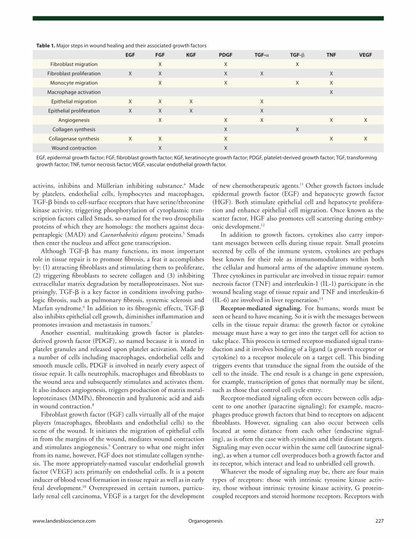
Growth factors and cytokines. Growth factors are specialized polypeptide molecules that bind to receptors on target cells and deliver messages regarding migration, proliferation, differentia-tion, survival and secretion. The list of growth factors and their attendant functions is so long that it would tax even the most capable memorizer. Herein we limit our discussion to the pri-mary growth factors associated with each stage of tissue repair (Table 1).
One of the most critical growth factors in tissue repair is trans-forming growth factor beta (TGF-β). This growth factor belongs to a superfamily that also includes a number of other factors with wide-ranging functions, such as bone morphogenetic proteins,
of the liver is resected for donation, for example, the remaining liver cells proliferate at such a rate that the liver reaches a size similar to that prior to resection. While this process is commonly described as regeneration, it is more accurately viewed as com-pensatory growth, since the original lobe itself does not regrow. A few types of tissue are composed of cells that have left the cell cycle permanently, and are therefore unable to proliferate. These nondividing tissues (or permanent tissues) include cardiac and skeletal muscle. Tissue repair in these tissues always leaves per-manent evidence of injury, such as a scar.1
Dramatis Personae
The cast of characters in the tissue repair drama is large and var-ied. Here we list the major actors, with a focus on those that may be less known to the reader. Central in the drama are the tissue’s own lost or damaged cells, which in most cases are terminally dif-ferentiated and incapable of replication. In nondividing tissues, such as myocardial tissue, lost cells are simply never replaced. In other tissues, however, replacement is possible. Continuously dividing tissues are particularly adept at self-renewal, undergoing innumerable cycles of cell loss and replacement during a normal human lifespan. The regenerative capacity of these tissues lies not in their parenchymal cells (which are terminally differenti-ated and thus unable to replicate), but in stem cells located deep within the tissue.
Stem cells are unique for two reasons: (1) they have the abil-ity to self-renew and (2) they have the capacity to generate more than one cell type. Self-renewal occurs either by symmetric rep-lication (in which a stem cell gives rise to two daughter stem cells, equally capable of self-renewal) or asymmetric replication (in which one daughter cell remains a self-renewing stem cell and the other daughter cell differentiates and matures).
The capacity of a stem cell to give rise to multiple lineages of cells is most striking in embryonic stem cells. These cells, which are denoted as pluripotent, are capable of generating cells from any of the tissues of the body. Adult (or somatic) stem cells are designated as multipotent, and give rise to a more restricted array of cell types. As expected, somatic stem cells have been found in continuously dividing tissues, such as skin, gastrointestinal epi-thelial lining, cornea and hematopoietic tissue. However, they have also been discovered in certain quiescent tissues, such as liver, pancreas and adipose tissue, in which they do not normally produce differentiated cells. Most surprising is the recent discov-ery of stem cells residing in certain parts of the central nervous system, an organ system whose tissues have long been thought to be incapable of proliferating.2
Most somatic stem cells are located in niches, micro-environ-ments within a tissue comprised of both stem cells and non-stem cells. Neighboring non-stem cells signal the stem cell to divide when necessary, a task that the stem cell generally performs very slowly. In the skin, stem cells located in a niche within the hair follicle bulge give rise to all the cells comprising the hair follicle and contribute to the production of new surface epithelial cells after wounding.3 Stem cells of the small intestine are located within monoclonal, stem cell-derived crypts that are completely
www.landesbioscience.com Organogenesis 227
of new chemotherapeutic agents.11 Other growth factors include epidermal growth factor (EGF) and hepatocyte growth factor (HGF). Both stimulate epithelial cell and hepatocyte prolifera-tion and enhance epithelial cell migration. Once known as the scatter factor, HGF also promotes cell scattering during embry-onic development.12
In addition to growth factors, cytokines also carry impor-tant messages between cells during tissue repair. Small proteins secreted by cells of the immune system, cytokines are perhaps best known for their role as immunomodulators within both the cellular and humoral arms of the adaptive immune system. Three cytokines in particular are involved in tissue repair: tumor necrosis factor (TNF) and interleukin-1 (IL-1) participate in the wound healing stage of tissue repair and TNF and interleukin-6 (IL-6) are involved in liver regeneration.13
Receptor-mediated signaling. For humans, words must be seen or heard to have meaning. So it is with the messages between cells in the tissue repair drama: the growth factor or cytokine message must have a way to get into the target cell for action to take place. This process is termed receptor-mediated signal trans-duction and it involves binding of a ligand (a growth receptor or cytokine) to a receptor molecule on a target cell. This binding triggers events that transduce the signal from the outside of the cell to the inside. The end result is a change in gene expression, for example, transcription of genes that normally may be silent, such as those that control cell cycle entry.
Receptor-mediated signaling often occurs between cells adja-cent to one another (paracrine signaling); for example, macro-phages produce growth factors that bind to receptors on adjacent fibroblasts. However, signaling can also occur between cells located at some distance from each other (endocrine signal-ing), as is often the case with cytokines and their distant targets. Signaling may even occur within the same cell (autocrine signal-ing), as when a tumor cell overproduces both a growth factor and its receptor, which interact and lead to unbridled cell growth.
Whatever the mode of signaling may be, there are four main types of receptors: those with intrinsic tyrosine kinase activ-ity, those without intrinsic tyrosine kinase activity, G protein- coupled receptors and steroid hormone receptors. Receptors with
activins, inhibins and Müllerian inhibiting substance.4 Made by platelets, endothelial cells, lymphocytes and macrophages, TGF-β binds to cell-surface receptors that have serine/threonine kinase activity, triggering phosphorylation of cytoplasmic tran-scription factors called Smads, so-named for the two drosophilia proteins of which they are homologs: the mothers against deca-pentaplegic (MAD) and Caenorhabeitis elegans proteins.5 Smads then enter the nucleus and affect gene transcription.
Although TGF-β has many functions, its most important role in tissue repair is to promote fibrosis, a feat it accomplishes by: (1) attracting fibroblasts and stimulating them to proliferate, (2) triggering fibroblasts to secrete collagen and (3) inhibiting extracellular matrix degradation by metalloproteinases. Not sur-prisingly, TGF-β is a key factor in conditions involving patho-logic fibrosis, such as pulmonary fibrosis, systemic sclerosis and Marfan syndrome.6 In addition to its fibrogenic effects, TGF-β also inhibits epithelial cell growth, diminishes inflammation and promotes invasion and metastasis in tumors.7
Another essential, multitasking growth factor is platelet-derived growth factor (PDGF), so named because it is stored in platelet granules and released upon platelet activation. Made by a number of cells including macrophages, endothelial cells and smooth muscle cells, PDGF is involved in nearly every aspect of tissue repair. It calls neutrophils, macrophages and fibroblasts to the wound area and subsequently stimulates and activates them. It also induces angiogenesis, triggers production of matrix metal-loproteinases (MMPs), fibronectin and hyaluronic acid and aids in wound contraction.8
Fibroblast growth factor (FGF) calls virtually all of the major players (macrophages, fibroblasts and endothelial cells) to the scene of the wound. It initiates the migration of epithelial cells in from the margins of the wound, mediates wound contraction and stimulates angiogenesis.9 Contrary to what one might infer from its name, however, FGF does not stimulate collagen synthe-sis. The more appropriately-named vascular endothelial growth factor (VEGF) acts primarily on endothelial cells. It is a potent inducer of blood vessel formation in tissue repair as well as in early fetal development.10 Overexpressed in certain tumors, particu-larly renal cell carcinoma, VEGF is a target for the development
Table 1. Major steps in wound healing and their associated growth factors
EGF FGF KGF PDGF TGF-α TGF-β TNF VEGF
Fibroblast migration X X X
Fibroblast proliferation X X X X X
Monocyte migration X X X X
Macrophage activation X
Epithelial migration X X X X
Epithelial proliferation X X X X
Angiogenesis X X X X X
Collagen synthesis X X
Collagenase synthesis X X X X X
Wound contraction X X
EGF, epidermal growth factor; FGF, fibroblast growth factor; KGF, keratinocyte growth factor; PDGF, platelet-derived growth factor; TGF, transforming growth factor; TNF, tumor necrosis factor; VEGF, vascular endothelial growth factor.
228 Organogenesis Volume 6 Issue 4
translation. To move into the S phase and begin the process of replication, cells must pass a checkpoint at the end of G1. Several such checkpoints built into the cycle operate as quality-control monitors, validating that the cell has accurately completed one phase of the cell cycle before allowing the cell to progress to the next phase. The G1 checkpoint assesses the integrity of DNA before allowing cells to pass through to S phase. It also serves as a decision point, determining whether the cell should divide immediately, divide at a later point or enter a resting stage. Another checkpoint at the end of G2 evaluates DNA after repli-cation to see if the cell can safely enter mitosis. A separate meta-phase checkpoint monitors the alignment of chromosomes at the mitotic plate and when alignment is achieved, allows chromo-somes to separate into their sister chromatids. After splitting into two daughter cells, the cell again enters G1.
Because the correct functioning of the cell cycle is crucial to its integrity, there are multiple controls built into the system, including activators, inhibitors and sensors that control check-points in the cycle. Proteins called cyclins, together with asso-ciated cyclin-dependent protein kinases (CDKs), drive the cell cycle by phosphorylating proteins that are essential for cell cycle transitions.17 One such protein is the retinoblastoma (RB) pro-tein, which in its normal, unphosphorylated state stalls the cell at the G1/S transition by binding and inactivating the transcription factor E2F.18 When RB is phosphorylated by cyclins, it releases (and activates) E2F, allowing transcription of genes necessary for cell cycle progression. Cyclin proteins are themselves regulated by CDK inhibitors.
The Stage
The stage upon which the drama unfolds is a special type of tis-sue known as extracellular matrix (ECM). ECM exists in two forms: interstitial matrix (a gelatinous, amorphous intercellular material) and basement membrane (a thin, highly-organized, plate-like layer upon which epithelial cells rest). Although it may appear inert and static, ECM has a long list of responsibili-ties and functions. Beyond its obvious physical characteristics—imparting resilience to soft tissues and firmness to bone—it also stores and presents growth factors, acts as a scaffold to which migrating cells can adhere and establish polarity and facilitates cell growth both in physiologic and tissue repair settings. ECM is the ever-changing background for regeneration and wound healing, but it also accompanies other processes such as mor-phogenesis, chronic fibrotic processes and tumor invasion/metastasis.
The many constituents of the ECM can be grouped into three types of molecules, according to their main functions: structural molecules (such as collagen and elastin), adhesive molecules (such as integrins, fibronectin and laminin) and resilient com-ponents (such as proteoglycans and hyaluronan). The interstitial matrix is composed primarily of collagen, elastin, fibronectin, proteoglycans and hyaluronan, whereas the basement membrane is composed largely of nonfibrillar collagen, laminin and pro-teoglycans.1 A short description of each major ECM component follows.
intrinsic tyrosine kinase activity are common targets for growth factors. When a growth factor binds to this type of receptor, the receptor dimerizes, causing phosphorylation of tyrosine residues and activation of the receptor. The activated receptor in turn phosphorylates other molecules, inducing them to carry the mes-sage (or “signal”) into the nucleus, resulting in a change of gene expression. Examples of actions mediated by this type of receptor include: production of growth factors, production of growth fac-tor receptors, production of proteins that control entry of the cell into the cell cycle and activation of cell proliferation and survival (through inhibition of apoptosis).14
Receptors lacking intrinsic tyrosine kinase activity use a simi-lar phosphorylation-activation system to transmit messages into the cell; however, since they lack phosphorylating capability, they must recruit a separate kinase system, called the JAK-STAT path-way, to complete the job. The receptor transmits the signal from the ligand (often a cytokine) on the surface of the cell to a Janus kinase (JAK) protein inside the cell. Once activated, the JAK protein in turn phosphorylates and activates cytoplasmic tran-scription factors called STATs (signal transducer and activator of transcription). As their name implies, STATs transduce the signal from the JAK protein to the nucleus of the cell, where they activate the transcription of certain genes.15
The largest family of signal transduction cell membrane recep-tors is comprised of G protein-coupled receptors. Many different types of ligands bind to this type of receptor, such as chemokines, vasopressin, serotonin, histamine, epinephrine, norepinephrine, calcitonin, glucagon, parathyroid hormone, corticotropin and rhodopsin.16 When the receptor receives a signal from a ligand, its seven transmembrane alpha helices change conformation, allow-ing it to interact with a G protein. Once activated, the G protein transmits the signal to a second messenger molecule, such as 3', 5' cyclic adenosine monophosphate (cAMP), which carries the signal to the nucleus of the cell. Such cAMP-mediated pathways are involved in vision and olfactory sensing. Steroid hormone receptors are unique in that they are located inside the cell rather than on the cell membrane. Their ligands, which include steroid hormones, thyroid hormone, vitamin D and retinoids, must dif-fuse through the cell membrane in order to bind to the receptor, which binds to DNA and alters gene expression.
The cell cycle. One of the main actions in the tissue repair script is cell proliferation. In order to heal after injury—whether by regeneration or scarring—cells must enter and progress through the cell cycle, a tightly-regulated process that consists of two main activities: DNA replication and mitosis. Continuously proliferating cells are always moving through the cell cycle, whereas quiescent cells must be called into the cell cycle by growth factors or cytokines (via receptor-mediated signal trans-duction) or by ECM components (via integrins).
The cycle consists of four consecutive phases: the G1 (presyn-thetic) phase, the S (DNA synthesis) phase, the G2 (premitotic) phase and the M (mitotic) phase. Cells may begin their journey through the cycle from G1 or G0 (a resting phase outside the cell cycle). In order to move from G0 to G1, cells must activate numer-ous previously-silent genes including proto-oncogenes, genes required for ribosome synthesis and genes required for protein
www.landesbioscience.com Organogenesis 229
In a process akin to regeneration, some organs are able to grow in size in response to resection. If a lobe of the liver is resected, the remaining liver will grow larger in response. A wave of repli-cation occurs as hepatocytes are pulled from their quiescent state and enter the cell cycle. Following a second wave of replication in nonparenchymal cells (such as Kupffer cells and endothelial cells), hepatocytes return to their quiescent state. This process, though often termed “regeneration,” is not true regeneration, but compensatory growth. True regeneration of lost organs is, at this time, a process relegated to certain animal species and science fiction movies, but perhaps in the not-so-distant future stem cells will be used for this purpose.21
If an injury damages only parenchymal cells in a continu-ously-dividing or quiescent tissue, repair by regeneration is pos-sible. However, if an injury is so severe as to damage not only the parenchymal cells but also the underlying stromal framework of the tissue (as occurs in most injuries) or if the injury occurs in non-dividing tissues, regeneration is impossible. In these instances, the tissue is repaired by the deposition of fibrous tissue in the defect created by the wound. Regeneration involves res-titution of tissue components; repair involves “patching” rather than restoring. The amount of regeneration vs. repair that occurs depends on the proliferative capacity of the cells, the integrity of the stromal framework and the duration of the injury and inflammatory response.1
Repair by connective tissue involves the influx of debris-removing inflammatory cells, formation of granulation tissue (a substance consisting of fibroblasts and delicate capillaries in a loose extracellular matrix) and conversion of said granulation tis-sue into fibrous tissue that is remodeled over time to form a scar. There are five major components in this process: inflammation, new vessel formation, fibroblast proliferation, collagen synthesis and scar remodeling.
Though the general principles are the same no matter what type of wound is being healed, the extent of granulation tissue, inflammation and scarring vary depending on the size and type of wound. In the case of skin wounds, there are two types of wound healing: first-intention healing and second-intention healing.22 Wounds healing by first intention are relatively small, with lim-ited epithelial and connective tissue damage, such as paper cuts, well-approximated surgical incisions and superficial stab wounds. Healing in this type of wound is generally rapid, since the area of the defect is relatively small. Inflammation and granulation tissue are present but not abundant and scarring is minimal.
In contrast, wounds healing by second intention are larger wounds with margins that are not easily approximated, such as burns, infarctions, ulcers and large excisional skin wounds. This type of wound necessarily heals more slowly, as the defect to be repaired is larger. The abundance of fibrin, necrotic debris and exudate necessitates a more intense inflammatory response and consequently the chance of inflammation-mediated injury is greater than it is in first-intention healing. Granulation tissue is abundant and wound contraction and scarring is maximal. Despite these differences between first and second intention heal-ing, the underlying drama is very similar. We will present the
Collagen, the most common protein in the animal world, con-sists of three chains that combine to form a triple helix trimer. Although 27 types of collagen have been identified, less than half of these have major roles in the human body. Fibrillar collagens (types I, II, III, V and IX) are found in many types of hard and soft tissues. Type IV collagen, which is arranged in long sheets rather than fibrils, is a primary component of basement mem-branes. Collagen imparts strength to tissues, but the ability to stretch and snap back into shape is provided by elastic fibers. Elastic fibers are composed of a central core of elastin surrounded by fibrillin, a glycoprotein that controls the availability of TGF-β within the ECM.
Adhesive molecules, such as integrins, fibronectin and lam-inin, provide connections between cells or between cells and ECM components. Integrins are transmembrane glycoproteins with an extracellular domain that attaches to ECM compo-nents, such as laminin and fibronectin and an intracellular domain that links to cytoskeletal complexes.19 They are opera-tive in the earliest stages of wound healing, helping leukocytes adhere to vascular endothelium in preparation for their trans-migration through the vessel. Fibronectin helps stabilize the initial blood clot filling the wound by binding to fibrin; it also provides a framework for building the collagen matrix during wound healing. Laminin provides a connection between cells and the ECM; it also binds with type IV collagen to form the basement membrane.
The resilient nature of the ECM is provided by proteoglycans and hyaluronan. Both of these substances are composed of gly-cosaminoglycans (GAGs), long repeating polymers of disaccha-rides. Proteoglycans, which consist of GAGs linked to a protein core, have many different roles: regulation of ECM permeability, mediation of inflammatory and immune responses and control of cell growth. Hyaluronan, comprised of many repeats of a single dissacharide, binds huge amounts of water and provides strength and turgor to connective tissue.
The Drama
Preface: regeneration vs. scarring. True regeneration, in which new cells replace damaged or dead cells such that the tissue is restored to its original state, occurs infrequently in humans. Two conditions must be met for a tissue to undergo pure regen-eration (without scarring): (1) the injured cells must be capable of proliferation (as is the case in continuously dividing and qui-escent tissues) and (2) the underlying stromal framework must be intact.
Regeneration occurs in a physiologic manner in continuously dividing tissues. Sloughed-off gastrointestinal epithelial cells, for example, are replaced by new cells arising from stem cells in the intestinal crypts. Pathologic examples of pure regeneration, how-ever, are few and far between. One example occurs when the liver undergoes acute, toxic injury from acetaminophen overdose. In this type of injury, hepatocytes are destroyed, but the underlying stromal framework remains intact, allowing for the possibility of complete regeneration of the injured tissue.20

230 Organogenesis Volume 6 Issue 4
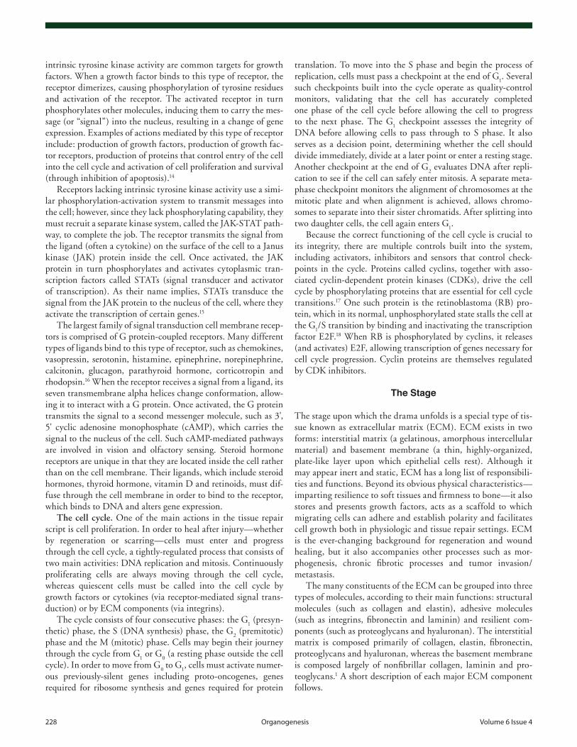
scattered fibroblasts. Initially, the new vessels are leaky and the granulation tissue has an edematous, loose appearance (Fig. 1). As the vessels grow stronger and fibroblasts begin to lay down collagen, it takes on a more substantial appearance, reaching its maximum vascularity by day 5.1
Macrophages—large, multitasking cells derived from mono-cytes—now replace the neutrophils that were so numerous in the immediate aftermath of the wounding. As their name suggests, a primary function of macrophages is the ingestion of unwanted materials: bacteria, cell remnants, debris, fibrin and foreign mate-rial. But macrophages also serve as directors for many other parts of the drama. They call in and stimulate fibroblasts and kera-tinocytes (through release of PDGF, TGF-β, TNF, IL-1 and KGF), stimulate angiogenesis (by secretion of VEGF, FGF and PDGF) and direct the laying down and remodeling of extracel-lular matrix [through the use of TGF-β, PDGF, tumor necrosis factor (TNF), osteopontin (OPN), IL-1, collagenase and matrix metalloproteinases (MMPs)].22
The formation of new blood vessels (angiogenesis) begins within the first few days following injury, aided by VEGF and FGF.27 The process is unexpectedly aided by a small number of bone-marrow-derived endothelial precursor cells which home to the wound by mysterious mechanisms. Their contribution to the overall angiogenesis effort, however, is minimal.28 Formation of the correct blood vessel structure is aided by integrins on the surface of endothelial cells; surrounding perithelial cells are recruited with the help of angiopoietin.
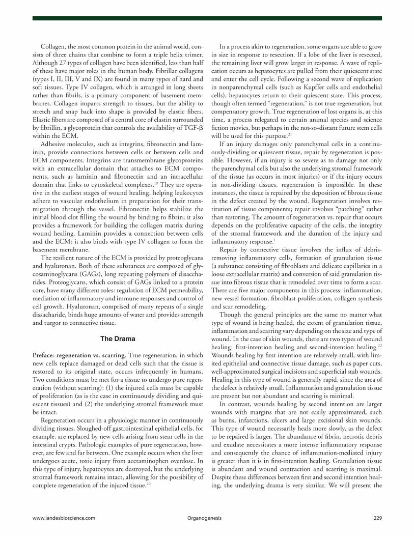
The end point toward which the entire drama is directed is the formation of a scar, a strong collagen filler that bridges the gap left by tissue destruction, restoring strength and integrity to the tissue. This process begins with the entrance of the fibroblast between days one and three, called to action by many growth fac-tors and cytokines, including PDGF, EGF and TGF-β. At first, the type III collagen secreted by fibroblasts is arrayed vertically at the margins of the incision. Later, the collagen combines with fibrin and plasma fibronectin (which deposition is promoted by the vascular permeability inherent in angiogenesis); this provi-sional matrix provides a framework for further fibroblast and endothelial cell ingrowth (Fig. 2).29
drama as it occurs in first intention wounds, noting second inten-tion differences where necessary.
Act I: inflammation. Immediately after the wound is inflicted, the most urgent task is not to repair the damaged tissue, but to stop the flow of blood from the wound. Within seconds of the injury, exposed collagen activates coagulation in the region of the injury. Platelets form an initial plug, coagulation cascade factors interact to form fibrin (which seals and solidifies the plug) and a blood clot soon appears at the skin surface. Not only does the clot stop the bleeding, but it serves as a scaffold for incoming cells and a repository of cytokines and growth factors.22,23
At the skin surface, all appears static and calm as the clot dries and forms a scab. Underneath the surface, however, a flurry of activity begins. An army of neutrophils rushes to the scene through dilated local blood vessels, called to the area by IL-1, TNF, TGF-β and other growth factors. Within 24 hours, the neutrophils appear at the edges of the wound incision, using the fibrin scaffolding of the clot to invade the region of destruc-tion, their primary tasks being to break down debris and kill bacteria.
Epithelial cells, meanwhile, begin their arduous process of repair by crawling from the basal layers of the epidermis into the wounded region, depositing basement membrane components as they migrate. They are called to begin this process by EGF and TGF-!, secreted by activated platelets and macrophages. In addition, fibroblasts secrete KGF (keratinocyte growth factor) and IL-6, factors that enhance keratinocyte migration and pro-liferation.24,25 Fusing in the midline underneath the inert scab, these epithelial cell pioneers form the first thin, continuous layer of epithelium upon which the rest of the new epithelium will be built.26
Act II: proliferation. Following the initial flurry of clotting and neutrophil activity, the work of cell proliferation begins in earnest. The characteristic feature of this act in the drama is the formation of a substance called granulation tissue, a pink, soft material so-named for its irregular, grainy appearance. Consisting of new blood vessels and fibroblasts in an extracel-lular matrix, granulation tissue is laid down over a period of a few days, beginning with a few small, tenuous blood vessels and
Figure 1. Skin ulcer showing early-stage granulation tissue with numer-ous blood vessels and scattered fibroblasts in a loose extracellular matrix. Numerous neutrophils are present within the granulation tissue.
Figure 2. Early granulation tissue with blood vessels in a loose matrix of collagen and fibrin.
www.landesbioscience.com Organogenesis 231
down by a family of enzymes known as tissue inhibitors of metal-loproteinases (TIMPs). Produced by mesenchymal cells, TIMPs help reign in collagen (and other ECM component) breakdown so that scar formation can proceed.
Ultimately, strength is achieved by increased collagen synthe-sis, followed by post-synthetic modifications of collagen, includ-ing cross-linking and increased fiber size. Early in wound healing, the collagen is relatively thin and oriented parallel to the skin; over time, the initial collagen fibrils are resorbed and replaced with thicker fibrils aligned with stress lines.22 Wound strength increases rapidly during the first month, then slows, reaching a plateau of 70–80% of the original tensile strength by the end of the third month. Although full-thickness epithelial regeneration is possible, any skin appendages lost during the initial injury will not be reformed.
Given their easily-approximated margins, wounds healing by first intention generally have minimal scarring. Wounds healing by second intention, however, are larger, with more epithelial destruction, necessitating more collagen deposition to close the wound. The process of wound contraction eases the fibroblasts’ burden a bit. Specialized contractile, smooth-muscle-like cells called myofibroblasts appear within the wound and pull the mar-gins of the wound toward each other. These cells may be derived from tissue fibroblasts under the influence of FGF and PDGF; they may also develop from epithelial cells or from bone marrow precursor cells called fibrocytes. So efficient are these myofibro-blasts that large wounds may decrease in size by up to 80%.32
When the drama ends badly. Several systemic factors may adversely affect the performance of the tissue repair drama. Patients with diabetes mellitus tend to have less granulation tis-sue formation, less collagen deposition and defects in collagen maturation, leading to slow and ineffective wound maturation.33 While this predisposition has been attributed to the microangi-opathy inherent in diabetes, recent research indicates that other factors, such as collagen glycosylation and pericapillary albumin deposition, may play a role.22,34 Poor nutrition also retards the healing process. Vitamin C, in particular, is necessary for col-lagen synthesis. Deprivation of this vitamin results in failure of activation of proline and lysine hydroxylases and formation of unhydroxylated procollagen peptides, resulting in an unstable,
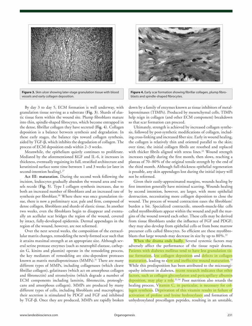
By day 3 to day 5, ECM formation is well underway, with granulation tissue serving as a substrate (Fig. 3). Shards of elas-tic tissue form within the wound site. Plump fibroblasts mature into thin, spindle-shaped fibrocytes, which become entrapped in the dense, fibrillar collagen they have secreted (Fig. 4). Collagen deposition is a balance between synthesis and degradation. In these early stages, the balance tips toward collagen synthesis, aided by TGF-β, which inhibits the degradation of collagen. The process of ECM deposition ends within 2–3 weeks.
Meanwhile, the epithelium quietly continues to proliferate. Mediated by the aforementioned KGF and IL-6, it increases in thickness, eventually regaining its full, stratified architecture and keratinized surface some time between 1 and 3 weeks (or later in second-intention healing).27
Act III: maturation. During the second week following the incision, leukocytes gradually abandon the wound area and ves-sels recede (Fig. 5). Type I collagen synthesis increases, due to both an increased number of fibroblasts and an increased rate of synthesis per fibroblast.30 Where there was once granulation tis-sue, there is now a preliminary scar, pale and firm, composed of dense collagen, fibroblasts and shards of elastic tissue. In another two weeks, even the fibroblasts begin to disappear and eventu-ally an acellular scar bridges the region of the wound, covered by intact, fully-developed epidermis. Dermal appendages in the region of the wound, however, are not reformed.
Over the next several weeks, the composition of the extracel-lular matrix changes, remodeling the newly-formed scar such that it attains maximal strength at an appropriate size. Although sev-eral serine protease enzymes (such as neutrophil elastase, cathep-sin G, kinins and plasmin) operate in the remodeling process, the key mediators of remodeling are zinc-dependent proteases known as matrix metalloproteinases (MMPs).31 There are many different types of MMPs, including collagenases (which cleave fibrillar collagen), gelatinases (which act on amorphous collagen and fibronectin) and stromelysins (which degrade a number of ECM components including laminin, fibronectin, proteogly-cans and amorphous collagen). MMPs are produced by many different types of cells, including fibroblasts and macrophages; their secretion is stimulated by PDGF and FGF and inhibited by TGF-β. Once they are produced, MMPs are rapidly broken
Figure 3. Skin ulcer showing later-stage granulation tissue with blood vessels and early collagen deposition.
Figure 4. Early scar formation showing fibrillar collagen, plump fibro-blasts and spindle-shaped fibrocytes.
Daniela del pilar Torrejon Peces
Daniela del pilar Torrejon Peces
Daniela del pilar Torrejon Peces
Daniela del pilar Torrejon Peces
Daniela del pilar Torrejon Peces
Daniela del pilar Torrejon Peces
Daniela del pilar Torrejon Peces
Daniela del pilar Torrejon Peces

232 Organogenesis Volume 6 Issue 4
poorly cross-linked collagen helix. Conditions in which blood flow is compromised—such as cardiac insufficiency or arterio-sclerosis—delay the delivery of necessary cells and factors to the scene of the wound. Certain drugs may also slow wound healing; glucocorticoids, for example, inhibit inflammation and collagen formation.
Local factors can also greatly affect the quality of wound heal-ing. The size of the wound is important (small injuries heal faster than large ones) as is the location (wounds in richly-vascularized areas of the body, such as the face, heal relatively quickly). The presence of foreign bodies may prolong healing, as may motion
Figure 5. Wide excision of skin. (A) Previous punch biopsy site (center) in late stages of repair. (B) Dense, acellular, normal dermal collagen bundles (bottom) abutting new, still-cellular fibrous tissue of the repair process (top). (C) Blood vessel in process of involution.
or pressure at the wound site. However, the single most impor-tant cause of delayed healing of wounds is infection.1 If any beta-hemolytic Streptococcus organisms are present or if there are over 105 organisms of any bacterial species per gram of tissue, the wound will not heal.35 More common in second-intention heal-ing, infection prolongs the inflammatory phase of wound heal-ing, impeding epithelialization, collagen deposition and wound contraction. Bacterial endotoxins stimulate collagenase secretion, leading to degradation of not only the forming scar, but of sur-rounding normal tissue.22
On the other hand, sometimes acts in the drama are played so well, so exuberantly, that the end result is an abnormally-healed wound. Occasionally, the balance of collagen formation and deg-radation tips in favor of formation, leading to hypertrophic scars or keloids. Hypertrophic scars are raised scars that remain con-fined to the region of the wound. They generally occur within 4 weeks of injury (often a severe traumatic or thermal injury to the dermis) and may regress over time.22,36 An aberrant autocrine loop in which myofibroblasts produce excessive TGF-β and, hence, collagen, appears to contribute to their formation.36 Keloids are scars that have overgrown the boundaries of the initial incision, presenting as nodules or masses of fibrous tissue (Fig. 6). While keloids generally appear within one year of the inciting injury, some may begin growing years later.22 For unknown reasons,
Figure 6. Keloid showing dense, haphazardly-arranged collagen bundles.
Figure 7. Proud flesh with exuberant granulation tissue.
Daniela del pilar Torrejon Peces

www.landesbioscience.com Organogenesis 233
References1. Kumar V, Abbas AK, Fausto N, Aster JC. Tissue
renewal, regeneration and repair. In: Robbins and Cotran, eds. Pathologic Basis of Disease. Eighth ed. Philadelphia: Elsevier 2010; 79-110.
2. Conover JC, Notti RQ. The neural stem cell niche. Cell Tissue Res 2008; 331:211-24.
3. Tumbar T, Guasch G, Greco V, Blanpain C, Lowry W, Rendl M, et al. Defining the epithelial stem cell niche in skin. Science 2004; 303:359-63.
4. Bessa PC, Casal M, Reis RL. Bone morphogenetic pro-teins in tissue engineering: the road from laboratory to the clinic. Part 1-basic concepts. J Tiss Eng Regen Med 2008; 2:1-13.
5. Itoh F, Asao H, Sugamura K, Heldin CH, ten Dijke P, Itoh S. Promoting bone morphogenetic protein signal-ing through negative regulation of inhibitory Smads. EMBO J 2001; 20:4132-42.
6. Horan GS, Wood S, Ona V, Li DJ, Lukashev ME, Weinreb PH, et al. Partial inhibition of integrin αvβ prevents pulmonary fibrosis without exacerbat-ing inflammation. Am J Resp Crit Care Med 2008; 177:56-65.
7. Attisano L, Wrana JL. Signal transduction by the TGFbeta superfamily. Science 2002; 296:1646-7.
8. Andrae J, Gallini R, Betsholtz C. Role of platelet-derived growth factors in physiology and medicine. Genes Dev 2008; 22:1276-312.
9. Cao R, Brakenhielm E, Pawliuk R, Wariaro D, Post MJ, Wahlberg E, et al. Angiogenic synergism, vascular stability and improvement of hind-limb ischemia by a combination of PDGF-BB and FGF-2. Nat Med 2003; 9:604-13.
10. Holmes K, Roberts OL, Thomas AM, Cross MJ. Vascular endothelial growth factor receptor-2: Structure, function, intracellular signaling and thera-peutic inhibition. Cell Signal 2007; 19:2003-12.
11. Escudier B. Anti-VEGF therapy for renal cell carci-noma. Clin Adv Hematol Oncol 2007; 5:530-1.
12. Schmidt C, Bladt F, Goedecke S, Brinkmann V, Zschiesche W, Sharpe M, et al. Scatter factor/hepato-cyte growth factor is essential for liver development. Nature 1995; 373:699-702.
Conclusion
Wound healing is a carefully-scripted drama performed count-less times throughout a human lifespan. Though occasionally the drama does not go as planned, in most cases damage is con-tained, dead cells and tissue are removed and a scar restores integrity to the injured tissue—a triumphant performance indeed.
13. Fedyk ER, Jones D, Critchley HO, Phipps RP, Blieden TM, Springer TA. Expression of stromal-derived fac-tor-1 is decreased by IL-1 and TNF and in dermal wound healing. J Immunol 2001; 166:5749-54.
14. Robinson DR, Wu YM, Lin SF. The protein tyrosine kinase family of the human genome. Oncogene 2000; 19:5548-57.
15. Rawlings JS, Rosler KM, Harrison DA. The JAK/STAT signaling pathway. J Cell Science 2004; 117:1281-3.
16. Cherezov V, Rosenbaum DM, Hanson MA, Rasmussen SG, Thian FS, Kobilka TS, et al. High-resolution crys-tal structure of an engineered human β2-adrenergic G protein-coupled receptor. Science 2007; 318:1258-65.
17. Nigg EA. Cyclin-dependent protein kinases: key regu-lators of the eukaryotic cell cycle. Bioessays 1995; 17:471-80.
18. Münger K, Howley PM. Human papillomavirus immortalization and transformation functions. Virus Res 2002; 89:213-28.
19. Hynes RO. Integrins: bidirectional, allosteric signaling machines. Cell 2002; 110:673-87.
20. Apte U, Singh S, Zeng G, Cieply B, Virji MA, Wu T, et al. Beta-catenin activation promotes liver regenera-tion after acetaminophen-induced injury. Am J Pathol 2009; 175:1056-65.
21. Rafii S, Lyden D. Therapeutic stem and progenitor cell transplantation for organ vascularization and regenera-tion. Nat Med 2003; 9:702-12.
22. Broughton G, Janis JE, Attinger CE. Wound healing: An overview. Plast Reconstr Surg 2006; 117:1-32.
23. Witte MB, Barbul A. General principles of wound healing. Surg Clin North Am 1997; 77:509-28.
24. Smola H, Thiekötter, Fusenig NE. Mutual induction of growth factor gene expression by epidermal-dermal interaction. J Cell Biol 1993; 122:417-29.
25. Xia YP, Zhao Y, Marcus J, Jiminez PA, Ruben SM, Moore PA, et al. Effects of keratinocyte growth factor-2 (KGF-2) on wound healing in an ischaemia-impaired rabbit ear model and on scar formation. J Pathol 1999; 188:431-8.
keloids occur much more frequently in patients with darkly-pigmented skin; certain people seem to have a predisposition towards their formation.
Formation of granulation tissue may likewise occur in an excessive fashion. In a lesion known as proud flesh, the process of wound healing is interrupted by masses of granulation tissue protruding above the skin surface, preventing re-epithelialization and appropriate scar formation (Fig. 7).
26. Lawrence WT, Diegelmann RF. Growth factors in wound healing. Clin Dermatol 1994; 12:157-69.
27. Werner S, Grose R. Regulation of wound healing by growth factors and cytokines. Physiol Rev 2003; 83:835-70.
28. Bluff J, Ferguson M, O’Kane S, Ireland G. Bone marrow-derived endothelial progenitor cells do not contribute significantly to new vessels during incisional wound healing. Exp Hematol 2007; 35:500-6.
29. Pierce GF, Mustoe TA, Altrock BW, Deuel TF, Thomason A. Role of platelet-derived growth factor in wound healing. J Cell Biochem 2004; 45:319-26.
30. Diegelmann R. Analysis of collagen synthesis. Methods Mol Med 2003; 78:349-58.
31. Pilcher BK, Wang M, Qin X, Parks WC, Senior RM, Welgus HG. Role of matrix metalloproteinases and their inhibition in cutaneous wound healing and aller-gic contact hypersensitivity. Ann NY Acad Sci 2006; 878:12-24.
32. Lorentz HP, Longaker MT. Wounds: Biology, pathol-ogy and management. In: Norton JA, Barie PS, Bollinger RR, Chang AE, Lowry S, Mulvihill SJ, et al. Eds. Surgery: Basic science and clinical evidence. 2nd edition. New York: Springer 2008; 191-208.
33. Chow LW, Loo WT, Yuen KY, Cheng C. The study of cytokine dynamics at the operation site after mastec-tomy. Wound Repair Regen 2003; 11:326-30.
34. Bucalo B, Eaglstein WH, Falanga V. Inhibition of cell proliferation by chronic wound fluid. Wound Rep Regen 2002; 1:181-6.
35. Robson MC. Wound infection: A failure of wound healing caused by an imbalance of bacteria. Surg Clin North Am 1997; 77:637-50.
36. Dabiri G, Tumbarello DA, Turner CE, Van De Water L. Hic-5 promotes the hypertrophic scar myofibroblast phenotype by regulating the TGF-β1 autocrine loop. J Invest Dermatol 2008; 128:2518-25.