Knee Osteoarthritis and Associated Periarticular ...cdn.intechweb.org/pdfs/30693.pdf · 14 Knee...
14
14 Knee Osteoarthritis and Associated Periarticular Conditions: Iliotibial Band Friction and Baker Cyst Violeta Vasilevska 1 , Ulrike Szeimies 2 , Milan Samardziski 1 and Axel Stäbler 2 1 University Surgical Clinic “St Naum Ohridski” Skopje, 2 Radiologie in München Harlaching, Munich, 1 Rep. Macedonia 2 Germany 1. Introduction Osteoarthritis (OA) is a highly prevalent disease with markedly increasing impact worldwide because of the aging of populations (Center for disease control and prevention (CDC); Murphy L.et al., 2008). It affects more than 21 milion people in the U.S. (Handout on Health: Osteoarthritis), with 36% of elderly aged 70 or older having some degree of radiographic knee OA (D’Ambrosia et al., 2005; Felson et al., 1987). It is a major public health problem, with prevalence in the knee of approximately 30% in those over 65 years old (Felson et al., 1987). The cause of knee pain in patients with OA remains unclear. Because hyaline cartilage has no innervations (Dye et al., 1998), the primary pathologic abnormality in OA (hyaline cartilage loss) could occur without pain. In MRI studies is reported an increase the prevalence of subchondral bone marrow edema, knee joint effusion, and synovial thickening in patients with symptomatic knee OA compared with patients with no symptoms (Hill et al., 2001; Felson et al., 2001). Knee with OA are biomechanically altered, and these changes may put stress on ligament and tendon insertion sites in and around the knee joint, creating pain (Hill et al., 2003). Some of the pain does not emanate from the joint itself but rather from the structures near the joint that contain pain fibers. Wide ranges of periarticular lesions occur around the knee joint, including popliteal Baker cyst (BC) (Vasilevska et.al., 2008, Janzen et al., 1994) and friction of the iliotibial band (ITBF) (Vasilevska et al., 2009). Iliotibial band friction syndrome (ITBFS) is an inflammatory overuse disorder affecting soft tissue, interposed between the iliotibial band and the lateral femoral condyle, caused by chronic friction (Muhle et al., 1999). Recently, an anatomic study disclosed a fibrous anchorage of the iliotibial band to the femur preventing rolling over the epicondyle; therefore ITBFS is mainly caused by increased pressure to the richly innervated and vascularized fat and loose connective tissue beneath the tract (Fairclough et al., 2006, 2007). Either ITBFS has been shown to cause lateral knee pain in athletes, it may be a consequence of gait changes induced by knee OA and may occur together with symptomatic knee OA (presented only 3 cases with low grade ITBF, only one with symptom) (Hill et al., 2003). www.intechopen.com
-
Upload
duongtuong -
Category
Documents
-
view
220 -
download
0
Transcript of Knee Osteoarthritis and Associated Periarticular ...cdn.intechweb.org/pdfs/30693.pdf · 14 Knee...
14
Knee Osteoarthritis and Associated Periarticular Conditions:
Iliotibial Band Friction and Baker Cyst
Violeta Vasilevska1, Ulrike Szeimies2, Milan Samardziski1 and Axel Stäbler2 1University Surgical Clinic “St Naum Ohridski” Skopje,
2Radiologie in München Harlaching, Munich, 1Rep. Macedonia
2Germany
1. Introduction
Osteoarthritis (OA) is a highly prevalent disease with markedly increasing impact worldwide because of the aging of populations (Center for disease control and prevention (CDC); Murphy L.et al., 2008). It affects more than 21 milion people in the U.S. (Handout on Health: Osteoarthritis), with 36% of elderly aged 70 or older having some degree of radiographic knee OA (D’Ambrosia et al., 2005; Felson et al., 1987). It is a major public health problem, with prevalence in the knee of approximately 30% in those over 65 years old (Felson et al., 1987). The cause of knee pain in patients with OA remains unclear. Because hyaline cartilage has no innervations (Dye et al., 1998), the primary pathologic abnormality in OA (hyaline cartilage loss) could occur without pain. In MRI studies is reported an increase the prevalence of subchondral bone marrow edema, knee joint effusion, and synovial thickening in patients with symptomatic knee OA compared with patients with no symptoms (Hill et al., 2001; Felson et al., 2001). Knee with OA are biomechanically altered, and these changes may put stress on ligament and tendon insertion sites in and around the knee joint, creating pain (Hill et al., 2003). Some of the pain does not emanate from the joint itself but rather from the structures near the joint that contain pain fibers. Wide ranges of periarticular lesions occur around the knee joint, including popliteal Baker cyst (BC) (Vasilevska et.al., 2008, Janzen et al., 1994) and friction of the iliotibial band (ITBF) (Vasilevska et al., 2009). Iliotibial band friction syndrome (ITBFS) is an inflammatory overuse disorder affecting soft tissue, interposed between the iliotibial band and the lateral femoral condyle, caused by chronic friction (Muhle et al., 1999). Recently, an anatomic study disclosed a fibrous anchorage of the iliotibial band to the femur preventing rolling over the epicondyle; therefore ITBFS is mainly caused by increased pressure to the richly innervated and vascularized fat and loose connective tissue beneath the tract (Fairclough et al., 2006, 2007). Either ITBFS has been shown to cause lateral knee pain in athletes, it may be a consequence of gait changes induced by knee OA and may occur together with symptomatic knee OA (presented only 3 cases with low grade ITBF, only one with symptom) (Hill et al., 2003).
www.intechopen.com
Osteoarthritis – Diagnosis, Treatment and Surgery
254
Recent reports have suggested a 60% reduction in cartilage volume in severe knee osteoarthritis (Vahlensieck et al., 2001; Fritschy et al., 2006). Medial compartment cartilage loss leads to varus deformity, which can affect knee biomechanics by altering the relationship of the iliotibial band and the lateral epicondyle with the possibility of an increased friction and pressure between these structures. In contrary it is well known that popliteal (Baker) cyst is the most frequent encountered lesion around the knee. Among older individual with asymptomatic OA, popliteal cyst have a high prevalence (20,8%) (Hill et al., 2001). Cystic lesions around the knee may present as a painless palpable mass (Kornaat et al., 2006; Hill et al., 2003), with pain, with symptoms of tenderness in the posterior fossa (Hill et al., 2003) or to be detected during the routine MR imaging of the knee with suspected internal joint derangement (Mc Carthy et al., 2004), eventually when is large can be painful. Multiple studies confirmed that intraarticular derangement play an important role in pathogenesis of popliteal cyst. MR studies of popliteal cyst demonstrated connection to one or more intraarticular lesions in 87-98% of the cases, like osteoarthritis or inflammatory arthritis; often joint effusion, meniscus tear and degenerative disease of the joint are found (Miller et al., 1996). During the routine practice in cases with advanced isolated medial osteoarthritis (with
subsequent genu varum) presence of MR signs of friction of ITB have been noted. The
purpose of the study was to describe the frequency of fibrovascular tissue between the
iliotibial tract and the lateral epicondyle in patients with severe isolated medial
compartment osteoarthritis of the knee (Vasilevska et al., 2009). From this cases were
selected those with Baker cyst, and the correlation between sizes of Baker cyst in patients
suffering from medial compartment osteoarthritis of the knee was recognized and
evaluated. The purpose was to describe the significance of the associated medial
compartment knee osteoarthritis: cartilage degeneration, different degree of medial
meniscus degeneration, bone edema and knee effusion.
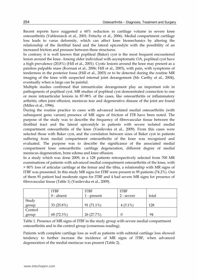
In a study which was done 2009, in a 128 patients retrospectively selected from 700 MR
examinations of patients with advanced medial compartment osteoarthritis of the knee, with
> 80% loss of articular cartilage at the femur and the tibia, a relationship with MR signs of
ITBF was presented. In this study MR signs for ITBF were present in 95 patients (74.2%). Out
of them 91 patient had moderate signs for ITBF and 4 had severe MR signs for presence of
fibrovascular tissue (Table 1) (Vasilevska et al., 2009).
ITBF 0 - absent
ITBF 1 - present
ITBF 2 - severe
total
Study group
33 (25.8%)
91 (71.1%)
4 (3.1%)
128
Control group
68 (72.3%)
26 (27.7%)
0
94
Table 1. Presence of MR signs of ITBF in the study group with severe medial compartment
osteoarthritis and in the control group (consensus reading).
Patients with complete cartilage loss as well as patients with subtotal cartilage loss showed
tendency to further increase the incidence of MR signs of ITBF, when advanced
degeneration of the medial meniscus was present (Table 2).
www.intechopen.com
Knee Osteoarthritis and Associated Periarticular Conditions: Iliotibial Band Friction and Baker Cyst
255
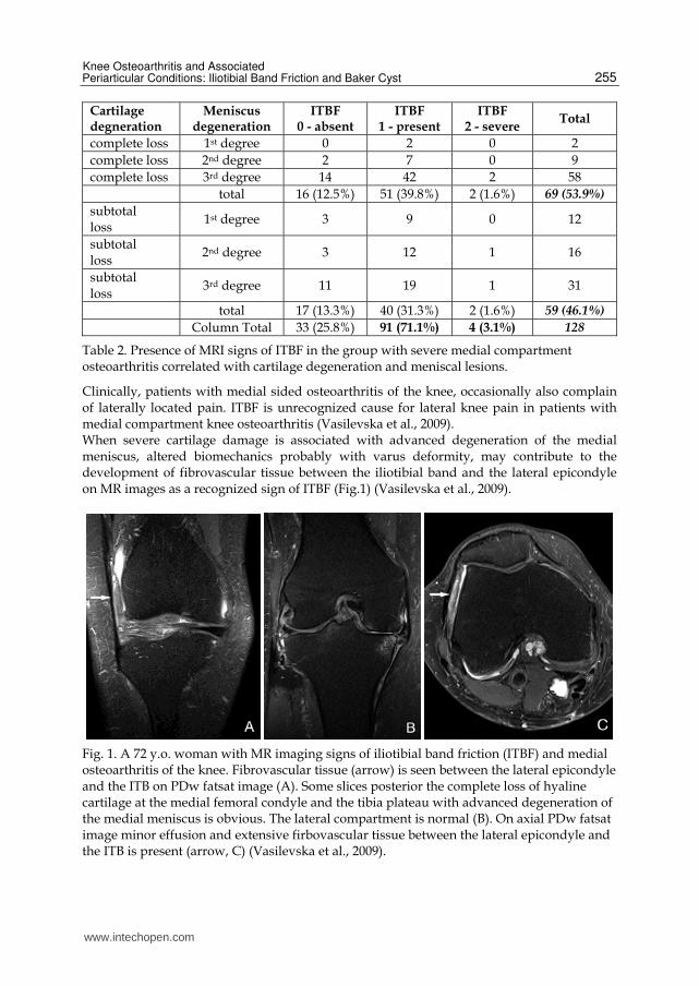
Cartilage degneration
Meniscus degeneration
ITBF 0 - absent
ITBF 1 - present
ITBF 2 - severe
Total
complete loss 1st degree 0 2 0 2
complete loss 2nd degree 2 7 0 9
complete loss 3rd degree 14 42 2 58
total 16 (12.5%) 51 (39.8%) 2 (1.6%) 69 (53.9%)
subtotal loss
1st degree 3 9 0 12
subtotal loss
2nd degree 3 12 1 16
subtotal loss
3rd degree 11 19 1 31
total 17 (13.3%) 40 (31.3%) 2 (1.6%) 59 (46.1%)
Column Total 33 (25.8%) 91 (71.1%) 4 (3.1%) 128
Table 2. Presence of MRI signs of ITBF in the group with severe medial compartment osteoarthritis correlated with cartilage degeneration and meniscal lesions.
Clinically, patients with medial sided osteoarthritis of the knee, occasionally also complain of laterally located pain. ITBF is unrecognized cause for lateral knee pain in patients with medial compartment knee osteoarthritis (Vasilevska et al., 2009). When severe cartilage damage is associated with advanced degeneration of the medial meniscus, altered biomechanics probably with varus deformity, may contribute to the development of fibrovascular tissue between the iliotibial band and the lateral epicondyle on MR images as a recognized sign of ITBF (Fig.1) (Vasilevska et al., 2009).
Fig. 1. A 72 y.o. woman with MR imaging signs of iliotibial band friction (ITBF) and medial osteoarthritis of the knee. Fibrovascular tissue (arrow) is seen between the lateral epicondyle and the ITB on PDw fatsat image (A). Some slices posterior the complete loss of hyaline cartilage at the medial femoral condyle and the tibia plateau with advanced degeneration of the medial meniscus is obvious. The lateral compartment is normal (B). On axial PDw fatsat image minor effusion and extensive firbovascular tissue between the lateral epicondyle and the ITB is present (arrow, C) (Vasilevska et al., 2009).
www.intechopen.com
Osteoarthritis – Diagnosis, Treatment and Surgery
256
In another study 66 cases were retrospectively evaluated its MR study of the knee with
medial compartment knee osteoarthritis and MR signs of Baker cyst. The median age was
56,42 years, (age range 34-84 years). We selected two groups according to the size of the
Baker cyst on MRI. The first group was with palpable soft tissue mass on medial aspect of
popliteal fosa with a large Baker cyst, and in the other group the Baker cyst was small and
detected only on MRI (Table 3).
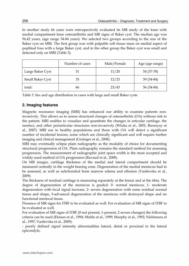
Number of cases Male/Female Age (age range)
Large Baker Cyst 31 11/20 54 (37-78)
Small Baker Cyst 35 12/23 59 (34-84)
total: 66 23/43 56 (34-84)
Table 3. Sex and age distribution in cases with large and small Baker cysts
2. Imaging features
Magnetic resonance imaging (MRI) has enhanced our ability to examine patients non-
invasively. This allows us to assess structural changes of osteoarthritis (OA) without risk to
the patient. MRI enables to visualize and quantitate the changes in articular cartilage, the
menisci, and other periarticular structures non-invasively (Wluka et al., 2001; Baranyay et
al., 2007). MRI use in healthy populations and those with OA will detect a significant
number of incidental lesions, some which are clinically significant and will require further
imaging and clinical management (Grainger et al., 2008).
MRI may eventually eclipse plain radiography as the modality of choice for documenting
structural progression of OA. Plain radiography remains the standard method for assessing
progression. The measurement of radiographic joint space width is the most accepted and
widely-used method of OA progression (Ravaud et al., 2008).
On MR images, cartilage thickness of the medial and lateral compartment should be
measured centrally in the weight bearing zone. Degeneration of the medial meniscus had to
be assessed, as well as subchondral bone marrow edema and effusion (Vasilevska et al.,
2009).
The thickness of residual cartilage is measuring separately at the femur and at the tibia. The
degree of degeneration of the meniscus is graded: 0- normal meniscus, 1- moderate
degeneration with focal signal increase, 2- severe degeneration with some residual normal
tissue and shape, 3-advanced degeneration of the meniscus with destroyed shape and no
functional meniscal tissue.
Presence of MR signs for ITBF to be evaluated as well. For evaluation of MR signs of ITBF to
be evaluated as well.
For evaluation of MR signs of ITBF (0-not present, 1-present, 2 severe changes) the following
criteria can be used (Ekman et al., 1994; Muhle et al., 1999; Murphy et al., 1992; Nishimura et
al., 1997; Vasilevska et al., 2009):
- poorly defined signal intensity abnormalities lateral, distal or proximal to the lateral epicondyle;
www.intechopen.com
Knee Osteoarthritis and Associated Periarticular Conditions: Iliotibial Band Friction and Baker Cyst
257
- signal intensity abnormalities superficial or deep to the ITT; - localized fluid collection lateral, distal or proximal to the lateral epicondyle; (Fig.2). To conclude that the reason of the lateral knee pain is ITBF in patients with osteoarthritis of
the knee, which showed advanced medial compartment osteoarthritis with complete or
subtotal (>80%) loss of articular cartilage at the femur and the tibia, lateral compartment
should be normal including the articular cartilage and the lateral meniscus, without
meniscal lesion or cartilage abnormalities (Vasilevska et al., 2009).
MR imaging allows confirmation of diagnosis of ITBFS and exclusion of other causes of
lateral knee pain - such as meniscal tears or ligament injuries. Axial images are necessary to
differentiate intraarticular fluid from ITBFS (Murphy et al., 1992).
Popliteal cysts on MR imaging are usually well defined, extending between the tendon of
semimembranosus and the medial head of gastrocnemius into the gastrocnemius-
semimembranosus bursa, situated superficial to the medial gastrocnemius muscle, along the
medial side of the popliteal fosa (Torreggiani et al., 2002; Steiner et al., 1996). As cyst
enlarge, the cystic fluid may extend in any direction. Inferomedial expansion is relatively
common with a superficial location, which results in cysts becoming palpable (Torreggiani
et al., 2002; Steiner et al., 1996). Baker cyst can be presented as a palpable soft tissue masses
on the medial aspect of the popliteal fossa when it is distended and large (Fig.3) (Vasilevska
et al., 2008).
On MRI, Baker cyst is presenting as a circumscribed mass with low signal on T1-weigted
image, intermedal signal intensity on proton density (PD) image and high signal intensity
comparing with skeletal muscle on PD-weithed fatsat image. The size of Baker cyst can be
assessed by measuring the distension of the cyst, and when is thickened more than 1cm is a
large cyst (Fig.4) (Vasilevska et al., 2008).
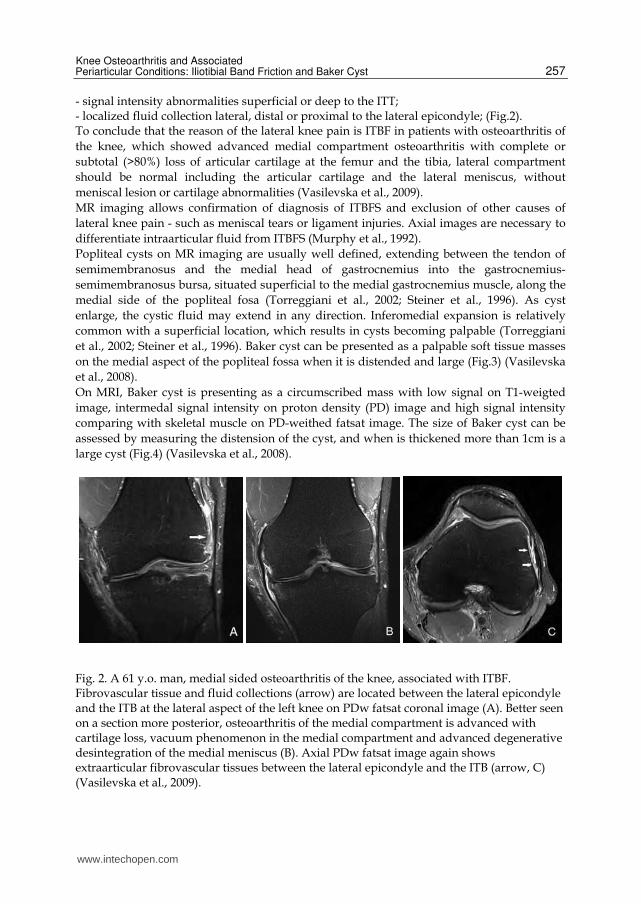
Fig. 2. A 61 y.o. man, medial sided osteoarthritis of the knee, associated with ITBF. Fibrovascular tissue and fluid collections (arrow) are located between the lateral epicondyle and the ITB at the lateral aspect of the left knee on PDw fatsat coronal image (A). Better seen on a section more posterior, osteoarthritis of the medial compartment is advanced with cartilage loss, vacuum phenomenon in the medial compartment and advanced degenerative desintegration of the medial meniscus (B). Axial PDw fatsat image again shows extraarticular fibrovascular tissues between the lateral epicondyle and the ITB (arrow, C) (Vasilevska et al., 2009).
www.intechopen.com
Osteoarthritis – Diagnosis, Treatment and Surgery
258
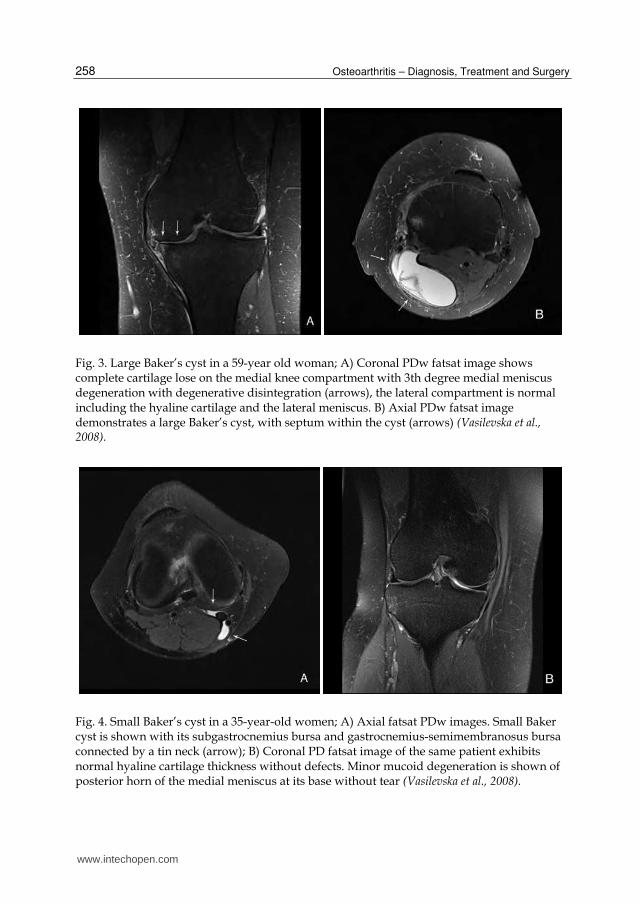
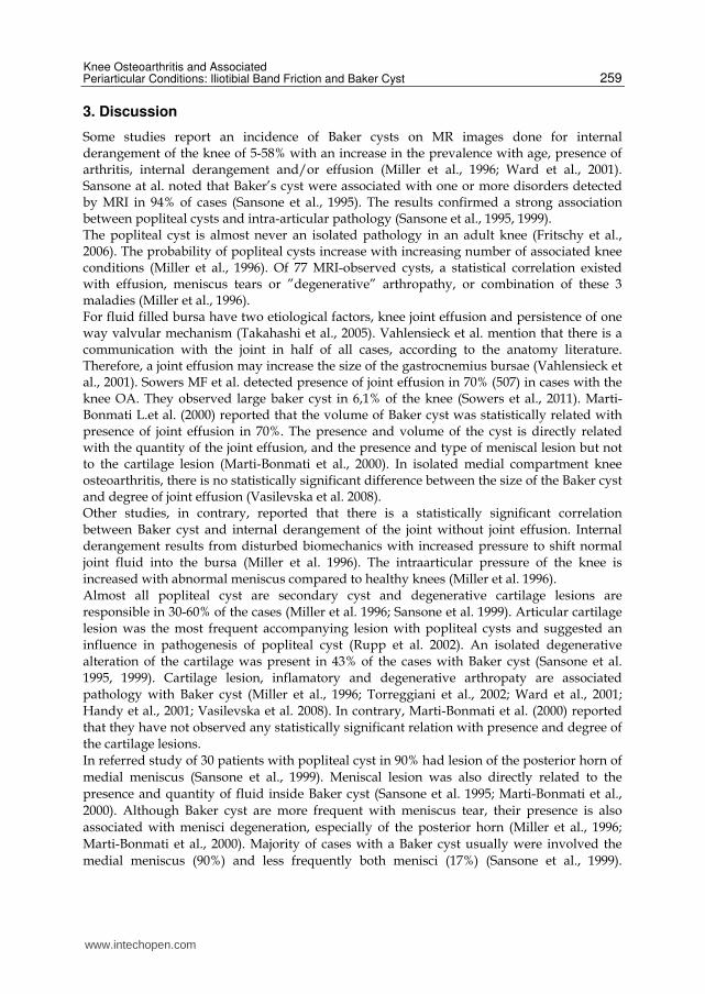
Fig. 3. Large Baker’s cyst in a 59-year old woman; A) Coronal PDw fatsat image shows complete cartilage lose on the medial knee compartment with 3th degree medial meniscus degeneration with degenerative disintegration (arrows), the lateral compartment is normal including the hyaline cartilage and the lateral meniscus. B) Axial PDw fatsat image demonstrates a large Baker’s cyst, with septum within the cyst (arrows) (Vasilevska et al., 2008).
Fig. 4. Small Baker’s cyst in a 35-year-old women; A) Axial fatsat PDw images. Small Baker cyst is shown with its subgastrocnemius bursa and gastrocnemius-semimembranosus bursa connected by a tin neck (arrow); B) Coronal PD fatsat image of the same patient exhibits normal hyaline cartilage thickness without defects. Minor mucoid degeneration is shown of posterior horn of the medial meniscus at its base without tear (Vasilevska et al., 2008).
www.intechopen.com
Knee Osteoarthritis and Associated Periarticular Conditions: Iliotibial Band Friction and Baker Cyst
259
3. Discussion
Some studies report an incidence of Baker cysts on MR images done for internal derangement of the knee of 5-58% with an increase in the prevalence with age, presence of arthritis, internal derangement and/or effusion (Miller et al., 1996; Ward et al., 2001). Sansone at al. noted that Baker’s cyst were associated with one or more disorders detected by MRI in 94% of cases (Sansone et al., 1995). The results confirmed a strong association between popliteal cysts and intra-articular pathology (Sansone et al., 1995, 1999). The popliteal cyst is almost never an isolated pathology in an adult knee (Fritschy et al., 2006). The probability of popliteal cysts increase with increasing number of associated knee conditions (Miller et al., 1996). Of 77 MRI-observed cysts, a statistical correlation existed with effusion, meniscus tears or ”degenerative” arthropathy, or combination of these 3 maladies (Miller et al., 1996). For fluid filled bursa have two etiological factors, knee joint effusion and persistence of one way valvular mechanism (Takahashi et al., 2005). Vahlensieck et al. mention that there is a communication with the joint in half of all cases, according to the anatomy literature. Therefore, a joint effusion may increase the size of the gastrocnemius bursae (Vahlensieck et al., 2001). Sowers MF et al. detected presence of joint effusion in 70% (507) in cases with the knee OA. They observed large baker cyst in 6,1% of the knee (Sowers et al., 2011). Marti-Bonmati L.et al. (2000) reported that the volume of Baker cyst was statistically related with presence of joint effusion in 70%. The presence and volume of the cyst is directly related with the quantity of the joint effusion, and the presence and type of meniscal lesion but not to the cartilage lesion (Marti-Bonmati et al., 2000). In isolated medial compartment knee osteoarthritis, there is no statistically significant difference between the size of the Baker cyst and degree of joint effusion (Vasilevska et al. 2008). Other studies, in contrary, reported that there is a statistically significant correlation between Baker cyst and internal derangement of the joint without joint effusion. Internal derangement results from disturbed biomechanics with increased pressure to shift normal joint fluid into the bursa (Miller et al. 1996). The intraarticular pressure of the knee is increased with abnormal meniscus compared to healthy knees (Miller et al. 1996). Almost all popliteal cyst are secondary cyst and degenerative cartilage lesions are responsible in 30-60% of the cases (Miller et al. 1996; Sansone et al. 1999). Articular cartilage lesion was the most frequent accompanying lesion with popliteal cysts and suggested an influence in pathogenesis of popliteal cyst (Rupp et al. 2002). An isolated degenerative alteration of the cartilage was present in 43% of the cases with Baker cyst (Sansone et al. 1995, 1999). Cartilage lesion, inflamatory and degenerative arthropaty are associated pathology with Baker cyst (Miller et al., 1996; Torreggiani et al., 2002; Ward et al., 2001; Handy et al., 2001; Vasilevska et al. 2008). In contrary, Marti-Bonmati et al. (2000) reported that they have not observed any statistically significant relation with presence and degree of the cartilage lesions. In referred study of 30 patients with popliteal cyst in 90% had lesion of the posterior horn of
medial meniscus (Sansone et al., 1999). Meniscal lesion was also directly related to the
presence and quantity of fluid inside Baker cyst (Sansone et al. 1995; Marti-Bonmati et al.,
2000). Although Baker cyst are more frequent with meniscus tear, their presence is also
associated with menisci degeneration, especially of the posterior horn (Miller et al., 1996;
Marti-Bonmati et al., 2000). Majority of cases with a Baker cyst usually were involved the
medial meniscus (90%) and less frequently both menisci (17%) (Sansone et al., 1999).
www.intechopen.com
Osteoarthritis – Diagnosis, Treatment and Surgery
260
Authors in study of 66 Baker cyst in cases with isolated medial compartment knee
osteoarthritis concluded that in the cases with large Baker cyst, there is statistically
significant difference between different degree of medial meniscus degeneration and
distension of the cyst. The degree of medial meniscal degeneration has no influence on the
distension of Baker cyst generally but an influence was found, when there is significant
cartilage degeneration (Vasilevska et al., 2008). The same authors reported that the
combination of medial compartment cartilage degeneration and medial meniscus
degeneration are associated with large Baker cyst in 84%, but only 48% with small Baker
cyst. In the group with large Baker cyst, isolated medial meniscus degeneration is present in
16%, comparing with association of medial meniscus degeneration in 52% from the cases
with small cyst (Vasilevska et al., 2008).
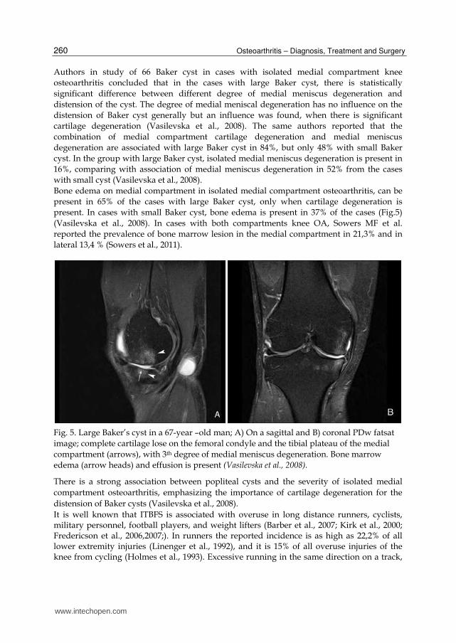
Bone edema on medial compartment in isolated medial compartment osteoarthritis, can be
present in 65% of the cases with large Baker cyst, only when cartilage degeneration is
present. In cases with small Baker cyst, bone edema is present in 37% of the cases (Fig.5)
(Vasilevska et al., 2008). In cases with both compartments knee OA, Sowers MF et al.
reported the prevalence of bone marrow lesion in the medial compartment in 21,3% and in
lateral 13,4 % (Sowers et al., 2011).
Fig. 5. Large Baker’s cyst in a 67-year –old man; A) On a sagittal and B) coronal PDw fatsat
image; complete cartilage lose on the femoral condyle and the tibial plateau of the medial
compartment (arrows), with 3th degree of medial meniscus degeneration. Bone marrow
edema (arrow heads) and effusion is present (Vasilevska et al., 2008).
There is a strong association between popliteal cysts and the severity of isolated medial
compartment osteoarthritis, emphasizing the importance of cartilage degeneration for the
distension of Baker cysts (Vasilevska et al., 2008).
It is well known that ITBFS is associated with overuse in long distance runners, cyclists, military personnel, football players, and weight lifters (Barber et al., 2007; Kirk et al., 2000; Fredericson et al., 2006,2007;). In runners the reported incidence is as high as 22,2% of all lower extremity injuries (Linenger et al., 1992), and it is 15% of all overuse injuries of the knee from cycling (Holmes et al., 1993). Excessive running in the same direction on a track,
www.intechopen.com

Knee Osteoarthritis and Associated Periarticular Conditions: Iliotibial Band Friction and Baker Cyst
261
downhill running, a lack of running experience, long distance running are the most often mentioned etiologic factors for ITBFS (Linenger et al., 1992; Messier et al., 1995). Weakness of the hip abductor muscles is also believed to play a role in the development of ITBFS (Fredericson et al., 2006; MacMahon et al., 2000). Football players and weight lifters can also suffer from chronic inflammation and fibrovascular tissue at the ITB proximal to its insertion into the anterolateral tibia. The causes of the ITBFS can be extrinsic (related to training technique) or intrinsic (related to the patient’s anatomic alignment) (Farell et al., 2003; Fredericson et al., 2006; Nishimura et al., 2003). Farell et al. (2003) emphasized that ITBFS usually occurs as a result of overuse. If, however, the patient has certain anatomical conditions (leg length discrepancies, varus knee alignment or excessive pronation and external tibial rotation of more than 20%), he/she will be more inclined to experience ITBFS. They concluded that knee-flexion repetition was more likely to result in the onset of the overuse injury ITBFS during cycling (Farell et al., 2003). ITBFS is predominantly a clinical diagnosis. Several other anatomical abnormalities, including leg length discrepancy and functional overpronation of the food have been postulated as predisposing factors (Nishimura et al., 2003). Nishimura et al. (2003) described also two no athletic patients with ITBFS. As a possible factor contributing to the development of ITBFS, genus varus has been described, in runners and in athlete that may increase the tension and thus a frictional force over the lateral femoral condyle (Jones et al., 1987; Sutker et al., 1981). The finding of predominant cartilage degeneration on the medial rather than the lateral tibia plateau side suggests a close relation to the varus- knee osteoarthritis present in most of the cases (Kleemann et al., 2005). Recent reports have suggested a 60% reduction in cartilage volume in severe knee osteoarthritis (Burgkart et al., 2001; Cicuttini et al., 2001). Significant correlation between joint space narrowing and cartilage volume was reported (Cicuttini et al., 2001). Medial compartment cartilage loss leads to varus deformity, which can affect knee biomechanics by altering the relationship of the iliotibial band and the lateral epicondyle with the possibility of an increased friction and pressure between these structures. An association of ITBFS with genu varum in runners has been previously established (Farell et al., 2001). There is only one study in literature of 128 MR of the knee, we published a 2009, that describe the correlation of advanced isolated medial compartment knee osteoarthritis (with subsequent genu varum) and MR signs of friction of ITB (Vasilevska et al., 2009). Cartilage volume was not measured but the cartilage thickness in the weight bearing zone, to assess if there was a significant difference on cartilage thickness between the medial and the lateral compartment. There was a significant difference in cartilage thickness between medial and lateral compartment which led to joint space narrowing when standing and varus deformity of the knee. Varus knee alignment may be one of the causes for permanent stretching of the ITB and even during walking, to cause friction of the ITB on the femoral lateral condyle which leads to inflammation of the fibrovascular tissue, thus presenting signs of ITBF on MRI (Vasilevska et al., 2009).
4. Conclusion
Advanced reduction of cartilage thickness combined with severe degeneration of the meniscus at the medial compartment probably leads to biomechanical changes, and varus knee alignment. It may be the cause for stretching of the ITB, and may be one of the reasons
www.intechopen.com
Osteoarthritis – Diagnosis, Treatment and Surgery
262
for ITBF. A latest statement for the so frequent presence of MR signs of ITBF in patients with isolated medial compartment knee osteoarthritis, give us a rights to put this entity in the list of an important associated entities with knee osteoarthritis. We should always think about it as the reason for lateral knee pain in those cases. This can be an explanation for the presence of lateral knee pain, when lateral knee compartment is unaffected. Baker cyst is not a single joint lesion, but it is associated with cartilage and meniscus degeneration on the medial compartment of the knee joint. Its size is strongly correlated with degenerative changes of the cartilage on the medial compartment and medial meniscus degeneration. It is not connected with a joint effusion. The size of Baker cyst had a strong correlation with degenerative changes of the cartilage and with the degree of meniscus degeneration on the medial compartment of the knee joint. Presence of distended Baker cyst can be one of the reasons of the pain and discomfort in the posterior aspect of the knee.
5. References
Baranyay FJ, Wang Y, Wluka AE, English DR, Giles GG, Sullivan RO, Cicuttini FM. (2007) Association of bone marrow lesions with knee structures and risk factors of bone marrow lesions in the knee of clinically healthy, community-based adults. Semin Arthritis Reum 37:112-118.
Barber FA, Boothby MH, Troop RL. (2007) Z-plasty lengthening for iliotibial band friction syndrome. J Knee Surg 20:281-284
Burgkart R, Glaser C, Hyhlik-Drr A, Englmeier K, Reiser M, Eckstein F. (2001) Magnetic resonance imaging based assessment of cartilage loss in severe osteoarthritis: accuracy, precision and diagnostic value. 44:2072-2077
Center for disease control and prevention (CDC). (2003) Public health and aging: projected prevalence of self reported arthritis or chronic joint symptoms among persons aged >65-United States,2005-2030.MMWR Morb Mortal Wkly Rep.52:489-91.
Cicuttini FM, Wluka AE, Stuckey SL. (2001) Tibial and femoral cartilage changes in knee osteoarthritis. ANN Rheum Dis 60:977-980
D’Ambrosia RD. (2005) Epidemiology of osteoarthritis. Orthopedics 28(suppl.):s201-205 Dye SF, Vaupel GL, Dye CC. (1998) Conscious neurosurgery mapping of the internal
structures of the human knee without intraarticular anesthesia. Am J Sports Med 26:773-7
Ekman EF, Pope T, Martin DF, Curl WW. (1994) Magnetic resonance imaging of iliotibial band syndrome. Am J Sports Med 22:851-854
Fairclough J, Hayashi K, Toumi H, Lyons K, Bydder G, Phillips N, Best TM, Benjamin M. (2006) The functional anatomy of the iliotibial band during flexion and extension of the knee: implications for understanding iliotibial band syndrome. J Anat 208:309-316
Fairclough J, Hayashi K, Toumi H, Lyons K, Bydder G, Phillips N, Best TM, Benjamin M. (2007) Is iliotibial band syndrome really a friction syndrome? J Sci Med Sport 10:74-76
Farell KC, Reisinger KD, Tillman MD. (2003) Force and repetition in cycling: possible implications for iliotibial band friction syndrome. The Knee 10:103-109
Felson DT, Naimark A, Anderson J, Kazis L, Castelli W, Meenan RF. (1987) The prevalence of knee osteoarthritis in elderly. The Framingham Osteoarthritis Study. Arthritis Rheum 30(8):914-918
www.intechopen.com
Knee Osteoarthritis and Associated Periarticular Conditions: Iliotibial Band Friction and Baker Cyst
263
Felson DT, Chaisson CE, Hill CL, Totterman SM, Gale ME, Skinner KM, et al. (2001) The association of bone marrow lesion with pain in knee osteoarthritis. Ann Intern Med 134:541-9.
Fredericson M, Weir A. (2006) Practical management of iliotibial band friction syndrome in runners. Clin J Sport Med 16:261-268
Fredericson M, Misra AK. (2007) Epidemiology and aetiology of marathon running injuries. Sports Med 37:437-439
Fritschy D, Fasel J, Umbert JC, Bianchi S, Verdonk R, Wirth CJ. (2006) The popliteal cyst. Knee Surg Sport Traumatol Arthrosc 14:623-628.
Grainger R, Stuckey S, O’Sullivan R, Davis SR, Ebeling PR, Wluka AE. (2008) What is the clinical and ethical importance of incidental abnormalities found by knee MRI? Arthritis Research & Therapy 10:R18
Handout on Health: Osteoarthritis. www.niams.nih.gov Handy JR. (2001) Popliteal cysts in adults: a review. Semin Arthritis Rheum 31:108-118. Hill CL, Gale DG, Chaisson CE, Skinner K, Kazis L, Gale ME, et al. (2001) Knee effusion,
popliteal cyst, and synovial thickening : association with knee pain in osteoarthritis. J Reumatol28:1330-7.
Hill CL, Gale DG, Chaisson CE, Skinner K, Kazis L, Gale ME, Felson DT. (2003) Periarticular lesions detected on magnetic resonance imaging. prevalence in knees with and without symptoms. Arthritis & Reumatism 48:2836-2844.
Holmes JC, Pruitt AL, Whalen NJ. (1993)Iliotibial band syndrome inIliotibial band friction syndrome in cyclists. Am Orthop Soc Sports Med 21:419-424
Janzen DL, Peterfy CG, Forbes JR, Tirman PF, Genant HK. (1994) Cystic lesions around the knee joint: MR imaging findings. AJR Am J Roendgenol163:155-61
Jones DC, James SL. (1987) Overuse injuries of the lower extremity: shin splints, iliotibial band friction syndrome and exertional compartment syndromes. Clin Sports Med 6:273-290
Kirk LK, Kuklo T, Klemme W. (2000) Iliotibial band friction syndrome. Orthopedics 23:1209-1214
Kleemann RU, Krocker D, Cedraro A, Tuischer J, Duda GN. (2005) Altered cartilage mechanics and histology in the knee osteoarthritis: relation to clinical assessment (ICRS Grade). OsteoArthritis and Cartilage 13:958-963
Kornaat PR, Bloem JL, Ceulemans RY, Riyazi N, Rosendaal FR, Nelissen RG, Carter WO, Le Graverand MPH, Kloppenburg M. (2006) Osteoarthritis of the knee: Association between Clinical Features and MR Imaging Findings. Radiology vol.239: No 3-June.
Linenger IMCC. (1992) Is iliotibial band syndrome overlooked? Phys Sport Med 20:98-108 MacMahon JM, Chaudhari AM, Andriacchi TP. (2000) Biomechanical injury predictors for
marathon runners; striding towards iliotibial band syndrome injury prevention. Conference of the International Society of Biomechanics in Sports, Hong Kong.
Marti-Bonmati L, Molla E, Dosda R, Casillas C, Ferrer P. (2000) MR imaging of Baker cyst-prevalence and relation to internal derangement of the knee. Magnetic Resonance Materials in Physics, Biology and Medicine 10:205-210.
Mc Carthy C.L., Mc Nally E.G. (2004) The MRI appearance of cystic lesions around the knee. Skeletal Radiol 33: 187-209.
Messier SP, Edwards DG, Martin DF, et al. (1995) Etiology of iliotibial band friction syndrome in distance runners. Med Sci Sports Exerc 27:951-960
www.intechopen.com
Osteoarthritis – Diagnosis, Treatment and Surgery
264
Miller TT, Staron RB, Koenigsberg T, Levin TL, Feldman F. (1996) MR imaging of Baker cysts:association withinternal derangement, effusion and degenerative arthropathy. Radiology 201:247-450.
Murphy L, Schwartz TA, Helmick CG, Renner JB, Tudor G, Koch G, Dragomir A, Kalsbeek WD, Luta G, Jordan JM. (2008) Lifetime risk of symptomatic knee osteoarthritis. Arthritis Rheum. 59:1207-13.
Muhle C, Ahn JM, Yeh L, Bergman GA, Boutin RD, Schweitzer M, Jacobson JA, Haghighi P, Trudell DJ, Resnick D. (1999) Iliotibial band friction syndrome: MR imaging findings in 16 patients and MR arthrographic study of six cadaveric knees. Radiology 212:103-110
Murphy BJ, Hechtman KS, Uribe JW, Selesnick H, Smith RL, Zlatkin MB. (1992) Iliotibial band friction syndrome: MR imaging findings. Radiology 185:569-571
Nishimura G, Yamato M, Tamai K, Takahashi J, Uetani M. (1997) MR findings in iliotibial band syndrome. Skeletal Radiol 26:533-537
Ravaud P, Giraudeau B, Aulely GR, Chastang C, Poiraudeau S, Ayral X, et al. (1996) Radiographic assessment of the knee osteoarthritis: reproducibility and sensitivity to change. J Reumatol 23(10):1756-1764.
Rupp S, Seil R, Jochum P, Kohn D. (2002) Popliteal cyst in adults. Prevalence, associated intraarticular lesions and results after arthroscopic treatment. Am J Sport Med 30:112-115.
Sansone V, de Ponti GM, del Maschio A. (1995) Popliteal cyst and associated disorder of the knee: critical review with MR imaging. Int Orthop 19:275-279
Sansone V, De Ponti A. (1999) Arthroscopic treatment of popliteal cyst and associated intra-articular knee disorders in adults. Arthroscopy15:368-372.
Steiner E, Steinbach LS, Schnarkowski P, Tirman PFJ, Ganant HK. (1996) Ganglia and cysts around joints. Radiol Clin North Am 34:400-410.
Sowers MF, Karvonen-Gutierrez CA, Jacobson JA, Jiang Y, Yosef M. (2011) Associations of anatomical measures from MRI with radiographically defined knee osteoarthritis, score, pain and physical function. J Bone Joint Surg Am93:241-51.
Sutker AN, Jackson DW, Pagliano JW. (1981) Iliotibial band syndrome in long distance runners. Phys Sportsmed 9:69-73
Takahashi M, Nagano A. (2005) Arthroscopic treatment of popliteal cyst and visualization of its cavity through the posterior portal of the knee. The Journal of Arthroscopic and Related Surgery vol 21, No 5: 638e1-638e4.
Torreggiani WC, Al-Ismail K, Munk PL, et al. (2002) The imaging spectrum of Baker’s (popliteal) cysts. Clin Radiol 57:681-691.
Vahlensieck M, Linneborn G, Schild HH, Schmidt HM. (2001) Magnetic resonance imaging(MRI) of the bursa around the knee joint[in German]. Rofo Fortschr Geb -Rontgenstr Neuen Bildgeb Verfahr173:195-199.
Vasilevska V, Szeimies U, Staebler A. (2008) MRI diagnosis of Baker cyst and significance of associated medial compartment knee osteoarthritis. Radiol Oncol 42(2);51-8.
Vasilevska V, Szeimies U, Staebler A. (2009) Magnetic resonance imaging signs of ilitibial band friction in patients with isolated medial compartment osteoarthritis of the knee. Skeletal Radiol 38:871-875.
Ward EE, Jacobson JA, Fessel DP, Hayes CW, Van Holsbeeck M. (2001) Sonographic detection of Baker’s cysts: comparison with MR imaging. AJR Am J Roentgenol 176:373-380.
Wluka AE, Stuckey S, Snaddon J, Cicuttini FM. (2002) The determinants of change in tibial cartilage volume in osteoarthritic knees. Arthritis Rheum 46:2065-2072.
www.intechopen.com
Osteoarthritis - Diagnosis, Treatment and SurgeryEdited by Prof. Qian Chen
ISBN 978-953-51-0168-0Hard cover, 404 pagesPublisher InTechPublished online 02, March, 2012Published in print edition March, 2012
InTech EuropeUniversity Campus STeP Ri Slavka Krautzeka 83/A 51000 Rijeka, Croatia Phone: +385 (51) 770 447 Fax: +385 (51) 686 166www.intechopen.com
InTech ChinaUnit 405, Office Block, Hotel Equatorial Shanghai No.65, Yan An Road (West), Shanghai, 200040, China
Phone: +86-21-62489820 Fax: +86-21-62489821
Osteoarthritis is one of the most debilitating diseases affecting millions of people worldwide. However, there isno FDA approved disease modifying drug specifically for OA. Surgery remains an effective last resort torestore the function of the joints. As the aging populations increase worldwide, the number of OA patientsincreases dramatically in recent years and is expected to increase in many years to come. This is a book thatsummarizes recent advance in OA diagnosis, treatment, and surgery. It includes wide ranging topics from thecutting edge gene therapy to alternative medicine. Such multifaceted approaches are necessary to developnovel and effective therapy to cure OA in the future. In this book, different surgical methods are described torestore the function of the joints. In addition, various treatment options are presented, mainly to reduce thepain and enhance the life quality of the OA patients.
How to referenceIn order to correctly reference this scholarly work, feel free to copy and paste the following:
Violeta Vasilevska, Ulrike Szeimies, Milan Samardziski and Axel Stäbler (2012). Knee Osteoarthritis andAssociated Periarticular Conditions: Iliotibial Band Friction and Baker Cyst, Osteoarthritis - Diagnosis,Treatment and Surgery, Prof. Qian Chen (Ed.), ISBN: 978-953-51-0168-0, InTech, Available from:http://www.intechopen.com/books/osteoarthritis-diagnosis-treatment-and-surgery/knee-osteoarthritis-and-associated-conditions-baker-cyst-and-iliotibial-band-friction

© 2012 The Author(s). Licensee IntechOpen. This is an open access articledistributed under the terms of the Creative Commons Attribution 3.0License, which permits unrestricted use, distribution, and reproduction inany medium, provided the original work is properly cited.