KNEE INJURIES IN FEMALE SOCCER PLAYERS: A FOCUS ON THE ACL...
68
KNEE INJURIES IN FEMALE SOCCER PLAYERS: A FOCUS ON THE ACL Item Type text; Electronic Thesis Authors PEÑA, VANESSA NICOLE Publisher The University of Arizona. Rights Copyright © is held by the author. Digital access to this material is made possible by the University Libraries, University of Arizona. Further transmission, reproduction or presentation (such as public display or performance) of protected items is prohibited except with permission of the author. Download date 03/08/2018 20:27:31 Link to Item http://hdl.handle.net/10150/613415
Transcript of KNEE INJURIES IN FEMALE SOCCER PLAYERS: A FOCUS ON THE ACL...
KNEE INJURIES IN FEMALE SOCCERPLAYERS: A FOCUS ON THE ACL
Item Type text; Electronic Thesis
Authors PEÑA, VANESSA NICOLE
Publisher The University of Arizona.
Rights Copyright © is held by the author. Digital access to this materialis made possible by the University Libraries, University of Arizona.Further transmission, reproduction or presentation (such aspublic display or performance) of protected items is prohibitedexcept with permission of the author.
Download date 03/08/2018 20:27:31
Link to Item http://hdl.handle.net/10150/613415

1
KNEE INJURIES IN FEMALE SOCCER PLAYERS: A FOCUS ON THE ACL
By
VANESSA PEÑA
____________________
A Thesis Submitted to the Honors College
In Partial Fulfillment of the Bachelors degree
With Honors in
Physiology
THE UNIVERSITY OF ARIZONA
MAY 2016
Approved by:
____________________________
Dr. Claudia Stanescu
Department of Physiology
1
TABLE OF CONTENTS:
ABSTRACT…………………………………………………………………………………… 3
INTRODUCTION…………………………………………………………………………....... 4
CHAPTER 1: NORMAL ANATOMY AND BIOMECHANICS…………………………...... 5
1.1: OVERVIEW…………………………………………………………………….... 5
1.2: STATIC STABILIZERS………………………………………………………...... 5
1.3: DYNAMIC STABILIZERS……………………………………………………..... 11
1.4: BIOMECHANICS OF EXTENSION AND FLEXION………………………...... 14
1.5: BIOMECHANICS OF ABDUCTION AND ADDUCTION…………….............. 16
1.6: BIOMECHANICS OF THE MENISCI................................................................... 17
CHAPTER 2: KNEE INJURIES IN SOCCER........................................................................... 19
2.1: OVERVIEW………………………………………………………………………. 19
2.2: CONTACT INJURIES………………………………………………..................... 19
2.3: NON-CONTACT INJURIES…………………………………………………....... 20
CHAPTER 3: GENDER DIFFERENCES…………………………………………………...... 24
3.1: OVERVIEW……………………………................................................................. 24
3.2: ANATOMICAL DIFFERENCES…………………................................................ 24
3.3: BIOMECHANICAL DIFFERENCES……………................................................. 28
3.4: NEUROMUSCULAR DIFFERENCES………….................................................. 31
3.5: HORMONAL DIFFERENCES………………………………………................... 34
CHAPTER 4: DIAGNOSIS AND TREATMENTS OF SOCCER KNEE INJURIES….......... 38
4.1: OVERVIEW……………………………………………………………................ 38
4.2: DIAGNOSIS………………………………………………………….................... 38

2
4.3: TORN PCL TREATMENT……………………………………............................. 42
4.4: TORN ACL TREATMENT……………………………........................................ 43
4.5: TORN MCL TREATMENT……………………................................................... 46
4.6: TORN LCL TREATMENT………........................................................................ 47
4.7: TORN MENISCUS TREATMENT………........................................................... 48
CHAPTER 5: KNEE INJURY PREVENTION…………………………................................ 50
5.1: OVERVIEW………………………………........................................................... 50
5.2: PREVENTION PROGRAMS………………………............................................ 50
REFERENCES.......................................................................................................................... 53

3
ABSTRACT
Knee injuries are extremely prevalent in high pivoting sports such as soccer, with non-
contact anterior cruciate ligament (ACL) injuries being the most common. Female athletes are up
to eight times more likely to experience ACL injuries compared to males. The purpose of this
study was to investigate the possible reasons why females are so much more likely to experience
ACL injuries and identify methods which can be used to prevent such injuries. A review of
textbooks and articles regarding the anatomy and biomechanics of the knee was conducted
followed by a review of articles on the topic of anatomical, biomechanical, neuromuscular, and
hormonal differences between males and females. This investigation identified multiple risk
factors under each category which place females at an increased risk of ACL tear. A later review
of the diagnosis and treatment showed that ACL injuries are the most well understood of the
major ligament tears which occur in the knee. Treatment options include non-operative methods
and surgical methods, depending on the patient and extent of the injury. Finally, a review of the
literature regarding prevention programs demonstrated that it is possible to decrease the risk of
ACL injury in females through neuromuscular and biomechanical training.

4
INTRODUCTION
Soccer is one of the many sports which involves quick turns and pivoting. The anterior
cruciate ligament (ACL) is crucial for the stabilization of the knee during such movements.
Therefore, it should not be surprising that non-contact ACL injuries are the most common
injuries in soccer. Players generally accept this as a risk they are willing to take for the sake of
the game they love. This is especially true for competitive female soccer players who are warned
by their coaches that they are at an increased risk for ACL injury compared to males.
Unfortunately, this warning does not come with an explanation or a way to counteract this risk.
Based on a review of multiple studies, there are multiple possible explanations why
females are at an increased risk for ACL injuries, and these have been used to generate
prevention programs which are effective in decreasing this risk. Therefore, the purpose of this
study is to present these findings, so that female soccer players no longer have to simply accept
that they are at an increased risk for ACL tears. Instead, they can understand why and how to
prevent ACL injury. In order to better understand the literature, which discusses these gender
specific risk factors which may put females at risk for injury, this thesis will also include sections
which discuss knee anatomy and biomechanics as well as diagnosis and treatment of various
knee injuries to get a full picture of the beauty and complexity of the knee.

5
CHAPTER 1: NORMAL ANATOMY AND BIOMECHANICS
1.1: OVERVIEW
The knee is one of the largest and most complex joints in the human body. Located
between the femur and the tibia, its strategic design gives the joint both durability and flexibility
which are crucial to maintaining support and locomotion. Compared to other hinge joints, which
include the interphalangeal joints, the elbow, and the ankle, the knee is minimally reinforced by
the neighboring bones which gives the knee more freedom in its movement 83. The importance of
this additional flexibility of the knee is exhibited in the many quick turns that are constantly
occurring in various sporting activities. How is the knee able to be so flexible yet stable enough
to not give out every time an athlete pivots? The answer is muscle and connective tissue. The
knee is composed of many ligaments and cartilage structures which serve as static stabilizers.
The bones of the knee are the site of attachment of multiple muscles via tendons which are the
dynamic stabilizers of the joint 81. Each one of these structures can be damaged, and as a result,
the knee is susceptible to a wide range of injuries which can leave an athlete off the field or court
for many weeks.
1.2: STATIC STABILIZERS
The femur, tibia, and patella (knee cap) articulate at the knee via three important joints:
medial tibiofemoral joint, lateral tibiofemoral joint, and patellofemoral joint. While the fibula
does not directly articulate at the knee joint, it is important in the stabilization of the knee
because it is the insertion point of the lateral collateral ligament 83. The medial tibiofemoral joint
is the articulation of the femur and tibia via the medial femoral condyle (knob-like structure) and
the medial tibial plateau (flat surface). Both of these structures are covered by a layer of hyaline
cartilage, also known as articular cartilage, which reduces
6
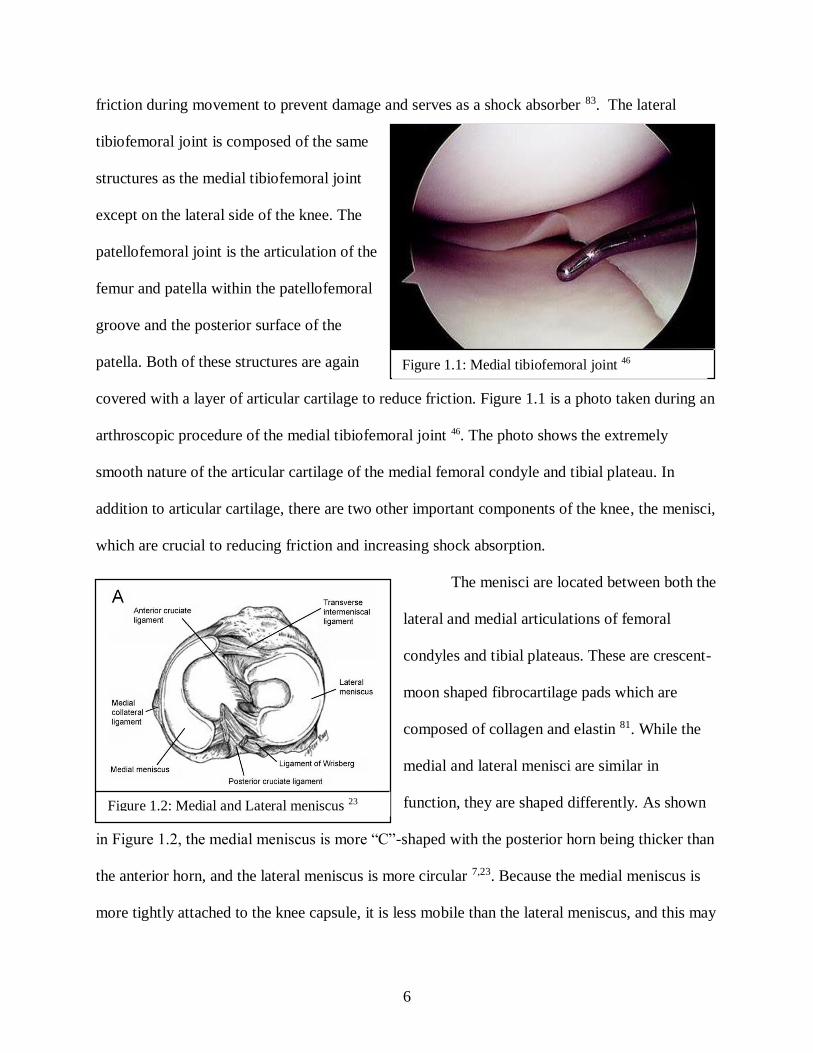
friction during movement to prevent damage and serves as a shock absorber 83. The lateral
tibiofemoral joint is composed of the same
structures as the medial tibiofemoral joint
except on the lateral side of the knee. The
patellofemoral joint is the articulation of the
femur and patella within the patellofemoral
groove and the posterior surface of the
patella. Both of these structures are again
covered with a layer of articular cartilage to reduce friction. Figure 1.1 is a photo taken during an
arthroscopic procedure of the medial tibiofemoral joint 46. The photo shows the extremely
smooth nature of the articular cartilage of the medial femoral condyle and tibial plateau. In
addition to articular cartilage, there are two other important components of the knee, the menisci,
which are crucial to reducing friction and increasing shock absorption.
The menisci are located between both the
lateral and medial articulations of femoral
condyles and tibial plateaus. These are crescent-
moon shaped fibrocartilage pads which are
composed of collagen and elastin 81. While the
medial and lateral menisci are similar in
function, they are shaped differently. As shown
in Figure 1.2, the medial meniscus is more “C”-shaped with the posterior horn being thicker than
the anterior horn, and the lateral meniscus is more circular 7,23. Because the medial meniscus is
more tightly attached to the knee capsule, it is less mobile than the lateral meniscus, and this may
Figure 1.1: Medial tibiofemoral joint 46
Figure 1.2: Medial and Lateral meniscus 23

7
be related to the observation that medial meniscal tears
occur twice as often as lateral meniscus tears 7. Blood is
supplied to the menisci from the superior and inferior
geniculate arteries, but because these are located around
the periphery of the menisci, the central sections are
mostly avascular and are supplied with nutrients from
the synovial fluid 23. Figure 1.3 shows the geniculate
arteries of the knee 23. Therefore, these areas do not heal well after an injury.
Both the medial and lateral menisci serve many important functions. First, the menisci
provide the knee with additional stability due to the fact that they form a concave structure
relative to the femoral condyles which prevents them from rolling off the tibial plateau 81. Figure
1.4, A and B, shows a simplified structure of the menisci between the femoral condyle and the
tibial plateau before and after a meniscectomy, respectively 45. Second, they act as shock
absorbers by dispersing the force
due to the weight of the body over
a larger area 23. They also decrease
contact stress between the tibial
plateau and femoral condyles
which acts to prevent damage to
the articular cartilage 23. Figure
1.4, C and D, shows the relative
measurements for peak contact
pressures of regions of the tibiofemoral joints before and after a meniscectomy, respectively 45.
Figure 1.3: Geniculate arteries 23
Figure 1.4: Structure of the meniscus and contact pressure
distribution before and after meniscectomy 45
C D
8
After the meniscectomy, the contact pressure increases significantly at the central aspect of the
joint which supports the menisci function as shock absorbers. Third, they contain nerve endings
which are important for proprioception 81.
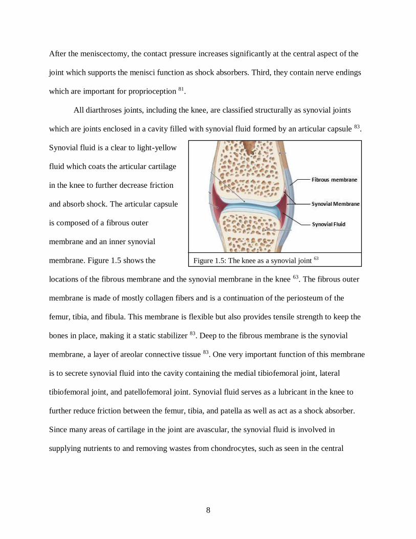
All diarthroses joints, including the knee, are classified structurally as synovial joints
which are joints enclosed in a cavity filled with synovial fluid formed by an articular capsule 83.
Synovial fluid is a clear to light-yellow
fluid which coats the articular cartilage
in the knee to further decrease friction
and absorb shock. The articular capsule
is composed of a fibrous outer
membrane and an inner synovial
membrane. Figure 1.5 shows the
locations of the fibrous membrane and the synovial membrane in the knee 63. The fibrous outer
membrane is made of mostly collagen fibers and is a continuation of the periosteum of the
femur, tibia, and fibula. This membrane is flexible but also provides tensile strength to keep the
bones in place, making it a static stabilizer 83. Deep to the fibrous membrane is the synovial
membrane, a layer of areolar connective tissue 83. One very important function of this membrane
is to secrete synovial fluid into the cavity containing the medial tibiofemoral joint, lateral
tibiofemoral joint, and patellofemoral joint. Synovial fluid serves as a lubricant in the knee to
further reduce friction between the femur, tibia, and patella as well as act as a shock absorber.
Since many areas of cartilage in the joint are avascular, the synovial fluid is involved in
supplying nutrients to and removing wastes from chondrocytes, such as seen in the central
Figure 1.5: The knee as a synovial joint 63

9
portions of the menisci. It also contains phagocytes responsible for the uptake of debris due to
normal wear-and-tear and microbes 83.
The abundant ligaments of the knee joint are also important static stabilizers. The first
type of ligaments in the knee are intracapsular, and these include the anterior cruciate ligament
(ACL) and the posterior cruciate ligament (PCL). Based on their name and location alone, it
should be known that these ligaments are located inside the articular capsule of the knee where
they cross over each other. Even though they are inside the capsule, the ACL and PCL are not
actually soaked in synovial fluid because there are folds in the synovial membrane which keeps
the ligaments isolated 83. The ACL extends from the anterior tibial eminence through the
intercondylar notch to the posteromedial side of the lateral condyle 81. The ACL is composed of
an anteromedial (AM) bundle and a posterolateral (PL) bundle which are named based on their
relative insertion locations on the tibia 84. The AM bundle contributes more to anteroposterior
knee stability while the PL bundle contributes more to rotational stability. While the main
function of the ACL is preventing the
anterior translation of the tibia from under
the femur (anterior dislocation), it has many
other secondary functions, such as
preventing internal rotation and varus,
medial flexion of the knee which is shown
in Figure 1.6 81,9. The PCL extends from the
posterior area of the tibia through the intercondylar notch to the lateral portion of the medial
condyle 83. The PCL, like the ACL, is also composed of an AL bundle and PM bundle which
differ in laxity depending on the degree of knee flexion, and contribute to the overall function of
Figure 1.6: Common deformities of the knee 9

10
the PCL, to prevent the tibia from sliding posteriorly from under the femur (posterior
dislocation) 81. Secondarily, it also functions in resisting medial flexion, lateral flexion, and
external rotation 81. Interestingly, the PCL is approximately 120-150% thicker than the ACL
which makes it less likely to tear 11.
In addition to these two intracapsular
ligaments, there are many more extracapsular
ligaments which contribute to the medial and
lateral stability of the knee.
The medial collateral ligament (MCL), also
known as the tibial collateral ligament, is a
wide, flat ligament which extends from the
medial epicondyle of the femur to the medial
condyle of the tibia 83. The deep layer of the
MCL is also firmly attached to the medial
meniscus 3. The MCL prevents lateral flexion
(valgus) of the knee 83. The posterior oblique
ligament (POL), shown in Figure 1.7, is a
thickening of the MCL which extends from the
adductor tubercle, a protuberance located
proximal to the medial condyle, to the posterior
tibia, and it also functions as a medial stabilizer
to prevent valgus 81,69. The lateral collateral ligament (LCL), also known as the fibular collateral
ligament, is a rounded ligament that extends from the lateral condyle of the femur to the lateral
Figure 1.7: View of the posterior oblique
ligament 69
Figure 1.8: Major static stabilizers of the
knee 57

11
side of the fibular head 83. The function of the LCL is to prevent medial flexion (varus) of the
knee 83. While all the ligaments are crucial to the normal function of the knee, the ACL, PCL,
MCL, and LCL are considered the major ligaments which are all shown in Figure 1.8 84,57.
1.3: DYNAMIC STABILIZERS
The hamstrings provide dynamic stabilization to the posterior aspect of the knee. These
include the semimembranosus, biceps femoris, and semitendinosus which are shown in Figure
1.9 31. The semimembranosus of the hamstrings provides posteromedial support to the knee 81.
Because it originates on the ischial
tuberosity of the hip and inserts
posteriorly at the medial condyle of the
tibia, its primary function is flexion of
the lower leg, but an extension of the
tendon at the tibia inserts more
anteriorly which gives it a secondary
function, internal rotation of the knee
49,38. Therefore, it resists excessive
external rotation of the knee. The distal
end of the semimembranosus tendon is
also connected to the posterior horn of
the medial meniscus which acts to pull
the meniscus during flexion to keep it from being pinched by the medial femoral condyle and
tibial plateau which can lead to meniscal damage 18. Another important hamstring is the biceps
femoris which originates on the ischial tuberosity and inserts at the head of the fibula and lateral
Figure 1.9: Hamstrings 31
0
0
0
semimembranosus
semitendinosus biceps femoris
(long head)
biceps femoris
(short head)

12
tibial condyle via a multiple layered tendon 18. The functions of the biceps femoris are flexion of
knee and external rotation when the knee is flexed 18. Thus, it has the opposite function of the
semimembranosus, in terms of rotation which makes sense since their insertions are on opposite
sides of the tibia 83. The third hamstring muscle is the semitendinosus which originates at the
ischial tuberosity, like the other hamstrings, and inserts medial to the tibial tuberosity at the pes
anserinus, the same location of insertion as the sartorius and gracilis 18. Some of the functions of
the semitendinosus are knee flexion and, if the knee is flexed, medial rotation 18. The tendons
which insert at the pes anserinus are important since they provide medial stability and resist
lateral stresses (valgus) 18. Two other ligaments which contribute to knee flexion are the gracilis
and sartorius, shown in Figure 1.11 62,80.
The calf muscle is another important muscle which
provides dynamic stability to the knee which include the
medial and lateral head of the gastrocnemius, shown in
Figure 1.10 50. The medial gastrocnemius originates on the
posterior aspect of the medial femoral condyle and the lateral
gastrocnemius originates on the posterior aspect of the lateral
femoral condyle, but both heads of the gastrocnemius share a
common tendon, the Achilles tendon, which inserts into the
posterior aspect of the calcaneus 83. Thus, the gastrocnemius
functions in both plantar flexion and knee flexion which are
important in dynamic stability during walking and running 83. Figure 1.10: Calf muscle 50

13
The quadriceps are additional muscles
which are important in knee extension and proper
movement of the patella. The quadriceps are
composed of the vastus medialis, vastus lateralis,
vastus intermedius, and rectus femoris 83. The
vastus medialis originates at the intertrochateric
line of the hip as well as the medial aspect of the
linea aspera of the femur 83. The vastus lateralis
originates at the greater trochanter and the lateral
aspect of the linea aspera 83. The vastus intermedius
originates at the anteriolateral surfaces of the femur shaft 83. Lastly, the rectus femoris originates
at the anterior aspect of the inferior iliac spine as well as the ilium 83. All of the quadriceps
muscles share a common tendon which inserts onto the distal patella which is connected to the
tibia via the patellar ligament 83. Therefore, all quadriceps, shown in Figure 1.11, are muscles are
involved in knee extension 62.
Two other less well known muscles involved in knee movement located deep to the
gastrocnemius are the popliteus and plantaris 83. The popliteus originates on the lateral side of the
lateral femoral condyle and inserts posteriorly at the proximal end of the tibia 83. Based on its
location, the popliteus functions in flexion and internal rotation of the knee 83. Therefore, the
popliteus is important in resisting excess external rotation. The plantaris originates at the distal
end of the femur on the supracondylar ridge of the femur and inserts posteriorly at the calcaneus
83. Thus, it is involved in plantar flexion of the foot as well as flexion of the knee 83.
Figure 1.11: Quadriceps, gracilis, and
sartorius62

14
1.4: BIOMECHANICS OF EXTENSION AND FLEXION
Contrary to popular belief, the most important function of the quadriceps, hamstrings,
and calf muscles are to produce passive forces to balance external forces acting upon the knee in
order to attain mechanical equilibrium 56. These external forces are often the result of reactive
forces due to the foot-ground contact, according to Newton’s Third Law of Motion. This ground
reaction force acts to flex knee. In response to this, the major extensors, the quadriceps will
generate tension in order to balance this force. The direction and amount of force the quadriceps
are able to exert is dependent on the angle of the patellar tendon 56. This angle is dependent on
the position of the patella which is dependent on the angle of flexion of the knee. Thus, the
amount of torque the quads are able to produce at the knee is determined by the complex
relationship between muscle fiber recruitment and degree of flexion.
In the event that the force of the quadriceps is insufficient due to the position of the
patella, the ACL and PCL are the key to preventing the tibia from sliding anteriorly or
posteriorly, respectively 56. For example, if the
flexion angle is small, the angle of the patellar
tendon is large, and the horizontal translational
force contributed by the quadriceps (labeled TH in
Figure 1.12a) is greater than that generated by the
external force (labeled W in Figure 1.12a).
Therefore, the ACL generates a tension force
(labeled A in Figure 1.12a) that is necessary in
order to keep the tibia from sliding anteriorly 56.
Also, if the flexion angle is large, the angle of the quadriceps tendon is small, and the horizontal
Figure 1.12a and 1.12b: Forces acting at the
knee while walking 56
a b
TH
TH

15
translational force generated by the external force (labeled W in Figure 1.12b) is greater than that
generated by the quadriceps force (labeled TH in Figure 1.12b). Thus, the PCL must generate a
force (labeled P in Figure 1.12b) to keep the tibia from sliding posteriorly 56. Based on both
figures 1.12a and 1.12b, it should be clear that the vertical component of the forces generated by
the quadriceps and ACL/PCL are balanced by the tibiofemoral contact force (not shown in
Figures 1.12a and 1.12b) which is distributed across the surface of the tibia by the menisci.
Because the hamstrings and gastrocnemius act as flexors,
they can also contribute to the balancing of the quadriceps and
external forces which decreases the load on the cruciate ligaments
providing a protective function to them; the amount of their
contribution relative to the cruciate ligaments is dependent on the
angle of flexion of the knee 56. For example, there is a critical
flexion angle where the forces from the major muscle and the antagonistic muscle balance each
other, and no cruciate ligament forces are necessary 56. This angle will vary depending on an
individual’s muscle strength and pattern of activation. While antagonistic muscles can serve a
protective function to a certain extent, they can also replace the function of ligaments when they
are damaged, but this can have negative consequences which will be discussed later 56. Figure
1.13 shows the hamstrings (labeled as H) acting as antagonistic muscles to the quadriceps
(labeled as T) 56.
Passive flexion of the knee is complemented by internal rotation of the tibia about its
long axis which suggests activation of the popliteus 56. The MCL is the primary restraint to
internal rotation, and the ACL is the major secondary restraint.
Figure 1.13: Dynamic forces
acting on the knee 56

16
It is important to note that the roles of the muscles can be reversed if the external force
acts to extend the knee rather than flex it. In that case, the hamstrings would be the major flexor
acting to balance the external force, and the quads could act as the antagonistic muscles which
could unload the cruciate ligaments. Passive extension of the knee is complemented by external
rotation of the tibia about its long axis which suggests activation of the biceps femoris 56. The
LCL and other lateral ligaments serve as the primary restraint to internal rotation, and the PCL is
the major secondary restraint.
While this is a simplified model since it does not include collateral ligaments and it
considers the heads of the muscles as a single collective unit, it is sufficient in demonstrating the
importance of the interaction between muscles and ligaments while walking or running.
1.5: BIOMECHANICS OF ABDUCTION AND ADDUCTION
In addition to external forces which act in the sagittal plane, there are also many external
forces which can act in the coronal plane which can
lead to abduction (valgus) and adduction (varus) of
the tibia. These forces can again be due to reactant
forces of foot-ground contact. If these forces only
have a small component in the lateral direction,
contact forces between the lateral and medial tibial
plateau and femoral condyles (labeled FL and FM,
respectively) can balance the external force
(labeled W), as shown in figure 1.14 56. However, this diagram does not consider the pressure
distribution due to the meniscus. If the load (labeled W) is directed more medially, as shown in
figure 1.15a, the lateral tibial plateau and femoral condyle are no longer in contact, so to prevent
Figure 1.14: Forces acting on the knee upon
application of slight adduction force 56

17
varus of the knee, the external force and the medial
contact force is balanced by the force generated by
the ACL (labeled A) and LCL (labeled L) instead
56. This is only possible because the tensile forces
generated by both the ACL and LCL have
components directed upward and laterally, shown in
Figure 1.15b 56. In contrast, if the load is directed
more laterally, the medial plateau and femoral
condyle are no longer in contact, so to prevent valgus of the knee, the external force and the
lateral contact force is balanced by the PCL and MCL instead 56.
It is important to note that the theoretical models described above are only applicable
when the knee is in full extension. When the knee is flexed, there is internal rotation of the tibia
which changes the angle of the ACL and, therefore, its ability to balance external forces and
muscular forces.
1.6: BIOMECHANICS OF THE MENISCI
As demonstrated in Figure 1.16,
application of a load to the menisci causes
the tissue to be pushed axially toward the
outside of the space between the tibia and
femur 12. In a normal, healthy meniscus, the
majority of the collagen fibers are oriented
circumferentially which enables the
meniscus to generate a circumferential force that is able to balance the axial force that pushes the
Figure 1.15: Forces acting on the knee
upon application of extreme adduction
force 56
Figure 1.16: Forces acting on the menisci when
bearing weight 12

18
menisci outward 44. The proper function of the menisci is dependent on their strength and
attachments to various structures of the knee via meniscal ligaments 44. The strategic placement
of these meniscal ligaments is crucial in controlling meniscal movement as the tibia and femur
move relative to each other which is necessary to keep pressures exerted on the bone surfaces to
a minimum 44.
19
CHAPTER 2: KNEE INJURIES IN SOCCER
2.1: OVERVIEW
Every soccer game is filled with a wide variety of stresses to the lower extremities of the
body. These stresses can range in their severity to cause either acute or overuse injuries. In
soccer, acute injuries (muscular and ligamentous strain) occur more often than overuse injuries
(tendonitis and synovitis) 67,55. Compared to the many injuries which can occur due to soccer,
knee injuries are known to have the most severe effects in the years following the injury. The
acute injuries to the knee can be classified as non-contact or contact in which contact refers to a
force applied directly to the knee due to contact with another player. The many quick turns and
cutting movements made consistently throughout a game leave soccer players vulnerable to
various non-contact injuries. As a contact sport, there are also multiple opportunities for contact
injuries. Of these two types of injuries, non-contact injuries tend to be more common as a result
of stress from excessive twisting of the knee 101.
2.2: CONTACT INJURIES
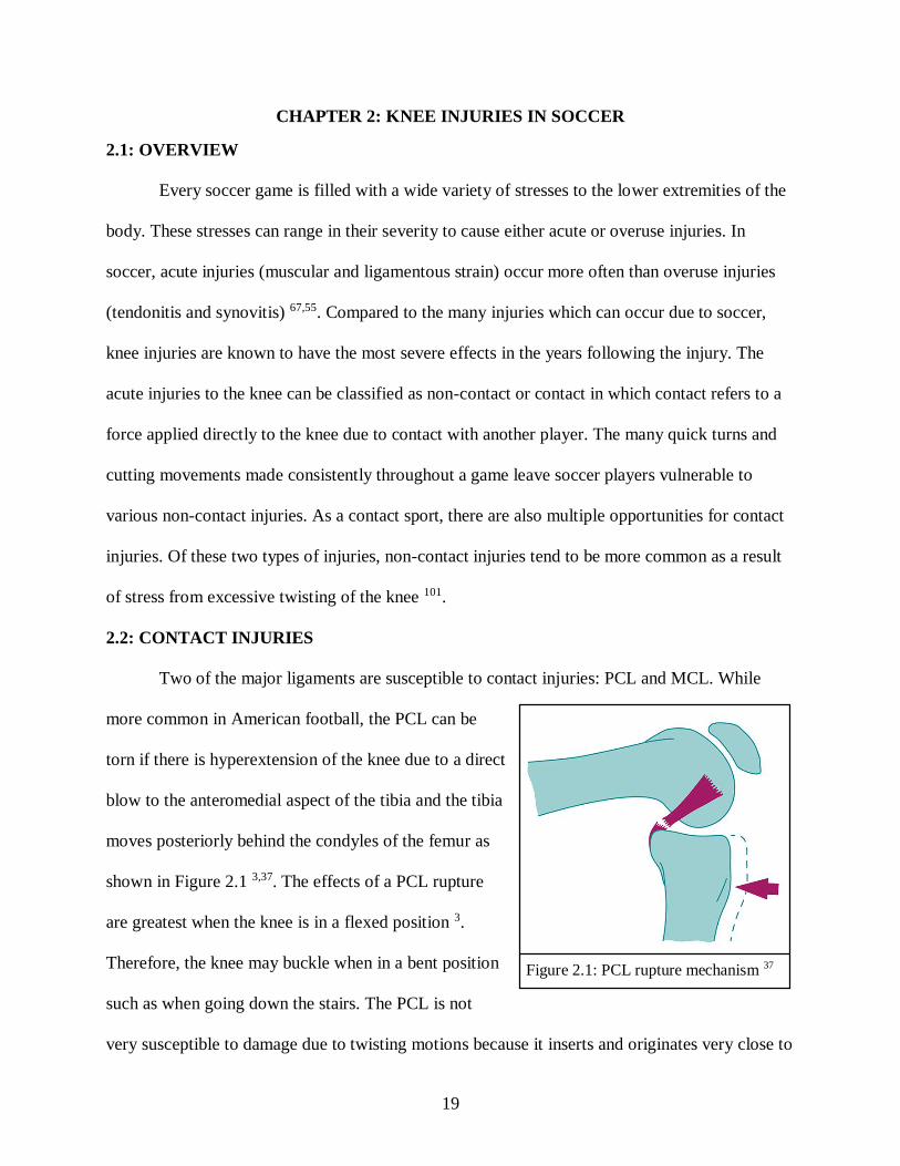
Two of the major ligaments are susceptible to contact injuries: PCL and MCL. While
more common in American football, the PCL can be
torn if there is hyperextension of the knee due to a direct
blow to the anteromedial aspect of the tibia and the tibia
moves posteriorly behind the condyles of the femur as
shown in Figure 2.1 3,37. The effects of a PCL rupture
are greatest when the knee is in a flexed position 3.
Therefore, the knee may buckle when in a bent position
such as when going down the stairs. The PCL is not
very susceptible to damage due to twisting motions because it inserts and originates very close to
Figure 2.1: PCL rupture mechanism 37

20
the axis of rotation which means the force exerted on the ligament during rotation are relatively
small 3. While a torn PCL can be an inconvenience, it may not cause pain 54.
The most common cause of an
MCL tear is a direct blow to the lateral
side of the knee which results in
stretching of the MCL past its
physiological limits, causing it to tear,
shown in Figure 2.2 54,58. Upon tearing,
the individual may hear a pop, and both
swelling and pain are common. While
the LCL can be torn in the same manner
but on the opposite side, this is less likely because the medial side of the knee is less exposed
than the lateral side. The torn MCL lacks the same amount of tensile force which causes the knee
to buckle to the side.
2.3: NON-CONTACT INJURIES
The most common injuries in soccer are non-contact and involve quick twisting motions
which frequently occur in soccer 101. The two structures of the knee most susceptible to damage
due to twisting motion are the menisci and ACL 54.
As discussed previously, the key to normal function of the menisci is their ability to
generate circumferential force in order to balance the radial force produced due to a vertical load
on the knee joint. In the event that the circumferential fibers of the menisci are weakened or the
strength of their tibial insertional ligaments are weakened, a tear is likely to occur 12,44. Because
the menisci act as secondary restraints to rotation of the knee along a vertical axis, the menisci
Figure 2.2: MCL tear mechanism 58
21
can also be torn in the event of excessive or quick rotation of the tibia which is the most common
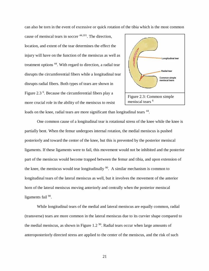
cause of meniscal tears in soccer 44,101. The direction,
location, and extent of the tear determines the effect the
injury will have on the function of the meniscus as well as
treatment options 44. With regard to direction, a radial tear
disrupts the circumferential fibers while a longitudinal tear
disrupts radial fibers. Both types of tears are shown in
Figure 2.3 6. Because the circumferential fibers play a
more crucial role in the ability of the meniscus to resist
loads on the knee, radial tears are more significant than longitudinal tears 44.
One common cause of a longitudinal tear is rotational stress of the knee while the knee is
partially bent. When the femur undergoes internal rotation, the medial meniscus is pushed
posteriorly and toward the center of the knee, but this is prevented by the posterior meniscal
ligaments. If these ligaments were to fail, this movement would not be inhibited and the posterior
part of the meniscus would become trapped between the femur and tibia, and upon extension of
the knee, the meniscus would tear longitudinally 90. A similar mechanism is common to
longitudinal tears of the lateral meniscus as well, but it involves the movement of the anterior
horn of the lateral meniscus moving anteriorly and centrally when the posterior meniscal
ligaments fail 90.
While longitudinal tears of the medial and lateral meniscus are equally common, radial
(transverse) tears are more common in the lateral meniscus due to its curvier shape compared to
the medial meniscus, as shown in Figure 1.2 90. Radial tears occur when large amounts of
anteroposteriorly directed stress are applied to the center of the meniscus, and the risk of such
Figure 2.3: Common simple
meniscal tears 6
22
stresses can increase when there is decreased mobility of the meniscus 90. One example of this
could be if the anterior horn of the meniscus became lodged between the tibial plateau and
femoral condyle during flexion which would prevent the meniscus from sliding backward upon
extension causing excess tension in the circumferential fibers 89.
Upon initial injury of the meniscus, people generally feel pain when the knee is
straightened 54. This could possibly be attributed to the decreased ability of the meniscus to
distribute pressure evenly on the tibiofemoral surface. If there is a loose fragment of the
meniscus, severe pain will be experienced whenever it is caught between the tibia and femur.
They will also experience catching or locking of the knee. Many people also experience swelling
of the knee which is due to damaged blood vessels or synovium, the synovial membrane 54.
While the symptoms can disappear over time, they often do not and will require treatment 54.
Similar to all the other ligaments of the knee, the ACL has a limit to the amount of tensile
force it is able to withstand, and when that force is exceeded, there is tearing of the ligament. As
described in chapter 1, the knee moves in such a way that the external forces and forces produced
by the muscles are able to be balanced by the tensile forces generated by the ligaments. Thus,
excessive forces applied to the knee by the muscles and external forces result in excess loading
of the ACL 97. In soccer, the ACL is subjected to the greatest stresses when a player is pivoting
or cutting where the knee is in a position of valgus, adduction, and internal rotation 51. The
anterior shear force generated by the quadriceps is the major contributor to ACL loading while
the valgus position of the knee further strains the ACL 97. Another aspect to consider is that
while pivoting, the knee is almost at full extension 8. This is important because as the angle of
flexion decreases, loading of the ACL increases which is partly due to the increase in the angle

23
of the ACL relative to the tibial plateau and the posterior direction of the tibia during extension
of the knee 97.
Interestingly, in some cases of ACL injury, a person may not experience pain and they
may hear a popping noise 54. Even if the injury is not painful, because the ACL is a major
stabilizer of the knee, injury to this ligament can cause severe instability which will require
treatment, either physical therapy or surgery depending on the extent of the tear 54.

24
CHAPTER 3: GENDER DIFFERENCES OF THE KNEE
3.1: OVERVIEW
The idea that female athletes are at a greater risk to injure their ACLs compared to males
is nothing new. In 1995, a 5-year evaluation of ACL injuries in collegiate level men’s and
women’s soccer showed that women were twice as likely to sustain a contact ACL injury and
three times as likely to sustain a non-contact ACL injury compared to men 5. Since then, the
number of females participating in sports at a competitive level has continued to increase and so
has the chance that women will withstand ACL injury compared to men. Some studies have
obtained data which show that women are up to 10 times more likely to sustain ACL injuries 15.
These significant epidemiological studies have led to numerous investigations of the possible
reasons for this increased prevalence of ACL injuries in females. The reasons can be classified as
anatomical, neuromuscular, hormonal, and biomechanical differences between the male and
female knee 28.
3.2: ANATOMICAL DIFFERENCES
One anatomical difference between the male and female
knee is that women may be more likely to have an increased
posterior tibial slope (PTS) which is defined as the angle
between the line perpendicular to the tibial shaft (A-B) and the
line parallel to the posterior to anterior inclination of the tibial
plateau (A-C), as shown in Figure 3.1 of the medial PTS 15,82.
Because the medial and lateral sides of the tibial plateau are
asymmetrical, the effects of both the medial PTS and lateral
PTS have been investigated. If the lateral PTS is steeper than the medial PTS, weight bearing is
Figure 3.1: How to
measure medial PTS 15

25
likely to cause external rotation of the femur and
internal rotation of the tibia because the lateral
femoral condyle slips off the tibial plateau which
leaves the contact point between the medial femoral
condyle and medial tibial plateau to serve as the axis
of rotation, as demonstrated in Figure 3.2 15,76. In
addition to rotation, an increased lateral PTS causes
significant anterior tibial translation 15,25. Since the
function of the ACL is to oppose internal rotation and
anterior tibial translation, it is clear why increased
lateral PTS puts excess strain on the ACL. The cutting
movements which occur on a regular basis in soccer
involve anterior tibial translation and internal rotation, so these movements are further
accentuated in people with an increase PTS.
Based on the anatomy of the tibiofemoral
joint, it is not surprising that the meniscus may also
contribute to risk factors for ACL injury in women,
specifically the meniscal slope (MS)—the angle
between the line tangential to the most superior
aspects of ipsilateral posterior and anterior menisci
and the line perpendicular to the longitudinal axis
of the tibia 15. Figure 3.3 shows the lines used to
measure the MS and PTS in the same knee 30.
Figure 3.2: Internal tibial rotation due to
increased PTS 15
Figure 3.3: How to measure medial MS
and PTS 30

26
Multiple studies have been performed which show the importance of the meniscal slope to knee
stability as described in chapter 1. In 2011, a clinical research study investigating the association
between noncontact ACL injuries and meniscal slope was completed by Hudek et al. They
observed that the lateral meniscal slope was greater among both male and female subjects with
ACL injuries compared to the control subjects, those without ACL injury 30. They also observed
that female control subjects had greater overall meniscal slopes compared to the male control
subjects 30. Thus, the meniscal slope could also possibly play a role in ACL injury mechanisms
in females. One large difference between the meniscal slope and the tibial slope is that the
meniscal slope changes depending on the knee’s degree of flexion and axial load because of the
mobility of the menisci. Unfortunately, the study described above did not account for this, as
they measured only the lateral and medial meniscal slopes at full extension without weight
bearing. Therefore, further study is required to determine more accurately the effect of the
meniscal slope on ACL injury in females compared to males.
Another difference of the tibiofemoral articulation between females and males is the size
of the femoral notch. Studies have shown that women tend to have smaller femoral notches
compared to men 15,21,72,93. Figure 3.4 demonstrates the large difference between a narrow (a) and
wide (b) femoral notch 33. While there have been many studies investigating narrow
intercondylar notch as a risk factor
for ACL injury, the methods and
results have differed. However, one
recent meta-analysis study by Zeng
et al. resulted in convincing data
that a narrow intercondylar notch is
a
b
Figure 3.4: X-ray images of a narrow intercondylar notch
(a) and much wider intercondylar notch (b) 33
27
increasingly associated with ACL injury and some have obtained data which support this
hypothesis 100,15. One proposed mechanism of ACL injury upon non-contact twisting of the knee
is that the ACL becomes pinched between the border of the femoral notch and the tibial plateau
15. Therefore, a narrower intercondylar notch would increase the likelihood of impingement of
the ACL upon twisting. Many orthopaedic surgeons support this theory as many notchplasties
are performed during ACL repair surgery to decrease the chance of re-injury 15.
Another possible anatomical difference between the structure of the male and female
knee is the actual size and strength of the ACL. According to a study performed by Anderson
and colleagues which measured the ACL cross-sectional area in 50 male and 50 female
basketball players, the ACL is smaller in females 4. While other studies have produced similar
results, the methods are not consistent, so further study is needed to come to this conclusion 15. A
study by Lipps et al. on cadaveric knees found that in addition to women having a smaller ACL
cross-sectional area compared to men, they also had fewer collagen fibrils per unit area 40. Since
collagen contributes to the tensile strength of ligaments, this observation suggests that the female
ACL is unable to withstand as great of strain. In addition, a smaller ACL experiences a greater
amount of strain in response to applied forces at the knee compared to a larger ACL which could
be because the force is not distributed across as large of a cross-sectional area 26. Based on the
information about femoral notch width and ACL cross-sectional area, there is an unfortunate
relationship between the two. If the ACL cross-sectional area is large, it experiences less strain,
but it is more likely to be impinged by the femoral condyle 15. Then if the ACL cross-sectional
area is small, it is less likely to be impinged on, but it experiences more strain 15. More studies
are needed to determine if it is worse to have a smaller, weaker ACL that is less likely to be
impinged or a larger, stronger ACL that is more likely to be impinged.

28
One final anatomical difference which has been attributed to the increased occurrence of
ACL injuries in females compared to males involves the
Quadriceps (Q) angle, the angle between the line that connects
the anterior superior iliac spine of the hip bone to the midpoint
of the patella and the line connecting the tibial tubercle to the
midpoint of the patella, as shown in Figure 3.5 26. Due to the
fact that women have wider hips than men, it is not surprising
that women have a larger Q angle as well 29. There has been
some evidence that a larger Q angle is associated with athletic
knee injury 71,26. This may be attributed to the idea that if the Q
angle is large then upon contraction of the quadriceps, there will be an increased lateral force on
the knee which could increase the amount of strain on the ACL 71,26.
3.3: BIOMECHANICAL DIFFERENCES
The biomechanical differences between the male and female knee are related to
musculature and general laxity of the knee joint. As described in Chapter 1, the major dynamic
stabilizers of the knee, including the quadriceps and hamstrings, can be activated to decrease the
strain exerted on the ACL. Studies have shown that the cross-sectional areas and overall strength
of the leg muscles associated with the knee are greater in males than females 92. This would
suggest that males are better able to decrease tension in the ACL by activating larger and
stronger muscles. In addition to differences in the overall strength of leg muscles in males
compared to females, there is also a difference in the quadriceps to hamstring strength ratio in
males and females 1. Some studies have shown that the hamstrings are significantly weaker than
the quadriceps in females while males do not exhibit this quadriceps dominance 60,79,1.
Figure 3.5: How to measure
Q angle 47

29
Contraction of the quadriceps muscles pulls the tibia anteriorly, and contraction of the hamstring
muscles pulls the tibia posteriorly. The insertion sites of the hamstring tendon on the tibia and
quadriceps tendon on the patella which is connected to the tibia via the patellar tendon are shown
in Figure 3.6 59. Therefore, if the anterior force generated by the quadriceps is greater than the
posterior force generated by the
hamstrings, the forces acting on the
ACL will increase resulting in an
increased risk of rupture. This
discovery could have a significant
impact on treatment and prevention of
such injuries because it could indicate
that hamstring strengthening could be
the key to decreasing the incidence of
ACL injuries in females 1. In addition to weaker hamstrings in females compared to males, the
results of a study using the sit-and-reach test (where a patient sits down with their legs out
straight in front of them and they try to reach past their toes) demonstrated that hamstring
flexibility increases in females after puberty while it decreases in males 28. Based on the
importance of the hamstring muscles in opposing anterior strain of the ACL, it is not surprising
that the few studies performed which have investigated the effect of hamstring flexibility
indicate that it could contribute to a decrease in dynamic stability 28. One proposed reason for
this is that lax hamstrings could lead to delayed activation resulting in quadriceps dominance 28.
While the leg muscles play an important role in dynamic stability of the knee, the
musculotendinous structures which cross the knee joint may also provide passive resistance to
Figure 3.6: Diagram of the anatagonistic relationship
between the hamstrings and quadriceps 59

30
displacement through joint compression 74. A study by Shultz and colleagues demonstrated a
strong association between lower extremity lean (muscle) mass and laxity in the frontal and
transverse planes of the knee 74. Laxity in the frontal plane means the knee is more susceptible to
forces which result in varus or valgus, and laxity in the transverse plane means the knee is unable
to generate the torsional stiffness necessary to oppose forces which cause internal or external
rotation 74,92. In addition to the musculotendionous structures, the ligaments and capsule of the
knee also play a large role in knee laxity, a measure of the ability to resist a displacing load 74.
An important study by Scerpella and colleagues assessed generalized ligamentous laxity in
collegiate athletes with non-contact ACL injuries and those without, as a control 70. For their
study, they used the Beighton nine-point scale which attributes points for the following:
hyperextension of the knee beyond -10°, hyperextension of the elbow beyond -10°, ability of the
thumb to touch the forearm with
wrist flexion, extension of the fifth
MCP (metacarpophalangeal) joint
past 90°, and ability of the palms to
touch the floor with complete
extension of the knees; they also
measured AP (anterior-posterior) tibial translation 70. Some of these movements are shown in
Figure 3.7 20. The study demonstrated a significant association between generalized ligamentous
laxity and AP laxity with non-contact ACL injuries, and it also showed an increased generalized
ligamentous laxity and AP laxity in females compared to males 70. As a pivoting sport, soccer
requires rapid deceleration and acceleration movements which exert an extreme amount of forces
on the tibia. Since there is increase AP laxity in females, the tibia could possibly translate further
Figure 3.7: Tests to measure generalized ligamentous laxity 20
31
before the muscles can be activated to oppose the displacement 28. The further the tibia
translates, the greater the strain on the ACL. While there is little evidence, another interesting
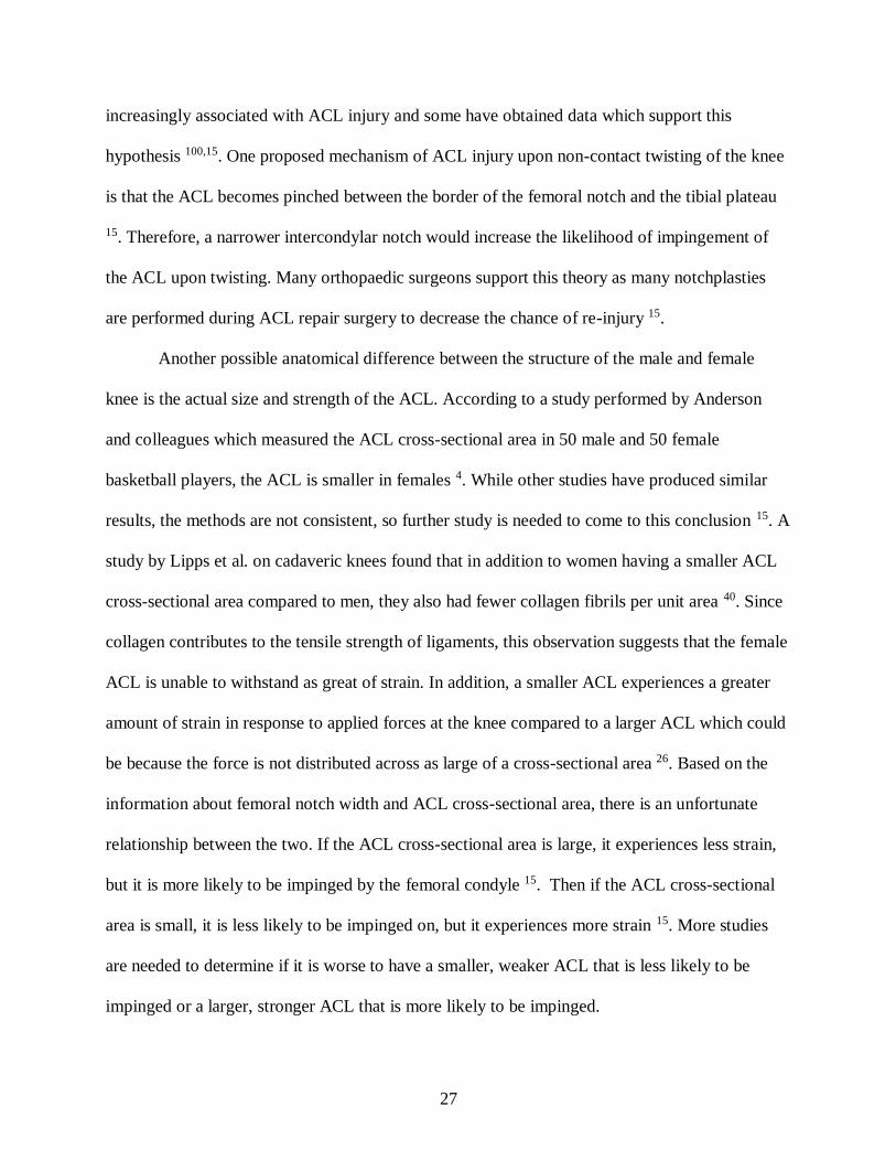
example of joint laxity’s effect on tibial translation is related to increased foot pronation. In
1996, a study performed by
Janice Loudon and colleagues
showed excessive navicular
drop, a measure of foot
pronation shown in Figure
3.8, in female athletes with
ACL injuries as opposed to those without 28,42,73. In 2002, another study performed by Trimble
and colleagues indicated that increased foot pronation could result in anterior translation of the
tibia 28,85. Given the importance of both muscle strength, muscle mass, and ligamentous laxity in
knee stability, it is easy to see how females could be at a disadvantage when it comes to both
passive and dynamic stability and why there is an increased incidence of ACL injuries in
females.
3.4: NEUROMUSCULAR DIFFERENCES
Although muscle mass and overall strength play an important role in dynamic stability of
the knee, the recruitment pattern of muscle fibers is equally significant. As indicated previously,
if there is an imbalance in the forces generated by the hamstrings and quadriceps, there is
increased stress on the ACL. This can also happen if there is disproportional recruitment of these
muscles, a neuromuscular problem. Multiple studies have revealed such neuromuscular
imbalances in females compared to males using electromyography, such as increased activation
of the quadriceps and decreased activation of hamstrings relative to each other 28. In addition to
Figure 3.8: Diagram demonstrating navicular drop 73

32
proportional recruitment of quadricep and hamstring muscle fibers, the pattern of recruitment is
also important. One study by Cowling and Steele showed that while female hamstrings are
activated sooner than male hamstrings during landing exercises, they suspect that the male
activation pattern shows more synchrony between the quadriceps and hamstrings 16,28. If the
quadriceps and hamstrings are not activated at the same time, then there is an imbalance of the
anterior-posterior forces acting on the knee.
In addition to differences in the activation patterns of both the quadriceps and hamstrings
in females, there are also differences in the activation patterns of, specifically, the medial and
lateral quadriceps. A study performed in 2005 by Gregory Myer and colleagues demonstrated
decreased activation of the medial quadriceps compared to the lateral quadriceps in females
compared to their male counterparts 53. Co-contraction of medial and lateral quadriceps cause
compression of the knee joint which increases the articular contact forces between the tibal
plateau and the femoral chondyles 53,28. These forces contribute to the ability of the knee to
oppose valgus-varus movement and anterior-posterior translation 53,28. If there is a decreased
medial-to-lateral quadriceps activation ratio, there will be less medial compression which can
contribute to dynamic knee valgus demonstrated by females when executing cutting maneuvers
in soccer 53. On top of unbalanced medial-lateral quadriceps activation, multiple studies have
also shown increased lateral hamstring activation in females 28,68. This will decrease medial
compression and limit the knee’s ability to resist valgus even more.
Because the ACL is highly innervated and has many mechanoreceptors, it plays an active
role in dynamic stabilization of the knee. In 1999, a study by Susan Rozzi and colleagues showed
decreased proprioception at the knee in females compared to males because women took
significantly longer to detect knee joint extension 28,68. Rozzi et al. infer that the decreased

33
proprioception in females may be due to increased ligamentous laxity which makes sense since
stretch receptors would undoubtedly be affected if the ligaments are inherently less taut in
females 68. A test performed by Solomonow and colleagues showed that ACL stress moderately
inhibits the quadriceps and activates the hamstrings, showing that the ACL is involved in a
stretch reflex 43.
Along with decreased proprioception to extension of the knee joint, a recent study
performed by Song Lee and colleagues showed that females have lower proprioception in
pivoting under weightbearing conditions using
an offaxis elliptical trainer 39. Women took
longer to detect both internal and external
pivoting movements controled by a motor which
moved the footplate 1°/second 39. Figure 3.9
shows an overhead view of the pivoting
footplates 65. This important discovery suggests
that women may be less able to detect pivoting
positions which could potentially cause injury 39. Again, this difference in proprioception could
potentially be due to the generalized ligamentous laxity exhibited in females 68. Lee and
colleagues also tested lower leg pivoting stiffness by using motors to generate pivoting
perturbations and having the subjects try to maintain a target position 68,39. From this test, they
were the first scientists to show that females generate lower leg pivoting stiffness than males
which could be an indicator of decreased neuromuscular control 68.
Figure 3.9: Overhead view of the pivoting
footplates used to measure proprioception 65

34
3.5: HORMONAL DIFFERENCES
Considering the most obvious difference between males and females involves the
reproductive system, the hormonal differences between males and females is likely to play a role
in the increased prevalence of knee injuries in females. Unfortunately, the actual effects of the
different sex hormones in males and females are the most controversial compared to the
anatomical, biomechanical, and neuromuscular differences. The two main types of female
hormones secreted by the ovaries are estrogens (estradiol and estrone) and progesterone, and the
male hormone secreted by the testes is testosterone 60,83. Figure 3.10 shows the very similar
chemical structures
of estradiol,
progesterone, and
testosterone 86. These
sex hormones are all
steroid hormones which have a wide variety of functions. Estrogens and progesterone function to
regulate oogenesis and the female reproductive cycle, maintain pregnancy, and promote the
development and maintenance of female secondary sex characteristics 83. Testosterone regulates
spermatogenesis and promotes the development and maintenance of male secondary sex
characteristics 83. As described previously, increased knee joint laxity in females may contribute
to their increased ACL injury rates. Some recent studies have shown that the rhythmic changes in
hormone levels during the menstrual cycle may have a significant influence on knee joint laxity
61.
Between 1996 and 1997, multiple discoveries were made regarding sex hormone
receptors in fibroblasts in the human ACL. In 1996, Stephen H. Liu and colleagues discovered
Figure 3.10: Structures of female and male steroid hormones 86

35
the existence of estrogen and progesterone receptors, and in 1997, Ward P. Hamlet and
colleagues discovered androgen receptors 41,27. Both studies utilized immunolocalization 41,27.
Because fibroblasts produce and secrete collagen, they are responsible for the majority of the
load-bearing function of the ACL 60. Therefore, the presence of estrogen and androgen receptors
in fibroblasts suggests estrogen, progesterone, and testosterone could have an impact on the
laxity of the ACL.
Another study by Liu et al. showed a significant decrease in fibroblast proliferation and
collagen synthesis in rabbit ACLs when exposed to increased, but still physiologic, levels of
17β-estradiol, an estrogen 41. Thus, the net effect of increased estrogen during the follicular
phase could alter the composition of the ACL, increasing the risk for injury 78,28. While the study
by Liu et al. investigated the effects of estrogen alone, a study by Warren D. Yu and colleagues
investigated the effects of estrogen and progesterone separately and in combination on fibroblast
proliferation and collagen synthesis 98,79. This study is important because, as demonstrated in
Figure 3.11, estrogens and progesterone are released in combination but at different levels during
the female reproductive cycle 28. The study by Yu et al. showed that increased estradiol levels
alone resulted in decreased fibroblast proliferation and procollagen type I synthesis. However, as
levels of progesterone
were increased in
combination with
estrogen, there was a
decreased inhibitory
effect which implies
that progesterone lessens the effects of estrogen 98. One interesting explanation for this
Figure 3.11: Graph of various hormone levels versus menstrual day/cycle 28

36
antagonistic relationship between estrogen and progesterone was described by W. Lee Kraus and
colleagues using a model system in 1996 98,36. Their model showed that an activated
progesterone receptor complex interferes with an estrogen receptor’s ability to interact properly
with transcription factors, a process referred to as quenching 98,36. Unlike estrogen and
progesterone levels in females which are cyclic, testosterone levels in males peak at 25 years old
and decrease with age 27. In 1977, Yamamuro et al. found a positive correlation between collagen
content and levels of testosterone in the hip joint capsule of male rats 27,96. This could suggest a
similar relationship between collagen content and testosterone levels on the knee joint.
There have been many studies which investigate knee laxity during the different phases
of the menstrual cycle, but the results have widely varied. Some studies show increased laxity of
the ACL near ovulation or post-ovulation while others showed no significant change in any of
the phases 61,99. Based on the studies which showed that estrogen decreases fibroblast
proliferation and collagen synthesis, it is logical that ACL
laxity would be lowest near ovulation when estrogen
levels peak. In a systematic review of nine studies which
investigate the hormone cycle and ACL laxity relationship,
Zazulak and colleagues identified multiple limitations
which could possibly explain their conflicting results 61,99.
These limitations include small sample size, unspecific
subject criteria, and variable testing methods. The meta-
analysis performed by Zazulak et al. suggested the
menstrual cycle may have a significant effect on ACL
laxity 99. The results of one study performed by Park et al. which addressed the limitations
Figure 3.12: Chart showing the
average displacement of the
ACL at different phases of the
menstrual cycle 61

37
described by Zazulak et al. showed that when an anterior displacement force of 89 N was applied
to the knee of female subjects in the ovulation phase, anterior tibial displacement was signicantly
greater than when measured in the luteal phase (see Figure 3.13) 61.
In addition to inconsistencies in studies regarding knee laxity and the hormone cycle,
there are inconsistencies between studies about knee laxity and injury findings. For example,
studies about injury indicate that ACL injuries occurred more often during the pre-ovulatory
phase 99. This conflicts with the hypothesis that the increased prevalence of knee injuries in
females is due to increased laxity since laxity is expected to be least when estrogen is low, such
as during the pre-ovulatory phase. Thus, the injury data supports the case that changes in
hormone concentrations during the menstrual cycle do not influence knee laxity. However, given
the data that demonstrates a direct relationship between estrogen levels and fibroblast
proliferation and collagen synthesis, the injury data could simply suggest that there are multiple
factors involved. This is a more likely explanation considering the many studies which
investigate the anatomical, biomechanical, and neuromuscular differences between the male and
female knee.

38
CHAPTER 4: DIAGNOSIS AND TREATMENT OF SOCCER KNEE INJURIES
4.1: OVERVIEW
While it is never pleasant to tear a knee ligament or meniscus, it is an injury that is easily
diagnosed and treatable with time. Unlike with a disease or infection, doctors are almost always
able to see or feel the problem. The special tests orthopaedic surgeons use on a daily bases are
almost as accurate as an MRI, and both options are non-invasive. Treatment may not be quite as
simple as the diagnosis, but the advancements that have been made in surgical techniques have
made surgery a valid option, especially for ACL reconstruction where a golden standard has
been set. Even though surgery has become more effective, physicians and patients have to
always consider nonoperative treatment which can be just as effective and comes with fewer
risks.
4.2: DIAGNOSIS
The first step to any diagnosis is talking to the patient to learn about their history. Some
of the important questions asked by
physicians are shown in Table 4.1 13. It is
imperative for the physician to know about
both the nature of the injury, symptoms, and
any problems experienced before the
incidence to come to the correct diagnosis 13.
The next step is a comprehensive physical
examination of both the uninjured leg and the injured leg. It is imperative that the physician first
examines the uninjured leg to establish the patient’s relative norms. This thorough physical
examination involves observation, assessment of range of motion, palpation, a neurovascular
Table 4.1: Key questions asked by orthopaedic
surgeons following a knee injury 13

39
examination, and special tests 13. X-rays and MRIs can
also serve as important tools for diagnosing ligament
tears, meniscal tears, bone bruises, and more 88. While
each of the aspects of the physical examination play an
important role in diagnosing soccer related injuries, the
focus in the following paragraphs will be on the special
tests described by Kerri Browne, MSBS, PA-C and
Christopher A. Kurtz, MD in their CME (continuing
medical education) article. The valgus stress test and the
varus stress test are used to assess the integrity of the
MCL and LCL, respectively. In the valgus stress test, the physician pushes the knee inward with
one hand while pushing the ankle outward with the other hand, as shown in Figure 4.1 13. They
do this first with the knee at 0° of flexion and then again at 30° of flexion. In the varus stress test,
the physician pushes the knee outward while pushing the ankle inward, the opposite of the valgus
stress test 13. Again, they do this with the knee at 0° of flexion and then again at 30° of flexion.
Increased laxity at 30° indicates an isolated collateral ligament injury (MCL or LCL depending
on the test) while increased laxity at 0° indicates cruciate ligament injury in addition to collateral
ligament injury 13.
Observations, range of motion (ROM), palpation, and multiple tests are the key to
diagnosing meniscal tears. If a patient is unable to fully extend the knee and/or they are sensitive
to palpation along the lateral or medial joint line, this is an indication of a meniscal tear 13. In the
Apley compression test, the patient lies on their stomach with their knee flexed 90°. The
physician then rotates the tibia externally and internally while applying downward pressure, as
Figure 4.1: Valgus stress test 13

40
shown in Figure 4.2 13. If there is clicking or the patient
experiences pain, the test indicates a meniscus injury 1.
McMurray’s test is another method used to diagnose a
meniscal tear 13. In this test, the patient lies on their back
with the knee fully flexed. The doctor rotates the foot
outward and applies an inward force while passively
straightening the leg 13. Any clicking is indicative of a
lateral meniscal tear. If the doctor, instead, rotates the foot
inward and applies an outward force while passively
straightening the leg, this tests for a medial meniscal tear
13. There are three tests used to diagnose ACL tears. The first is the anterior drawer test 13. The
patient lies in the supine position with the knee flexed at a right angle, and the doctor stabilizes
the patient’s foot by sitting on it 13.
To test the integrity of the ACL, the physician pulls the tibia anteriorly at the proximal
end of the tibia while palpating the knee along the
medial and lateral anterior joint lines to determine the
extent of anterior tibia translation, as shown in Figure
4.3 13. Increased laxity of the injured leg is suggestive of
an ACL injury. The second and preferred test is the
Lachman’s test 13. In this test, the patient lies in the
supine position with their leg at rest over the physician’s
leg. The physician flexes the knee between 20° and 30°, stabilizes the femur with their hand, and
pulls the tibia anteriorly with the other hand, as shown in Figure 4.4 13. Again, increased laxity of
Figure 4.2: Apley compression
test 13
Figure 4.3: Anterior drawer test 13

41
the injured leg is suggestive of an ACL injury. The
pivot shift test is a third test which evaluates the
capacity of the ACL to resist tibia rotation 13. In this
test, the patient lies on their back with their knee fully
extended. Similar to the valgus stress test, the
physician pushes the knee inward and rotates the tibia,
but they also apply an upwards force 13. The physician
slowly bends the knee, and if the physician feels
anterior shift of the tibia or hears a clunk at about 30° of flexion, this is a positive test for an
ACL tear 13.
There are also three tests used to diagnose PCL tears. The first is the posterior drawer test
which is performed in similar manner as the anterior drawer test except the physician wraps their
hands around the tibia and pushes it forward into posterior translation 13. Increased laxity of the
injured leg compared to the other is suggestive of a PCL injury. The second test is the reverse
pivot shift test 13. In this test, the patient lies in the supine position while the physician holds their
leg at 45° of flexion, rotates their tibia outward, and applies an inward force on their tibia while
moving the knee into extension 13. Again, if the
physician feels the tibia shift anteriorly or hears a
clunk, this result is indicative of PCL insufficiency.
The posterior sag test is the third test in which the
patient lies on their back with their hip and knee flexed
at 90° 13. If the PCL is insufficient, the tibia will sag
below the femur, as shown in Figure 4.5 13.
Figure 4.5: Posterior sag test 13
Figure 4.4: Lachman’s test 13

42
4.3: TORN PCL TREATMENT
Treatment of the PCL is dependent on a combination of factors: severity of the knee
injury, the grade of the tear, symptoms, and activity level. The first treatment option is
conservative, consisting of physical therapy which focuses on strengthening the quadriceps and
proprioceptive training. By increasing the strength of the quadriceps, there will be increased
dynamic posterior control of the tibia, putting less stress on the PCL. The proprioceptive training
will enhance the ability of the body to sense posterior translation of the tibia and adequately
activate the quadriceps. Conservative treatment is recommended if the knee injury is isolated to
the PCL and the tear is only partial 19. Conservative treatment is initially recommended even if
the injury is isolated to the PCL but is more severe, almost completely torn. In fact, the treatment
is even more conservative because the risk of re-injury is increased 19. Traditionally, the knee is
completely immobilized for 2-4 weeks with the leg extended or a dynamic PCL brace is worn for
3 months 19. When immobilization is recommended, the leg is always extended because the PCL
is in a relaxed position, making the ligament tauter upon healing 19. If the patient’s symptoms do
not improve with therapy and they experience prolonged pain, the injury may eventually require
PCL reconstruction 19.
The second treatment option is surgery in which the PCL is reconstructed. This treatment
is recommended when damage occurs to both the PCL and posterolateral corner, the collection
of tendons, ligaments, and muscles which are located in the posterolateral corner of the knee 19.
This is because this injury generally causes physical limitations to movement and can possibly
lead to arthritis 19. Unfortunately, unlike with ACL reconstruction, there is no “golden standard”
for PCL reconstruction 19. Due to variations in PCL reconstruction results, there is much debate
over single versus double-bundle reconstruction, graft tension, tibial insertion location, and

43
femoral tunnel placement 19. Similar to ACL reconstructions, grafts can either come from
tendons in the patient (autograft) or another person (allograft). Allografts are opted for when the
patient demonstrates general ligamentous laxity, but they have some disadvantages 19. They take
much longer to revascularize and mature 19.
Following surgical treatment, activation of the hamstrings should be avoided because this
can cause further injury or stretch the graft, decreasing its strength. Instead, physical therapy
would focus on strengthening the quadriceps. Athletes are able to play sports once they have
adequate quadriceps strength and no pain which, for a typical double-bundle PCL reconstruction,
takes between 6 to 9 months 19.
4.4: TORN ACL TREATMENT
Similar to PCL injuries, there are two treatment options for ACL injuries: conservative
and surgical management. Physicians decide on the treatment type based on the patient’s overall
health, symptoms and lifestyle 88. Conservative treatment is opted for when the patient has
serious comorbidities which makes failure of the graft likely or the surgery dangerous 88.
Nonoperative treatment is also chosen when the patient is asymptomatic with daily activities and
leads a sedentary lifestyle 88. Therefore, surgery is unnecessary for a sedentary individual but
very much necessary for an athlete who play sports where jumping, pivoting, and cutting are a
common occurrence 88. Another indication for ACL reconstruction surgery are patients which
return to moderate activity and often have incidents of their knee giving way 88. This giving way
can cause meniscal tears and pain. Another case which requires surgery is patients with
hypermobility, lax secondary restraints 88. These patients often experience instability with their
daily activities.

44
If a patient is diagnosed with an ACL tear and their orthopaedic surgeon determines that
surgery is the best treatment option, the patient generally has to wait two months before surgery.
Surgeons elect to have their patients have an initial period of rehabilitation for their knee motion
and quadriceps strength to return to normal 88. Studies have shown that delayed surgery is
associated with a decreased rate of surgical complications and quicker return of knee movement
and muscle function after the surgery 88. In those two months before surgery, the physician and
the patient select a type of graft for the reconstruction. As discussed previously, the options are
an autograft or allograft. The autograft source options are the patellar tendon, hamstring tendon,
or quadriceps tendon 88. Each source has its advantages and disadvantages but the advantages of
the patellar tendon graft make it the “gold standard.” It has great initial and long term fixation
and overall stability. While the disadvantages of the patellar tendon graft include increased
anterior knee pain and quadriceps atrophy, this could possibly be an advantage for women since
they tend to exhibit quadriceps dominance 88,1. The allograft source options are the Achille’s
tendon, patellar tendon, quadriceps tendon, tibialis tendon, and hamstring tendon 19. Some
benefits of allografts include shorter surgery times, the ability to perform multiple ligament
reconstruction, and the availability of a large size range 88. One of the problems with an allograft
is the increased possibility of disease transmission. While the likelihood of this is extremely
small with modern sterilization procedures, irradiation and ethylene oxide, these same
procedures significantly weaken the graft 88. Thus, allografts have a higher failure rate and a
slower healing rate 88.
Rather than discuss the step by step technique used by orthopaedic surgeons to
reconstruct an ACL, it is more useful to discuss the important aspects of the technique used by
surgeons and how this relates back to the normal anatomy and biomechanics of the knee. One

45
important aspect of the ACL reconstruction technique is the topic of notchplasty, surgical
procedure in which the size of the intercondylar notch is increased 88. Some surgeons perform
this for better visualization during surgery as well as ensure the graft is not impinged or abraded
88. For some patients, notchplasty may not be necessary.
The second important aspect of an ACL reconstruction surgery which can vary slightly
depending on the patient and surgeon is the tunnel placement for the graft 88. When drilling into
the tibia, the tunnel should begin medial to the tibial tubercle where the bone quality is best for
drilling, and the angle should be at specifically 60° or 65° 88. A drill guide is used to assist with
proper placement in the tibia. Upon exiting the tibia at the articular cartilage, the tunnel exit
should be slightly medial to the original ACL tibial insertion 24. The tibial tunnel placement is
very important for ensuring the graft is not impinged. The orientation of the femoral tunnel is
also important for the successful function of the graft. If the tunnel is placed at the 11 o’clock
position in the right leg or 1 o’clock position in the left leg, the graft replicates the anteromedial
bundle, resisting anterior translation 24. If the tunnel is placed at the 10 o’clock or 2 o’clock
position, the graft replicates the posteromedial bundle, resisting rotational knee movement 24.
One very controversial topic in ACL reconstruction is the idea of single versus double
bundle reconstruction 88. While a traditional ACL reconstruction only consists of a single-bundle,
there is a push more recently towards double-bundle grafts that resemble the AM and PL bundles
of the original ACL 88. This sounds reasonable in theory, but recent research suggests that the
double-bundle technique does not yield superior results in terms of knee stability 88.
The final variable aspect of ACL reconstruction surgery is how much the graft is
tensioned upon fixation 88. This depends on multiple factors: the type of graft, laxity of the knee,
and the fixation technique 88. For instance, a patellar tendon graft is very stiff, so it needs to be

46
fixated when the leg is extended to ensure that the graft stretches enough 88. For other grafts,
fixation is done while the leg is flexed because the graft will reach proper tension upon extending
the leg 88.
Following the initial recuperation phase after surgery, patients perform exercises to
strengthen the quadriceps and hamstrings. Some exercises include walking, stationary cycling,
and swimming 88. About 3-4 months after surgery, patients can generally start to run. The time to
return to sports is roughly 6 months 88. Recovery time is completely dependent on the patient.
4.5: TORN MCL TREATMENT
Treatment of the MCL is dependent on the severity of the knee injury, grade of the tear,
and symptoms. Usually, isolated MCL injuries occur when there is a direct hit to the lateral side
of the knee. This high impact injury generally results in a grade III tear to the MCL. Similar to
the treatment of PCL tears, treatment of grade III MCL tears is controversial 77,94. Traditionally,
grade III tears were treated surgically, but recent research suggests that nonoperative treatment
could be a viable option 77,94. In contrast, low-grade MCL tears, generally the result of non-
contact injuries, are treated non-operatively 77,94.
Treatment of MCL tears becomes even more controversial when other ligaments or
structures of the knee are involved. If both the ACL and MCL are torn, there are is a sequence of
treatment followed. First, depending on the grade of the tears, both ligaments are treated
nonoperatively with a brace 77. If the MCL heals, and the knee is medially stable, the ACL is
reconstructed 77. If the MCL does not heal, and the knee is still not medially stable, both
ligaments are reconstructed 77. If the MCL and the posteromedial capsule are injured, they should
be surgically repaired or reconstructed because the PMC contributes largely to medial stability of

47
the knee 77. In addition, studies have shown that a damaged PMC leads to poor outcomes of
MCL repairs/reconstructions 77.
As usual, there are multiple techniques used and
graft options for reconstructing the MCL. One interesting
technique presented by Kim et al. is reconstruction of both
the MCL and POL, a component of the posterior medial
capsule, using a single autograft from the semitendinosus
tendon 35,77. The graft remains attached to the tibia in its
original anatomic location, and using a screw and washer
inserted on the medial epicondyle, the graft is wrapped
around the screw and sutured to the head of the
semimembranosus tendon, as shown in Figure 4.6 35,77. Such as for all grafts, tensioning of the
graft varies. This technique has proved successful in restoring medial stability of the knee 35,77.
4.6: TORN LCL TREATMENT
Isolated grade I and II LCL tears are treated nonoperatively while treatment of a grade III
tear is controversial 14. One study by Bushnell et al. about LCL tears in football players showed
that nonoperative treatment of isolated grade III tears allowed players to return to football sooner
than with surgery 14. Thus, patients can avoid the dangers of surgery and have an equal chance of
returning to football in a shorter amount of time. As tempting as this treatment option sounds, the
study used a small sample size, so more studies are needed before a “golden rule” for isolated
grade III LCL tears can be identified 14. Isolated LCL injuries are rare, and other structures of the
knee are often damaged as well 14. The more complex the injury, the more complex the
treatment.
Figure 4.6: Proposed technique for
MCL reconstruction 35
48
If surgery is necessary, there are multiple techniques and graft options for surgeons to
choose from. Some of these options include a semitendinosus graft, quadriceps tendon graft,
Achilles tendon graft 14,22. These grafts can be used with or without a bone plug 14.
Unfortunately, there have been minimal clinical studies that compare different LCL
reconstruction techniques, and for those studies that have, the patients have had associated ACL,
PCL, or other injuries 14.
4.7: TORN MENISCUS TREATMENT
Treatment of meniscal tears is dependent on the patient’s symptoms as well as the
location and severity of the tear 48. Non-operative treatment is effective for meniscal injuries,
particularly those that are degenerative and complex 48. Conservative treatment involves
exercises that strengthen the muscles of the leg, increase flexibility, and improve proprioception
48. Because this treatment is often effective, most orthopaedic surgeons initially suggest non-
operative treatment, and only if the patient continues to experience pain and symptoms does a
physician recommend surgery 48.
There are two options for surgery: meniscectomy and meniscal repair. The surgeon’s
recommendation depends on the location and severity of the tear 95. Because of the importance of
the meniscus in knee stability, surgeons try to conserve as much of the meniscus as possible 95.
The peripheral portions of the meniscus, referred to as the red-red portion by most texts, is rich
in blood supply due to the adjacent geniculate arteries. This blood supply makes this region the
most likely region to heal 95. Therefore, tears in the peripheral region are repaired with a suture
95. If the tear occurs in a more central portion of the meniscus, a partial meniscectomy is
performed because the injury is unlikely to heal. It is important that the rim of the meniscus is
preserved because this is the most important region for knee stability 95. While the initial

49
outcomes of partial meniscectomy procedures are overall positive, studies have shown that they
result in accelerated degenerative changes at the knee 95. There has been a recent push towards
meniscal repair over meniscectomy when possible which has led to advancements in repair
techniques 95. For instance, recently
developed devices allow sutures to be
added without accessory incisions
that could be damaging to the neuro-
vasculature 95. However, there are
instances where the location of the
tear makes techniques using accessory incisions necessary, the inside out or outside in techniques
95. Figure 4.7 shows the location of the cannula and sutures used in the inside out, outside in, and
all inside techniques 95,91.
Figure 4.7: Three techniques used to repair meniscus
tears 91

50
CHAPTER 5: KNEE INJURY PREVENTION
5.1: OVERVIEW
As discussed in chapter 2, there are two types of knee injury mechanisms: contact and
non-contact. The only way athletes can prevent contact injuries is to avoid physical contact with
other athletes which is impossible when playing contact sports, such as soccer, basketball, and
football. In contrast, there is evidence that non-contact ACL injuries can be prevented with
various programs.
5.2: PREVENTION PROGRAMS
According to a recent review which investigated the effects of prevention programs on
knee injuries, it was concluded that the prevention programs studied were ineffective at reducing
knee injuries overall and ACL injuries in male soccer players 2. In addition, the review also
concluded that the prevention programs were not effective at reducing overall knee injuries in
female soccer players but were effective at reducing ACL injuries 2. The focus of the prevention
programs was on neuromuscular training and muscle strengthening which target the
biomechanical and neuromuscular risk factors which put females at risk for ACL tears. Unlike
the female anatomical and hormonal risk factors, the biomechanical and neuromuscular risk
factors are more readily modifiable.
There are multiple types of exercises used for neuromuscular training. Two important
examples are balance and plyometric exercises. The balance board, shown in Figure 5.1, used in
physical therapy sessions is a good way to work on proprioception, 2,87. This and other exercises
that target proprioception are especially important in females because they have shown decreased
proprioception compared to their male counterparts. Plyometric training involves jumping
exercises which use only body weight as the resistance 10. These exercises exploit the stretch-

51
shortening .cycle of muscles which allows the
muscles to exert more force upon stretching due to
their elastic properties 10. Plyometrics are an excellent
way to enhance muscle recruitment and mechanics 2.
Figure 5.2 shows an example of a plyometric exercise
34. These exercises are extremely important in females
because they have shown increased activation of the
quadriceps compared to the hamstrings and initial
activation of the quadriceps before the hamstrings.
Plyometric exercises address these risk factors by
training the muscles, nervous system, and connective
tissue to appropriately respond to ligamentous
stretching and promote improved body mechanics 2.
It is important to progressively add perturbations in
order to check for proper form when the athlete is in a
more game like situation 2.
Strength training is another important
component of the prevention programs which reduce
the risk of ACL injury in females. Because females
have shown quadriceps dominance, hamstring
strengthening exercises are particularly important. By
strengthening the hamstring muscles, less stress is put on the ACL since they are better able to
counteract the anterior force generated by the quadriceps. Figure 5.3 demonstrates some possible
Figure 5.1: Side view and overhead
view of a balance board 87
Figure 5.2: Example of plyometric
exercise 34

52
hamstring strengthening exercises 66. In addition to the
hamstrings, it is also important to strengthen the core muscles
which stabilize the trunk 2. A stronger core makes athletes better
able to maintain their balance and keep the trunk rigid and over
the pelvis and legs, making injury less likely 2.
Figure 5.3: Examples of
hamstring strengthening
exercises 66

53
REFERENCES
1. Ahmad CS, Clark AM, Heilmann N, Schoeb JS, Gardner TR, Levine WN. Effect of Gender and
Maturity on Quadriceps-to-Hamstring Strength Ratio and Anterior Cruciate Ligament Laxity. Am J Sports
Med. 2006; 2005;34:370-374.
2. Alentorn-Geli E, Mendiguchía J, Samuelsson K, et al. Prevention of non-contact anterior cruciate
ligament injuries in sports. Part II: systematic review of the effectiveness of prevention programmes in
male athletes. Knee Surgery, Sports Traumatology, Arthroscopy. 2014; 2013;22:16-25.
3. Amis AA, Bull AMJ, Gupte CM, Hijazi I, Race A, Robinson JR. Biomechanics of the PCL and related
structures: posterolateral, posteromedial and meniscofemoral ligaments. Knee Surgery, Sports
Traumatology, Arthroscopy. 2003;11:271-281.
4. Anderson AF, Dome DC, Gautam S, Awh MH, Rennirt GW. Correlation of Anthropometric
Measurements, Strength, Anterior Cruciate Ligament Size, and Intercondylar Notch Characteristics to Sex
Differences in Anterior Cruciate Ligament Tear Rates. Am J Sports Med. 2001;29:58-66.
5. Arendt E, Dick R. Knee Injury Patterns Among Men and Women in Collegiate Basketball and Soccer:
NCAA Data and Review of Literature. Am J Sports Med. 1995;23:694-701.
6. Baring T, Vardi R, Little Z, Bhanot K. Other joint disorders. Available at:
http://www.juniorbones.com/other-joint-disorders.html. Accessed May 2, 2016.
7. Beaufils P, Verdonk R. The Meniscus. New York; Heidelberg: Springer-Verlag; 2010.
8. Boden BP, Dean CS, Feagin Jr JA, Garrett Jr WE. Mechanisms of anterior cruciate ligament injury.
Orthopedics. 2000;23:573-578.

54
9. Bon Secours Hampton Roads Health System & Primary Care Sports Medicine. ACL INJURIES IN
WOMEN: CAUSE AND PREVENTION. Available at:
http://www.hamptonroadssportsmedicine.com/acl-injuries-in-women/. Accessed May 2, 2016.
10. Booth M, Orr R. Effects of Plyometric Training on Sports Performance. STRENGTH AND
CONDITIONING JOURNAL. 2016;38:30-37.
11. Bowman KF, Sekiya JK. Anatomy and Biomechanics of the Posterior Cruciate Ligament and Other
Ligaments of the Knee. Operative Techniques in Sports Medicine [posterior cruciate ligament].
2009;17:126-134.
12. Brinckmann P, Frobin W, Leivseth G. Musculoskeletal Biomechanics. New York; Stuttgart: Thieme;
2002.
13. Browne K, Kurtz CA. How to perform a comprehensive examination of the knee. JAAPA : official
journal of the American Academy of Physician Assistants. 2009;22:20-25.
14. Bushnell B. Treatment of magnetic resonance imaging-documented isolated grade III lateral collateral
ligament injuries in National Football League athletes. Am J Sports Med. 2010;38:86-91.
15. Cheung E, Boguszewski D, Joshi N, Wang D, McAllister D. Anatomic Factors that May Predispose
Female Athletes to Anterior Cruciate Ligament Injury. CURRENT SPORTS MEDICINE REPORTS.
2015;14:368-372.
16. Cowling EJ, Steele JR. Is lower limb muscle synchrony during landing affected by gender?
Implications for variations in ACL injury rates. Journal of Electromyography and Kinesiology.
2001;11:263-268.

55
17. Delincé P, Ghafil D. Anterior cruciate ligament tears: conservative or surgical treatment? A critical
review of the literature. Knee Surgery, Sports Traumatology, Arthroscopy. 2012;20:48-61.
18. Ellenbecker T, Carlo MD, DeRosa C. Muscular stabilization exercises for the knee. In: Effective
Functional Progressions in Sport Rehabilitation. Champaign, IL: Human Kinetics; 2009.
19. Ellman M, LaPrade RF. Current concepts in the treatment of posterior cruciate ligament injuries. In: ;
2015.
20. Erika VH, Buxton S. Beighton score. Available at: http://www.physio-
pedia.com/Beighton_score#cite_note-Clinch-6. Accessed April 13, 2016.
21. Everhart JS, Flanigan DC, Chaudhari AMW. Anteromedial ridging of the femoral intercondylar
notch: an anatomic study of 170 archival skeletal specimens. Knee Surgery, Sports Traumatology,
Arthroscopy. 2014; 2012;22:80-87.
22. Fanelli GC. The Multiple Ligament Injured Knee: A Practical Guide to Management. 2nd ed. New
York, NY: Springer; 2013.
23. Fox AJS, Bedi A, Rodeo SA. The Basic Science of Human Knee Menisci: Structure, Composition,
and Function. Sports Health: A Multidisciplinary Approach. 2012; 2011;4:340-351.
24. Fu FH, Bennett CH, Ma CB, Menetrey J, Lattermann C. Current Trends in Anterior Cruciate
Ligament Reconstruction: Part II. Operative Procedures and Clinical Correlations. Am J Sports Med.
2000;28:124-130.
25. Giffin JR, Vogrin TM, Zantop T, Woo SL, Harner CD. Effects of Increasing Tibial Slope on the
Biomechanics of the Knee. Am J Sports Med. 2004;32:376-382.

56
26. Giugliano DN, Solomon JL. ACL Tears in Female Athletes. Phys Med Rehabil Clin N Am.
2007;18:417-438.
27. Hamlet WP, Liu SH, Panossian V, Finerman GAM. Primary immunolocalization of androgen target
cells in the human anterior cruciate ligament. Journal of Orthopaedic Research. 1997;15:657-663.
28. Hewett TE, Myer GD, Ford KR. Anterior Cruciate Ligament Injuries in Female Athletes: Part 1,
Mechanisms and Risk Factors. Am J Sports Med. 2006;34:299-311.
29. Horton MG, Hall TL. Quadriceps Femoris Muscle Angle: Normal Values and Relationships with
Gender and Selected Skeletal Measures. Phys Ther. 1989;69:897-901.
30. Hudek R, Fuchs B, Regenfelder F, Koch PP. Is Noncontact ACL Injury Associated with the Posterior
Tibial and Meniscal Slope? Clin Orthop. 2011;469:2377-2384.
31. Kaeding CC, Borchers JR. Hamstring and Quadriceps Injuries in Athletes: A Clinical Guide. New
York: Springer; 2014.
32. Keays SL, Keays R, Newcombe PA. Femoral intercondylar notch width size: a comparison between
siblings with and without anterior cruciate ligament injuries. Knee Surgery, Sports Traumatology,
Arthroscopy. 2014;24:672-679.
33. Keays SL, Keays R, Newcombe PA. Femoral intercondylar notch width size: a comparison between
siblings with and without anterior cruciate ligament injuries. Knee Surgery, Sports Traumatology,
Arthroscopy. 2016; 2014;24:672-679.
34. Kibele A, Classen C, Muehlbauer T, Granacher U, Behm DG. Metastability in plyometric training on
unstable surfaces: a pilot study. BMC sports science, medicine and rehabilitation. 2014; 2015;6:30-30.

57
35. Kim S-, Lee D-, Kim T-, Choi N-. Concomitant reconstruction of the medial collateral and posterior
oblique ligaments for medial instability of the knee. Journal of Bone and Joint Surgery - Series B.
2008;90:1323-1327.
36. Kraus WL, Weis KE, Katzenellenbogen BS. Inhibitory cross-talk between steroid hormone receptors:
differential targeting of estrogen receptor in the repression of its transcriptional activity by agonist- and
antagonist-occupied progestin receptors. Mol Cell Biol. 1995;15:1847-1857.
37. Landreau P. PCL Injury Diagnosis and Treatment Options. Aspetar Sports Medicine Journal [serial
online]. 2016:October 18, 2016.
38. Larson RV. Anatomy of the medial and lateral sides of the knee. In: Callaghan JJ, Rosenberg AG,
Rubash HE, eds. Adult Knee. 1st ed. Philadelphia, PA: Lippincott Williams & Wilkins; 2003:40.
39. Lee SJ, Ren Y, Kang SH, Geiger F, Zhang L. Pivoting neuromuscular control and proprioception in
females and males. Eur J Appl Physiol. 2015; 2014;115:775-784.
40. Lipps DB, Oh YK, Ashton-Miller JA, Wojtys EM. Morphologic Characteristics Help Explain the
Gender Difference in Peak Anterior Cruciate Ligament Strain During a Simulated Pivot Landing. Am J
Sports Med. 2012;40:32-40.
41. Liu SH, Al-Shaikh R, Panossian V, et al. Primary immunolocalization of estrogen and progesterone
target cells in the human anterior cruciate ligament. Journal of Orthopaedic Research. 1996;14:526-533.
42. Loudon JK, Jenkins W, Loudon KL. The relationship between static posture and ACL injury in
female athletes. J Orthop Sports Phys Ther. 1996;24:91-97.

58
43. M. Solomonow R. Baratta B.H. Zhou H. Shoji W. Bose C. Beck R. D'Ambrosia. The synergistic
action of the anterior cruciate ligament and thigh muscles in maintaining joint stability. Am J Sports Med.
1987;15:207-213.
44. Masouros SD, McDermott ID, Bull AMJ, Amis AA. Biomechanics. In: Beaufils P, Verdonk R, eds.
The Meniscus. New York; Heidelberg: Springer-Verlag; 2010:29-37.
45. McDermott ID, Masouros SD, Amis AA. Biomechanics of the menisci of the knee. Current
Orthopaedics. 2008;22:193-201.
46. McKeon BP, Bono JV, Richmond JC, MD. Knee Arthroscopy. New York; London: Springer; 2009.
47. Mehta TD. Q-Angle. Available at: http://orthopaedicsplus.in/2012/11/q-angle-and-patellar-
instability/q_angle/. Accessed April 13, 2016.
48. Mordecai SC, Al-Hadithy N, Ware HE, Gupte CM. Treatment of meniscal tears: An evidence based
approach. World journal of orthopedics. 2014;5:233-241.
49. Mosby. Mosby's Dictionary of Medicine, Nursing, & Health Professions. Philadelphia, PA: Elsevier
Health Sciences; 2012.
50. Mosby's Medical Dictionary. Gastrocnemius. . 2009.
51. Murray MM, Vavken P, Fleming B. The ACL Handbook: Knee Biology, Mechanics, and Treatment.
New York, NY: Springer; 2013.
52. Musahl V. The effect of medial versus lateral meniscectomy on the stability of the anterior cruciate
ligament-deficient knee. Am J Sports Med. 2010;38:1591-1597.
59
53. Myer GD, Ford KR, Hewett TE. The effects of gender on quadriceps muscle activation strategies
during a maneuver that mimics a high ACL injury risk position. Journal of Electromyography and
Kinesiology. 2005;15:181-189.
54. National Institute of Arthritis and Musculoskeletal and Skin Diseases. Knee injuries. In: Shannon JB,
ed. Sports Injuries Sourcebook. 2nd ed. Detroit, MI: Omnigraphics; 2002:125-144.
55. Nielsen AB, Yde J, MD. Epidemiology and traumatolorgy of injuries in soccer. Am J Sports Med.
1989;17:803-807.
56. O'Connor J, Shercliff T, FitzPatrick D, Biden E, Goodfellow J. Mechanics of the knee. In: Daniel D,
Akeson W, O'Connor J, eds. Knee Ligaments: Structure, Function, Injury, and Repair. 1st ed. New York,
NY: Raven Press; 1990:201-223.
57. Orchard Health Clinic. Anatomy of the Knee- A Simple Understanding. Available at:
https://singaporeosteopathy.com/2015/04/08/anatomy-of-the-knee-a-simple-understanding/. Accessed
10/25, 2015.
58. Orchard Health Clinic. Knee Pain. . 2015.
59. Orthopaedic Associates of Coeur d'Alene. Knee. Available at: http://cdaortho.com/procedures/knee/.
Accessed April 13, 2016.
60. Park SK. The influence of knee joint laxity and hormones on dynamic joint function in healthy
females. ProQuest Dissertations Publishing; 2008.
61. Park S. Changing hormone levels during the menstrual cycle affect knee laxity and stiffness in healthy
female subjects. Am J Sports Med. 2009;37:588-598.

60
62. Patton, Thibodeau. Sartorius. 2009. Available from: http://medical-
dictionary.thefreedictionary.com/sartorius+muscles. Accessed May 2, 2016.
63. Pearson E. The Structure of Synovial Joints. Available at:
http://www.highlands.edu/academics/divisions/scipe/biology/faculty/harnden/2121/images/synovial.jpg.
Accessed 10/25, 2015.
64. Puddu G, Cipolla M, Cerullo G, Franco V, Giannì E. Which osteotomy for a valgus knee? Int Orthop.
2010;34:239-247.
65. Rehabilitation Engineering Research Center. Top View of Foot Plate. Available at:
http://www.tech4pod.org/eqtd/#d1_top_view. Accessed March 11, 2016.
66. RelayHealth. Hamstring Strain Exercises. Available at:
http://www.summitmedicalgroup.com/library/adult_health/sma_hamstring_strain_tear_exercises/.
Accessed April 26, 2016.
67. Robinson P, F.R.C.R., White LM, M.D. The Biomechanics and Imaging of Soccer Injuries. Semin
Musculoskelet Radiol. 2005;9:397-420.
68. Rozzi SL, Lephart SM, Gear WS, Fu FH. Knee Joint Laxity and Neuromuscular Characteristics of
Male and Female Soccer and Basketball Players. Am J Sports Med. 1999;27:312-319.
69. Saldanha G, Lanca N, Oliveira N. Knee injuries in the barrel: how heavy and deep can you go?
Available at: http://www.surfingdoctorseurope.com/wp-content/gallery/2013/07/knee.png. Accessed May
2, 2016.
70. Scerpella TA, Stayer TJ, Makhuli BZ. Ligamentous laxity and non-contact anterior cruciate ligament
tears: A gender-based comparison. Orthopedics. 2005;28:656-660.

61
71. Shambaugh JP, Shambaugh JP, Klein A, Klein A, Herbert JH, Herbert JH. Structural measures as
predictors of injury in basketball players. Med Sci Sports Exerc. 1991;23:522-527.
72. Shelbourne KD, Davis TJ, Klootwyk TE. The Relationship Between Intercondylar Notch Width of
the Femur and the Incidence of Anterior Cruciate Ligament Tears. Am J Sports Med. 1998;26:402.
73. Shrader JA, John M Popovich J, Gracey GC, Danoff JV. Navicular Drop Measurement in People
With Rheumatoid Arthritis: Interrater and Intrarater Reliability. Phys Ther. 2005;85:656-664.
74. Shultz SJ, Pye ML, Montgomery MM, Schmitz RJ. Associations Between Lower Extremity Muscle
Mass and Multiplanar Knee Laxity and Stiffness: A Potential Explanation for Sex Differences in Frontal
and Transverse Plane Knee Laxity. Am J Sports Med. 2012;40:2836-2844.
75. Siebold R, Dejour D, Zaffagnini S. Anterior Cruciate Ligament Reconstruction: A Practical Surgical
Guide. Heidelberg: Springer; 2014.
76. Simon RA, Everhart JS, Nagaraja HN, Chaudhari AM. A case-control study of anterior cruciate
ligament volume, tibial plateau slopes and intercondylar notch dimensions in ACL-injured knees. J
Biomech. 2010;43:1702-1707.
77. Stannard JP. Medial and posteromedial instability of the knee: Evaluation, treatment, and results.
Sports Medicine and Arthroscopy Review. 2010;18:263-268.
78. Stephen H. Liu Raad A. Al-Shaikh Vahé Panossian Gerald A. M. Finerman Joseph M. Lane.
Estrogen Affects the Cellular Metabolism of the Anterior Cruciate Ligament. Am J Sports Med.
1997;25:704.
79. Sutton KM, Bullock JM. Anterior Cruciate Ligament Rupture: Differences Between Males and
Females. J Am Acad Orthop Surg. 2013;21:41-50.

62
80. Taylor T. Sartorius Muscle. Available at: http://www.innerbody.com/image_musfov/musc20-
new.html. Accessed May 2, 2016.
81. Thompson JC, Dr. leg/knee. In: Netter's Concise Orthopaedic Anatomy. 2nd ed. Philadelphia, PA:
Saunders Elsevier; 2010:285-336.
82. Todd M. The relationship between posterior tibial slope and anterior cruciate ligament injuries. Am J
Sports Med. 2010;38:63-67.
83. Tortora GJ, Derrickson B. Joints. In: Principles of Anatomy & Physiology. 13th ed. Hoboken, NJ:
John Wiley & Sons, Inc.; 2012:289-326.
84. Tria AJ,Jr., Alicea JA. Ligaments and capsular structures. In: Tria AJ,Jr., ed. Ligaments of the Knee.
1st ed. New York, NY: Churchill Livingstone Inc.; 1995:15-26.
85. Trimble MH, Bishop MD, Buckley BD, Fields LC, Rozea GD. The relationship between clinical
measurements of lower extremity posture and tibial translation. Clin Biomech. 2002;17:286-290.
86. Tutorvista. Types of Lipids. Available at: http://chemistry.tutorvista.com/biochemistry/types-of-
lipids.html. Accessed March 1, 2016.
87. Valle MS, Casabona A, Cavallaro C, Castorina G, Cioni M. Learning Upright Standing on a
Multiaxial Balance Board: e0142423. PLoS One. 2015;10.
88. Valverde-Villar AM, Gomez-Cardero P, Rodriguez-Merchan EC. Anterior cruciate ligament (ACL)
injuries. In: Rodriguez-Merchan EC, ed. Traumatic Injuries of the Knee. New York; Milan: Springer;
2013:99-109.
89. Vedi V, Williams A, Tennant SJ, Spouse E, Hunt DM, Gedroyc WMW. Meniscal movement. An in-
vivo study using dynamic MRI. Journal of Bone and Joint Surgery - Series B. 1999;81:37-41.
63
90. Verdonk P, Vererfve P. Traumatic lesions: Stable knee, ACL knee. In: Beaufils P, Verdonk R, eds.
The Mensicus. Vol 1. Springer; 2010:45-49.
91. Vieira HD. Meniscus Repair. Available at: http://harlesonortopedista.com.br/index.php/reparo-
meniscal/. Accessed March 25, 2016.
92. Wojtys EM, Huston LJ, Schock HJ, Boylan JP, Ashton-Miller JA. Gender Differences in Muscular
Protection of the Knee in Torsion in Size-Matched Athletes. The Journal of Bone & Joint Surgery.
2003;85:782-789.
93. Wolters F, Vrooijink SHA, Van Eck CF, Fu FH. Does notch size predict ACL insertion site size?
Knee Surgery, Sports Traumatology, Arthroscopy. 2011;19:17-21.
94. Woo SL-, Vogrin TM, Abramowitch SD. Healing and Repair of Ligament Injuries in the Knee. J Am
Acad Orthop Surg. 2000;8:364-372.
95. Xu C, Zhao J. A meta-analysis comparing meniscal repair with meniscectomy in the treatment of
meniscal tears: the more meniscus, the better outcome? Knee Surgery, Sports Traumatology, Arthroscopy.
2015; 2013;23:164-170.
96. Yamamuro T, Hama H, Takeda T, Shikata J, Sanada H. Biomechanical and hormonal factors in the
etiology of congenital dislocation of the hip joint. Int Orthop. 1977;1:231-236.
97. Yu B, Garrett WE. Mechanisms of non-contact ACL injuries. Br J Sports Med. 2007;41:i47-i51.
98. Yu WD, Panossian V, Hatch JD, Liu SH, Finerman GAM. Combined effects of estrogen and
progesterone on the anterior cruciate ligament. Clin Orthop. 2001;383:268-281.
99. Zazulak BT, Paterno M, Myer GD, Romani WA, Hewett TE. The Effects of the Menstrual Cycle on
Anterior Knee Laxity: A Systematic Review. Sports Medicine. Cham: Adis International; 2006;36:847-

64
862. Available from:
http://arizona.summon.serialssolutions.com.ezproxy2.library.arizona.edu/2.0.0/link/0/eLvHCXMwlV1bi9
QwFA4iKoJ4X60XyIO-CJU2l6b1bRlchHVAUOdhX0LbpDAPzizTCqu_3u-
kaWdcRdyXUjon6WnOyblMTr4wJsXbLL1kE5wXSnqNXEA0WjSqE1nn2lLXdWOKqqJ9w2efqtVKni
3FaTz5a6p9nxYog92m0uW80AF00cBdk5QLiRgg7I8OwKDksSht_7za13qU4RQd6LJIkR_Ekp5_dvSbn
4rWer_fafZWN2jzyPf-YB31rx4seKuTe3NVyVSlMkNC7tY_EXkfVHEfwEFefRTus7sxnuXHowI-
YNf85iG7tYwr9o_YO-ghHyGSe77tOOJNvvQBthbNFj_QiG83_Jjku97u-
OnGe_6xvkBy8Jh9PXn_ZfEhjec1pK0whUrp8Ji21Q5GwUmT5xVyUxjbTLjcNYgbHMTeuVrDZOABN
MA3DfyjVp1svO6cPGJ3aqrr3wxh_597ynhG2AVFl8FmK1WbplS6Kp1QPpdKibZMWD6JyZ6P-
BwWeQ2Njp1Gx86jEx6phD0heVqawsOubq0oDNLGwoiE6ShiW7t1P176by70EBoTfk-
8wWswu6ceR4XY80DHBpQK_L0eNWT-Rdhe2MxKaVQlEUOYwg4XA3q4RKeUQq5XgKc3k-
z3fRA7GF1TWbADRsLXIdBIWPYH8UiHQE_YqAwzuT13XcJeHerr3Ix8JSIczJb4ifn_kC0isjwhKgzPrs
L6c3Y7_L0VCiVfsOvQQ_-S3Ywz4xda8Dux.
100. Zeng C, Gao S, Wei J, et al. The influence of the intercondylar notch dimensions on injury of the
anterior cruciate ligament: a meta-analysis. Knee Surgery, Sports Traumatology, Arthroscopy.
2013;21:804-815.
101. Zoltan D, M.D. Soccer injuries hit below the belt. In: Shannon JB, ed. Sports Injuries Sourcebook.
Detroit, MI: Omnigraphics; 2002:203-205.

65

66