Knee Axis of Rotation Correlates with Sagittal Plane ...
1
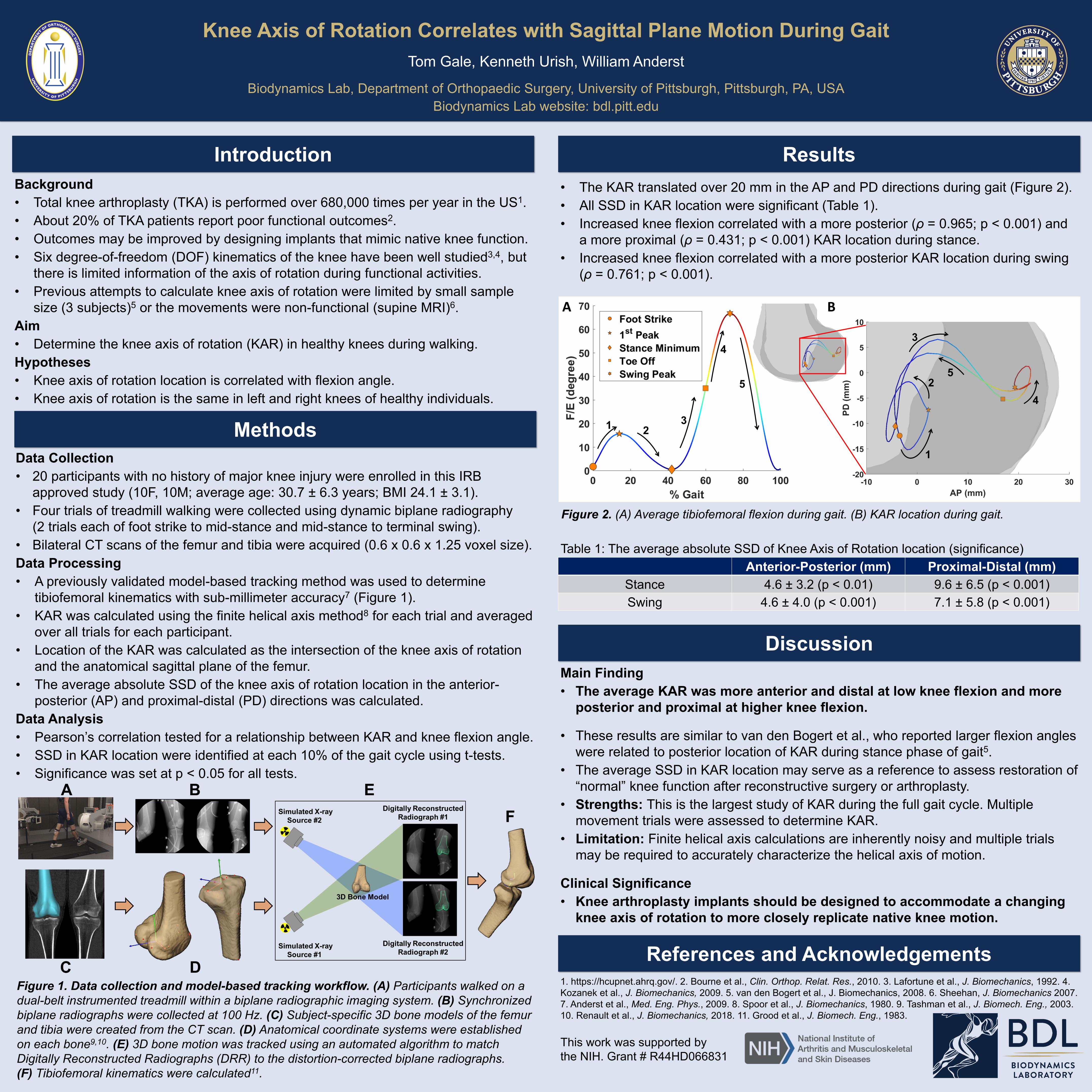
Background • Total knee arthroplasty (TKA) is performed over 680,000 times per year in the US 1 . • About 20% of TKA patients report poor functional outcomes 2 . • Outcomes may be improved by designing implants that mimic native knee function. • Six degree-of-freedom (DOF) kinematics of the knee have been well studied 3,4 , but there is limited information of the axis of rotation during functional activities. • Previous attempts to calculate knee axis of rotation were limited by small sample size (3 subjects) 5 or the movements were non-functional (supine MRI) 6 . Aim • Determine the knee axis of rotation (KAR) in healthy knees during walking. Hypotheses • Knee axis of rotation location is correlated with flexion angle. • Knee axis of rotation is the same in left and right knees of healthy individuals. Data Collection • 20 participants with no history of major knee injury were enrolled in this IRB approved study (10F, 10M; average age: 30.7 ± 6.3 years; BMI 24.1 ± 3.1). • Four trials of treadmill walking were collected using dynamic biplane radiography (2 trials each of foot strike to mid-stance and mid-stance to terminal swing). • Bilateral CT scans of the femur and tibia were acquired (0.6 x 0.6 x 1.25 voxel size). Data Processing • A previously validated model-based tracking method was used to determine tibiofemoral kinematics with sub-millimeter accuracy 7 (Figure 1). • KAR was calculated using the finite helical axis method 8 for each trial and averaged over all trials for each participant. • Location of the KAR was calculated as the intersection of the knee axis of rotation and the anatomical sagittal plane of the femur. • The average absolute SSD of the knee axis of rotation location in the anterior- posterior (AP) and proximal-distal (PD) directions was calculated. Data Analysis • Pearson’s correlation tested for a relationship between KAR and knee flexion angle. • SSD in KAR location were identified at each 10% of the gait cycle using t-tests. • Significance was set at p < 0.05 for all tests. Knee Axis of Rotation Correlates with Sagittal Plane Motion During Gait Tom Gale, Kenneth Urish, William Anderst Biodynamics Lab, Department of Orthopaedic Surgery, University of Pittsburgh, Pittsburgh, PA, USA Biodynamics Lab website: bdl.pitt.edu Introduction Methods Results Discussion References and Acknowledgements • The KAR translated over 20 mm in the AP and PD directions during gait (Figure 2). • All SSD in KAR location were significant (Table 1). • Increased knee flexion correlated with a more posterior (ρ = 0.965; p < 0.001) and a more proximal (ρ = 0.431; p < 0.001) KAR location during stance. • Increased knee flexion correlated with a more posterior KAR location during swing (ρ = 0.761; p < 0.001). Main Finding • The average KAR was more anterior and distal at low knee flexion and more posterior and proximal at higher knee flexion. • These results are similar to van den Bogert et al., who reported larger flexion angles were related to posterior location of KAR during stance phase of gait 5 . • The average SSD in KAR location may serve as a reference to assess restoration of “normal” knee function after reconstructive surgery or arthroplasty. • Strengths: This is the largest study of KAR during the full gait cycle. Multiple movement trials were assessed to determine KAR. • Limitation: Finite helical axis calculations are inherently noisy and multiple trials may be required to accurately characterize the helical axis of motion. Clinical Significance • Knee arthroplasty implants should be designed to accommodate a changing knee axis of rotation to more closely replicate native knee motion. 1. https://hcupnet.ahrq.gov/. 2. Bourne et al., Clin. Orthop. Relat. Res., 2010. 3. Lafortune et al., J. Biomechanics, 1992. 4. Kozanek et al., J. Biomechanics, 2009. 5. van den Bogert et al., J. Biomechanics, 2008. 6. Sheehan, J. Biomechanics 2007. 7. Anderst et al., Med. Eng. Phys., 2009. 8. Spoor et al., J. Biomechanics, 1980. 9. Tashman et al., J. Biomech. Eng., 2003. 10. Renault et al., J. Biomechanics, 2018. 11. Grood et al., J. Biomech. Eng., 1983. Figure 2. (A) Average tibiofemoral flexion during gait. (B) KAR location during gait. This work was supported by the NIH. Grant # R44HD066831 Anterior-Posterior (mm) Proximal-Distal (mm) Stance 4.6 ± 3.2 (p < 0.01) 9.6 ± 6.5 (p < 0.001) Swing 4.6 ± 4.0 (p < 0.001) 7.1 ± 5.8 (p < 0.001) Figure 1. Data collection and model-based tracking workflow. (A) Participants walked on a dual-belt instrumented treadmill within a biplane radiographic imaging system. (B) Synchronized biplane radiographs were collected at 100 Hz. (C) Subject-specific 3D bone models of the femur and tibia were created from the CT scan. (D) Anatomical coordinate systems were established on each bone 9,10 . (E) 3D bone motion was tracked using an automated algorithm to match Digitally Reconstructed Radiographs (DRR) to the distortion-corrected biplane radiographs. (F) Tibiofemoral kinematics were calculated 11 . Table 1: The average absolute SSD of Knee Axis of Rotation location (significance) A B 1 2 3 4 5 5 4 3 2 1
Transcript of Knee Axis of Rotation Correlates with Sagittal Plane ...
Background• Total knee arthroplasty (TKA) is performed over 680,000 times per year in the US1.• About 20% of TKA patients report poor functional outcomes2.• Outcomes may be improved by designing implants that mimic native knee function. • Six degree-of-freedom (DOF) kinematics of the knee have been well studied3,4, but
there is limited information of the axis of rotation during functional activities.• Previous attempts to calculate knee axis of rotation were limited by small sample
size (3 subjects)5 or the movements were non-functional (supine MRI)6.Aim• Determine the knee axis of rotation (KAR) in healthy knees during walking.Hypotheses• Knee axis of rotation location is correlated with flexion angle.• Knee axis of rotation is the same in left and right knees of healthy individuals.
Data Collection• 20 participants with no history of major knee injury were enrolled in this IRB
approved study (10F, 10M; average age: 30.7 ± 6.3 years; BMI 24.1 ± 3.1). • Four trials of treadmill walking were collected using dynamic biplane radiography
(2 trials each of foot strike to mid-stance and mid-stance to terminal swing). • Bilateral CT scans of the femur and tibia were acquired (0.6 x 0.6 x 1.25 voxel size).Data Processing• A previously validated model-based tracking method was used to determine
tibiofemoral kinematics with sub-millimeter accuracy7 (Figure 1). • KAR was calculated using the finite helical axis method8 for each trial and averaged
over all trials for each participant.• Location of the KAR was calculated as the intersection of the knee axis of rotation
and the anatomical sagittal plane of the femur. • The average absolute SSD of the knee axis of rotation location in the anterior-
posterior (AP) and proximal-distal (PD) directions was calculated.Data Analysis• Pearson’s correlation tested for a relationship between KAR and knee flexion angle.• SSD in KAR location were identified at each 10% of the gait cycle using t-tests.• Significance was set at p < 0.05 for all tests.
Knee Axis of Rotation Correlates with Sagittal Plane Motion During GaitTom Gale, Kenneth Urish, William Anderst
Biodynamics Lab, Department of Orthopaedic Surgery, University of Pittsburgh, Pittsburgh, PA, USABiodynamics Lab website: bdl.pitt.edu
Introduction
Methods
Results
Discussion
References and Acknowledgements
• The KAR translated over 20 mm in the AP and PD directions during gait (Figure 2).• All SSD in KAR location were significant (Table 1). • Increased knee flexion correlated with a more posterior (ρ = 0.965; p < 0.001) and
a more proximal (ρ = 0.431; p < 0.001) KAR location during stance. • Increased knee flexion correlated with a more posterior KAR location during swing
(ρ = 0.761; p < 0.001).
Main Finding• The average KAR was more anterior and distal at low knee flexion and more
posterior and proximal at higher knee flexion.
• These results are similar to van den Bogert et al., who reported larger flexion angles were related to posterior location of KAR during stance phase of gait5.
• The average SSD in KAR location may serve as a reference to assess restoration of “normal” knee function after reconstructive surgery or arthroplasty.
• Strengths: This is the largest study of KAR during the full gait cycle. Multiple movement trials were assessed to determine KAR.
• Limitation: Finite helical axis calculations are inherently noisy and multiple trials may be required to accurately characterize the helical axis of motion.
Clinical Significance• Knee arthroplasty implants should be designed to accommodate a changing
knee axis of rotation to more closely replicate native knee motion.
1. https://hcupnet.ahrq.gov/. 2. Bourne et al., Clin. Orthop. Relat. Res., 2010. 3. Lafortune et al., J. Biomechanics, 1992. 4. Kozanek et al., J. Biomechanics, 2009. 5. van den Bogert et al., J. Biomechanics, 2008. 6. Sheehan, J. Biomechanics 2007. 7. Anderst et al., Med. Eng. Phys., 2009. 8. Spoor et al., J. Biomechanics, 1980. 9. Tashman et al., J. Biomech. Eng., 2003. 10. Renault et al., J. Biomechanics, 2018. 11. Grood et al., J. Biomech. Eng., 1983.
Figure 2. (A) Average tibiofemoral flexion during gait. (B) KAR location during gait.
This work was supported by the NIH. Grant # R44HD066831
Anterior-Posterior (mm) Proximal-Distal (mm)Stance 4.6 ± 3.2 (p < 0.01) 9.6 ± 6.5 (p < 0.001)Swing 4.6 ± 4.0 (p < 0.001) 7.1 ± 5.8 (p < 0.001)
Figure 1. Data collection and model-based tracking workflow. (A) Participants walked on a dual-belt instrumented treadmill within a biplane radiographic imaging system. (B) Synchronized biplane radiographs were collected at 100 Hz. (C) Subject-specific 3D bone models of the femur and tibia were created from the CT scan. (D) Anatomical coordinate systems were established on each bone9,10. (E) 3D bone motion was tracked using an automated algorithm to match Digitally Reconstructed Radiographs (DRR) to the distortion-corrected biplane radiographs. (F) Tibiofemoral kinematics were calculated11.
Table 1: The average absolute SSD of Knee Axis of Rotation location (significance)
A B
1 23
4
55
4
3
2
1