Klaassen et al 1999
28
Annu. Rev. Pharmacol. Toxicol. 1999. 39:267–94 Copyright c 1999 by Annual Reviews. All rights reserved METALLOTHIONEIN: An Intracellular Protein to Protect Against Cadmium Toxicity Curtis D. Klaassen, Jie Liu, and Supratim Choudhuri 1 Center for Environmental Health and Occupational Medicine, Department of Pharmacology, Toxicology, and Therapeutics, University of Kansas Medical Center, Kansas City, Kansas 66160; e-mail: [email protected] KEY WORDS: MT-transgenic and knockout animals, cadmium, zinc, free radical scavenge ABSTRACT Metallothioneins (MT) are low-molecular-weight, cysteine-rich, metal-binding proteins. MT genes are readily induced by various physiologic and toxicologic stimuli. Because the cysteines in MT are absolutely conserved across species, it was suspected that the cysteines are necessary for function and MT is essential for life. In attempts to determine the function(s) of MT, studies have been performed using four different experimental paradigms: (a) animals injected with chemicals known to induce MT; (b) cells adapted to survive and grow in high concentrations of MT-inducing toxicants; (c) cells transfected with the MT gene; and (d ) MT- transgenic and MT-null mice. Most often, results from studies using the first three approaches have indicated multiple functions of MT in cell biology: MT (a) is a “storehouse” for zinc, (b) is a free-radical scavenger, and (c) protects against cadmium (Cd) toxicity. However, studies using MT-transgenic and null mice have not strongly supported the first two proposed functions but strongly support its function in protecting against Cd toxicity. Repeated administration of Cd to MT-null mice results in nephrotoxicity at one tenth the dose that produces nephrotoxicity in control mice. Human studies indicate that 7% of the general population have renal dysfunction from Cd exposure. Therefore, if humans did not have MT, “normal” Cd exposure would be nephrotoxic to humans. Thus, it appears that during evolution, the ability of MT to protect against Cd toxicity might have taken a more pivotal role in the maintenance of life processes, as 1 Current address: Department of Internal Medicine, Wayne State University School of Medicine, Detroit, Michigan 48201. 267 0362-1642/99/0415-0267$08.00
Transcript of Klaassen et al 1999
P1: KKK/ary P2: KKK/vks QC: KKK/kba T1: KKK
February 18, 1999 9:47 Annual Reviews AR079-12
Annu. Rev. Pharmacol. Toxicol. 1999. 39:267–94Copyright c© 1999 by Annual Reviews. All rights reserved
METALLOTHIONEIN: An IntracellularProtein to Protect AgainstCadmium Toxicity
Curtis D. Klaassen, Jie Liu, and Supratim Choudhuri1
Center for Environmental Health and Occupational Medicine, Department ofPharmacology, Toxicology, and Therapeutics, University of Kansas Medical Center,Kansas City, Kansas 66160; e-mail: [email protected]
KEY WORDS: MT-transgenic and knockout animals, cadmium, zinc, free radical scavenge
ABSTRACT
Metallothioneins (MT) are low-molecular-weight, cysteine-rich, metal-bindingproteins. MT genes are readily induced by various physiologic and toxicologicstimuli. Because the cysteines in MT are absolutely conserved across species, itwas suspected that the cysteines are necessary for function and MT is essential forlife. In attempts to determine the function(s) of MT, studies have been performedusing four different experimental paradigms: (a) animals injected with chemicalsknown to induce MT; (b) cells adapted to survive and grow in high concentrationsof MT-inducing toxicants; (c) cells transfected with the MT gene; and (d ) MT-transgenic and MT-null mice. Most often, results from studies using the firstthree approaches have indicated multiple functions of MT in cell biology: MT(a) is a “storehouse” for zinc, (b) is a free-radical scavenger, and (c) protectsagainst cadmium (Cd) toxicity. However, studies using MT-transgenic and nullmice have not strongly supported the first two proposed functions but stronglysupport its function in protecting against Cd toxicity. Repeated administration ofCd to MT-null mice results in nephrotoxicity at one tenth the dose that producesnephrotoxicity in control mice. Human studies indicate that 7% of the generalpopulation have renal dysfunction from Cd exposure. Therefore, if humans didnot have MT, “normal” Cd exposure would be nephrotoxic to humans. Thus, itappears that during evolution, the ability of MT to protect against Cd toxicitymight have taken a more pivotal role in the maintenance of life processes, as
1Current address: Department of Internal Medicine, Wayne State University School of Medicine,Detroit, Michigan 48201.
2670362-1642/99/0415-0267$08.00

P1: KKK/ary P2: KKK/vks QC: KKK/kba T1: KKK
February 18, 1999 9:47 Annual Reviews AR079-12
268 KLAASSEN, LIU & CHOUDHURI
compared with its other proposed functions (i.e. storehouse for zinc and freeradical scavenger).
INTRODUCTION
The discovery of a cadmium (Cd)-binding, cysteine-rich protein from horsekidney by Margoshes & Vallee (1) was the seminal finding that marked thebirth of a field of research focused on the study of a low-molecular-weightpolypeptide superfamily, the metallothioneins (MTs). MTs are low-molecular-weight (6–7 kDa), nonenzymatic proteins ubiquitous in the animal kingdom(2, 3). MT has an unusual amino acid composition: It does not contain aromaticamino acids, and most important, one third of its residues are cysteines.
Although MT was discovered over 40 years ago, its physiological functionsare still unclear. Studies aimed at determining physiological function(s) ofMTs have made use of four different model systems: (a) animals injected withchemicals known to induce MT; (b) cells adapted to survive and grow in highconcentrations of toxicants; (c) cells transfected with the MT gene; and (d )MT-transgenic and -null animals. Among these, the use of MT-transgenic andMT-null animals has provided the most persuasive evidence for the importanceof MT in detoxification and protection against Cd and other heavy metals.Here, we emphasize the recent advances in understanding of the regulation andfunctional significance of MT.
STRUCTURE AND OCCURRENCEOF METALLOTHIONEIN
MTs have been found throughout the animal kingdom, in higher plants, ineukaryotic microorganisms, and in many prokaryotes (3, 4). Based on theirstructural similarities, MTs have been divided into three classes: class I, II, andIII. Class I MTs, which include mammalian MTs and any polypeptide fromother phyla with related primary structure, are the focus of this review.
The amino acid sequences of MTs from many mammalian sources reveal thatall contain approximately 61 amino acids of remarkably similar composition.More important, all contain 20 cysteine residues that remain invariant alongthe amino acid sequence. All cysteines are known to participate in the coordi-nation of 7 mol of Cd or zinc (Zn) per mol of MT (5). Coordination of thesecysteine residues results in a high binding affinity for Zn (10−18) and Cd (10−22)(5). Detailed structural properties of the individual mammalian MT metal co-ordinating sites have been obtained from113Cd-nuclear magnetic resonance(6–8). The seven atoms of bound Cd are arranged in two separate polynuclear
P1: KKK/ary P2: KKK/vks QC: KKK/kba T1: KKK
February 18, 1999 9:47 Annual Reviews AR079-12
METALLOTHIONEIN IN TOXICOLOGY 269
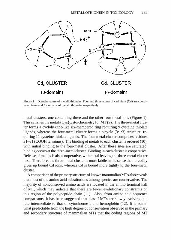
Figure 1 Domain nature of metallothionein. Four and three atoms of cadmium (Cd) are coordi-nated inα- andβ-domains of metallothionein, respectively.
metal clusters, one containing three and the other four metal ions (Figure 1).This satisfies the metal7(Cys)20stoichiometry for MT (9). The three-metal clus-ter forms a cyclohexane-like six-membered ring requiring 9 cysteine thiolateligands, whereas the four-metal cluster forms a bicyclo [3:1:3] structure, re-quiring 11 cysteine thiolate ligands. The four-metal cluster comprises residues31–61 (COOH terminus). The binding of metals to each cluster is ordered (10),with initial binding to the four-metal cluster. After these sites are saturated,binding occurs at the three-metal cluster. Binding in each cluster is cooperative.Release of metals is also cooperative, with metal leaving the three-metal clusterfirst. Therefore, the three-metal cluster is more labile in the sense that it readilygives up bound Cd ions, whereas Cd is bound more tightly to the four-metalcluster.
A comparison of the primary structure of known mammalian MTs also revealsthat most of the amino acid substitutions among species are conservative. Themajority of nonconserved amino acids are located in the amino terminal halfof MT, which may indicate that there are fewer evolutionary constraints onthis region of the polypeptide chain (11). Also, from amino acid sequencecomparisons, it has been suggested that class I MTs are slowly evolving at arate intermediate to that of cytochromec and hemoglobin (12). It is some-what predictable from the high degree of conservation observed in the primaryand secondary structure of mammalian MTs that the coding regions of MT
P1: KKK/ary P2: KKK/vks QC: KKK/kba T1: KKK
February 18, 1999 9:47 Annual Reviews AR079-12
270 KLAASSEN, LIU & CHOUDHURI
genes are also strongly homologous, whereas the noncoding sequences are moredivergent (2).
Taken together, these facts about MT structure probably can be distilledinto one simple principle: MT is functionally a very important protein and itsstructural conservation is dictated by its functional requirement. In addition tothe need for positional constancy of the cysteines, the importance of the overallstructural conservation of MT can be further demonstrated by the finding thatsimply changing the length of the interdomain hinge leads to a decline in itsmetal-binding ability. Thus, it cannot be overemphasized that the structure ofmammalian MTs is the product of a functionally driven, evolutionarily selectedprocess.
REGULATION OF MT GENE EXPRESSION
The current understanding of MT gene structure and regulation initially stem-med from studies on mouse and human MT genes. Although the mouse MTmultigene family consists of four known members (MT-I through -IV) that arelocated on chromosome 8, human MTs are encoded by a multigene family of atleast 15 members located on chromosome 16. They include one MT-II, MT-III,and MT-IV gene each and at least 13 MT-I genes (13–15). Although mouseMT-I and MT-II genes are coordinately regulated, MT-III is expressed in adultbrain, and MT-IV in differentiating stratified squamous epithelium (14). How-ever, all four isoforms are expressed in placenta (16). In humans, the MT-IIisoform gene (MT-IIA) is the most highly expressed gene, accounting for almost50% of total MT expression (17).
MT gene expression is controlled primarily at the level of transcription (18).In cells selected for resistence to Cd, enhanced expression of MT protein canalso be achieved through MT gene amplification (19). The 5′ end of MT-Iand MT-II genes contain a TATA box (core promoter element) and numerouscis-acting response elements (promoter proximal elements). Thesecis-actingresponse elements include the metal responsive elements (MREs), glucocor-ticoid responsive elements, and antioxidant response elements (18–22). Inaddition to the metal and glucocorticoid responsive elements, human MT genepromoters contain a number of other response elements, including the follow-ing: basal level enhancer elements, GC-box, interferon responsive elements,TPA responsive elements, and AP2 binding sites (2, 15, 23–25). Whereas MTpromotercis-acting elements may be unique to one gene or species, the singlecommon motif from invertebrates to vertebrates is the MREs, which are alwayspresent in multiple copies in the MT-gene promoter (15).
Some of the response elements in the MT promoter are binding sites for puta-tive transcription factors. For example, TPA responsive elements are recognized
P1: KKK/ary P2: KKK/vks QC: KKK/kba T1: KKK
February 18, 1999 9:47 Annual Reviews AR079-12
METALLOTHIONEIN IN TOXICOLOGY 271
by AP1 and AP2, and the GC-box is the binding site for Sp1 transcription fac-tor (15, 24, 25). However, studies aimed at identifying specific transcriptionfactors have mainly focused on identifying the metal transcription factors thatmediate MT gene expression by metals.
Several groups have reported proteins that bind to the metal-responsive DNAelements in mouse MTs (26). The candidate MRE-binding protein, termedMTF-1 for metal transcription factor-I (27), was subsequently cloned and foundto be a Zn-finger (Cys2His2) transcription factor (26, 28). Targeted disruption ofboth copies of MTF-1 allele in mouse embryonic stem cells resulted in silencingof constitutive as well as metal-mediated expression of MT-I and MT-II genes(26). Using a different approach, Palmiter (29) also demonstrated that MTF-1 isindispensable for basal and metal-mediated MT-gene expression. Thus, MTF-1appears to be the only transcription factor that mediates metal responsivenessof the MT genes (29). However, MTF-1 itself appears to be under the controlof a Zn-sensitive inhibitor. Thus, the current model for MT gene regulation bymetals depicts that in the absence of Zn, MTF-1 is complexed with an inhibitor,termed MTI (metallothionein transcription inhibitor). In the presence of Zn,MTI dissociates from MTF-1, which allows it to interact with the MREs in theMT promoter to activate transcription. The newly synthesized MT binds Znand the MTF-1/MTI complex reforms (29).
An attempt to generate MTF-1–null mutant mice resulted in embryos thatdied on day 14 of gestation (30). However, in the embryos, an absense ofMTF-1 abolished the transcription of MT-I and MT-II genes and reduced thetranscription ofγ -glutamylcysteine synthetase, a key enzyme in glutathionesynthesis. MTF-1–null embryos showed increased susceptibility to Cd and hy-drogen peroxide and had liver degeneration. Thus, although MT is not essentialfor life, the metal transcription factor is.
REGULATION OF MT PROTEIN DEGRADATION
Degradation of MT protein is also an important aspect of MT regulation (31).There are tremendous differences in the half-lives of MT synthesized as a resultof chemical induction of the MT gene. For example, the half-life of Zn-MT isapproximately 18–20 h, whereas that of Cd-MT is about 3 days (32). Studiesfrom our laboratory (33) showed that the half-life for constitutive MT in adultrats is about 4 h, whereas in neonates it is 49 h. MT induced by ethanol, ZnCl2or CdCl2 also has widely different half-lives, approximately 9, 25, and 60 h,respectively (33), indicating that the degradation of MT is dependent on the ageof the animals and the metal bound to MT.
Using cultured hepatocytes, Chen & Failla (34) showed that the degradationof MT is primarily regulated by cellular Zn content, and it occurs in both
P1: KKK/ary P2: KKK/vks QC: KKK/kba T1: KKK
February 18, 1999 9:47 Annual Reviews AR079-12
272 KLAASSEN, LIU & CHOUDHURI
lysosomal and non-lysosomal compartments. The importance of cathepsin B inlysosomal fraction was demonstrated when cathepsin B–specific inhibitors werefound to suppress apo-MT degradation by 80% (35). This was further confirmedusing purified cathepsin B (36). It was also shown that MT is degraded rapidlywhen there are fewer than five atoms of metal associated with each molecule ofMT (35, 36). Concurrent titration studies indicated that at lysosomal pH, mostof the Zn is released from MT whereas most of the Cd is not (36). This couldbe the reason Cd MT has a higher half-life in vivo than does Zn MT.
Steinbach & Wolterbeek (37) demonstrated that an intracellular MT poolexists in at least two forms, cytosolic apo-MT and lysosomal metal-boundMT, each being depleted and replenished at different rates. Subsequent studiesshowed that cytosolic apo-MT can be degraded by the cytosolic 26S proteasomecomplex (38). The evidence that the intracellular MT pool can exist in indepen-dent, compartmentalized pools has important implications for the intracellularfunctions of MT.
During fetal development, tissue MT-I and MT-II concentrations changedramatically. Both MT-I and MT-II are detected in rat fetal liver by day 18 ofgestation, reaching maximum hepatic concentrations at birth (39). MT con-centrations in liver of newborn rats are 20-fold higher than in adult rats. Thishigh level of hepatic MT is maintained during the first 2 weeks postpartum.Thereafter, the hepatic concentration of MT decreases, with adult expressionlevels exhibited by 35 days of age (39–41). One explanation for a consistentlyhigh level of MT during development is that MT is localized in the nucleusduring development, and thus it is not available to the intracellular degrada-tion machinery (31). Both MT-I and MT-II isoforms are coordinately regulatedduring development. MT levels in kidney, spleen, heart, lung, pancreas, andstomach are three- to ten-fold higher in 1-day-old rats than in adult rats and fallsteadily to adult values over a 3–4 week period. In contrast, neonatal brain haslower (∼50%) MT concentrations and increases to adult level by day 21 (42).
PROPOSED FUNCTIONS OF METALLOTHIONEIN
Role of Metallothionein in Essential Metal HomeostasisZINC Zn is a physiologically important metal and the most abundant metalbound to constitutive MT. Zn provides essential structural and catalytic func-tions to a wide variety of proteins. More than 300 different enzymes dependon Zn for proper protein folding and biological function. Zn is also crucial inthe regulation of gene expression because numerous transcription factors have“zinc finger motifs” that are maintained by Zn.
Apo-MT (metallothionein with no metals bound) is a Zn acceptor because ofthe abundance of free sulfhydryl groups and their high affinity for Zn. However,
P1: KKK/ary P2: KKK/vks QC: KKK/kba T1: KKK
February 18, 1999 9:47 Annual Reviews AR079-12
METALLOTHIONEIN IN TOXICOLOGY 273
the sulfhydryl groups are highly reactive, and Zn, although bound with highaffinity, can undergo exchange reactions, which allows Zn to be transferredfrom MT to other proteins (43–46). The affinity of sulfhydryl groups for Zncan also make MT an efficient metal ion scavenger. This implies a possibleregulatory role of MT in the activation or inactivation of various moleculareffectors. Such a possibility was demonstrated by showing that apo-MT canchelate Zn out of the transcription factor IIIA (TFIIIA), a process that inac-tivates TFIIIA (47). Therefore, it is tempting to speculate that MT might beessential for Zn homeostasis by regulating Zn absorption, or as a donor of Znto various enzymes and transcription factors during development or proteinsynthesis.
The role of MT in intestinal Zn absorption was recently reevaluated usingboth MT-transgenic and MT-null mouse models (48). At 2 h after a single oraldose of Zn, serum Zn concentrations were twofold higher in MT-null mice,while in MT-transgenic mice, serum Zn concentrations were only one-thirdthat of controls, which suggests that MT reduces Zn absorption. Intestinal Znwas higher in MT-null mice but was unchanged in MT-transgenic mice, whichsuggests that MT does not reduce Zn absorption simply by sequestration of Znin the mucosa (48).
MT has been suggested to provide a biologically important pool of Zn duringperiods of extreme Zn deficiency, as the teratogenic effects observed in fetusesof dams placed on a Zn-deficient diet was lower in MT-I transgenic mice thanin control mice (49). Hepatic concentrations of Zn were 60% less in newbornMT-null mice than in control mice, and kidney development in the MT-nullpups was retarded when they were fed a Zn-deficient diet (50). However, theserenal abnormalities resolved with time and did not impair kidney function (51).When adult MT-null mice were challenged with a toxic dose of Zn, they hada greater incidence of pancreatic acinar cell degeneration compared with con-trols (50). During endotoxemia, MT-null mice were also less responsive thancontrol mice to hepatic Zn accumulation and to reduction in plasma Zn levels(52, 53).
Thus, MT may aid in maintaining Zn homeostasis and protecting againstexcess Zn-induced toxicity. However, MT-null animals appear “normal,” andthus MT does not appear to play the most crucial role in enabling Zn to performits important roles in growth and development. However, it should be empha-sized that the apparent “normal” appearance of MT-null mice could be due tothe presence of back-up systems and thus does not necessarily argue againstMT having a physiological role in Zn homeostasis.
COPPER Copper (Cu) is an essential metal for structural and catalytic prop-erties of many enzymes, such as Cu/Zn superoxide dismutase, cytochrome
P1: KKK/ary P2: KKK/vks QC: KKK/kba T1: KKK
February 18, 1999 9:47 Annual Reviews AR079-12
274 KLAASSEN, LIU & CHOUDHURI
c oxidase, and copper-responsive transcription factors. However, excess Cucan be toxic, particularly when associated with a deficit in Cu excretion (54).There are two diseases in humans caused by abnormal transport of Cu:Wilson’s disease and Menkes’ disease. Wilson’s disease is a hypercupremicstate, whereas Menkes’ disease is a hypocupremic state. Wilson’s disease isdue to a defect of a P-type ATPase (ATP7B), which is located on autosome13 in humans and autosome 16 in rats (55). As a result of the mutation in theCu-efflux transporter (56), there is an inability to transport Cu across the bilecanalicular membrane, and thus Cu accumulates in liver during aging. Menkes’disease gene is located on the X chromosome and is characterized by a mutationof a copper efflux ATPase (ATP7A) (57). This results in decreased transport ofCu across the placenta to the fetus, as well as in decreased efflux of Cu from theintestine into the blood, thereby creating a copper-deficient state. Both copper-efflux ATPases and MT are important mechanisms affecting copper toxicity(58).
Analysis of Wilson’s disease patients suggests that MT is the major Cu-binding protein in their liver, and increased liver MT levels assist in the detoxi-cation of the accumulated Cu (59, 60). Thus, the beneficial effects produced byZn administration to Wilson’s patients could be due to induction of MT (61).Indeed, MT-rich cells are resistant to Cu toxicity (62–65). It has been proposedthat MT protects against Cu toxicity by sequestration of Cu from critical cellulartargets (58, 61).
Cu is a transition metal and can exist in three oxidation states: Cu, Cu+, andCu2+. It appears that MT containing Zn and Cu functions as an antioxidant;however, when MT is saturated with Cu, it becomes a prooxidant and maycause oxidative liver damage (66–68).
The role of MT in Menkes’ disease has been studied by crossing MT-I-and -II–null mice with mice carrying the Mottled-Brindled allele (Mo-BrJ), amurine model for X-linked Menkes’ disease (69). It is believed that Mo-BrJmales die from Cu-deficiency because of an inability to transport Cu from theintestine into the circulation. Because MT avidly binds Cu, elimination of MTin the intestine might enhance passage of Cu into the circulation, resulting inprolonged survival. Contrary to expectation, on an MT−/− background, mostoffspring of Mo-BrJ mice die before gestational day 11. These results suggestthat MT protects against Cu toxicity in the embryo (69).
Because Cu is an essential metal that binds to MT, one might anticipate thatMT would have an essential role in the normal biological activity of Cu inthe body. However, because mice with a targeted deletion of MT appear tobe “normal,” the results suggest that MT is not essential for Cu homeostasis.However, it does appear that MT can protect against the toxicity of Cu underextreme conditions.

P1: KKK/ary P2: KKK/vks QC: KKK/kba T1: KKK
February 18, 1999 9:47 Annual Reviews AR079-12
METALLOTHIONEIN IN TOXICOLOGY 275
Role of Metallothionein in Protection AgainstMetal ToxicityCADMIUM Cd is an environmental pollutant toxic to a number of tissues. Acuteexposure to Cd produces hepatic, pulmonary, and testicular injury, whereaschronic exposure results in renal and bone injury and cancer, as well as toxicityto other organs (70). Numerous studies have suggested that MT plays animportant role in Cd disposition and detoxication.
The factors that influence Cd absorption, distribution, and elimination are notwell understood, but it is known that Cd is poorly absorbed after oral ingestion(71). The role of MT in Cd disposition has been examined in MT-transgenicmice. Using this model, MT does not inhibit intestinal Cd absorption, nordoes it affect initial Cd distribution to various tissues (72, 73). However, MTdecreases Cd elimination through the bile (74) and is a major factor for tissueretention of Cd (73, 75).
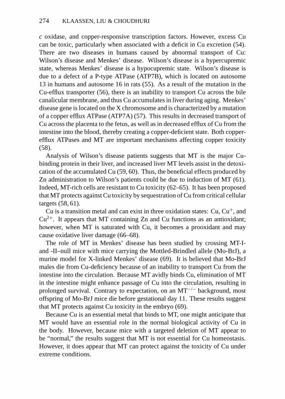
The correlation between Cd resistance in cultured cells and cellular MT levelsis strong and has been extensively documented (64). Pretreatment of animalswith low doses of Cd (76, 77), Zn (76), ethanol (78), diethylmaleate (79), ortriterpenoids (80), all of which are known to increase MT, protect against acuteCd-induced lethality and hepatotoxicity. Newborn animals have a high levelof hepatic MT and are resistant to Cd-induced hepatotoxicity (81, 82). Simi-larly, MT-I–transgenic mice, which have concentrations of hepatic MT ten-foldhigher than that of control mice (83), are resistant to Cd-induced lethality andhepatotoxicity (84). In comparison, MT-null mice show increased susceptibilityto Cd-induced lethality (85, 86) and liver injury (86, 87). Furthermore, Zn pre-treatment, which cannot increase hepatic MT in MT-null mice, failed to protectagainst Cd-induced hepatotoxicity in MT-null mice. These data support the hy-pothesis that Zn-induced tolerance to Cd is also due to induction of MT (76, 87).Collectively, both constitutive cellular MT and induction of MT by chemicalsare important for the detoxication of Cd. MT-mediated hepatoprotection is dueto the high-affinity sequestration of Cd by MT in the cytosol, thus reducing theamount of Cd available to injure other critical organelles (Figure 2) (77, 84).
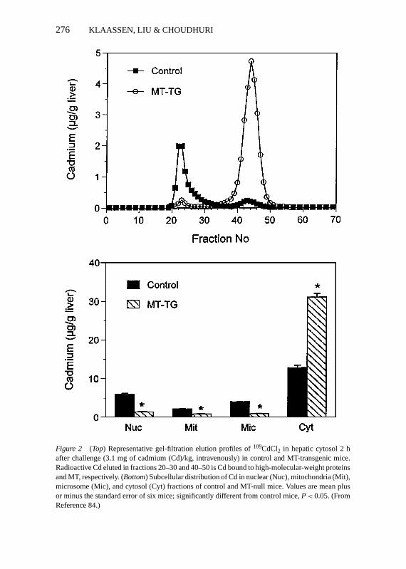
Figure 3 illustrates how Cd is thought to produce renal toxicity. Cd is initiallytaken up by the liver. In the liver, Cd can bind with glutathione (GSH) and beexcreted into bile. More important, Cd can bind to MT and be stored. SomeCd bound to MT leaks into the plasma and then is taken up by the kidney.Circulating Cd-MT complex is a potent nephrotoxicant (88, 89). In lysosomesof the kidney, Cd is released and can bind to preformed MT in the kidney. Whena critical concentration of Cd is reached in the kidney, renal injury occurs.Therefore, whether MT is beneficial or detrimental for chronic Cd-inducednephrotoxicity has been debatable (70, 90, 91).
P1: KKK/ary P2: KKK/vks QC: KKK/kba T1: KKK
February 18, 1999 9:47 Annual Reviews AR079-12
276 KLAASSEN, LIU & CHOUDHURI
Figure 2 (Top) Representative gel-filtration elution profiles of109CdCl2 in hepatic cytosol 2 hafter challenge (3.1 mg of cadmium (Cd)/kg, intravenously) in control and MT-transgenic mice.Radioactive Cd eluted in fractions 20–30 and 40–50 is Cd bound to high-molecular-weight proteinsand MT, respectively. (Bottom) Subcellular distribution of Cd in nuclear (Nuc), mitochondria (Mit),microsome (Mic), and cytosol (Cyt) fractions of control and MT-null mice. Values are mean plusor minus the standard error of six mice; significantly different from control mice,P< 0.05. (FromReference 84.)
P1: KKK/ary P2: KKK/vks QC: KKK/kba T1: KKK
February 18, 1999 9:47 Annual Reviews AR079-12
METALLOTHIONEIN IN TOXICOLOGY 277
Figure 3 Current theory of cadmium (Cd)-induced nephropathy. (From Reference 203.)
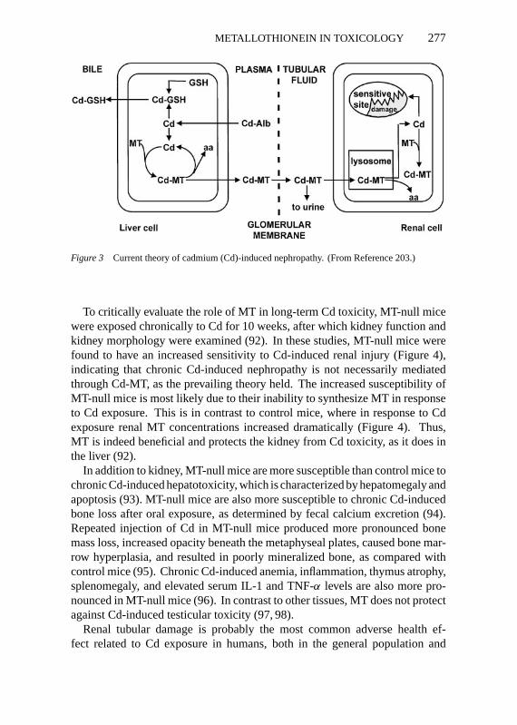
To critically evaluate the role of MT in long-term Cd toxicity, MT-null micewere exposed chronically to Cd for 10 weeks, after which kidney function andkidney morphology were examined (92). In these studies, MT-null mice werefound to have an increased sensitivity to Cd-induced renal injury (Figure 4),indicating that chronic Cd-induced nephropathy is not necessarily mediatedthrough Cd-MT, as the prevailing theory held. The increased susceptibility ofMT-null mice is most likely due to their inability to synthesize MT in responseto Cd exposure. This is in contrast to control mice, where in response to Cdexposure renal MT concentrations increased dramatically (Figure 4). Thus,MT is indeed beneficial and protects the kidney from Cd toxicity, as it does inthe liver (92).
In addition to kidney, MT-null mice are more susceptible than control mice tochronic Cd-induced hepatotoxicity, which is characterized by hepatomegaly andapoptosis (93). MT-null mice are also more susceptible to chronic Cd-inducedbone loss after oral exposure, as determined by fecal calcium excretion (94).Repeated injection of Cd in MT-null mice produced more pronounced bonemass loss, increased opacity beneath the metaphyseal plates, caused bone mar-row hyperplasia, and resulted in poorly mineralized bone, as compared withcontrol mice (95). Chronic Cd-induced anemia, inflammation, thymus atrophy,splenomegaly, and elevated serum IL-1 and TNF-α levels are also more pro-nounced in MT-null mice (96). In contrast to other tissues, MT does not protectagainst Cd-induced testicular toxicity (97, 98).
Renal tubular damage is probably the most common adverse health ef-fect related to Cd exposure in humans, both in the general population and
P1: KKK/ary P2: KKK/vks QC: KKK/kba T1: KKK
February 18, 1999 9:47 Annual Reviews AR079-12
278 KLAASSEN, LIU & CHOUDHURI
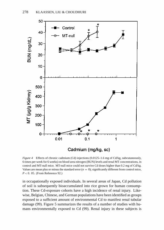
Figure 4 Effects of chronic cadmium (Cd) injections (0.0125–1.6 mg of Cd/kg, subcutaneously,6 times per week for 6 weeks) on blood urea nitrogen (BUN) levels and renal MT concentrations, incontrol and MT-null mice. MT-null mice could not survive Cd doses higher than 0.2 mg of Cd/kg.Values are mean plus or minus the standard error (n = 8); significantly different from control mice,P< 0. 05. (From Reference 92.)
in occupationally exposed individuals. In several areas of Japan, Cd pollutionof soil is subsequently bioaccumulated into rice grown for human consump-tion. These Cd-exposure cohorts have a high incidence of renal injury. Like-wise, Belgian, Chinese, and German populations have been identified as groupsexposed to a sufficient amount of environmental Cd to manifest renal tubulardamage (99). Figure 5 summarizes the results of a number of studies with hu-mans environmentally exposed to Cd (99). Renal injury in these subjects is

P1: KKK/ary P2: KKK/vks QC: KKK/kba T1: KKK
February 18, 1999 9:47 Annual Reviews AR079-12
METALLOTHIONEIN IN TOXICOLOGY 279
Figure 5 The relationship between human exposure to cadmium and renal injury. (Adapted fromReference 99.)
indicated by proteinuria. Because Cd injures proximal tubules of kidney, anincreased excretion of proteins into urine is observed.
Also noted in Figure 5 are data on recent human exposure to Cd. As can beseen, present-day human exposure results in renal concentrations of Cd that areknown to produce renal injury. It has been calculated that 7% of the generalpopulation has Cd-induced kidney damage (99).
MERCURY Inorganic mercury (HgCl2) is a relatively potent inducer of MT(100, 101). Hg can readily substitute for Zn in the seven-metal thiolate cluster(102) and binds to both MT-I and MT-II isoforms (65, 103).
MT-rich cells are resistant to the toxicity of HgCl2 (64, 65, 104). Induction ofMT in the kidney protects against HgCl2-mediated nephrotoxicity (105). Theprotective effects of MT against HgCl2 toxicity appear to be due to MT bindingHg in the cytosol (65, 106). Mice deficient in MT-I/II are more susceptible thancontrols to HgCl2-induced renal injury (107). Thus, MT can protect against thetoxicity of HgCl2.
Metallothionein as a Trap for Reactive Oxygen SpeciesMT has a high (30%) content of cysteine residues. It is reasonable to expectthat sulfhydryl-rich MT may function in a manner similar to GSH, wherein MTprovides an intracellular nucleophilic “sink” to trap electrophiles, alkylating
P1: KKK/ary P2: KKK/vks QC: KKK/kba T1: KKK
February 18, 1999 9:47 Annual Reviews AR079-12
280 KLAASSEN, LIU & CHOUDHURI
agents, and free radicals (108–110). MT can serve as a sacrificial scavenger forhydroxyl radicals and superoxide anion in vitro (111, 112) and can assume thefunction of superoxide dismutase in yeast (113). The multiple cysteine residuesof MT can be oxidized during oxidative stress, and the subsequent release ofZn has been proposed to be important in protection against oxidative damage(114, 115). However, oxidation of MT in vivo has been difficult to demonstrate,and there have been controversial reports on the role of MT during oxidativestress.
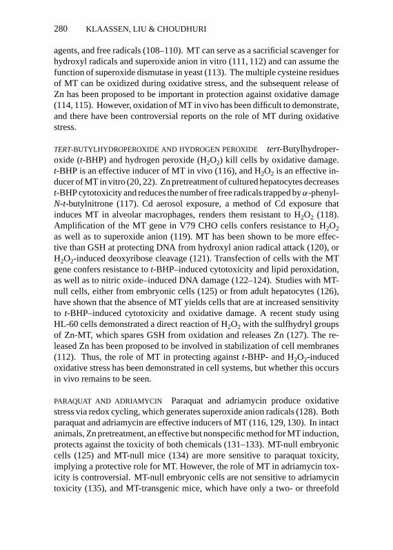
TERT-BUTYLHYDROPEROXIDE AND HYDROGEN PEROXIDE tert-Butylhydroper-oxide (t-BHP) and hydrogen peroxide (H2O2) kill cells by oxidative damage.t-BHP is an effective inducer of MT in vivo (116), and H2O2 is an effective in-ducer of MT in vitro (20, 22). Zn pretreatment of cultured hepatocytes decreasest-BHP cytotoxicity and reduces the number of free radicals trapped byα-phenyl-N-t-butylnitrone (117). Cd aerosol exposure, a method of Cd exposure thatinduces MT in alveolar macrophages, renders them resistant to H2O2 (118).Amplification of the MT gene in V79 CHO cells confers resistance to H2O2as well as to superoxide anion (119). MT has been shown to be more effec-tive than GSH at protecting DNA from hydroxyl anion radical attack (120), orH2O2-induced deoxyribose cleavage (121). Transfection of cells with the MTgene confers resistance tot-BHP–induced cytotoxicity and lipid peroxidation,as well as to nitric oxide–induced DNA damage (122–124). Studies with MT-null cells, either from embryonic cells (125) or from adult hepatocytes (126),have shown that the absence of MT yields cells that are at increased sensitivityto t-BHP–induced cytotoxicity and oxidative damage. A recent study usingHL-60 cells demonstrated a direct reaction of H2O2 with the sulfhydryl groupsof Zn-MT, which spares GSH from oxidation and releases Zn (127). The re-leased Zn has been proposed to be involved in stabilization of cell membranes(112). Thus, the role of MT in protecting againstt-BHP- and H2O2-inducedoxidative stress has been demonstrated in cell systems, but whether this occursin vivo remains to be seen.
PARAQUAT AND ADRIAMYCIN Paraquat and adriamycin produce oxidativestress via redox cycling, which generates superoxide anion radicals (128). Bothparaquat and adriamycin are effective inducers of MT (116, 129, 130). In intactanimals, Zn pretreatment, an effective but nonspecific method for MT induction,protects against the toxicity of both chemicals (131–133). MT-null embryoniccells (125) and MT-null mice (134) are more sensitive to paraquat toxicity,implying a protective role for MT. However, the role of MT in adriamycin tox-icity is controversial. MT-null embryonic cells are not sensitive to adriamycintoxicity (135), and MT-transgenic mice, which have only a two- or threefold
P1: KKK/ary P2: KKK/vks QC: KKK/kba T1: KKK
February 18, 1999 9:47 Annual Reviews AR079-12
METALLOTHIONEIN IN TOXICOLOGY 281
higher level of MT in the heart, are not protected against adriamycin toxicity(136). In contrast, mice engineered to express MT in heart (10- to 150-fold)are protected from adriamycin cardiotoxicity (137) and ischemia-reperfusionheart injury (138). Neonatal cardiomyocytes isolated from these heart-specificMT-transgenic mice are also resistant to adriamycin-induced cytotoxicity andoxidative stress (139). Whether the discrepancy in the role of MT in adriamycintoxicity is due to varying levels of intracellular MT or to a combination of otherfactors needs further clarification.
RADIATION Irradiation byγ -, x-, and ultraviolet rays produces reactive oxygenspecies by the radiolysis of water in living cells. Because of its hydroxyl radicalscavenging ability in vitro (111), MT might be expected to provide protectionagainst the toxic effects of radiation. Pretreatment of mice with Zn protectsagainst X-radiation–induced lethality (140, 141). However, transfection of cellswith MT genes failed to confer radio-resistance (142, 143). In intact animals,MT-transgenic mice were not protected fromγ -radiation–induced lethality andhematotoxicity (144). In agreement with these findings, MT-null mice do nothave an increased sensitivity to radiation-induced damage to cellular DNA, pro-tein, and lipids (145). At very low doses of radiation (0. 1–1. 0 Gy), MT-nullmice appear to be more sensitive to radiation-induced leukocyte reduction, butthey suffered similar damage at doses higher than 3.0 Gy (146). Furthermore,Zn treatment of MT-null mice protected against the lethal effects of radiation,indicating that Zn-induced protection was not mediated through the inductionof MT (145).
It is proposed that MT localized in the cytosol may function in metal detox-ication and protection from oxidative stress, whereas MT localized in thenucleus may provide protection against DNA-damaging electrophiles (147).Indeed, MT-null cells are more susceptible to Cd-,t-BHP–, and anti-cancerdrug–induced apoptotic lesions (92, 148), have enhanced spontaneous muta-tions (149), and are sensitive to Cd-induced protooncogene (c-jun) and tumorsuppressor gene (p53) expression (150). Whether such a susceptibility is relatedto cytosolic versus nuclear MT localization requires further investigation.
CONCLUSIONS Although biochemical data indicate that MT can quench vari-ous reactive oxygen species and other electrophiles, current data from MT-nullanimals are less than convincing with regard to MT having a major role inprotecting cells against reactive oxygen species. In addition, reactive oxygenspecies are thought to be important in aging, cancer, and neurodegenerativediseases, yet MT-null mice do not appear to suffer from premature aging, anincreased incidence of cancer, or neurodegenerative disease. Thus, it does notappear likely that the conservation of MT resulted from evolutionary pressure
P1: KKK/ary P2: KKK/vks QC: KKK/kba T1: KKK
February 18, 1999 9:47 Annual Reviews AR079-12
282 KLAASSEN, LIU & CHOUDHURI
to protect cells from reactive oxygen species or other electrophiles. In addition,if the main function of MT is to bind reactive chemical species, why would thelocation of the cysteine residues be conserved, and why would the sulfhydrylmoieties not be freely available to bind electrophiles instead of being bound tometal?
Metallothionein-Mediated Protection Againstthe Toxicity of Other ChemicalsCISPLATIN cis-Diaminedichloroplatinum (cisplatin) is an effective anticancerdrug containing the metal platinum. Binding of cisplatin to MT has beendemonstrated (151), but the role of MT in cisplatin resistance has been anissue of debate (110, 152).
Some tumor cell lines with acquired resistance to cisplatin overexpress MT(153), whereas other cisplatin-resistant cell lines do not have increased MTlevels (154). Pretreatment of mice with MT-inducers, such as Zn and bismuth,protects against the lethal and nephrotoxic effects of cisplatin (155). However,in other studies, induction of MT in rats failed to protect against the toxic-ity of cisplatin (156). Transfection of cells with the hMT-IIA gene confersresistance to cisplatin toxicity in some cell lines (143, 157) but not in others(142, 158, 159). Targeted deletion of the MT gene renders cells and animalsmore vulnerable to cisplatin toxicity (148, 160, 161). In contrast, overexpressionof MT in MT-transgenic mice does not confer resistance to cisplatin-inducednephrotoxicity (144).
Overall, studies with cisplatin suggest that MT protection against cisplatintoxicity is equivocal at best. Therefore, it seems unlikely that MT plays a criticalrole in modulating cisplatin toxicity.
CARBON TETRACHLORIDE Hepatic MT can be induced by carbon tetrachlo-ride (CCl4) administration (162), indicating a possible role for this protein inproviding tolerance to CCl4 hepatotoxicity. Induction of MT by Zn (163, 164),or by inflammation (165, 166), protects against CCl4 hepatotoxicity. Similarto the binding of Cd to MT, more14C from 14CCl4 is bound to MT in theMT-induced animals than in controls, with a concomitant reduction of cova-lent binding of14CCl4 to cellular protein and lipids (163). Recent studies withMT-null mice suggest that a lack of MT also renders animals more susceptibleto CCl4 hepatotoxicity (146, 167), providing further evidence of a role for MTas a cellular mechanism in decreasing CCl4 toxicity. However, the ability ofMT to scavenge trichloromethyl radicals in vivo could not be confirmed by thespin-trap chemical phenyl N-tert-butylnitrone (PBN) (168). Thus, it has beensuggested that the oxidation of MT by CCl4, with subsequent Zn release, ratherthan covalent binding, is responsible for protection (169).
P1: KKK/ary P2: KKK/vks QC: KKK/kba T1: KKK
February 18, 1999 9:47 Annual Reviews AR079-12
METALLOTHIONEIN IN TOXICOLOGY 283
ACETAMINOPHEN MT is induced following acetaminophen administration(170), and induction of MT has been associated with protection against ac-etaminophen hepatotoxicity (171–173). However, there is evidence neitherfor the binding of acetaminophen to MT nor for an altered subcellular dis-tribution of acetaminophen following induction of MT by Zn (172), or bytriterpenoids (173). MT-I/II–null mice are more susceptible than control miceto acetaminophen hepatotoxicity (174, 175). The increased susceptibility isnot due to altered acetaminophen bioactivation, as P-450 enzymes, and ac-etaminophen metabolites appear to be unaltered in these MT-null animals. Thecellular GSH content and covalent binding of acetaminophen to 44- and 58-kDaproteins are also similar in MT-null and control mice (174, 175). The increasedsensitivity to acetaminophen in MT-null mice has been suggested to be due toincreased oxidative stress, asN-acetyl-benzoquinoimine, a reactive intermedi-ate of acetaminophen biotransformation, produced more oxidative damage toMT-null than to control hepatocytes (175).
STREPTOZICIN, ALLOXAN, AND CAERULEIN Pancreas has the highest basalconcentration of MT (84, 176). Pancreatic MT can be further increased bymetals and chemicals that produce diabetes. Because of the high basal levels,the percentage increase in MT is not as pronounced as in liver and kidney.Induction of pancreatic MT by Zn has been associated with protection againststreptozotocin- and caerulein-induced acute pancreatitis (177–179) but not pro-tect against alloxan-induced injury to endocrine cells of the pancreas (180).Both MT-transgenic and MT-null mice have been used to study the role of MTin chemical-induced pancreatic injury. MT-I–transgenic mice are more resis-tant, whereas MT-null mice are more sensitive to caerulein-induced pancreatitisthan are corresponding control mice (181). However, the differences are sub-tle. Treatment of MT-null mice with Zn protects against streptozocin-inducedpancreatitis, which suggests that Zn, rather than MT, is more important in thisprotection (182).
Metallothionein and Protection AgainstNeurodegenerative DiseaseMT in brain has been proposed to play a role in Zn homeostasis and neurodegen-erative diseases (183–186). Brain MT-III was discovered as a growth-inhibitingfactor, inhibiting neuronal sprouting in culture (187). MT-III was originallythought to be down-regulated in Alzheimer patients (187, 188). However, fur-ther studies could not confirm the association of MT-III with Alzheimer’s dis-ease (189, 190).
Transgenic mice that overexpress human MT-III have also been engineered(191). They have a ninefold increase in MT-III in cerebral cortex, a three- to
P1: KKK/ary P2: KKK/vks QC: KKK/kba T1: KKK
February 18, 1999 9:47 Annual Reviews AR079-12
284 KLAASSEN, LIU & CHOUDHURI
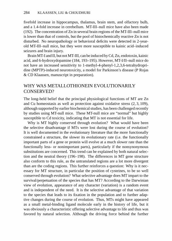
fivefold increase in hippocampus, thalamus, brain stem, and olfactory bulb,and a 1.4-fold increase in cerebellum. MT-III–null mice have also been made(192). The concentration of Zn in several brain regions of the MT-III–null miceis lower than that of controls, but the pool of histochemically reactive Zn is notdisturbed. No neuropathology or behavioral deficits were detected in 2-year-old MT-III–null mice, but they were more susceptible to kainic acid–inducedseizures and brain injury.
Brain MT-I and II, but not MT-III, can be induced by Cd, Zn, endotoxin, kainicacid, and 6-hydroxydopamine (184, 193–195). However, MT-I/II–null mice donot have an increased sensitivity to 1-methyl-4-phenyl-1,2,3,6-tetrahydropri-dine (MPTP)-induced neurotoxicity, a model for Parkinson’s disease (P Rojas& CD Klaassen, manuscript in preparation).
WHY WAS METALLOTHIONEIN EVOLUTIONARILYCONSERVED?
The long-held belief that the principal physiological functions of MT are Znand Cu homeostasis as well as protection against oxidative stress (2, 3, 109),although supported by earlier biochemical studies, has been challenged recentlyby studies using MT-null mice. These MT-null mice are “normal” but highlysusceptible to Cd toxicity, indicating that MT is not essential for life.
Why is MT highly conserved through evolution? What would have beenthe selective disadvantage if MTs were lost during the course of evolution?It is well documented in the evolutionary literature that the more functionallyconstrained a structure, the slower its evolutionary rate (i.e. the functionallyimportant parts of a gene or protein will evolve at a much slower rate than thefunctionally less- or nonimportant parts), particularly if the nonsynonymoussubstitutions are concerned. This trend can be explained by both natural selec-tion and the neutral theory (196–198). The differences in MT gene structurealso conform to this rule, as the untranslated regions are a lot more divergentthan are the coding regions. This further reinforces a question: Why is it nec-essary for MT structure, in particular the position of cysteines, to be so wellconserved through evolution? What selective advantage does MT impart to thesurvival/perpetuation of the species that has MT? According to the Darwinianview of evolution, appearance of any character (variation) is a random eventand is independent of the need. It is the selective advantage of that variationto the species that leads to its fixation in the population and to further adap-tive changes during the course of evolution. Thus, MTs might have appearedas a small metal-binding ligand molecule early in the history of life, but itwas obviously a characteristic offering selective advantage to life and thus wasfavored by natural selection. Although the driving force behind the further
P1: KKK/ary P2: KKK/vks QC: KKK/kba T1: KKK
February 18, 1999 9:47 Annual Reviews AR079-12
METALLOTHIONEIN IN TOXICOLOGY 285
evolution of MT is subject to speculation, its metal-binding ability appears tohave been a major determinant in its subsequent evolution. This is evidencedby the overall structural conservation of MT, particularly the positions of thecysteine residues, and the resulting slow evolutionary rate.
A snapshot of MT structure and function in prokaryotes and lower eukary-otes, mollusks, and mammals offers some clues to its structural and functionalevolutionary trend. It appears that early in evolution, the main function of MT(the class II MTs) (2–4) was to preferentially bind to and be induced by physio-logically important metals. For example, studies on the regulation of MT genefrom the cyanobacteriaSynechococcus(199, 200) indicate that as in mammals,various metal ions including Cd, Zn, and Cu increase the abundance of thetranscript (200). However, unlike mammalian MT, the most potent inducer ofSynechococcusMT in vivo is Zn, followed by Cu and Cd. Similarly, the mainfunctions of MT in the lower eukaryote yeastSaccharomycesis to detoxify Cuby maintaining low levels of the free ion (201). In terrestrial gastropods, thereare two distinct structural and functional types of MTs, one that preferentiallybinds Cu and one that binds Cd. Evidently, these two MT types serve twodifferent functions (202). The story of mammalian MT is best known and hasbeen discussed in preceding sections. Thus, MT evolution has made it a per-fect metal-binding molecule whose inducibility and metal-binding ability haveundergone changes throughout the course of evolution from lower to higherforms. Events such as gene duplication and subsequent evolution of the du-plicated genes following split from the ancestral one have led to the structuraldivergence of species-specific MT genes (2, 3). Nevertheless, the metal-bindingability of MT protein remains unaltered.
If MT is so well conserved because it is an important protein, why are MT-nullmice normal? An answer to this question leads us to think that MT may not havea critical role in normal Zn biology, or there exists a parallel/back-up systemto compensate for the loss of MT. In contrast, the increased susceptibility ofMT-null mice to Cd reinforces the protective role of MT against Cd toxicity.These explanations are actually in contrast to the long-held belief that the pri-mary function of MT is in maintaining Zn and/or Cu homeostasis, and that pro-tection from Cd toxicity is an adjunct function dictated merely by its structure.
Although the appearance and evolution of MT was probably not dictated byCd, with the evolution of higher life forms, it appears, MT became more indis-pensible for protection against Cd and other heavy-metal toxicity than for per-forming the other suggested functions. It also appears that additional/alternativemechanisms to compensate for the loss of MT have evolved in mammals, butno such mechanisms have evolved to protect against Cd toxicity. That is whyMT-null mice are normal but are highly susceptible to Cd toxicity. This the-sis, however, does not negate other suggested functions of MT. MT in normal
P1: KKK/ary P2: KKK/vks QC: KKK/kba T1: KKK
February 18, 1999 9:47 Annual Reviews AR079-12
286 KLAASSEN, LIU & CHOUDHURI
mice may still be among the first line of effector molecules modulating inter-molecular Zn transfer, and it may form a significant line of defense againstoxidative stress. But whereas these functions are not impaired in the absenceof MT, there is no mechanism to protect the animals from Cd toxicity.
Although the physiological functions of MT are still elusive, and a num-ber of functions have been attributed to it, they are all subject to debate. Onthe contrary, the only function of MT that has almost been unequivocally es-tablished (that has not been contradicted) is its role in protection against Cdtoxicity. It has been calculated that 7% of the general population have Cd-induced kidney alterations due to chronic Cd exposure (99). Incidents of chronichuman Cd exposure in developed as well as developing countries are well doc-umented. In each case, chronic Cd exposure manifested in renal proximaltubular cell damage (99). So what would the significance be if humans didnot have MT? If one would examine the human data from Figure 5 and im-pose renal dysfunction at one-tenth the amount known to produce renal dys-function in humans (because Cd produces nephrotoxicity in MT-null mice atone-tenth the dose in control mice), there would be a major overlap in theincidence of Cd exposure and altered renal function. That is to say, a largepercentage of the human population would be victims of Cd-induced nephropa-thy. Thus, MT also appears to be critical for human health for protection from Cdtoxicity.
ACKNOWLEDGMENTS
This work was supported by NIH grant ES-01142 and ES-06190.
Visit the Annual Reviews home pageathttp://www.AnnualReviews.org
Literature Cited
1. Margoshes M, Vallee BL. 1957. A cad-mium protein from equine kidney cortex.J. Am. Chem. Soc.79:1813–14
2. Hamer DH. 1986. Metallothionein.Annu.Rev. Biochem.55:913–51
3. Kagi JHR. 1993. Evolution, structure andchemical activity of class I metalloth-ioneins: An overview. InMetallothioneinIII: Biological Roles and Medical Im-plications, ed. KT Suzuki, N Imura, MKimura, pp. 29–56. Berlin: Birkhauser
4. Kojima Y, Hunziker PE. 1991. Aminoacid analysis of metallothionein.MethodsEnzymol.205:419–21
5. Kagi JHR, Vallee BL. 1960. Metalloth-ionein, a cadmium- and zinc-containing
protein from equine renal cortex.J. Biol.Chem.235:3460–65
6. Otvos JD, Armitage IM. 1980. Structureof the metal clusters in rabbit liver met-allothionein.Proc. Natl. Acad. Sci. USA77:7094–98
7. Boulanger Y, Armitage IM, MiklossyKA, Winge DR. 1982.113Cd NMR studyof a metallothionein fragment. Evidencefor a two-domain structure.J. Biol. Chem.257:13717–19
8. Winge DR, Miklossy KA. 1982. Dif-ferences in the polymorphic forms ofmetallothionein.Arch. Biochem. Biophys.214:80–88
9. Kagi JH, Himmelhoch SR, Whanger PD,

P1: KKK/ary P2: KKK/vks QC: KKK/kba T1: KKK
February 18, 1999 9:47 Annual Reviews AR079-12
METALLOTHIONEIN IN TOXICOLOGY 287
Bethune JL, Vallee BL. 1974. Equine hep-atic and renal metallothioneins. Purifica-tion, molecular weight, amino acid com-position, and metal content.J. Biol. Chem.249:3537–42
10. Nielson KB, Winge DR. 1983. Order ofmetal binding in metallothionein.J. Biol.Chem.258:13063–69
11. Kagi JH, Schaffer A. 1988. Biochem-istry of metallothionein.Biochemistry27:8509–15
12. Hunziker PE, Kagi JHR. 1985. Metal pro-teins with non-redox roles. InMetallo-proteins, ed. PM Harrison, 2:149–81.Berlin: Chemie
13. West AK, Stallings R, Hildebrand CE,Chiu R, Karin M, et al. 1990. Human met-allothionein genes: structure of the func-tional locus at 16q13.Genomics8:513–18
14. Quaife C, Findley SD, Erickson JC, KellyEJ, Zambrowicz BP, Palmiter RD. 1994.Induction of a new metallothionein iso-form (MT-IV) occurs during differentia-tion of stratified squamous epithelia.Bio-chemistry33:7250–59
15. Samson SL, Gedamu L. 1998. Molecularanalyses of metallothionein gene regula-tion. Prog. Nucleic Acids Res. Mol. Biol.59:257–88
16. Liang L, Fu K, Lee DK, Sobieski RJ, Dal-ton T, Andrews GK. 1996. Activation ofthe complete mouse metallothionein genelocus in the maternal deciduum.Mol. Re-prod. Dev.43:25–37
17. Skroch P, Buchman C, Karin M. 1993.Regulation of human and yeast metalloth-ionein gene transcription by heavy metalions.Prog. Clin. Biol. Res.380:113–28
18. Palmiter RD. 1987. Molecular biology ofmetallothionein gene expression.Experi-entia52(Suppl.):63–80
19. Andrews GK. 1990. Regulation of met-allothionein gene expression.Prog. FoodNutr. Sci.14:193–258
20. Dalton T, Palmiter RD, Andrews GK.1994. Transcriptional induction of themouse metallothionein-I gene in hy-drogen peroxide-treated Hepa cells in-volves a composite major late transcrip-tion factor/antioxidant response elementand metal response promoter elements.Nucleic Acids Res.22:5016–23
21. Kelly EJ, Sandgren EP, Brinster RL,Palmiter RD. 1997. A pair of adjacentglucocorticoid response elements regu-late expression of two mouse metalloth-ionein genes.Proc. Natl. Acad. Sci. USA94:10045–50
22. Dalton T, Paria BC, Fernando LP, Huet-Hudson YM, Dey SK, et al. 1997. Ac-tivation of the chicken metallothionein
promoter by metals and oxidative stress incultured cells and transgenic mice.Comp.Biochem. Physiol.116:75–86
23. Karin M, Haslinger A, Holtgreve H,Richards RI, Krauter P, et al. 1984. Char-acterization of DNA sequences throughwhich cadmium and glucocorticoid hor-mones induce human metallothionein-IIA gene.Nature308:513–19
24. Lee W, Haslinger A, Karin M, Tjian R.1987. Activation of transcription by twofactors that bind promoter and enhancersequences of the human metallothioneingene and SV40.Nature325:368–72
25. Harrington MA, Jones PA, Imagawa M,Karin M. 1988. Cytosine methylationdoes not affect binding of transcriptionfactor Sp1.Proc. Natl. Acad. Sci. USA85:2066–70
26. Heuchel R, Radtke F, Georgiev O, StarkG, Aguet M, Schaffner W. 1994. Thetranscription factor MTF-1 is essentialfor basal and heavy metal-induced metal-lothionein gene expression.EMBO J.12:1355–62
27. Westin G, Schaffner W. 1988. Zinc-responsive factor interacts with a metal-regulated enhancer element (MRE) of themouse metallothionein-I gene.EMBO. J.7:3763–70
28. Radtke F, Heuchel R, Georgiev O, Herg-ersberg M, Gariglio M, et al. 1993.Cloned transcription factor MTF-1 acti-vates the mouse metallothionein I pro-moter.EMBO J.12:1355–62
29. Pamiter RD. 1994. Regulation of met-allothionein genes by heavy metals ap-pears to be mediated by a zinc-sensitiveinhibitor that interacts wiith a constitu-tively active transcription factor, MTF-1.Proc. Natl. Acad. Sci. USA91:1219–23
30. Gunes C, Heuchel R, Georgiev O, MullerKH, Lichtlen P, et al. 1998. Embryoniclethality and liver degeneration in micelacking the metal-responsive transcrip-tional activator MTF-1.EMBO J. 17:2846–54
31. Klaassen CD, Choudhuri S, McKim JMJr, Lehman-McKeeman LD, KershawWC. 1994. In vitro and in vivo studies onthe degradation of metallothionein.Envi-ron. Health Perspect.102(Suppl. 3):141–46
32. Feldman SL, Failla ML, Cousins RJ.1978. Degradation of rat liver metalloth-ioneins in vitro.Biochim. Biophys. Acta544:638–46
33. Kershaw WC, Klaassen CD. 1992. Degra-dation and metal composition of hepaticisometallothioneins in rats.Toxicol. Appl.Pharmacol.112:24–31

P1: KKK/ary P2: KKK/vks QC: KKK/kba T1: KKK
February 18, 1999 9:47 Annual Reviews AR079-12
288 KLAASSEN, LIU & CHOUDHURI
34. Chen ML, Failla ML. 1989. Degradationof zinc-metallothionein in monolayer cul-tures of rat hepatocytes.Proc. Soc. Exp.Biol. Med.191:130–38
35. Choudhuri S, McKim JM Jr, KlaassenCD. 1992. Role of hepatic lysosomes inthe degradation of metallothionein.Toxi-col. Appl. Pharmacol.115:64–71
36. McKim JM Jr, Choudhuri S, KlaassenCD. 1992. In vitro degradation of apo-,zinc-, and cadmium-metallothionein bycathepsins B, C, and D.Toxicol. Appl.Pharmacol.116:117–24
37. Steinbach OM, Wolterbeek BT. 1992.Metallothionein biodegradation in rathepatoma cells: a compartmental analy-sis aided35S-radiotracer study.Biochim.Biophys. Acta1116:155–65
38. McKim JM Jr, Choudhuri S, KlaassenCD. 1993. Degradation of apo-metallo-thionein by neutral endopeptidase-24.5isolated from rat kidney.Toxicologist13:572 (Abstr.)
39. Wong KL, Klaassen CD. 1979. Isolationand characterization of metallothioneinwhich is highly concentrated in newbornrat liver.J. Biol. Chem.254:12399–403
40. Lehman-McKeeman LD, Andrews GK,Klaassen CD. 1988. Ontogeny and in-duction of hepatic isometallothioneins inimmature rats.Toxicol. Appl. Pharmacol.92:10–17
41. Kershaw WC, Lehman-McKeeman LD,Klaassen CD. 1990. Hepatic isometal-lothioneins in mice: induction in adultsand postnatal ontogeny.Toxicol. Appl.Pharmacol.104:267–75
42. Waalkes MP, Klaassen CD. 1984. Postna-tal ontogeny of metallothionein in variousorgans of the rat.Toxicol. Appl. Pharma-col. 74:314–20
43. Brady FO, Webb M, Mason R. 1982. Zincand copper metabolism in neonates: roleof metallothionein in growth and devel-opment in the rat.Dev. Toxicol. Environ.Sci.9:77–98
44. Jiang LJ, Maret W, Vallee BL. 1998. Theglutathione redox couple modulates zinctransfer from metallothionein to zinc-depleted sorbitol dehydrogenase.Proc.Natl. Acad. Sci. USA95:3483–88
45. Jacob C, Maret W, Vallee BL. 1998. Con-trol of zinc transfer between thionein,metallothionein, and zinc proteins.Proc.Natl. Acad. Sci. USA95:3489–94
46. Maret W, Vallee BL. 1998. Thiolate lig-ands in metallothionein confer redox ac-tivity on zinc clusters.Proc. Natl. Acad.Sci. USA95:3478–82
47. Zeng J, Vallee BL, Kagi JH. 1991. Zinctransfer from transcription factor IIIA
fingers to thionein clusters.Proc. Natl.Acad. Sci. USA88:9984–88
48. Davis SR, McMahon RJ, Cousins RJ.1998. Metallothionein knockout andtransgenic mice exhibit altered intestinalprocessing of zinc with uniform zinc-dependent zinc transporter-1 expression.J. Nutr.128:825–31
49. Dalton T, Fu K, Palmiter RD, AndrewsGK. 1996. Transgenic mice that overex-press metallothionein-I resist dietary zincdeficiency.J. Nutr.126:825–33
50. Kelly EJ, Quaife CJ, Froelick GJ, PalmiterRD. 1996. Metallothionein I and II protectagainst zinc deficiency and zinc toxicityin mice.J. Nutr.126:1782–90
51. Palmiter RD. 1998. The elusive functionof metallothionein.Proc. Natl. Acad. Sci.USA95:8428–30
52. Philcox JC, Coyle P, Michalska A, ChooKH, Rofe AM. 1995. Endotoxin-inducedinflammation does not cause hepatic zincaccumulation in mice lacking metal-lothionein gene expression.Biochem. J.308:543–46
53. Rofe AM, Philcox JC, Coyle PC. 1996.Trace metal, acute phase and metabolicresponse to endotoxin in metallothionein-null mice.Biochem. J.314:793–97
54. Bremner I. 1998. Manifestations of cop-per excess.Am. J. Clin. Nutr.67(Suppl.):1069–73S
55. Suzuki KT. 1995. Disordered coppermetabolism in LEC rats, an animal mod-el of Wilson disease: roles of metallo-thionein. Res. Commun. Mol. Pathol.Pharmacol.89:221–40
56. Bingham MJ, Ong TJ, Summer KH, Mid-dleton RB, McArdle HJ. 1998. Physi-ologic function of the Wilson diseasegene product, ATP7B.Am. J. Clin Nutr.67(Suppl. 5):982–87S
57. Harris ZL, Gitlin JD. 1996. Genetic andmolecular basis for copper toxicity.Am.J. Clin. Nutr.63:836–41S
58. Dameron CT, Harrison MD. 1998. Mech-anisms for protection against copper tox-icity. Am. J. Clin. Nutr.67:1091–97S
59. Nartey NO, Frei JV, Cherian MG. 1987.Hepatic copper and metallothionein dis-tribution in Wilson’s disease (hepato-lenticular degeneration).Lab. Invest.57:397–401
60. Mulder TP, Janssens AR, Verspaget HW,van Hattum J, Lamers CB. 1992. Met-allothionein concentration in the liver ofpatients with Wilson’s disease, primarybiliary cirrhosis, and liver metastasis ofcolorectal cancer.J. Hepatol.16:346–50
61. Brewer GJ, Yuzbasiyan-Gurkan V, LeeDY. 1990. Use of zinc-copper metabolic

P1: KKK/ary P2: KKK/vks QC: KKK/kba T1: KKK
February 18, 1999 9:47 Annual Reviews AR079-12
METALLOTHIONEIN IN TOXICOLOGY 289
interactions in the treatment of Wilson’sdisease.J. Am. Coll. Nutr.9:487–91
62. Ecker DJ, Butt TR, Sternberg EJ, NeeperMP, Debouck C, et al. 1986. Yeast metal-lothionein function in metal ion detoxifi-cation.J. Biol. Chem.261:16895–900
63. Thiele DJ, Walling MJ, Hamer DH. 1986.Mammalian metallothionein is functionalin yeast.Science231:854–56
64. Durnam DM, Palmiter RD. 1987. Anal-ysis of the detoxification of heavy metalions by mouse metallothionein.Experien-tia 52(Suppl.):457–63
65. Liu J, Kershaw WC, Klaassen CD. 1991.The protective effect of metallothioneinon the toxicity of various metals in ratprimary hepatocyte culture.Toxicol. Appl.Pharmacol.107:27–34
66. Stephenson GF, Chan HM, Cherian MG.1994. Copper-metallothionein from thetoxic milk mutant mouse enhances lipidperoxidation initiated by an organic hy-droperoxide.Toxicol. Appl. Pharmacol.125:90–96
67. Suzuki KT, Rui M, Ueda J, Ozawa T.1996. Production of hydroxyl radicals bycopper-containing metallothionein: rolesas prooxidant.Toxicol. Appl. Pharmacol.141:231–37
68. Deng DX, Ono S, Koropatnick J, CherianMG. 1998. Metallothionein and apopto-sis in the toxic milk mutant mouse.Lab.Invest.78:175–83
69. Kelly EJ, Palmiter RD. 1996. A murinemodel of Menkes’ disease reveals a physi-ological function of metallothionein.Nat.Genet.13:219–22
70. Goering PL, Waalkes MP, Klaassen CD.1995. Toxicology of cadmium. See Ref.204, pp. 189–213
71. Lehman LD, Klaassen CD. 1986. Dosage-dependent disposition of cadmium ad-ministered orally to rats.Toxicol. Appl.Pharmacol.84:159–67
72. Liu J, Klaassen CD. 1996. Absorptionand distribution of cadmium in meta-llothionein-I transgenic mice.Fundam.Appl. Toxicol.29:294–300
73. Tohyma C, Satoh M, Kodama N, Nishi-mura H, Choo A, et al. 1996. Reducedretention of cadmium in the liver of met-allothionein null mice.Environ. Toxicol.Pharmacol.1:213–16
74. Klaassen CD. 1978. Effect of metalloth-ionein on hepatic disposition of metals.Am. J. Physiol.234:E47–53
75. Liu J, Liu Y, Michalska AE, Choo KH,Klaassen CD. 1996. Distribution and re-tention of cadmium in metallothionein Iand II null mice.Toxicol. Appl. Pharma-col. 136:260–68
76. Leber AP, Miya TS. 1976. A mechanismfor cadmium- and zinc-induced toleranceto cadmium toxicity: involvement of met-allothionein. Toxicol. Appl. Pharmacol.37:403–14
77. Goering PL, Klaassen CD. 1984. Toler-ance to cadmium-induced hepatotoxicityfollowing cadmium pretreatment.Toxi-col. Appl. Pharmacol.74:308–13
78. Kershaw WC, Iga T, Klaassen CD. 1990.Ethanol decreases cadmium hepatotoxic-ity in rats: possible role of hepatic metal-lothionein induction.Toxicol. Appl. Phar-macol.106:448–55
79. Bauman JW, McKim JM Jr, Liu J,Klaassen CD. 1992. Induction of metal-lothionein by diethyl maleate.Toxicol.Appl. Pharmacol.114:188–96
80. Liu Y, Kreppel H, Liu J, Choudhuri S,Klaassen CD. 1993. Oleanolic acid pro-tects against cadmium hepatotoxicity byinducing metallothionein.J. Pharmacol.Exp. Ther.266:400–6
81. Wong KL, Cachia R, Klaassen CD. 1980.Comparison of the toxicity and tissuedistribution of cadmium in newborn andadult rats after repeated administration.Toxicol. Appl. Pharmacol.56:317–25
82. Goering PL, Klaassen CD. 1984. Resis-tance to cadmium-induced hepatotoxicityin immature rats.Toxicol. Appl. Pharma-col. 74:321–29
83. Iszard MB, Liu J, Liu Y, Dalton T, An-drews GK, et al. 1995. Characterization ofmetallothionein-I-transgenic mice.Toxi-col. Appl. Pharmacol.133:305–12
84. Liu YP, Liu J, Iszard MB, AndrewsGK, Palmiter RD, Klaassen CD. 1995.Transgenic mice that overexpress met-allothionein-I are protected from cad-mium lethality and hepatotoxicity.Toxi-col. Appl. Pharmacol.135:222–28
85. Michalska AE, Choo KHA. 1993. Target-ing and germ-line transmission of a nullmutation at the metallothionein I and IIloci in mouse.Proc. Natl. Acad. Sci. USA90:8088–92
86. Masters BA, Kelly EJ, Quaife CJ, Brin-ster RL, Palmiter RD. 1994. Targeted dis-ruption of metallothionein I and II genesincreases sensitivity to cadmium.Proc.Natl. Acad. Sci. USA91:584–88
87. Liu J, Liu YP, Michalska AE, ChooKHA, Klaassen CD. 1996. Metalloth-ionein plays less of a protective rolein CdMT-induced nephrotoxicity thanCdCl2-induced hepatotoxicity.J. Phar-macol. Exp. Ther.276:1216–23
88. Nordberg GF, Goyer R, Nordberg M.1975. Comparative toxicity of cadmium-metallothionein and cadmium chloride

P1: KKK/ary P2: KKK/vks QC: KKK/kba T1: KKK
February 18, 1999 9:47 Annual Reviews AR079-12
290 KLAASSEN, LIU & CHOUDHURI
on mouse kidney.Arch. Pathol.99:192–97
89. Dudley RE, Gammal LM, KlaassenCD. 1985. Cadmium-induced hepaticand renal injury in chronically exposedrats: likely role of hepatic cadmium-metallothionein in nephrotoxicity.Toxi-col. Appl. Pharmacol.77:414–26
90. Petering DH, Fowler BA. 1986. Rolesof metallothionein and related proteinsin metal metabolism and toxicity: prob-lems and perspectives.Environ. HealthPerspect.65:217–24
91. Cherian MG. 1995. Metallothionein andits interaction with metals. See Ref. 204,pp. 121–38
92. Liu J, Liu YP, Habeebu SM, Klaassen CD.1998. Susceptibility of MT-null mice tochronic CdCl2-induced nephrotoxicity in-dicates that renal injury is not mediated bythe CdMT complex.Toxicol. Sci.In press
93. Habeebu SS, Liu J, Liu YP, Klaassen CD.1997. Metallothionein null mice are vul-nerable to chronic CdCl2-induced hepa-totoxicity.4th Int. Metallothionein Meet.,Kansas City, Abstr. 147
94. Bhattacharrya MH, Blum CA, WilsonAK. 1998. The role of metallothioneinin cadmium-induced bone resorption. SeeRef. 205:473–76
95. Habeebu SSM, Liu J, Liu YP, KlaassenCD. 1998. Metallothionein-null mice aremore susceptible than controls to chroniccadmium-induced osteotoxicity.Toxicol.Sci.42(Suppl.):1606 (Abstr.)
96. Liu J, Liu YP, Habeebu SSM, KlaassenCD. 1998. Metallothionein-null mice aremore susceptible than control mice tothe hematotoxic effects from chronic cad-mium chloride exposure.Toxicol. Sci.42(Suppl.):1605 (Abstr.)
97. Dalton T, Fu K, Enders GC, Palmiter RD,Andrews GK. 1996. Analysis of the ef-fects of overexpression of metallothio-nein-I in transgenic mice on the repro-ductive toxicology of cadmium.Environ.Health Perspect.104:68–76
98. Klaassen CD, Liu J. 1996. Cadmium-induced testicular injury in metallothio-nein-null mice: roles of mouse strain andmetallothionein.Fundam. Appl. Toxicol.30(Suppl.):721
99. Jarup L, Berglund M, Elinder CG, Nord-berg G, Vahter M. 1998. Health effects ofcadmium exposure, a review of the liter-ature and a risk estimate.Scand. J. WorkEnviron. Health24(Suppl.):1–51
100. Waalkes MP, Klaassen CD. 1985. Con-centration of metallothionein in major or-gans of rats after administration of variousmetals.Fundam. Appl. Toxicol.5:473–77
101. Morcillo MA, Santamaria J. 1996.Mercury distribution and renal metalloth-ionein induction after subchronic oral ex-posure in rats.Biometals9:213–20
102. Waalkes MP, Harvey MJ, Klaassen CD.1984. Relative in vitro affinity of hepaticmetallothionein for metals.Toxicol. Lett.20:33–39
103. Morcillo MA, Santamaria J. 1993. Sep-aration and characterization of rat kid-ney isometallothioneins induced by expo-sure to inorganic mercury.J. Chromatogr.655:77–83
104. Evans RM, Patierno SR, Wang DS, Can-toni O, Costa M. 1983. Growth inhi-bition and metallothionein induction incadmium-resistant cells by essential andnon-essential metals.Mol. Pharmacol.24:77–83
105. Zalups RK, Cherian MG. 1992. Renalmetallothionein metabolism after a re-duction of renal mass. II. Effect of zincpretreatment on the renal toxicity andintrarenal accumulation of inorganicmercury.Toxicology71:103–17
106. Zalups RK, Cherian MG, Barfuss DW.1993. Mercury-metallothionein and therenal accumulation and handling of mer-cury.Toxicology83:61–78
107. Satoh M, Nishimura N, Kanayama Y,Naganuma A, Suzuki T, Tohyama C.1997. Enhanced renal toxicity by inorga-nic mercury in metallothionein-null mice.J. Pharmacol. Exp. Ther.283:1529–33
108. Klaassen CD, Cagen SZ. 1981. Metalloth-ionein as a trap for reactive organic inter-mediates.Adv. Exp. Med. Biol.136:633–46
109. Sato M, Bremner I. 1993. Oxygen freeradicals and metallothionein.Free Rad.Biol. Med.14:325–37
110. Lazo JS, Pitt BR. 1995. Metallothioneinsand cell death by anticancer drugs.Annu.Rev. Pharmacol. Toxicol.35:635–53
111. Thornalley PJ, Vas¨ak M. 1985. Possi-ble role for metallothionein in protec-tion against radiation-induced oxidativestress: kinetics and mechanism of its re-action with superoxide and hydroxyl rad-icals.Biochim. Biophys. Acta27:36–44
112. Thomas JP, Bachowsk GJ, Girotti AW.1986. Inhibition of cell membrane lipidperoxidation by cadmium- and zinc-metallothioneins.Biochim. Biophys. Acta884:448–61
113. Tamai KT, Gralla EB, Ellerby LM, Valen-tine JS, Thiele DJ. 1993. Yeast and mam-malian metallothioneins functionally sub-stitute for yeast copper-zinc superoxidedismutase.Proc. Natl. Acad. Sci. USA90:8013–17
P1: KKK/ary P2: KKK/vks QC: KKK/kba T1: KKK
February 18, 1999 9:47 Annual Reviews AR079-12
METALLOTHIONEIN IN TOXICOLOGY 291
114. Maret W. 1994. Oxidative metal releasefrom metallothionein via zinc-thiol/disulfide interchange.Proc. Natl. Acad.Sci. USA91:237–41
115. Maret W, Vallee BL. 1998. Thiolate lig-ands in metallothionein confer redox ac-tivity on zinc clusters.Proc. Natl. Acad.Sci. USA95:3478–82
116. Bauman JW, Liu J, Liu YP, KlaassenCD. 1991. Increase in metallothioneinproduced by chemicals that induce ox-idative stress.Toxicol. Appl. Pharmacol.110:347–54
117. Coppen DE, Richardson DE, CousinsRJ. 1988. Zinc suppression of free rad-icals induced in cultures of rat hepato-cytes by iron, t-butyl hydroperoxide, and3-methylindole. Proc. Soc. Exp. Biol.Med.189:100–9
118. Hart BA, Gong Q, Eneman JD, Durieux-Lu CC, Kimberly P, et al. 1996. In-creased oxidant resistance of alveolarmacrophages isolated from rats repeat-edly exposed to cadmium aerosols.Toxi-cology107:163–75
119. Mello-Filho AC, Chubatsu LS, Menegh-ini R. 1988. V79 Chinese-hamster cellsrendered resistant to high cadmium con-centration also become resistant to oxida-tive stress.Biochem. J.256:475–79
120. Abel J, Ruiter N. 1989. Inhibition ofhydroxyl-radical-generated DNA degra-dation by metallothionein.Toxicol. Lett.47:191–96
121. Min KS, Nishida K, Nakahara Y, OnosakaS. 1998. Protective effect of metal-lothionein on DNA damage inducedby hydrogen peroxide and ferric ion-nitrilotriacetic acid. See Ref. 205:529–34
122. Schwarz MA, Lazo LS, Yalowich JC,Allen WP, Whitmore M, et al. 1995. Met-allothionein protects against the cytotoxicand DNA-damaging effects of nitric ox-ide.Proc. Natl. Acad. Sci. USA92:4452–56
123. Schwarz MA, Lazo JS, Yalowich JC,Reynolds I, Kagan VE, et al. 1994. Cyto-plasmic metallothionein overexpressionprotects NIH 3T3 cells from tert-butyl hy-droperoxide toxicity.J. Biol. Chem.269:15238–43
124. Pitt BR, Schwarz M, Woo ES, Yee E,Wasserloos K, et al. Overexpression ofmetallothionein decreases sensitivity ofpulmonary endothelial cells to oxidant in-jury. Am. J. Physiol.273(4):L856–65
125. Lazo JS, Kondo Y, Dellapiazza D,Michalska AE, Choo KHA, Pitt BR. 1995.Enhanced sensitivity to oxidative stressin cultured embryonic cells from trans-genic mice deficient in metallothionein
I and II genes.J. Biol. Chem.270:5506–10
126. Zheng H, Liu J, Liu Y, Klaassen CD.1996. Hepatocytes from metallothionein-I and II knock-out mice are sensitive tocadmium- andtert-butylhydroperoxide-induced cytotoxicity.Toxicol. Lett. 87:139–45
127. Quesada AR, Byrnes RW, Krezoski SO,Petering DH. 1996. Direct reaction ofH2O2 with sulfhydryl groups in HL-60 cells: zinc-metallothionein and othersites.Arch. Biochem. Biophys.334:241–50
128. Kappus H, Sies H. 1981. Toxic drug ef-fects associated with oxygen metabolism:redox cycling and lipid peroxidation.Ex-perientia37:1233–41
129. Sato M. 1991. Dose-dependent increasesin metallothionein synthesis in the lungand liver of paraquat-treated rats.Toxicol.Appl. Pharmacol.107:98–105
130. Bauman JW, Madhu C, McKim JM Jr, LiuY, Klaassen CD. 1992. Induction of hep-atic metallothionein by paraquat.Toxicol.Appl. Pharmacol.117:233–41
131. Satoh M, Naganuma A, Imura N. 1988.Involvement of cardiac metallothioneinin prevention of adriamycin induced lipidperoxidation in the heart.Toxicology53:231–37
132. Satoh M, Naganuma A, Imura N. 1988.Metallothionein induction prevents toxicside effects of cisplatin and adriamycinused in combination.Cancer Chemother.Pharmacol.21:176–78
133. Satoh M, Naganuma A, Imura N. 1992.Effect of preinduction of metallothioneinon paraquat toxicity in mice.Arch. Toxi-col. 66:145–48
134. Sato M, Apostolova MD, Hayama M, Ya-maki J, Choo KHA, et al. 1996. Sus-ceptibility of metallothionein null miceto paraquat.Environ. Toxicol. Pharmacol.1:221–25
135. Kondo Y, Woo ES, Michalska AE, ChooKH, Lazo JS. 1995. Metallothionein nullcells have increased sensitivity to anti-cancer drugs.Cancer Res.55:2021–23
136. DiSilvestro RA, Liu J, Klaassen CD.1996. Transgenic mice overexpressingmetallothionein are not resistant to adri-amycin cardiotoxicity. Res. Commun.Mol. Pathol. Pharmacol.93:163–70
137. Kang YJ, Chen Y, Yu A, Voss-McCowanM, Epstein PN. 1997. Overexpression ofmetallothionein in the heart of transgenicmice suppresses doxorubicin cardiotoxi-city. J. Clin. Invest.100:1501–6
138. Kang YJ, Wang JF. 1998. Cardiac protec-tion by metallothionein against ischemia-
P1: KKK/ary P2: KKK/vks QC: KKK/kba T1: KKK
February 18, 1999 9:47 Annual Reviews AR079-12
292 KLAASSEN, LIU & CHOUDHURI
reperfusion injury and its possible relationto ischemic preconditioning. See Ref.205:511–16
139. Wang GW, Kang YJ. 1999. Inhibition ofdoxorubicin toxicity in cultured neonatalmouse cardiomyocytes with elevated met-allothionein levels.J. Pharmacol. Exp.Ther.In press
140. Matsubara J, Shida T, Ishioka K, EgawaS, Inada T, et al. 1986. Protective effect ofzinc against lethality in irradiated mice.Environ. Res.41:558–67
141. Matsubara J, Tajima Y, Karasawa M.1987. Metallothionein induction as a po-tent means of radiation protection in mice.Radiat. Res.111:267–75
142. Lohrer H, Robson T. 1989. Overexpres-sion of metallothionein in CHO cells andits effect on cell killing by ionizing radi-ation and alkylating agents.Carcinogen-esis10:2279–84
143. Kaina B, Lohrer H, Karin M, HerrlichP. 1990. Overexpressed human metalloth-ionein IIA gene protects Chinese ham-ster ovary cells from killing by alkylat-ing agents.Proc. Natl. Acad. Sci. USA87:2710–14
144. Liu J, Kimler BF, Liu YP, Klaassen CD.1999. Metallothionein-I transgenic miceare not protected from radiation.Toxicol.Lett. In press
145. Conrad CC, Grabowski DT, Walter CA,Richardson A. 1997. Metallothioneindoes not protect mice in vivo from ox-idative damage.4th Int. MetallothioneinMeet., Kansas City, Abstr. 161
146. Satoh M, Tohyama C. 1998. Susceptibil-ity to metals and radical-inducing chem-icals of metallothionein-null mice. SeeRef. 205:541–46
147. Woo ES, Lazo JS. 1997. Nucleocyto-plasmic functionality of metallothionein.Cancer Res.57:4236–41
148. Kondo Y, Rusnak JM, Hoyt DG, SettineriCE, Pitt BR, Lazo JS. 1997. Enhancedapoptosis in metallothionein null cells.Mol. Pharmacol.52:195–201
149. Rossman TG, Goncharova EI, Nadas A,Dolzhanskaya N. 1997. Chinese ham-ster cells expressing antisense to metal-lothionein become spontaneous mutators.Mutat. Res.373:75–85
150. Zheng H, Liu J, Choo KH, MichalskaAE, Klaassen CD. 1996. Metallothionein-I and -II knock-out mice are sensitiveto cadmium-induced liver mRNA expres-sion of c-jun and p53.Toxicol. Appl. Phar-macol.136:229–35
151. Lemkuil DC, Nettesheim D, Shaw CFIII, Petering DH. 1994. Reaction of Cd7-metallothionein with cis-dichlorodiamine
platinum (II).J. Biol. Chem.269:24792–97
152. Cherian MG, Howell SB, Imura N,Klaassen CD, Koropatnick J, et al. 1994.Role of metallothionein in carcinogene-sis.Toxicol. Appl. Pharmacol.126:1–5
153. Kasahara K, Fujiwara Y, Nishio K,Ohmori T, Sugimoto Y, et al. 1991. Met-allothionein content correlates with thesensitivity of human small cell lung can-cer cell lines to cisplatin.Cancer Res.51:3237–42
154. Farnworth P, Hillcoat B, Roos I. 1990.Metallothionein-like proteins and cellresistance tocis-dichlorodiamineplati-num(II) in L1210 cells.Cancer Chemo-ther. Pharmacol.25:411–17
155. Naganuma A, Satoh M, Imura N. 1997.Prevention of lethal and renal toxic-ity of cis-diaminedichloroplatinum(II) byinduction of metallothionein synthesiswithout compromising its antitumor ac-tivity in mice. Cancer Res.47:983–87
156. Suzuki CA, Cherian MG. 1990. The inter-actions of cis-diaminedichloroplatinumwith metallothionein and glutathione inrat liver and kidney.Toxicology64:113–27
157. Kelley SL, Basu A, Teicher BA, HackerMP, Hamer DH, et al. 1988. Overexpres-sion of metallothionein confers resistanceto anticancer drugs.Science241:1813–15
158. Morton KA, Jones BJ, Sohn MH, DatzFL, Lynch RE. 1993. Enrichment for met-allothionein does not confer resistance tocisplatin in transfected NIH/3T3 cells.J.Pharmacol. Exp. Ther.267:697–702
159. Koropatnick J, Pearson J. 1993. Alteredcisplatin and cadmium resistance and cellsurvival in Chinese hamster ovary cellsexpressing mouse metallothionein.Mol.Pharmacol.44:44–50
160. Liu J, Liu Y, Habeebu SS, Klaassen CD.1998. Metallothionein (MT)-null miceare sensitive to cisplatin-induced hepato-toxicity. Toxicol. Appl. Pharmacol.149:24–31
161. Satoh M, Aoki Y, Tohyama C. 1997b. Pro-tective role of metallothionein in renaltoxicity of cisplatinum.Cancer Chemo-ther. Pharmacol.40:358–62
162. Min KS, Terano Y, Onosaka S, Tanaka K.1992. Induction of metallothionein syn-thesis by menadione or carbon tetrachlo-ride is independent of free radical pro-duction.Toxicol. Appl. Pharmacol.113:74–79
163. Cagen SZ, Klaassen CD. 1979. Protectionof carbon tetrachloride-induced hepato-toxicity by zinc: role of metallothionein.Toxicol. Appl. Pharmacol.52:107–16
P1: KKK/ary P2: KKK/vks QC: KKK/kba T1: KKK
February 18, 1999 9:47 Annual Reviews AR079-12
METALLOTHIONEIN IN TOXICOLOGY 293
164. Clarke IS, Lui EM. 1986. Interaction ofmetallothionein and carbon tetrachlorideon the protective effect of zinc on hep-atotoxicity. Can. J. Physiol. Pharmacol.64:1104–10
165. DiSilvestro RA, Carlson GP. 1992. In-flammation, an inducer of metalloth-ionein, inhibits carbon-tetrachloride-in-duced hepatotoxicity in rats.Toxicol. Lett.60:175–81
166. DiSilvestro RA, Carlson GP. 1994.Effects of mild zinc deficiency, plus orminus acute phase response, on CCl4hepatotoxicity.Free Rad. Biol. Med.16:57–61
167. Liu YP, Hartley DP, Liu J. 1998. Pro-tection against carbon tetrachloride hep-atotoxicity by oleanolic acid is not me-diated through metallothionein.Toxicol.Lett.95:75–85
168. Hanna PM, Kadiiska MB, Jordan SJ, Ma-son RP. 1993. Role of metallothionein inzinc(II) and chromium(III) mediated tol-erance to carbon tetrachloride hepatotox-icity: evidence against a trichloromethylradical-scavenging mechanism.Chem.Res. Toxicol.6:711–17
169. Suntres ZE, Lui EM. 1990. Biochem-ical mechanism of metallothionein andcarbon tetrachloride interaction in vitro.Biochem. Pharmacol.39:833–40
170. Wormser U, Calp D. 1988. Increased lev-els of hepatic metallothionein in rat andmouse after injection of acetaminophen.Toxicology53:323–29
171. Szymanska JA, Swietlicka EA, Piotro-wski JK. 1991. Protective effect of zincin the hepatotoxicity of bromobenzeneand acetaminophen.Toxicology 66:81–91
172. Chengelis CP, Dodd DC, Means JR, Kot-sonis FN. 1986. Protection by zinc againstacetaminophen induced hepatotoxicityin mice. Fundam. Appl. Toxicol.6:278–84
173. Liu Y, Kreppel H, Liu J, Choudhuri S,Klaassen CD. 1993. Oleanolic acid pro-tects against cadmium hepatotoxicity byinducing metallothionein.J. Pharmacol.Exp. Ther.266:400–6
174. Rofe AM, Barry EF, Shelton TL, SheltonJC, Philcox PC. 1998. Paracetamol hep-atotoxicity in metallothionein-null mice.Toxicology125:131–40
175. Liu J, Liu YP, Hartley D, Klaassen CD,Shehin-Johnson SE, Lucas A, Cohen SD.1999. Metallothionein-I/II knockout miceare sensitive to acetaminophen-inducedhepatotoxicity.J. Pharmacol. Exp. Ther.In press
176. Onosaka S, Min KS, Fujita Y, Tanaka K,
Iguchi S, et al. 1988. High concentrationof pancreatic metallothionein in normalmice.Toxicology50:27–35
177. Ohly P, Gleichmann H. 1995. Metal-lothionein: in vitro induction with zincand streptozotocin in pancreatic islets ofmice. Exp. Clin. Endocrinol. Diabetes103(Suppl. 2):79–82
178. Yang J, Cherian MG. 1994. Protectiveeffects of metallothionein on strepto-zotocin-induced diabetes in rats.Life Sci.55:43–51
179. Wang ZH, Iguchi H, Ohshio G, ImamuraT, Okada N, et al. 1996. Increased pan-creatic metallothionein and glutathionelevels: protecting against caerulein- andtaurocholate-induced acute pancreatitis inrats.Pancreas13:173–83
180. Minami T, Tanaka H, Okazaki Y, Tohno S,et al. 1997. MT does not protect endocrinedamage induced by alloxan in pancreas ofmice. See Ref. 205:429–32
181. Fu K, Tomita T, Sarras MP Jr, DeLisle RC, Andrews GK. 1998. Metalloth-ionein protects against caerulein-inducedacute pancreatis: analysis using trans-genic mice.Pancreas.17:238–46
182. Apostolova MD, Choo KH, MichalskaAE, Tohyama C. 1997. Analysis of thepossible protective role of metalloth-ionein in streptozotocin-induced diabetesusing metallothionein-null mice.J. TraceElem. Med. Biol.11:1–7
183. Masters BA, Quaife CJ, Erickson JC,Kelly EJ, Froelick GJ, et al. 1994. Met-allothionein III is expressed in neuronsthat sequester zinc in synaptic vesicles.J. Neurosci.14:5844–57
184. Choudhuri S, Kramer KK, Berman NE,Dalton TP, Andrews GK, et al. 1995. Con-stitutive expression of metallothioneingenes in mouse brain.Toxicol. Appl.Pharmacol.131:144–54
185. Ebadi M, Leuschen MP, el Refaey H,Hamada FM, Rojas P. 1996. The antiox-idant properties of zinc and metalloth-ionein.Neurochem. Int.29:159–66
186. Aschner M, Cherian MG, Klaassen CD,Palmiter RD, Erickson JC, Bush AL.1997. Metallothioneins in brain: the rolein physiology and pathology.Toxicol.Appl. Pharmacol.142:229–42
187. Uchida Y, Takio K, Titani K, Ihara Y,Tomonaga M. 1991. The growth in-hibitory factor that is deficient in theAlzheimer’s disease brain is a 68 aminoacid metallothionein-like protein.Neuron7:337–47
188. Tsuji S, Kobayashi H, Uchida Y, IharaY, Miyatake T. 1992. Molecular cloningof human growth inhibitory factor cDNA
P1: KKK/ary P2: KKK/vks QC: KKK/kba T1: KKK
February 18, 1999 9:47 Annual Reviews AR079-12
294 KLAASSEN, LIU & CHOUDHURI
and its down-regulation in Alzheimer’sdisease.EMBO J.11:4843–50
189. Erickson JC, Sewell AK, Jensen LT,Winge DR, Palmiter RD. 1994. Enhancedneurotrophic activity in Alzheimer’s dis-ease cortex is not associated with down-regulation of metallothionein-III (GIF).Brain Res.649:297–304
190. Amoureux MC, VanGool D, Herrero MT,Dom R, Colpaert FC, et al. 1997. Regula-tion of metallothionein-III (GIF) mRNAin the brain of patients with Alzheimerdisease is not impaired.Mol. Chem. Neu-ropathol.32:101–21
191. Erickson JC, Masters BA, Kelly EJ, Brin-ster RL, Palmiter RD. 1995. Expression ofhuman metallothionein-III in transgenicmice.Neurochem. Int.27:35–41
192. Erickson JC, Hollopeter G, Thomas SA,Froelick GJ, Palmiter RD. 1997. Disrup-tion of the metallothionein-III gene inmice: analysis of brain zinc, behavior,and neuron vulnerability to metals, aging,and seizures.J. Neurosci.17:1271–81
193. Zheng H, Berman NE, Klaassen CD.1995. Chemical modulation of metalloth-ionein I and III mRNA in mouse brain.Neurochem. Int.27:43–58
194. Dalton T, Pazdernik TL, Wagner J,Samson F, Andrews GK. 1995. Tem-poral spatial patterns of expression ofmetallothionein-I and -III and other stressrelated genes in rat brain after kainic acid-induced seizures.Neurochem. Int.27:59–71
195. Rojas P, Cerutis DR, Happe HK, Mur-rin LC, Hao R, et al. 1996. 6-Hydroxy-dopamine-mediated induction of rat brainmetallothionein I mRNA.Neurotoxicol-ogy17:323–34
196. Kimura M. 1983.The Neutral Theoryof Molecular Evolution. London: Cam-bridge Univ. Press. 384 pp.
197. Li WH, Graur D. 1991.Fundamentals ofMolecular Evolution. Sunderland: Sin-auer. 284 pp.
198. Ridley M, ed. 1996.Evolution. Cam-bridge, UK: Blackwell Sci. 719 pp.
199. Olafson RW, McCubbin WD, Kay CM.1988. Primary- and secondary-structuralanalysis of a unique prokaryotic met-allothionein from aSynechococcussp.cyanobacterium.Biochem. J.251:691–99
200. Huckle JW, Morby AP, Turner JS, Robin-son NJ. 1993. Isolation of a prokary-otic metallothionein locus and analysis oftranscriptional control by trace metal ions.Mol. Microbiol. 7:177–87
201. Hamer D, Culotta V, Furst P, Hackett R,Hsu T, Hu S, et al. 1991. Function andregulation of yeast Cu-metallothionein. InMetallothionein in Biology and Medicine,ed. CD Klaassen, KT Suzuki, pp. 75–86.Boca Raton, FL: CRC. 414 pp.
202. Dallinger R, Berger B, Hunziker P, KagiJH. 1997. Metallothionein in snail Cd andCu metabolism.Nature38:237–38
203. Klaassen CD. 1996. Heavy metals andheavy-metal antagonists. InGoodman& Gilman’s The Pharmacological Ba-sis of Therapeutics, ed. JG Hardman,LE Limbird, PB Molinoff, RW Ruddon,AG Gilman, pp. 1649–72. New York:McGraw-Hill
204. Goyer RA, Cherian MG, eds. 1995.Toxi-cology of Metals: Biochemical Aspects.Handbook of Experimental Pharmacol-ogy, Vol. 115. New York: Springer-Verlag
205. Klaassen CD, ed. 1998.MetallothioneinIV. Switerland: Birkhauser