kjo-30-127
7
pISSN: 1011-8942 eISSN: 2092-9382 Korean J Ophthalmol 2016;30(2):127-133 http://dx.doi.org/10.3341/kjo.2016.30.2.127 © 2016 The Korean Ophthalmological Society This is an Open Access article distributed under the terms of the Creative Commons Attribution Non-Commercial License (http://creativecommons.org/licenses /by-nc/3.0/) which permits unrestricted non-commercial use, distribution, and reproduction in any medium, provided the original work is properly cited. 127 Original Article Clinical Characteristics of Juvenile-onset Open Angle Glaucoma Youngkyo Kwun, Eun Jung Lee, Jong Chul Han, Changwon Kee Department of Ophthalmology, Samsung Medical Center, Sungkyunkwan University School of Medicine, Seoul, Korea Purpose: To demonstrate the clinical characteristics of juvenile-onset open angle glaucoma (JOAG) and to eval- uate the prognostic factors for visual field (VF) progression in eyes with JOAG. Methods: The medical records of 125 eyes of 72 patients with JOAG were analyzed retrospectively. At least four reliable VF tests were required to determine the VF progression, and the progression was defined using the modified Anderson criteria. Comparisons in clinical manifestations among groups were performed using independent t-test, and generalized estimating equations were also conducted. Results: The mean follow-up duration was 94.4 ± 50.5 months. Patients with JOAG showed a male preponder- ance (64%), myopia (-4.99 ± 4.01 diopters) and a severe elevation of intraocular pressure (35.6 ± 10.8 mmHg). Forty-two JOAG patients (58%) had complained of symptoms associated with vision and pain; however, one- third presented with no definite symptoms. Fifty-seven patients were diagnosed with JOAG in both eyes, and they were significantly older ( p = 0.039) and had a greater family history ( p = 0.035) than patients with unilat- eral JOAG. The progression group exhibited a significantly higher intraocular pressure at the last visit (p = 0.023) than the non-progression group. Conclusions: Because patients with considerable JOAG had no definite symptoms, periodic eye examinations are needed. To prevent the VF’s progression, JOAG patients may require more careful management of intra- ocular pressure. Key Words: Family history, Juvenile-onset open angle glaucoma, Visual field progression Glaucoma is a chronic, progressive optic neuropathy with a characteristic optic nerve head change and corre- sponding visual field (VF) defects. It is the leading cause of irreversible blindness worldwide. Primary open angle glaucoma (POAG) is the most common type of glaucoma and occurs in elderly individuals with an open angle with- out gonioscopic abnormalities. The overall prevalence of POAG ranges from 1.0% to 3.9%, depending on the race, age and definitions used [1-3]. Juvenile-onset open angle glaucoma (JOAG) is an un- common subset of POAG characterized by an autosomal dominant pattern of inheritance [4,5]. It generally affects individuals during childhood or early adulthood but is dis- tinct from congenital glaucoma that presents with bu- phthalmos, megalocornea, Haab’s striae, and ocular or other systemic developmental anomalies. A recent popula- tion-based study reported that the incidence of JOAG was 0.38 per 100,000 residents between 4 and 20 years of age [6]. Received: October 3, 2014 Accepted: July 15, 2015 Corresponding Author: Changwon Kee, MD, PhD. Department of Oph- thalmology, Samsung Medical Center, Sungkyunkwan University School of Medicine, #81 Ilwon-ro, Gangnam-gu, Seoul 06351, Korea. Tel: 82-2- 3410-3548, Fax: 82-2-3410-0074, E-mail: [email protected]
-
Upload
refangga-lova-efendi -
Category
Documents
-
view
213 -
download
1
description
lkj
Transcript of kjo-30-127

pISSN: 1011-8942 eISSN: 2092-9382
Korean J Ophthalmol 2016;30(2):127-133http://dx.doi.org/10.3341/kjo.2016.30.2.127
© 2016 The Korean Ophthalmological SocietyThis is an Open Access article distributed under the terms of the Creative Commons Attribution Non-Commercial License (http://creativecommons.org/licenses /by-nc/3.0/) which permits unrestricted non-commercial use, distribution, and reproduction in any medium, provided the original work is properly cited.
127
Original Article
Clinical Characteristics of Juvenile-onset Open Angle Glaucoma
Youngkyo Kwun, Eun Jung Lee, Jong Chul Han, Changwon Kee
Department of Ophthalmology, Samsung Medical Center, Sungkyunkwan University School of Medicine, Seoul, Korea
Purpose: To demonstrate the clinical characteristics of juvenile-onset open angle glaucoma (JOAG) and to eval-
uate the prognostic factors for visual field (VF) progression in eyes with JOAG.
Methods: The medical records of 125 eyes of 72 patients with JOAG were analyzed retrospectively. At least
four reliable VF tests were required to determine the VF progression, and the progression was defined using
the modified Anderson criteria. Comparisons in clinical manifestations among groups were performed using
independent t-test, and generalized estimating equations were also conducted.
Results: The mean follow-up duration was 94.4 ± 50.5 months. Patients with JOAG showed a male preponder-
ance (64%), myopia (-4.99 ± 4.01 diopters) and a severe elevation of intraocular pressure (35.6 ± 10.8 mmHg).
Forty-two JOAG patients (58%) had complained of symptoms associated with vision and pain; however, one-
third presented with no definite symptoms. Fifty-seven patients were diagnosed with JOAG in both eyes, and
they were significantly older (p = 0.039) and had a greater family history (p = 0.035) than patients with unilat-
eral JOAG. The progression group exhibited a significantly higher intraocular pressure at the last visit (p = 0.023)
than the non-progression group.
Conclusions: Because patients with considerable JOAG had no definite symptoms, periodic eye examinations
are needed. To prevent the VF’s progression, JOAG patients may require more careful management of intra-
ocular pressure.
Key Words: Family history, Juvenile-onset open angle glaucoma, Visual field progression
Glaucoma is a chronic, progressive optic neuropathy with a characteristic optic nerve head change and corre-sponding visual field (VF) defects. It is the leading cause of irreversible blindness worldwide. Primary open angle glaucoma (POAG) is the most common type of glaucoma and occurs in elderly individuals with an open angle with-
out gonioscopic abnormalities. The overall prevalence of POAG ranges from 1.0% to 3.9%, depending on the race, age and definitions used [1-3].
Juvenile-onset open angle glaucoma (JOAG) is an un-common subset of POAG characterized by an autosomal dominant pattern of inheritance [4,5]. It generally affects individuals during childhood or early adulthood but is dis-tinct from congenital glaucoma that presents with bu-phthalmos, megalocornea, Haab’s striae, and ocular or other systemic developmental anomalies. A recent popula-tion-based study reported that the incidence of JOAG was 0.38 per 100,000 residents between 4 and 20 years of age [6].
Received: October 3, 2014 Accepted: July 15, 2015
Corresponding Author: Changwon Kee, MD, PhD. Department of Oph-thalmology, Samsung Medical Center, Sungkyunkwan University School of Medicine, #81 Ilwon-ro, Gangnam-gu, Seoul 06351, Korea. Tel: 82-2-3410-3548, Fax: 82-2-3410-0074, E-mail: [email protected]

128
Korean J Ophthalmol Vol.30, No.2, 2016
An epidemiological study from the Dallas Glaucoma Reg-istry reported that JOAG comprised about 4% of the cases of childhood glaucoma when JOAG was defined as an id-iopathic glaucoma arising in children older than three years of age [7]. JOAG is associated with myopia and se-verely elevated intraocular pressure (IOP) with large fluc-tuations [8,9].
The resulting visual impairment and blindness can sig-nificantly impair the patient’s quality of life and limit daily living activities [10]. Patients with JOAG are diagnosed at an early age and therefore have a longer life expectancy than the typical glaucoma patient. Because of the rarity of the disease, there are little data on its clinical characteris-tics and prognosis. In this study, we demonstrated the clin-ical characteristics and evaluated the prognostic factors for the progression of VF defects in patients with JOAG.
Materials and Methods
This retrospective study was approved by the institution-al review board of Samsung Medical Center and was car-ried out in accordance with the tenets of the Declaration of Helsinki. We reviewed the medical records of 125 eyes in 72 consecutive patients (15 eyes of 15 unilateral JOAG pa-tients and 110 eyes of 57 bilateral JOAG patients) who had been diagnosed with JOAG at Samsung Medical Center between 1996 and 2014. JOAG was also diagnosed when patients met the following criteria: elevated IOP ≥24 mmHg by Goldmann applanation tonometry at the initial hospital visit, open angle configuration on gonioscopy, glaucomatous optic neuropathy (neural rim thinning, focal notching or a vertical cup-to-disc ratio >0.6) and/or glau-comatous VF defects, and patients between the ages of 10 and 40 years. The results of the medical history and com-prehensive ophthalmologic examination, including slit-lamp biomicroscopy, best-corrected visual acuity, refrac-tive error, and central corneal thickness, were recorded. The IOP was measured on two separate occasions with a Goldmann applanation tonometer under topical anesthesia, and the average value of the two was determined and used. The baseline IOP was checked at the initial hospital visit, and the final IOP was calculated as the mean IOP mea-sured during the last two hospital visits. An automated VF test was evaluated by the 30-2 program Swedish interac-tive threshold algorithm standard on the Humphrey 740
Visual Field Analyzer (HFA 30-2; Carl Zeiss Meditec, Dublin, CA, USA). A dilated stereoscopic examination of the fundus with optic disc and red-free fundus photogra-phy for identifying a glaucomatous optic nerve head, reti-nal nerve fiber layer defects and retinal pathology, was re-viewed for all participants. The VF test, color optic disc photographs and red-free fundus photographs were ac-quired at an interval of one year. The evaluated medical history included the patient’s family history of glaucoma, sex, follow-up duration, numbers of medications used for the management of IOP, steroid use, chief complaint at the initial visit, and age at the diagnosis of JOAG. A positive family history of glaucoma was defined as the presence of one or more relatives (first- or second-degree) of the patient who reportedly had been diagnosed with glaucoma by ophthalmologists. The JOAG patients were grouped ac-cording to the laterality of the disease and the VF progres-sion.
Glaucomatous VF defects were defined as the presence of a cluster of three or more contiguous non-edge points on the pattern deviation probability plot with a probability less than 5%, with at least one of these having a probability less than 1%, which was confirmed on two consecutive tests. Unreliable test results were excluded if the fixation loss was more than 30%, or a false-positive or false-nega-tive was greater than 33%. VFs were evaluated for progres-sion using a modification of the criteria suggested by An-derson and Patella [11]. The first VF test was excluded to minimize the impact of the learning effect. At least four reliable VF datasets with a mean deviation better than -20.00 dB of the baseline VF test were required. Progres-sion was defined when one of the following was present compared to the baseline values: a reproducible reduction in the sensitivity of at least 10 dB in a cluster of ≥2 contig-uous locations and/or a deterioration of at least 5 dB in a cluster of ≥3 contiguous locations, at least one of which had deteriorated by ≥10 dB on two consecutive VF tests. The progression points were not the outermost points and did not cross the nasal meridian. Out of 125 eyes of 72 pa-tients, 30 eyes had less than four reliable VF tests; 15 eyes had a mean deviation worse than -20.00 dB or threat to fixation on the baseline VF test. The remaining 80 eyes were included for the analysis of the VF progression.
Exclusion criteria were as follows: (1) evidence of sec-ondary causes of elevated IOP; (2) a history of intraocular surgery, including laser treatment and refractive surgery;

129
Y Kwun, et al. Clinical Characteristics of JOAG
(3) pigmentation of the angle greater than grade 3 or pe-ripheral anterior synechia; (4) conditions other than glau-coma affecting the VF; (5) presence of any other retinal or neurological pathology; and (6) no light perception of visu-al acuity.
Statistical analyses were performed using SAS ver. 9.4 (SAS Institute, Cary, NC, USA) and R 3.0.3 (R Foundation for Statis-tical Computing, Vienna, Austria; http://www.R-project.org/). We used the independent t-test and chi-square test to com-pare the means of data within the JOAG subgroups. For a comparison between bilateral and unilateral JOAG pa-tients, eyes diagnosed with JOAG were included in our statistical analyses. In bilateral JOAG patients, the eye pre-senting with a higher IOP at the initial visit among both eyes was used. All eyes were diagnosed with JOAG, and both eyes in the case of bilateral JOAG patients were in-cluded in the statistical analysis to compare the groups when they were divided according to the VF progression. To identify the clinical parameters associated with VF pro-gression, each variable was first tested in a univariable model using logistic regression analysis. Those with a p-value less than 0.10 were then entered in a multivariable model. The correlations of the outcomes of both eyes in one patient were adequately adjusted. To do this, a gener-alized estimating equations method was adopted in our
analyses. A p-value less than 0.05 was considered statisti-cally significant.
Results
We enrolled 125 eyes of 72 patients with JOAG, of which there were 46 males and 26 females. Four eyes presenting with no light perception were excluded from this study be-cause they are typically not treated when the patients do not complain of pain. At the initial hospital visit, 15 pa-tients (21%) were diagnosed with unilateral JOAG. During the follow-up, nine fellow normal eyes were diagnosed with JOAG, and three of nine eyes demonstrated a glauco-matous VF defect. Patient characteristics and comparisons between bilateral and unilateral JOAG patients are shown in Table 1. Among the 72 JOAG patients, the mean age at diagnosis was 26.8 ± 7.3 years old, and the mean follow-up duration was 94.4 ± 50.5 months. Eighteen patients (28%) had a known family history of glaucoma, while 46 (72%) did not. Eight patients (11%) were not sure of their history of glaucoma. The mean refractive error corrected for spherical equivalence was -4.99 ± 4.01 diopters. Of these patients, 57 (79%) had glaucoma in both eyes, and they were defined as the bilateral group. The age at diagnosis
Table 1. Subject characteristics and comparisons between bilateral and unilateral juvenile-onset open angle glaucoma patients
Characteristics Total (n = 72) Unilateral JOAG (n = 15) Bilateral JOAG (n = 57) p-value*
Age at diagnosis (yr) 26.8 ± 7.3 23.3 ± 5.7 27.8 ± 7.4 0.039Male 46 (64%) 8 (53%) 38 (67%) 0.376Family history 18 (n = 64) 1 (n = 11) 17 (n = 53) 0.035Refractive error (diopter) -4.99 ± 4.01 -4.34 ± 2.29 -5.13 ± 4.29 0.794Central corneal thickness (µm) 536.7 ± 48.9 524.0 ± 35.5 539.6 ± 51.4 0.404Vertical cup-to-disc ratio 0.8 ± 0.2 0.8 ± 0.2 0.8 ± 0.2 0.994Baseline IOP (mmHg) 35.6 ± 10.8 39.3 ± 10.5 34.6 ± 10.8 0.148Last IOP (mmHg) 16.4 ± 4.5 15.3 ± 5.7 16.6 ± 4.2 0.145No. of surgical procedures 0.9 ± 1.0 0.9 ± 0.8 0.9 ± 1.0 0.509No. of eye drops at the last visit 1.7 ± 1.5 1.0 ± 1.3 1.9 ± 1.5 0.040Baseline MD (dB) -14.51 ± 11.47 -17.41 ± 12.89 -13.82 ± 11.12 0.387MD at last visit (dB) -12.62 ± 10.17 -17.27 ± 11.53 -11.32 ± 9.49 0.103Follow-up period (mon) 94.4 ± 50.5 97.0 ± 45.7 92.5 ± 51.6 0.841
Values are presented as mean ± SD unless otherwise indicated.JOAG = juvenile-onset open angle glaucoma; IOP = intraocular pressure; MD = mean deviation of visual field test. *Independent t-test between the affected eyes of unilateral JOAG patients and eyes that showed higher intraocular pressure at the initial visit in bilateral JOAG patients.

130
Korean J Ophthalmol Vol.30, No.2, 2016
was significantly older in the bilateral group (27.8 ± 7.4 years) than in the unilateral group (23.3 ± 5.7 years, p = 0.039), and the bilateral group (32%) more commonly showed a family history of glaucoma than the unilateral group (9%, p = 0.035). Other parameters were not signifi-cantly different between the bilateral and unilateral groups. Fifty-six eyes (44.8%) were successfully controlled with medical treatment, and the mean IOP at the last visit was 18.35 ± 3.50 mmHg with an average of 2.43 ± 1.13 eye drops. Sixty-nine eyes (55.2%) underwent surgical treat-ment, and the mean IOP at their last visit was 14.17 ± 4.72 mmHg with an average of 1.14 ± 1.50 eye drops.
The chief complaints of patients with JOAG at their ini-tial hospital visit are shown in Table 2. Blurred vision was the most common manifestation in 16 (22%), and 14 re-ported pain (19%). Twenty-one patients (29%) were diag-nosed with glaucoma by health promotion and pre-refrac-tive surgery examinations. Others included conjunctival injection, tearing, irritation, and foreign body sensation.
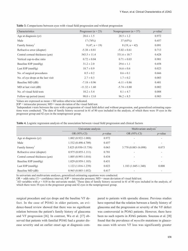
Table 3 shows a comparison of the clinical characteris-tics between 23 progressors and 57 nonprogressors in 80 eyes with JOAG. The progressor group (47%) represented a higher proportion of the family history than the nonpro-gressor group (19%, p = 0.091), but the difference was not significant. The last IOP was significantly higher in the progressor group (18.7 ± 0.9 mmHg) than in the nonpro-gressor group (16.6 ± 0.6 mmHg, p = 0.023), despite the significantly higher number of IOP-lowering eye drops be-ing used at the last visit in the progressor group (2.7 ± 0.3) vs. the nonprogressor group (1.7 ± 0.2, p = 0.003).
The univariate logistic regression analyses are shown in Table 4. The last IOP showed significant associations with the VF progression (odds ratio [OR], 1.122; p = 0.023).
Variables with p-values of less than 0.10 were included in the multivariate analysis to assess their joint effects on the VF progression. Using multivariate analyses, the VF pro-gressions were associated with the final IOP (OR, 1.183; p = 0.008).
Discussion
In this study, we demonstrated that JOAG patients had a male preponderance, myopic refractive state and severe el-evation of IOP; these findings were similar to those of pre-vious reports [8,12-14]. Bilateral JOAG patients were older and made up a higher proportion of those with a family history of glaucoma than unilateral JOAG patients. It is probable that unilateral JOAG patients were diagnosed earlier than bilateral JOAG patients, because 64% of the normal fellow eyes of unilateral JOAG patients at the ini-tial hospital visit were also diagnosed with JOAG during the follow-up in this study. In cases of normal tension glaucoma, one-fourth of patients developed VF loss in the fellow eyes that had an initially normal VF, and the esti-mate of the median time to VF loss onset was 95.1 months [15]. In this study, nine of 14 patients (64%) with unilateral JOAG developed JOAG in their fellow eyes, and the esti-mate of the median time to diagnosis was 12.3 months. Because the patients with unilateral JOAG had higher chance of developing JOAG in their fellow normal eyes, more caution is needed during follow-up to identify these patients so they can receive proper treatment.
Symptoms associated with visual acuity, including blurred vision and decreased visual acuity, were the major manifestation. However, one-third of the patients visited the clinics without definite symptoms and was diagnosed with JOAG by chance. Visual impairment and vision-relat-ed quality of life in working-age adults was related with impaired vision as well as with adverse social outcomes [10]. Because JOAG patients have a long life expectancy, the periodic IOP measurements conducted during the school’s physical examinations are important despite their rarity.
Our study showed that VF progression was associated with a higher IOP at the last hospital visit. A family histo-ry of glaucoma was weakly associated with a higher pro-portion of VF progression despite there being no signifi-cant differences in the baseline and last IOP, number of
Table 2. Presenting complaints of juvenile-onset open angle glaucoma patients at the initial hospital visit
Presenting complaint No. (%) Blurred vision 16 (22)Pain 14 (19)Health promotion examinations 13 (18)Decreased visual acuity 12 (17)Pre-refractive surgery examinations 8 (11)Others* 10 (14)
*Conjunctival injection, tearing, irritation, and foreign body sen-sation.
131
Y Kwun, et al. Clinical Characteristics of JOAG
surgical procedure and eye drops and the baseline VF de-fect. In the case of POAG in older patients, an evi-dence-based review showed that there was no significant relation between the patient’s family history of glaucoma and VF progression [16]. In contrast, Wu et al. [17] ob-served that patients with familial POAG had a greater dis-ease severity and an earlier onset age at diagnosis com-
pared to patients with sporadic disease. Previous studies have reported that the relation between a family history of glaucoma and the progression or severity of the VF defect was controversial in POAG patients. However, there have been no such reports in JOAG patients. Souzeau et al. [18] found that the prevalence of myocilin mutations in glauco-ma cases with severe VF loss was significantly greater
Table 4. Logistic regression analysis of the association between visual field progression and clinical factors
Univariate analysis Multivariate analysis*
OR (95% CI) p-value OR (95% CI) p-valueAge at diagnosis (yr) 1.002 (0.922-1.088) 0.972 - -Male 1.532 (0.498-4.709) 0.457 - -Family history† 3.825 (0.930-15.738) 0.063 3.770 (0.883-16.098) 0.073Refractive error (diopter) 0.975 (0.855-1.111) 0.701 - -Central corneal thickness (µm) 1.005 (0.993-1.016) 0.434 - -Baseline IOP (mmHg) 1.029 (0.959-1.103) 0.431 - -Last IOP (mmHg) 1.122 (1.016-1.239) 0.023 1.183 (1.045-1.340) 0.008Baseline MD (dB) 0.965 (0.885-1.052) 0.417 - -
In univariate and multivariate analyses, generalized estimating equations were conducted.OR = odds ratio; CI = confidence interval; IOP = intraocular pressure; MD = mean deviation of visual field test. *All variables with p < 0.10 in the univariate model; †These data of family history occurred in 61 of 80 eyes included in the analysis, of which there were 19 eyes in the progressor group and 42 eyes in the nonprogressor group.
Table 3. Comparisons between eyes with visual field progression and without progression
Characteristics Progressor (n = 23) Nonprogressor (n = 57) p-value*
Age at diagnosis (yr) 28.6 ± 1.5 28.5 ± 1.2 0.972Male 17 (74%) 37 (65%) 0.457Family history† 9 (47, n = 19) 8 (19, n = 42) 0.091Refractive error (diopter) -5.38 ± 0.81 -5.02 ± 0.61 0.713Central corneal thickness (µm) 543.5 ± 11.4 531.6 ± 10.7 0.428Vertical cup-to-disc ratio 0.72 ± 0.04 0.71 ± 0.03 0.901Baseline IOP (mmHg) 31.2 ± 2.0 29.6 ± 1.1 0.458Last IOP (mmHg) 18.7 ± 0.9 16.6 ± 0.6 0.023No. of surgical procedures 0.5 ± 0.2 0.6 ± 0.1 0.666No. of eye drops at the last visit 2.7 ± 0.3 1.7 ± 0.2 0.003Baseline MD (dB) -7.18 ± 0.96 -6.11 ± 0.86 0.401MD at last visit (dB) -11.32 ± 1.60 -5.74 ± 0.80 0.002No. of visual field tests 10.2 ± 5.4 8.1 ± 4.7 0.088Follow-up period (mon) 98.0 ± 13.0 96.2 ± 8.5 0.903
Values are expressed as mean ± SD unless otherwise indicated.IOP = intraocular pressure; MD = mean deviation of the visual field test. *Independent t-tests between the eyes with a progression of visual field defect and without progression, and generalized estimating equa-tions were conducted; †The data of family history occurred in 61 of 80 eyes included in the analysis, of which there were 19 eyes in the progressor group and 42 eyes in the nonprogressor group.

132
Korean J Ophthalmol Vol.30, No.2, 2016
than in nonadvanced glaucoma patients. In addition, the prevalence of a myocilin mutation of POAG patients ranged from 1.4% to 4.3%, worldwide [19-21], while 12.5% to 36% of cases of JOAG might have been linked to myoci-lin mutation [22,23]. Therefore, it is possible that a family history in JOAG patients might be associated with VF pro-gression.
This study had several limitations. First, because of its retrospective design, there were possibly confounding fac-tors and selection bias. In the event of poorly managed pa-tients, drop-out during follow-up may have occurred. However, there was no significant difference in the clinical backgrounds, including the age, sex, baseline IOP, duration of follow-up, and the severity of VF defect at initial visit between the VF progression and the nonprogression groups. The second limitation was the relatively small number of total patients due to its rarity, which made it difficult to detect differences among groups.
In conclusion, we confirmed that JOAG patients present-ed with a male preponderance, a myopic refractive state and severe elevation of IOP. Periodic eye examinations are needed because a considerable number of JOAG patients have no definite symptoms. Our data showed that the IOP at the last hospital visit was higher in patients were experi-encing a progression of VF defects. These findings indicat-ed that JOAG patients may require more careful manage-ment of IOP. For a definite conclusion, further large-scale prospective studies would be needed.
Conflict of Interest
No potential conflict of interest relevant to this article was reported.
References
1. Dielemans I, Vingerling JR, Wolfs RC, et al. The preva-lence of primary open-angle glaucoma in a popula-tion-based study in The Netherlands: the Rotterdam Study. Ophthalmology 1994;101:1851-5.
2. Iwase A, Suzuki Y, Araie M, et al. The prevalence of pri-mary open-angle glaucoma in Japanese: the Tajimi Study. Ophthalmology 2004;111:1641-8.
3. Wang YX, Xu L, Yang H, Jonas JB. Prevalence of glauco-
ma in North China: the Beijing Eye Study. Am J Ophthal-mol 2010;150:917-24.
4. Johnson AT, Drack AV, Kwitek AE, et al. Clinical features and linkage analysis of a family with autosomal dominant juvenile glaucoma. Ophthalmology 1993;100:524-9.
5. Wiggs JL, Haines JL, Paglinauan C, et al. Genetic linkage of autosomal dominant juvenile glaucoma to 1q21-q31 in three affected pedigrees. Genomics 1994;21:299-303.
6. Aponte EP, Diehl N, Mohney BG. Incidence and clinical characteristics of childhood glaucoma: a population-based study. Arch Ophthalmol 2010;128:478-82.
7. Fung DS, Roensch MA, Kooner KS, et al. Epidemiology and characteristics of childhood glaucoma: results from the Dallas Glaucoma Registry. Clin Ophthalmol 2013;7:1739-46.
8. Lotufo D, Ritch R, Szmyd L Jr, Burris JE. Juvenile glauco-ma, race, and refraction. JAMA 1989;261:249-52.
9. Park SC, Kee C. Large diurnal variation of intraocular pressure despite maximal medical treatment in juvenile open angle glaucoma. J Glaucoma 2007;16:164-8.
10. Rahi JS, Cumberland PM, Peckham CS. Visual impairment and vision-related quality of life in working-age adults: findings in the 1958 British birth cohort. Ophthalmology 2009;116:270-4.
11. Anderson DR, Patella VM, editors. Automated static pe-rimetry. 2nd ed. St. Louis: Mosby; 1999. p. 76-159, 204-11.
12. Goldwyn R, Waltman SR, Becker B. Primary open-angle glaucoma in adolescents and young adults. Arch Ophthal-mol 1970;84:579-82.
13. Perkins ES. Glaucoma in the younger age groups. Arch Ophthalmol 1960;64:882-91.
14. Gupta V, Ov M, Rao A, et al. Long-term structural and functional outcomes of therapy in juvenile-onset primary open-angle glaucoma: a five-year follow-up. Ophthalmo-logica 2012;228:19-25.
15. Fontana L, Armas R, Garway-Heath DF, et al. Clinical fac-tors influencing the visual prognosis of the fellow eyes of normal tension glaucoma patients with unilateral field loss. Br J Ophthalmol 1999;83:1002-5.
16. Ernest PJ, Schouten JS, Beckers HJ, et al. An evi-dence-based review of prognostic factors for glaucomatous visual field progression. Ophthalmology 2013;120:512-9.
17. Wu J, Hewitt AW, Green CM, et al. Disease severity of fa-milial glaucoma compared with sporadic glaucoma. Arch Ophthalmol 2006;124:950-4.
18. Souzeau E, Burdon KP, Dubowsky A, et al. Higher preva-

133
Y Kwun, et al. Clinical Characteristics of JOAG
lence of myocilin mutations in advanced glaucoma in com-parison with less advanced disease in an Australasian dis-ease registry. Ophthalmology 2013;120:1135-43.
19. Aldred MA, Baumber L, Hill A, et al. Low prevalence of MYOC mutations in UK primary open-angle glaucoma patients limits the utility of genetic testing. Hum Genet 2004;115:428-31.
20. Fingert JH, Heon E, Liebmann JM, et al. Analysis of myo-cilin mutations in 1703 glaucoma patients from five differ-ent populations. Hum Mol Genet 1999;8:899-905.
21. Sripriya S, Uthra S, Sangeetha R, et al. Low frequency of myocilin mutations in Indian primary open-angle glauco-ma patients. Clin Genet 2004;65:333-7.
22. Shimizu S, Lichter PR, Johnson AT, et al. Age-dependent prevalence of mutations at the GLC1A locus in primary open-angle glaucoma. Am J Ophthalmol 2000;130:165-77.
23. Yen YC, Yang JJ, Chou MC, Li SY. Identification of muta-tions in the myocilin (MYOC) gene in Taiwanese patients with juvenile-onset open-angle glaucoma. Mol Vis 2007; 13:1627-34.