Kirchhoff-Institut für Physik Molekulare Biophysik (F15) Gerrit L. Heuvelman Atomic Force...
22
Kirchhoff-Institut für Physik Molekulare Biophysik (F15) Gerrit L. Heuvelman Atomic Force Microscope
-
Upload
ursula-joseph -
Category
Documents
-
view
215 -
download
1
Transcript of Kirchhoff-Institut für Physik Molekulare Biophysik (F15) Gerrit L. Heuvelman Atomic Force...

Kirchhoff-Institut für Physik
Molekulare Biophysik (F15)
Gerrit L. Heuvelman
Atomic Force Microscope

Scanning Probe Microscopy
STM: scanning tunneling microscope
AFM: atomic force microscope
MFM: magnetic force microscope
SNOM: scanning near-field optical microscope
yx
tunneling of electrons between probe and surface
measuring of the force on the probe
AFM with magnetical probe
probe is a fiber; tunneling of fotonen
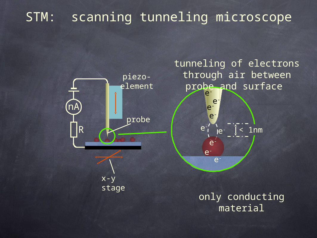
STM: scanning tunneling microscope
nA
R
piezo-element
e-e-
e-
e-e-
e-
e-
e-
e-
< 1nm
tunneling of electrons through air between probe
and surface
only conducting material
probe
x-y stage

STM: scanning tunneling microscope
nA
Icontrol
Itip
∆I
R
∆I -> ∆Vtransfer
piezo-element (changes length at different voltages)

AFM: atomic force microscope
AFM probe scans over the surface (in contact)
e.g. living cells, chromatin fibers
laser photodiode
piezo-element
probe
SFM: scanning force microscope
QuickTime™ and aGIF decompressor
are needed to see this picture.

MFM: magnetic force microscope
AFM with magnetic probe
e.g. hard disc, tape
magnetic tip
laser photodiode
piezo-element
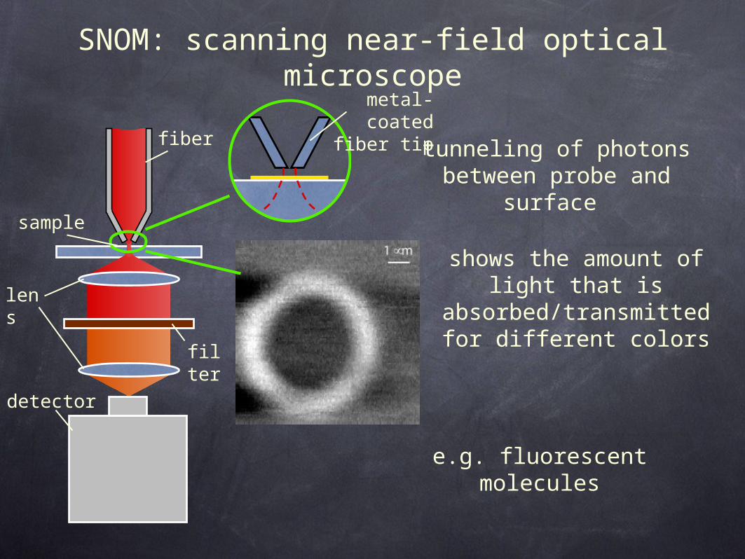
SNOM: scanning near-field optical microscope
fiber tunneling of photons between probe and surface
shows the amount of light that is
absorbed/transmitted for different colors
sample
lens
detector
filter
e.g. fluorescent molecules
metal-coated fiber
tip

Atomic Force Microscope
cantilever tip
laser
cantileverpiezo
y
z
x
photodiode

tipholder
motor control
SPM tip
sample
piezo translat
or
laser beam
mirror
photodiode
samplex,y,z piezo translator
fluid in fluid out
fluid cell
O-ring
Atomic Force Microscope
in air and in buffer solutions

path of AFM tip
AFM tip
superhelical DNA plasmid
DNA double helix
Mg2
+
negatively charged mica surface
Mg2
+
Mg2+Mg2+Mg2+Mg2+
movement of the AFM tip along the sample

AFM image of a 6.8 kb superhelical plasmid
AFM tip

AFM image of a nucleosome on a 614 base pair DNA
2 µm x 2 µm overview scan
zoom of mononucleosome complexes
Kepert, F., Fejes Tóth, K., Caudron, M., Mücke, N., Langowski, J. & Rippe, K., manuscript in preparation

AFM image of a nucleosome on a 614 base pair DNA
2 µm x 2 µm overview scan
zoom of mononucleosome complexes
Kepert, F., Fejes Tóth, K., Caudron, M., Mücke, N., Langowski, J. & Rippe, K., manuscript in preparation

0
20
40
60
80
100
120
140
160
0
10
20
30
40
50
60
DNA entry-exit angle
0
10
20
30
40
50
60
0
10
20
30
40
50
60
70
DNA entry-exit angle
DNA entry-exit angle of nucleosomes

movement of a 600 bp DNA fragment by AFM

different levels of chromatin packing
A
C
D
E
F
B
300 nm
700 nm11 nm
30 nm
2 nm
1400 nm
2 nm
11 nm
30 nm
300 nm
700 nm
1400 nm

chromatin structure400 nm x 400 nm

chromatin structure400 nm x 400 nm

chromatin structure

spectrin
C-terminal
Helix C
N-terminal
Helix B
Helix A
molecule that contributes to the mechanical properties,
especially the elasticity of the cells
measurement of its mechanical stability provides information
about the physiological function

stretching spectrin with an AFM
distance
force
1 2 3
surface
cantilever tip
4 repeated spectrin domain
1. adhesion force between cantilever tip and surface
2. dissociation from the folded state to the intermediate unfolded state
3. dissociation from the intermediate to the total unfolding state

0.15
100 30 40
0.05
20 50
0.10
0.20
0.00
unfolding force (pN)
pro
babili
ty
stretching spectrin with an AFMfo
rce (
pN
)
0 20 40 60 80
0
4020
608010
0
distance (nm)
-20


![ÜBERBLICK BIOPHYSIK Lebendiges Nichtgleichgewicht · und , , -, , , , , , , , , , , [13]](https://static.fdocuments.net/doc/165x107/5d63985588c993595c8b8ddc/ueberblick-biophysik-lebendiges-nichtgleichgewicht-und-.jpg)


![BIOPHYSIK Physik der Zelladhäsionbiophys/PDF/PJ2015.pdf · 4).) ((), (+ (– – [()() ] , / / . (), () ...](https://static.fdocuments.net/doc/165x107/5d56f76688c99392138b6b93/biophysik-physik-der-zelladhaesion-biophyspdfpj2015pdf-4-.jpg)