
Kinetic Characterization of Xenobiotic Reductase A from ... · Kinetic Characterization of...
9
pubs.acs.org/Biochemistry Published on Web 10/19/2009 r 2009 American Chemical Society 11412 Biochemistry 2009, 48, 11412–11420 DOI: 10.1021/bi901370u Kinetic Characterization of Xenobiotic Reductase A from Pseudomonas putida 86 † Olivia Spiegelhauer, ‡ Frank Dickert, § Sophia Mende, ‡ Dimitri Niks, ) Russ Hille, ) Matthias Ullmann, § and Holger Dobbek* ,‡ ‡ Bioinorganic Chemistry and § Structural Biology/Bioinformatics, University of Bayreuth, 95447 Bayreuth, Germany, and ) Department of Biochemistry, University of California, Riverside, California 92507 Received August 6, 2009; Revised Manuscript Received September 25, 2009 ABSTRACT: Xenobiotic reductase A (XenA) from Pseudomonas putida is a member of the old-yellow-enzyme family of flavin-containing enzymes and catalyzes the NADH/NADPH-dependent reduction of various substrates, including 8-hydroxycoumarin and 2-cyclohexenone. Here we present a kinetic and thermodyna- mic analysis of XenA. In the reductive half-reaction, complexes of oxidized XenA with NADH or NADPH form charge-transfer (CT) intermediates with increased absorption around 520-560 nm, which occurs with a second-order rate constant of 9.4 10 5 M -1 s -1 with NADH and 6.4 10 5 M -1 s -1 with NADPH, while its disappearance is controlled by a rate constant of 210-250 s -1 with both substrates. Transfer of hydride from NADPH proceeds 24 times more rapidly than from NADH. This modest kinetic preference of XenA for NADPH is unlike the typical discrimination between NADH and NADPH by binding affinity. Docking studies combined with electrostatic energy calculations indicate that the 2 0 -phosphate group attached to the adenine moiety of NADPH is responsible for this difference. The reductions of 2-cyclohexenone and coumarin in the oxidative half-reaction are both concentration-dependent under the assay conditions and reveal a more than 50-fold larger limiting rate constant for the reduction of 2-cyclohexenone compared to that of coumarin. Our work corroborates the link between XenA and other members of the old-yellow-enzyme family but demonstrates several differences in the reactivity of these enzymes. Xenobiotic reductases are bacterial enzymes of the old-yellow- enzyme (OYE) 1 family known to catalyze the reduction of the olefinic bond of R,β-unsaturated carbonyl compounds, including ketones and esters with NADH or NADPH as the electron source (1-7). Xenobiotic reductase A (XenA) from Pseudomonas putida catalyzes the NAD(P)H-dependent reduction of various biotic and abiotic compounds (8). Recently, it was shown that XenA catalyzes the reduction of the C3-C4 double bond of 8-hydroxycoumarin, indicating its involvement in the degrada- tion of quinoline, a ubiquitous N-heterocyclic pollutant with carcinogenic properties (9), along the 8-hydroxycoumarin path- way in P. putida 86 (10). The crystal structure of XenA has been determined for the enzyme alone and in complex with two different substrates (10). The structure reveals a dimeric arrangement with one (β/R) 8 barrel domain per monomer, which binds a FMN molecule on the solvent-exposed C-terminal side of the barrel. A tryptophan residue from the C-terminal helix of the neighboring monomer protrudes into the active site and forms one wall of the substrate- binding pocket. The active site is further lined by histidine and tyrosine residues, which presumably are needed to bind and orient the substrates, stabilize developing charges during turnover, and donate protons. A feature distinguishing XenA from most members of the OYE family is the presence of a cysteine residue in the active site near the N5 position of the isoalloxazine ring (10). XenA shares its overall arrangement and active site architecture with YqjM, and both have been suggested to form a new subfamily within the OYE family (11). Genome sequencing projects revealed the OYE-like enzymes to be a rapidly growing family, which in some organisms, such as Saccharomyces cerevisiae (12), Shewanella oneidensis (13), and P. putida KT2440 (14), occur in up to six copies. The modest substrate specificity of OYE-like enzymes has attracted the attention of biotechnologists (15), further motivating more de- tailed studies of the physiological function and catalytic mechani- sms of different variants. Therefore, several members of this enzyme family have been intensely studied, and the structural, kinetic, and thermodynamic characteristics of their interaction with various substrates have been elucidated, which have revealed that despite their similar structures they differ remarkably in their reactivities (11, 16-33). Here, we have investigated the thermo- dynamic characteristics of XenA and studied its reactivity with various substrates. Our data suggest an electrostatic basis for the kinetic preference of XenA for NADPH over NADH. EXPERIMENTAL PROCEDURES Chemicals and Enzymes. Complex microbial media were purchased from Roth and Otto Nordwald and were prepared as described by Sambrook et al. (35). All chemicals and enzymes were purchased from Fluka and AppliChem. 5-Deaza-10-methyl-3- sulfopropylisoalloxazine was a gift from P. M. H. Kroneck (University of Konstanz, Konstanz, Germany). The extinction † This work was funded by grants from the Deutsche Forschungsge- meinschaft (DFG, DO-785/2), the Fonds der chemischen Industrie, and the Bavarian-Californian Technology Centre. *To whom correspondence should be addressed: Universit€ atsstrasse 30, 95440 Bayreuth, Universit€ at Bayreuth, Germany. Telephone: (þþ49) 921-554364. Fax: (þþ49) 921-552432. E-mail: Holger.Dobbek@ uni-bayreuth.de. 1 Abbreviations: CT, charge transfer; EDTA, ethylenediaminetetra- acetate; FMN, flavin mononucleotide; OYE, old yellow enzyme; PDB, Protein Data Bank; XenA, xenobiotic reductase A.
Transcript of Kinetic Characterization of Xenobiotic Reductase A from ... · Kinetic Characterization of...
pubs.acs.org/Biochemistry Published on Web 10/19/2009 r 2009 American Chemical Society
11412 Biochemistry 2009, 48, 11412–11420
DOI: 10.1021/bi901370u
Kinetic Characterization of Xenobiotic Reductase A from Pseudomonas putida 86†
Olivia Spiegelhauer,‡ Frank Dickert,§ Sophia Mende,‡ Dimitri Niks, ) Russ Hille, ) Matthias Ullmann,§ andHolger Dobbek*,‡
‡Bioinorganic Chemistry and §Structural Biology/Bioinformatics, University of Bayreuth, 95447 Bayreuth, Germany, and )Departmentof Biochemistry, University of California, Riverside, California 92507
Received August 6, 2009; Revised Manuscript Received September 25, 2009
ABSTRACT: Xenobiotic reductase A (XenA) from Pseudomonas putida is a member of the old-yellow-enzymefamily of flavin-containing enzymes and catalyzes the NADH/NADPH-dependent reduction of varioussubstrates, including 8-hydroxycoumarin and 2-cyclohexenone. Here we present a kinetic and thermodyna-mic analysis of XenA. In the reductive half-reaction, complexes of oxidized XenA with NADH or NADPHform charge-transfer (CT) intermediates with increased absorption around 520-560 nm, which occurs with asecond-order rate constant of 9.4 � 105 M-1 s-1 with NADH and 6.4 � 105 M-1 s-1 with NADPH, while itsdisappearance is controlled by a rate constant of 210-250 s-1 with both substrates. Transfer of hydride fromNADPH proceeds 24 times more rapidly than from NADH. This modest kinetic preference of XenA forNADPH is unlike the typical discrimination between NADH and NADPH by binding affinity. Dockingstudies combined with electrostatic energy calculations indicate that the 20-phosphate group attached to theadenine moiety of NADPH is responsible for this difference. The reductions of 2-cyclohexenone andcoumarin in the oxidative half-reaction are both concentration-dependent under the assay conditions andreveal amore than 50-fold larger limiting rate constant for the reduction of 2-cyclohexenone compared to thatof coumarin. Our work corroborates the link between XenA and other members of the old-yellow-enzymefamily but demonstrates several differences in the reactivity of these enzymes.
Xenobiotic reductases are bacterial enzymes of the old-yellow-enzyme (OYE)1 family known to catalyze the reduction of theolefinic bond ofR,β-unsaturated carbonyl compounds, includingketones and esters with NADH or NADPH as the electronsource (1-7). Xenobiotic reductaseA (XenA) fromPseudomonasputida catalyzes the NAD(P)H-dependent reduction of variousbiotic and abiotic compounds (8). Recently, it was shown thatXenA catalyzes the reduction of the C3-C4 double bond of8-hydroxycoumarin, indicating its involvement in the degrada-tion of quinoline, a ubiquitous N-heterocyclic pollutant withcarcinogenic properties (9), along the 8-hydroxycoumarin path-way in P. putida 86 (10).
The crystal structure of XenA has been determined for theenzyme alone and in complex with two different substrates (10).The structure reveals a dimeric arrangement with one (β/R)8barrel domain per monomer, which binds a FMN molecule onthe solvent-exposed C-terminal side of the barrel. A tryptophanresidue from the C-terminal helix of the neighboring monomerprotrudes into the active site and forms one wall of the substrate-binding pocket. The active site is further lined by histidineand tyrosine residues, which presumably are needed to bindand orient the substrates, stabilize developing charges during
turnover, and donate protons. A feature distinguishing XenAfrom most members of the OYE family is the presence of acysteine residue in the active site near the N5 position of theisoalloxazine ring (10). XenA shares its overall arrangement andactive site architecture with YqjM, and both have been suggestedto form a new subfamily within the OYE family (11).
Genome sequencing projects revealed theOYE-like enzymes tobe a rapidly growing family, which in some organisms, such asSaccharomyces cerevisiae (12), Shewanella oneidensis (13), andP. putida KT2440 (14), occur in up to six copies. The modestsubstrate specificity of OYE-like enzymes has attracted theattention of biotechnologists (15), further motivating more de-tailed studies of the physiological function and catalytic mechani-sms of different variants. Therefore, several members of thisenzyme family have been intensely studied, and the structural,kinetic, and thermodynamic characteristics of their interactionwith various substrates have been elucidated, which have revealedthat despite their similar structures they differ remarkably in theirreactivities (11, 16-33). Here, we have investigated the thermo-dynamic characteristics of XenA and studied its reactivity withvarious substrates. Our data suggest an electrostatic basis for thekinetic preference of XenA for NADPH over NADH.
EXPERIMENTAL PROCEDURES
Chemicals and Enzymes. Complex microbial media werepurchased from Roth and Otto Nordwald and were prepared asdescribed by Sambrook et al. (35). All chemicals and enzymes werepurchased from Fluka and AppliChem. 5-Deaza-10-methyl-3-sulfopropylisoalloxazine was a gift from P. M. H. Kroneck(University of Konstanz, Konstanz, Germany). The extinction
†This work was funded by grants from the Deutsche Forschungsge-meinschaft (DFG, DO-785/2), the Fonds der chemischen Industrie, andthe Bavarian-Californian Technology Centre.*To whom correspondence should be addressed: Universit€atsstrasse
30, 95440 Bayreuth, Universit€at Bayreuth, Germany. Telephone:(þþ49) 921-554364. Fax: (þþ49) 921-552432. E-mail: [email protected].
1Abbreviations: CT, charge transfer; EDTA, ethylenediaminetetra-acetate; FMN, flavin mononucleotide; OYE, old yellow enzyme; PDB,Protein Data Bank; XenA, xenobiotic reductase A.

Article Biochemistry, Vol. 48, No. 48, 2009 11413
coefficients of XenA (ε280=71050 M-1 cm-1, and ε464=12200M-1 cm-1), NADH (ε340=6200M-1 cm-1), andNADPH (ε340=6220 M-1 cm-1) were used to calculate the concentrations of theenzyme, cofactor, and substrates.Protein Expression, Purification, and Activity Assay.
The gene of XenAwas isolated fromP. putida 86 and cloned intoa pET11a vector as described previously (10). XenA was ex-pressed inEscherichia coliRosetta(DE3)pLysS using LBmediumsupplementedwith 100 μg/mL ampicillin and 34 μg/mL chloram-phenicol, at 20 �C overnight.
Crude cell extracts were prepared in 50 mM Tris buffer (pH8.0) for a purification that included three chromatographic steps.The first column was a Q-Sepharose FF column, followed bySource 15-ISO and Sephacryl S-200 columns. Before size-exclu-sion chromatography was conducted, the protein was reconsti-tuted on ice with 5 mM FMN overnight. Eight liters of cellculture yielded 600 mg of enzyme with a purity exceeding 95%estimated via SDS-PAGE (data not shown). The pure enzymewas stored at a concentration of ∼50 mg/mL in 50 mM Trisbuffer (pH 8.0) at -80 �C.
The specific activity ofXenAwas determined fromabsorbancechanges at 340 nm due to the oxidation of NADPH. To preventthe oxidase activity of XenA, all kinetic measurements wereperformed under anoxic conditions. We made solutions anoxicby bubbling them with dinitrogen gas. The cuvettes were sealedby screw caps with a rubber septum and flushed with dinitrogengas before use. The typical oxidase activity found under theseconditions was 0.14 unit mg-1. Activity tests were performedusing an Analytik Jena Specord40 spectrophotometer at 25 �C.The reaction mixture contained 50 mM Tris-HCl (pH 8.0), 300μM 2-cyclohexenone, and 150 μM NADPH. The reaction wasstarted by addition of enzyme to the reaction mixture andconducted in a volume of 1 mL with a XenA concentration ofapproximately 1� 10-2 mgmL-1. One activity unit is defined asthe oxidation of 1 μmol of NADPH/min.
We determined the flavin content spectrophotometrically bySDS treatment according to the protocol by Aliverti et al. (36).Photoreduction of XenA. Photoreduction of XenA was
essentially conducted as described by Massey et al. (37). XenAwas photoreduced in a glass tonometer with a cuvette attached toa side arm. As a photoreductant, we used the potassium salt of5-deaza-10-methyl-3-sulfopropylisoalloxazine, a deazaflavin deri-vative, in catalytic amounts. The cuvette contained 5-deaza-10-methyl-3-sulfopropylisoalloxazine and EDTA in 100 mM Trisbuffer (pH 8.0) in a final volume of 1 mL. The tonometercontained XenA and phenosafranine in 100 mM Tris buffer(pH 8.0) in a final volume of 2 mL. Oxygen was removed byrepeated evacuation and flushing with dinitrogen gas. Afteroxygen removal, the two volumes were mixed, resulting in finalconcentrations of 15 mM EDTA, 30 μM XenA, and 1 μMphenosafranine. For illumination, a 100 W lamp from a slideprojector (Agfa, Opticus 100) was used. The reduction of XenAwas followed by recording absorption spectra directly after and3 min after each illumination.Determination of Reduction Potentials. Determination of
the reduction potential of XenA-boundFMN in the presence andabsence of 1 mM NADþ was conducted as described pre-viously (38). The reaction mixture contained 15 μM XenA,15 μM phenosafranine [E�m,D=-252 mV (39)] as a referencedye, 2 μMmethylviologen as an electron mediator, and 0.05 unitof xanthine oxidase. The reactionwas conducted under anaerobicconditions in a glass tonometer and started by addition of
xanthine with a final concentration of 300 μM. To calculatethe concentrations of oxidized XenA (Eox) and oxidized pheno-safranine (Dox), the absorbance values at 464 and 521 nm wereused and integrated in eqs 1 and 2.
A464 ¼ εEox464c
Eox þ εDox464c
Dox þ εEred464ðcEtot -cEoxÞ
þ εDred464ðcDtot -cDoxÞ ð1Þ
A521 ¼ εEox521c
Eox þ εDox521c
Dox þ εEred521ðcEtot -cEoxÞ
þ εDred521ðcDtot -cDoxÞ ð2Þ
Following are the values of the molar extinction coefficients:ε(oxidized enzyme at 464 nm)=12.2� 103 M-1 cm-1, ε(reducedenzyme at 464 nm)=0.6 � 103 M-1 cm-1, ε(oxidized enzyme at521 nm)=1.5 � 103 M-1 cm-1, ε(reduced enzyme at 521 nm)=0.6� 103M-1 cm-1, ε(oxidized dye at 464 nm)=12.9� 103M-1
cm-1, ε(reduced dye at 464 nm)=0.3� 103M-1 cm-1, ε(oxidizeddye at 521 nm)=44.9 � 103 M-1 cm-1, and ε(reduced dye at521 nm)=0.3 � 103 M-1 cm-1.
The reduction potential of XenA (E�m,E) can subsequently bedetermined from the difference (ΔE�) in the reduction potentialsof the enzyme and dye. The latter can be obtained from the plot oflog(Eox/Ered) versus log(Dox/Dred) using eq 3.
E�m;E ¼ E�m;D þΔE� ð3Þ
Steady-State Kinetics Experiments. The steady-state kine-tics of XenA reacting with various concentrations of 2-cyclohexe-none and NADPH were performed under anaerobic conditions.The concentration of 2-cyclohexenone was varied between 10 and160 μM, and the concentration of NADPHwas varied between 10and 400 μM. Each single measurement was performed three times.2-Cyclohexenone and NADPH were both added from a freshlyprepared 1mMstock solution, and the concentration ofXenAwasapproximately 250 nM. The apparent values for KmA, KmB, andVmax have been determined by multiple nonlinear regressionanalysis of the measured rates using eq 4 (40):
v ¼ Vmax½A�½B�KmB½A� þKmA½B� þ ½A�½B� ð4Þ
where [A] is the concentration of 2-cyclohexenone, [B] is theconcentration of NADPH, v is the observed rate, Vmax is the rateat saturating substrate concentrations, KmA is the Km for 2-cyclo-hexenone at saturating NADPH levels, and KmB is the Km forNADPH at saturating 2-cyclohexenone levels.Rapid Reaction Techniques. Measurements on the kinetics
of the reductive half-reaction were performed under anaerobicconditions using an Applied Photophysics SX-20MV kineticspectrophotometer with a 1 cm observation path length cuvettecoupled either to a diode array detector or to a monochromatorand photomultiplier. The reaction temperature was controlledwith a Haake F8/C25 thermostat. Standard reaction conditionswere 50 mM Tris (pH 8.0) at 20 �C. In a typical experiment,enzyme at a XenA concentration of approximately 10 μM wasmixed with an equal volume of substrate solution, the latter atconcentrations ranging from 50 to 5000 μM. Each experimentwas repeated at least five times for each substrate concentration.The reactions were monitored at 464 and 540 nm over anappropriate time scale. Observed kinetic transients at 464 nmwere fit to single exponentials, and transients at 540 nm,
11414 Biochemistry, Vol. 48, No. 48, 2009 Spiegelhauer et al.
monitoring formation and decay of charge-transfer complexes,were fit with double exponentials using Pro-Data (AppliedPhotophysics) to yield observed rate constants (kobs).
The reductive half-reaction sequence was modeled as shown ineq 5
AþBsFRsk1
k-1
C sFk2D ð5Þ
where A is XenAox, B is NAD(P)H, C is the XenA-NAD(P)Hcharge-transfer complex, and D is XenA containing the two-electron-reduced state of FMN and bound NAD(P)þ.
The oxidative half-reaction sequence was modeled as shown inthe general eq 6
EþFsFRsk3
k-3
G sFk4H ð6Þ
where E is XenAred, F is 2-cyclohexenone (coumarin), G is theXenAred-2-cyclohexenone (coumarin) charge-transfer complex,and H is XenAox with bound 2-cyclohexanone (chroman-2-one).
Hyperbolic plots of observed rate constants versus substrateconcentration were fitted using eq 7 to yield the limiting rate ofreaction at high [S], kX, and the dissociation constant KD (41).
kobs ¼ kX½S�=ðKD þ ½S�Þ ð7Þ
kobs ¼ k1½S� þ k-1 þ k2 ð8ÞRate constants for the formation of the charge-transfer complexbetween oxidized XenA and NADH/NADPH were approxi-mated by linear regression analysis with eq 8, where k2 is thelimiting rate of reduction of XenA with the respective nicotina-mide used.
To relate the individual limiting rate constants of the reductiveand oxidative half-reactions to the steady-state catalytic con-stants, eq 9 has been used:
kcat ¼ k2k4=ðk2 þ k4Þ ¼ kredkox=ðkred þ koxÞ ð9ÞSolutions of oxidized XenA were made anoxic in a glasstonometer as described above. Solutions of reduced XenA weremade anoxic in a tonometer with a cuvette side arm and titratedwith NADH to achieve complete flavin reduction. We made allsubstrate solutions anoxic by flushing them with dinitrogen gas.
Docking. NADPH was docked to the crystal structure ofXenA (PDB entry 2H8X) using DOCK 6 (42). The charges weretaken from the CHARMM27 force field (43). A “divide andconquer” approach was used to reduce the number of rotatablebonds. The nicotinamide was docked close to FMN usingchemical docking, which was used to incorporate informationabout the chemical complementarity of ligand and receptormoieties into the matching process. In a second step, adenosinewas independently docked within a radius of 25 A from FMN.The remaining parts of NAD(P)H (i.e., the ribose and phos-phates) were constructed geometrically and subsequently ener-getically minimized using CHARMM (44), while the rest of theprotein, the nicotinamide ring and the adenine ring, was keptfixed.Electrostatic Calculations. To study the effects of the
phosphate group of NADPH on the hydride-transfer energiesbetween the nicotinamide ring and the isoalloxazine ring, elec-trostatics were calculated using the program SOLINPROTof theMEAD package (45). We calculated the interaction of thenicotinamide ring and the isoalloxazine ring with the monopho-sphate group bound to the ribose before and after the electrontransfer. The XenA dimer was used to define the dielectricboundaries. For this calculation, only the charges of the nicotina-mide ring and the phosphate group of NADPH and theisoalloxazine ring of the FMN were considered. The dielectricconstants of protein and of water were set to 4.0 and 80.0,respectively. The ionic strength was set to 0.1 M. The calculationwas done in two focusing levels with grids of 181� 180� 180 gridpoints. The grid spacing was set to 1.0 and 0.25 A for the outerand the inner grid, respectively. The outer grid was centered at thecoordinate center of the protein; the inner gridwas centered at theN1 atom of FMN.
RESULTS
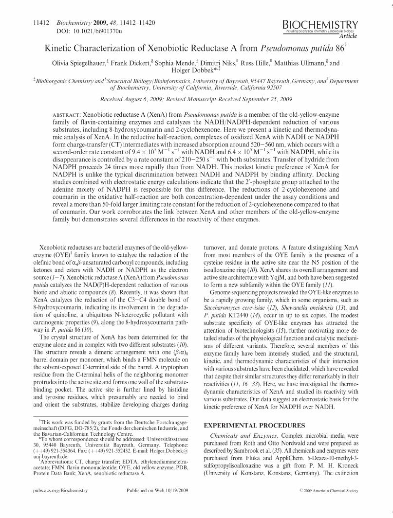
Photoreduction of XenA.XenAwas reduced using the light-mediated generation of electrons by the deazaflavin-EDTAcouple in the presence of phenosafranine as the redox mediator(Figure 1A). This method has been applied to ensure singleelectron transfer to allow for initial semiquinone formation.Photoreduction proceeds in a two-stepmechanism.Directly aftereach illumination period, we observe a signal increase around350 nm, which we assign to the formation of the red anionic
FIGURE 1: Photoreduction ofXenA. Conditions: 30 μMXenA, 15mMEDTA, 1 μMphenosafranine, 100mMTris buffer (pH 8.0), and traces of5-deaza-10-methyl-3-sulfopropylisoalloxazine as catalyst. (A)Time-dependent reduction ofXenAafter illumination.The figure shows the spectrarecorded before ( 3 3 3 ) and after (;) different periods of illumination. The spectrum of reoxidized XenA is displayed as a dashed line. (B) Spectrarecorded directly (---) and 3 min (;) after a single irradiation step.

Article Biochemistry, Vol. 48, No. 48, 2009 11415
semiquinone. Figure 1B shows the spectrum (dashed line) with acharacteristic peak around 400 nm and an increase in signalmagnitude between 500 and 550 nm. Three minutes afterillumination, the semiquinone signature was not observed anymore and the enzyme-bound flavin was converted to the di-hydroflavin form as a result of the dismutation of the semiqui-none. After complete reduction of the enzyme, the cuvette wasexposed to air, allowing XenA to reoxidize, resulting in aspectrum indistinguishable from the starting spectrum.Determination of Reduction Potential. The generation of
electrons by the xanthine oxidase-xanthine couple has been usedas an alternative method to reduce XenA. The presence of areference dye (phenosafranine; E�m =-252 mV) allowed thedetermination of the reduction potential of XenA-bound FMN.The small amount of xanthine oxidase ensured equilibriumconditions at all times, whereas both enzyme and dye take uptwo electrons. Reduction of XenA by xanthine oxidase in theabsence of phenosafranine showed the slow conversion of FMNfrom the quinone to the hydroquinone state without anydetectable formation of semiquinone species (data not shown).Spectra recorded during the reaction with phenosafranine showthat XenA and phenosafranine are reduced to similar extents(Figure 2). The absorbance values at 464 and 521 nmwere used tocalculate the amount of oxidizedXenAand dye using eqs 1 and 2.Equation 3 gives a reduction potential of -263 mV for XenA.The linear fit of the plot of log(Eox/Ered) versus log(Dox/Dred)shows a slope of -1, confirming that both XenA and pheno-safranine received the same amount of electrons and reactedunder equilibrium conditions. There was no difference observedin the presence of 1 mM NADþ and 1 mM NADPþ.Steady-State Kinetics. Catalytic turnover of XenA with
various concentrations of 2-cyclohexenone and NADPH underanaerobic conditions was analyzed to determine the values ofKmA,KmB, and Vmax. The inset in Figure 3 shows the rate dependenciesof 2-cyclohexenone for different NADPH concentrations andthe corresponding nonlinear fits using the normal Michaelis-Menten equation. The parallel lines in the double-reciprocal plot
(Figure 3) are consistent with a double-displacement (ping-pong)mechanism (40). Consequently, the measured rates were analyzedby multiple nonlinear regression analysis using eq 4, resulting inthe following values: kcat = 7.2 ( 0.3 s-1, KmA (the Km for2-cyclohexenone)=37.2( 2.4 μM, andKmB (theKm for NADPH)=200 ( 13.0 μM.Reductive Half-Reaction. The reaction of XenA with
NADH and NADPH was next examined following the spectralchange associated with reduction of the enzyme’s flavin(Figure 4A). No spectral changes indicating the formation of aMichaelis complex in the dead time of the stopped-flow experi-mentwere observed (Figure 4A). Formation of a CT complex, onthe other hand, can be discerned by an absorption increase at520-560 nm (Figure 4B, inset), which decayswith a rate constantcomparable to that of FMN reduction recorded at 464 nm(Figure 4B). The characteristic absorption around 464 nm showsthat the weak CT complex is indeed formed between oxidizedXenA and the NADH or NADPH and is not a complex ofthe reduced enzyme (Figure 4A). The observed rate constant forthe formation of the CT complex depends linearly on theconcentration of NADH or NADPH (Figure 4C). Using eq 8to fit the observed dependence of observed rate constants on theconcentration of NAD(P)H under pseudo-first-order conditionsgives a second-order rate constant (k1) of (9.4 ( 0.5) � 105 M-1
s-1 withNADHand (6.4( 0.3)� 105M-1 s-1 withNADPH forthe formation of the CT complex. The rate constants for thedissociation of NAD(P)H from the CT complex (k-1) have beenderived from the positive intercept on the ordinate, which isapproximated to be k-1 þ k2. The values for k-1 are very similarfor both nicotinamides with 256( 17 s-1 for NADH and 215 (11 s-1 for NADPH. The rate constants determined for the decayof the CT complex at long wavelengths (Figure 4B, inset)correspond to the observed rates measured for the reduction ofFMN seen at 464 nm and show a hyperbolic dependence on theconcentration of reduced nicotinamide (Figure 4D,E). The linear
FIGURE 2: Redox potential determination for the FMN-FMNH-
couple of XenA. Conditions: 15 μM XenA, 15 μM phenosafranine,2 μMmethylviologen, 0.05 unit of xanthine oxidase, and 50mMTrisbuffer (pH 8.0). The dotted line shows the spectrum of the reactionmixture before the addition of xanthine. The reaction was followedover 1.5 h (;). Absorbance values at 464 and 521 nm were used tocalculate the concentrations of oxidized XenA and the dye. The insetshows the plot of log(Eox/Ered) vs log(Dox/Dred). The solid linedisplays the linear fit with a slope of -1. The redox potential ofXenA was calculated to be -263 mV.
FIGURE 3: Steady-state kinetic data of theXenA-catalyzed reductionof 2-cyclohexenone by NADPH. Lineweaver-Burk plot with linearregression analysis using the simple Michaelis-Menten equation.Each point represents the mean of three independent measurements.The assays were performed in 50 mM Tris buffer (pH 8.0) at 25 �C.The final volume of 1 mL contained 250 nM XenA, varyingconcentrations of 2-cyclohexenone (10-160 μM), and varying con-centrations ofNADPH (10-400 μM). Each line displays one distinctNADPH concentration: (O) 10 μM NADPH, (b) 50 μM NADPH,(0) 100 μM NADPH, (9) 150 μM NADPH, (4) 200 μM NADPH,(2) 300 μM NADPH, and (3) 400 μM NADPH. The inset displaysthe direct rate dependencies of 2-cyclohexenone for the differentNADPH concentrations (see above) and the nonlinear fits using thesimple Michaelis-Menten equation.

11416 Biochemistry, Vol. 48, No. 48, 2009 Spiegelhauer et al.
correlation of 1/kobs with 1/[NADH] or 1/[NADPH] (Figure 4D,E, insets) indicates that the equilibrium condition for theXenA-NAD(P)H reaction (k-1 . k2) holds, as seen experimen-tally, and that eq 7 can be used to determine the limiting rateconstant for the reduction of FMN (k2) and the dissociationconstant (KD) of the complex (41). For the reaction of XenAox
with NADH, a rate constant (k2) of 1.50 ( 0.02 s-1 is obtained,while with NADPH, the rate constant is 24 times higher, 35.7 (0.6 s-1. The dissociation constants for the XenAox-NADHcomplex of 176( 14 μMand for the XenAox-NADPH complexof 256( 12 μMobtained using the rapid equilibriummodel are ingood agreement with the ratio between the rate constants for theformation of the CT complexes (XenAox-NADH, k-1/k1=272μM;XenAox-NADPH, k-1/k1=336 μM). Appreciable rates forthe back reactionwould cause the double-reciprocal plot of 1/kobsversus 1/[NAD(P)H] to curve down for high substrate concen-trations (41), which is not observed (Figure 4D,E, insets).Furthermore, an adaptation of the model to include the rateconstant of the back-reaction (k-2) did not improve the fit to theobserved rates, as judged from testing the goodness of fit. We
conclude that k-2 is very small and that reduction of XenA byeither NADH or NADPH is functionally irreversible.Oxidative Half-Reaction. To examine the reoxidation of
XenA, two different substrates have been studied, 2-cyclohexe-none and coumarin. 2-Cyclohexenone has been used as asubstrate in most rapid kinetic studies with members of theOYE family, notably with OYE (19), morphinone reductase (24,46), PETN reductase (29), and YqjM (34), and it is thereforepossible to compare the reactivity of XenA with the reactivitiesof these enzymes. However, since we have recently shownthat XenA participates in the degradation of quinoline alongthe 8-hydroxycoumarin pathway and is able to reduce theC3-C4 double bonds of heteroaromatic compounds such ascoumarin (10), we have also examined the reaction of XenA withthis substrate.
Spectral changes are observed directly after reduced XenAreacts with 50 μM2-cyclohexenone, as compared to the spectrumof reduced XenA with substrate (Figure 5A). The observedmaximum at 424 nm 5 ms after reduced XenA is mixed with2-cyclohexenone indicates that it is not due to a fast reaction
FIGURE 4: Reductive half-reactions of XenA with NADH and NADPH. (A) Time-dependent spectral changes of 35 μMXenAox reacting with800 μMNADH (corresponding to 70 μMXenAox and 1600 μMNADHbefore mixing). The dashed line represents the spectrum of XenAox. (B)Time-dependent absorbance change at 464 nm for the reactionof 5μMXenAwith 2500μMNADH.The inset displays the absorbance changes at540 nm (with 35μMXenAand 50μMNADH), whichwe attribute to the formation of theCT complex. (C)Dependencies of the observed rates at540 nm (CT formation) onNADH concentration (O) and NADPH concentration (0) for reactions with 35 μMXenA. (D and E) Concentrationdependence of the observed rates at 464 nm (5 μM XenA) for NADH (D) and NADPH (E), with the reciprocal plots in the insets. The curvesdisplay the best fits to the data using eq 7. All experiments were conducted under anaerobic conditions in 50 mM Tris buffer (pH 8.0) at 20 �C.

Article Biochemistry, Vol. 48, No. 48, 2009 11417
phase in which FMN is oxidized but rather that another reactionintermediate involving reduced XenA and 2-cyclohexenone isformed rapidly within the dead time of the instrument. Formationof a CT complex is discernible with both substrates by an initialabsorption increase around 650 nm (data not shown). The CTcomplex forms very rapidly and decays with the same rate asXenAbecomes oxidized. The low absorbance around 464 nm at veryshort times indicates that the CT complex involves reduced XenAand substrate. A larger absorption increase is subsequently ob-served at 464 nm (Figure 5B). The rate constant observed for themajority of the absorbance increase at 464 nm shows a hyperbolicdependence on the concentration of 2-cyclohexenone and cou-marin (Figure 5C,D). The linear relation between 1/kobs and1/[2-cyclohexenone] or 1/[coumarin] (Figure 5C,D, insets) indi-cates that it is justified to include the rapid equilibrium condition inour model (41). A rate constant (k4) of 13.1( 0.1 s-1 and a KD of86 ( 2 μM have been determined for 2-cyclohexenone. Thereduction of coumarin is slower than the reduction of 2-cyclohexe-none by a factor of 50with a rate constant (k4) of 0.243( 0.001 s-1,and the complex has a KD of 19.3 ( 0.2 μM.Modeling NADH and NADPH in the Active Site of
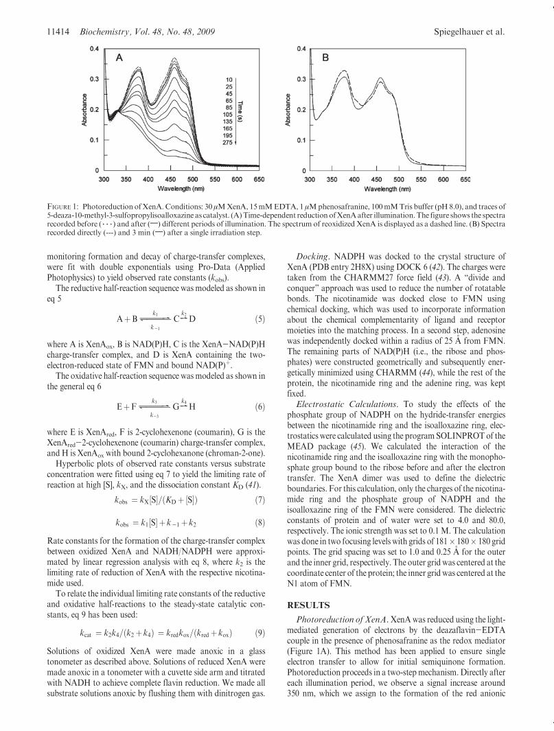
XenA. We next examined a model for both NADH andNADPH bound to oxidized XenA, generated using the DOCK6 program. In this model, NADH and NADPH bind both in thesame way toXenA. The nicotinamide ring ofNAD(P)H docks ina stacked conformation with the isoalloxazine ring of FMN(Figure 6) and is hydrogen-bonded to His178, His181, and
Cys25. The diphosphate forms a salt bridge with Lys106 and ahydrogen bond with Tyr183. The distance between the phos-phorus atom of the 20-phosphate of NADPH and N1 of thenicotinamide is 6.2 A. The 20-phosphate of NADPH is orientedtoward the solvent and does not interact with the protein matrixbut does form a hydrogen bond to the 20-OH group of the riboseattached to nicotinamide (Figure 6).
From electrostatic calculations using the Poisson-Boltzmannequation, we find that the free energy of the transfer of hydridefrom the nicotinamide to the isoalloxazine ring of the FMN isshifted by-1.83 kcal/mol due to the presence of the 20-phosphateon NADPH compared to NADH. Thus, the hydride transfer ismore favorable with NADPH than with NADH. Most of thisdifference in free energy manifests itself in a lower activationenergy for NADPH versus NADH as reflected in the relativelimiting rates of reduction (36 s-1 vs 1.5 s-1, respectively).
DISCUSSION
Recently, we have shown that XenA participates in thedegradation of quinoline and reacts with heteroaromatic com-pounds such as coumarin and 8-hydroxycoumarin using bothNADH and NADPH as electron sources (10). Here we haveexamined its reactivity with both reducing and oxidizing sub-strates, allowing us to compare it to related flavoenzymes.
No stable semiquinone formofXenAwas observed in the courseof the reductive titrations (using either the xanthine-xanthineoxidase or deazaflavin-light couple as the reductant), indicating
FIGURE 5: Oxidative half-reaction of reduced XenAwith 2-cyclohexenone and coumarin. (A) Time-dependent spectral changes of 35 μMXenA(NADH-reduced) reactingwith 800μM2-cyclohexenone.The dashed line represents the spectrumof reducedXenA. (B)Time-dependent spectralchanges at 464 nmfor the reactionof5μMXenAwith 25μM2-cyclohexenone.A reaction tracewith25μMcoumarin is shown in the inset ofpanelB. (C andD)Concentration dependence of the observed rate constants at 464 nm for the reactionof 5 μMXenAwith 2-cyclohexenone (C) and forcoumarin (D).The curves display the best fits to the datausing eq7.The reciprocal plots are shown in the insets.All experimentswere conducted in50 mM Tris buffer (pH 8.0) at 20 �C under anaerobic conditions.
11418 Biochemistry, Vol. 48, No. 48, 2009 Spiegelhauer et al.
that transiently formed red, anionic semiquinone rapidly dispro-portionated. The hydrogen bonding distance between the amidenitrogen of Cys25 and N5 of FMN (Figure 6) indicates that theamide nitrogen acts as hydrogen bond donor and N5 as hydrogenbond acceptor in the oxidized state. This interaction disfavors theformation of the neutral semiquinone as the hydrogen bond wouldbe brokenwhenN5becomes protonated. The anionic semiquinoneis therefore favored; however, as frequently observed, it is thermo-dynamically unstable. The protein environment thus does notstabilize the semiquinone state to any significant degree. Morphi-none reductase (24), PETN reductase (29), and YqjM (2) also failto form detectable amounts of semiquinone, although OYE forms15-20% of the anionic semiquinone species under equilibriumconditions (47).
The rate constants for the reaction of reductases with bothreducing and oxidizing substrates are critically dependent on thereduction potential of the flavin cofactor, which determineswhich reactions are thermodynamically feasible. The reductionpotential of the FMN-FMNH- couple in XenA is -263 mV,substantially lower than found for PETN reductase(-193 mV) (29), OYE (-230 mV) (47), and morphinonereductase (-242 mV) (26). XenA has several structural peculia-rities thatmay be responsible for this, including the presence of anactive site cysteine residue [Cys25 (Figure 6)] in place of aconserved threonine residue found in other members of theOYE family. The hydroxyl group of the threonine residue ofother family members forms a hydrogen bond with the C4oxygen atom of the isoalloxazine ring, and its replacement withalanine lowered the reduction potential of the FMN-FMNH-
couple from -230 to -263 mV in OYE (21) and from -242
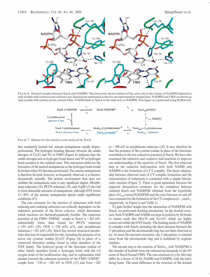
to -290 mV in morphinone reductase (26). It may therefore bethat the presence of the cysteine residue in place of the threoninecontributes to the low reduction potential of XenA.We have alsoexamined the reductive and oxidative half-reactions to improveour understanding of the reactivity of XenA. The first observedstep in the reductive half-reaction with both NADH andNADPH is the formation of a CT complex. The linear relation-ship between observed rates of CT complex formation and theconcentration of reductants is consistent with a simple bimole-cular reaction (Figure 7). There is good agreement between theapparent dissociation constants for the complexes betweenoxidized XenA and NAD(P)H obtained from the hyperbolicplots of kred versus [NAD(P)H] and the ratio between on and offrate constants for the formation of the CT complexes (k-1 and k1,respectively, in Figure 6 and Table 1).
To gain further insight into the interaction of NAD(P)H withXenA, we performed docking simulations. In the docked struc-ture, both NADPH andNADH are kept in position byH-bondsto amino acids like His178 and Tyr183, which are highlyconserved within theOYE family. The bindingmode ofNADPHin complex with XenA, including the short distance between the20-phosphate and the nicotinamide ring, has not been observed sofar. In most flavoprotein structures, the 20-phosphate is orientedaway from the nicotinamide ring and is stabilized by arginineresidues.
The second step in the reaction of XenAox with NAD(P)H isthe transfer of hydride from the reduced nicotinamide to the N5atom of XenA-bound FMN. The rate constants (k2) for this stepdiffer by a factor of 24 for NADH and NADPH, with the latterbeing faster. The main difference in the structure of the docked
FIGURE 6: Docked complex between XenA and NADPH. The stereoview shows residues of the active site in the vicinity of NADPH depicted asstickmodelswith carbonatoms colored cyan. Interactionsmentioned in the text are represented bydashed lines.NADPHandFMNare shown asstick models with carbon atoms colored white. NADHbinds to XenA in the same way as NADPH. This figure was generated using PyMol (48).
FIGURE 7: Scheme for the reaction cycle catalyzed by XenA.

Article Biochemistry, Vol. 48, No. 48, 2009 11419
complexes of NAD(P)H with oxidized XenA is the presence ofthe 20-phosphate group, which is situated above the 1,4-dihy-dropyridine ring of NADPH near the transferred hydrogen atC4. An additional negative charge above the pyridine ring couldfurther stabilize NADPþ and would therefore be expected todecrease the reduction potential of the NADPþ-NADPHcouple in the XenA-bound state. The calculated ΔΔG of-1.83 kcal/mol for the hydride-transfer reaction of XenA-boundNADPH to FMN compared to XenA-bound NADH indicatesthat the higher rate constant in the reaction with NADPH is dueto the interaction of the 20-phosphate group of NADPHwith thenicotinamide ring and the FMN. The lower affinity of XenA forNADPH compared to NADH is more than compensated by itshigher reactivity. Using k2/Kd as a criterion for the specificity ofXenA with the two nicotinamides, we obtain apparent second-order rate constants of 8.5� 103 M-1 s-1 for NADH and 1.39�105 M-1 s-1 for NADPH, indicating that under physiologicalconditions XenA reacts preferentially with NADPH. We notethat this is an exceptionally low level of selectivity for a flavo-protein. The last step of the reductive half-reaction would be therelease of NADþ or NADPþ. We do not observe CT complexesafter transfer of hydride to the flavin or any appreciable back-reaction of reduced XenA with NADþ or NADPþ (consistentwith the observation that during reductive titrations XenA can befully reduced with a stoichiometric amount of NADH).
We have also examined the oxidative half-reaction of XenAwith 2-cyclohexenone and coumarin. As 2-cyclohexenone is alsoa substrate of OYE, PETN reductase, and morphinone reduc-tase, we focus our discussion on this substrate. The oxidative half-reaction of the catalytic cycle is initiated by the formation of a CTcomplex between 2-cyclohexenone and reduced XenA for whichthe equilibrium dissociation constant could be determined (86μM).Only one intermediatewas included in our kineticmodel forthe oxidative half-reaction, although our data suggest the exi-stence of two different intermediates. The rapid formation of anintermediate in the reaction of reduced OYE and 2-cyclohexe-nonewithin the dead time of the stopped-flow spectrophotometerhas also been observed, having an absorbance maximum at455 nm, and was interpreted as a CT complex (19). Theobservation of two intermediates between reduced XenA and2-cyclohexenone indicates that their relative orientations arechanging from the encounter complex to the reactive complexbefore the hydride transfer occurs in the fourth step of thereaction. Again we have no indication that an appreciable back-reaction between 2-cyclohexanone and oxidized XenA occurs.The limiting rate constant of the reaction of NADH-reducedXenA with coumarin is 50-fold smaller than that for the reaction
with 2-cyclohexenone, while coumarin binds with a 4-fold higheraffinity. The lower reactivity with coumarin probably reflects theweaker electrophilicity of the β-carbon of coumarin compared to2-cyclohexenone. The good agreement of the catalytic constantderived from eq 4 with NADPH and 2-cyclohexenone as sub-strates in the steady-state assay (kcat=7.2 s-1) with the cata-lytic constant following from the limiting rate constants of thereductive and oxidative half-reactions with NADPH and2-cyclohexenone (eq 9; kcat = 5.3 s-1) indicates that the twoproduct release steps, which have not been observed by ourtransient kinetic analysis, occur rapidly and are not limiting thereaction rate of XenA.
The structures of XenA and othermembers of the OYE familyare very similar, but at the same time, some distinct features in thecomposition of the active site do exist. This analysis of nativeXenA revealed further remarkable properties of the enzyme suchas its low reduction potential and its only modest specificity forNADPH over NADH.
ACKNOWLEDGMENT
We thank Professor Peter M. H. Kroneck (University ofKonstanz) for kindly supplying the deazaflavin derivative.
REFERENCES
1. Binks, P. R., French, C. E., Nicklin, S., and Bruce, N. C. (1996)Degradation of pentaerythritol tetranitrate by Enterobacter cloacaePB2. Appl. Environ. Microbiol. 62, 1214–1219.
2. Fitzpatrick, T. B., Amrhein, N., and Macheroux, P. (2003) Charac-terization of YqjM, an Old Yellow Enzyme homolog from Bacillussubtilis involved in the oxidative stress response. J. Biol. Chem. 278,19891–19897.
3. Blehert, D. S., Knoke, K. L., Fox, B. G., and Chambliss, G. H. (1997)Regioselectivity of nitroglycerin denitration by flavoprotein nitroe-ster reductases purified from two Pseudomonas species. J. Bacteriol.179, 6912–6920.
4. Snape, J. R.,Walkley, N. A.,Morby, A. P., Nicklin, S., andWhite, G.F. (1997) Purification, properties, and sequence of glycerol trinitratereductase from Agrobacterium radiobacter. J. Bacteriol. 179, 7796–7802.
5. Miura, K., Tomioka, Y., Suzuki, H., Yonezawa, M., Hishinuma, T.,and Mizugaki, M. (1997) Molecular cloning of the nemA geneencoding N-ethylmaleimide reductase from Escherichia coli. Biol.Pharm. Bull. 20, 110–112.
6. French, C. E., and Bruce, N. C. (1994) Purification and characteriza-tion of morphinone reductase from Pseudomonas putida M10. Bio-chem. J. 301 (Part 1), 97–103.
7. Rohde, B. H., Schmid, R., andUllrich,M. S. (1999) Thermoregulatedexpression and characterization of an NAD(P)H-dependent 2-cyclo-hexen-1-one reductase in the plant pathogenic bacterium Pseudomo-nas syringae pv. glycinea. J. Bacteriol. 181, 814–822.
8. Blehert, D. S., Fox, B. G., and Chambliss, G. H. (1999) Cloning andsequence analysis of two Pseudomonas flavoprotein xenobiotic re-ductases. J. Bacteriol. 181, 6254–6263.
9. Fetzner, S., Tshisuaka, B., Lingens, F.,Kappl, R., andH€uttermann, J.(1998) Bacterial degradation of quinoline and derivatives: Pathwaysand their biocatalysts. Angew. Chem., Int. Ed. 37, 577–597.
10. Griese, J. J., Jakob, R. P., Schwarzinger, S., and Dobbek, H. (2006)Xenobiotic reductase A in the degradation of quinoline by Pseudo-monas putida 86: Physiological function, structure and mechanism of8-hydroxycoumarin reduction. J. Mol. Biol. 361, 140–152.
11. Kitzing, K., Fitzpatrick, T. B., Wilken, C., Sawa, J., Bourenkov,G. P., Macheroux, P., and Clausen, T. (2005) The 1.3 A crystalstructure of the flavoprotein YqjM reveals a novel class of Old YellowEnzymes. J. Biol. Chem. 280, 27904–27913.
12. Niino, Y. S., Chakraborty, S., Brown, B. J., and Massey, V. (1995) Anew old yellow enzyme of Saccharomyces cerevisiae. J. Biol. Chem.270, 1983–1991.
13. Brige, A., Van den Hemel, D., Carpentier, W., De Smet, L., and VanBeeumen, J. J. (2006) Comparative characterization and expressionanalysis of the four Old Yellow Enzyme homologues from Shewanella
Table 1: Stopped-Flow Kinetic Data
Reductive Half-Reaction
substrate Kd (μM) k1 (M-1 s-1) k-1 (s
-1) k2 (s-1)
NADH 176 ( 14 (9.4( 0.5)� 105 256 ( 17 1.50( 0.02
NADPH 256( 12 (6.4( 0.3)� 105 215 ( 11 35.7( 0.6
Oxidative Half-Reaction
substrate Kd (μM) k4 (s-1)
2-cyclohexenone 86( 2 13.1( 0.1
coumarin 19.3( 0.6 0.243 ( 0.001

11420 Biochemistry, Vol. 48, No. 48, 2009 Spiegelhauer et al.
oneidensis indicate differences in physiological function. Biochem. J.394, 335–344.
14. van Dillewijn, P., Wittich, R. M., Caballero, A., and Ramos, J. L.(2008) Subfunctionality of Hydride Transferases of the Old YellowEnzyme Family of Flavoproteins of Pseudomonas putida. Appl. En-viron. Microbiol. 74, 6703–6708.
15. Williams, R. E., and Bruce, N. C. (2002) ’New uses for an OldEnzyme’: The Old Yellow Enzyme family of flavoenzymes. Micro-biology 148, 1607–1614.
16. Abramovitz, A. S., andMassey, V. (1976) Interaction of phenols withold yellow enzyme. Physical evidence for charge-transfer complexes.J. Biol. Chem. 251, 5327–5336.
17. Fox, K. M., and Karplus, P. A. (1994) Old yellow enzyme at 2 Aresolution: Overall structure, ligand binding, and comparison withrelated flavoproteins. Structure 2, 1089–1105.
18. Karplus, P. A., Fox, K. M., and Massey, V. (1995) Flavoproteinstructure and mechanism. 8. Structure-function relations for oldyellow enzyme. FASEB J. 9, 1518–1526.
19. Kohli, R. M., and Massey, V. (1998) The oxidative half-reaction ofOld Yellow Enzyme. The role of tyrosine 196. J. Biol. Chem. 273,32763–32770.
20. Brown, B. J., Deng, Z., Karplus, P. A., and Massey, V. (1998) On theactive site of Old Yellow Enzyme. Role of histidine 191 and aspar-agine 194. J. Biol. Chem. 273, 32753–32762.
21. Xu, D., Kohli, R. M., andMassey, V. (1999) The role of threonine 37in flavin reactivity of the old yellow enzyme. Proc. Natl. Acad. Sci. U.S.A. 96, 3556–3561.
22. Fox, K. M., and Karplus, P. A. (1999) The flavin environment in oldyellow enzyme. An evaluation of insights from spectroscopic andartificial flavin studies. J. Biol. Chem. 274, 9357–9362.
23. Brown, B. J.,Hyun, J.W.,Duvvuri, S., Karplus, P.A., andMassey, V.(2002) The role of glutamine 114 in old yellow enzyme. J. Biol. Chem.277, 2138–2145.
24. Craig, D. H., Moody, P. C., Bruce, N. C., and Scrutton, N. S. (1998)Reductive and oxidative half-reactions ofmorphinone reductase fromPseudomonas putida M10: A kinetic and thermodynamic analysis.Biochemistry 37, 7598–7607.
25. Barna, T., Messiha, H. L., Petosa, C., Bruce, N. C., Scrutton, N. S.,and Moody, P. C. (2002) Crystal structure of bacterial morphinonereductase and properties of the C191Amutant enzyme. J. Biol. Chem.277, 30976–30983.
26. Messiha, H. L., Bruce, N. C., Sattelle, B. M., Sutcliffe, M. J., Munro,A. W., and Scrutton, N. S. (2005) Role of active site residues andsolvent in proton transfer and the modulation of flavin reductionpotential in bacterial morphinone reductase. J. Biol. Chem. 280,27103–27110.
27. Pudney, C. R., Hay, S., Pang, J., Costello, C., Leys, D., Sutcliffe,M. J., and Scrutton, N. S. (2007) Mutagenesis of morphinonereductase induces multiple reactive configurations and identifiespotential ambiguity in kinetic analysis of enzyme tunneling mechani-sms. J. Am. Chem. Soc. 129, 13949–13956.
28. Barna, T. M., Khan, H., Bruce, N. C., Barsukov, I., Scrutton, N. S.,and Moody, P. C. (2001) Crystal structure of pentaerythritol tetra-nitrate reductase: “Flipped” binding geometries for steroid substratesin different redox states of the enzyme. J. Mol. Biol. 310, 433–447.
29. Khan, H., Harris, R. J., Barna, T., Craig, D. H., Bruce, N. C.,Munro,A.W.,Moody, P. C., and Scrutton,N. S. (2002)Kinetic and structuralbasis of reactivity of pentaerythritol tetranitrate reductase withNADPH, 2-cyclohexenone, nitroesters, and nitroaromatic explosives.J. Biol. Chem. 277, 21906–21912.
30. Khan, H., Barna, T., Harris, R. J., Bruce, N. C., Barsukov, I.,Munro,A. W., Moody, P. C., and Scrutton, N. S. (2004) Atomic resolutionstructures and solution behavior of enzyme-substrate complexes ofEnterobacter cloacae PB2 pentaerythritol tetranitrate reductase. Mul-tiple conformational states and implications for the mechanism ofnitroaromatic explosive degradation. J. Biol. Chem. 279, 30563–30572.
31. Khan, H., Barna, T., Bruce, N. C., Munro, A. W., Leys, D., andScrutton, N. S. (2005) Proton transfer in the oxidative half-reaction ofpentaerythritol tetranitrate reductase. Structure of the reduced en-zyme-progesterone complex and the roles of residues Tyr186, His181,His184. FEBS J. 272, 4660–4671.
32. Breithaupt, C., Kurzbauer, R., Lilie, H., Schaller, A., Strassner, J.,Huber, R.,Macheroux, P., andClausen, T. (2006) Crystal structure of12-oxophytodienoate reductase 3 from tomato: Sself-inhibition bydimerization. Proc. Natl. Acad. Sci. U.S.A. 103, 14337–14342.
33. Breithaupt, C., Strassner, J., Breitinger, U., Huber, R., Macheroux, P.,Schaller, A., and Clausen, T. (2001) X-ray structure of 12-oxophyto-dienoate reductase 1 provides structural insight into substrate bindingand specificity within the family of OYE. Structure 9, 419–429.
34. Fitzpatrick, T. B., Auweter, S., Kitzing, K., Clausen, T., Amrhein, N.,andMacheroux, P. (2004) Structural and functional impairment of anOld Yellow Enzyme homologue upon affinity tag incorporation.Protein Expression Purif. 36, 280–291.
35. Sambrook, J., and Russel, D. (2001) Molecular Cloning: A Labora-tory Manual , Vol. 1, Cold Spring Harbor Laboratory Press, Plainview,NY.
36. Aliverti, A., Curti, B., and Vanoni, M. A. (1999) Identifying andquantitating FAD and FMN in simple and in iron-sulfur-containingflavoproteins. Methods Mol. Biol. 131, 9–23.
37. Massey, V., Stankovich, M., and Hemmerich, P. (1978) Light-mediated reduction of flavoproteins with flavins as catalysts. Bio-chemistry 17, 1–8.
38. Sucharitakul, J., Chaiyen, P., Entsch, B., and Ballou, D. P. (2005) Thereductase of p-hydroxyphenylacetate 3-hydroxylase fromAcinetobac-ter baumannii requires p-hydroxyphenylacetate for effective catalysis.Biochemistry 44, 10434–10442.
39. Loach, P. A. (1973) Oxidation-reduction potentials: Absorbancebands and molar absorbance of compounds used in biochemicalstudies. In Handbook of Biochemistry Selected Data for MolecularBiology (Sorber, H. A., Ed.) pp J33-J40, CRC Press, Cleveland, OH.
40. Segel, I. H. (1993) Enzyme Kinetics , John Wiley and Sons, New York.41. Strickland, S., Palmer, G., and Massey, V. (1975) Determination of
dissociation constants and specific rate constants of enzyme-substrate(or protein-ligand) interactions from rapid reaction kinetic data.J. Biol. Chem. 250, 4048–4052.
42. Lang, P. T., Brozell, S. R., Mukherjee, S., Pettersen, E. F., Meng,E. C., Thomas, V., Rizzo, R. C., Case, D. A., James, T. L., andKuntz,I. D. (2009) DOCK 6: Ccombining techniques to model RNA-smallmolecule complexes. RNA 15, 1219–1230.
43. MacKerell, A. D.Jr., Banavali, N., and Foloppe, N. (2000) Develop-ment and current status of theCHARMMforce field for nucleic acids.Biopolymers 56, 257–265.
44. Brooks, B. R., Brooks, C. L.III, Mackerell, A. D.Jr., Nilsson, L.,Petrella, R. J., Roux, B.,Won, Y., Archontis, G., Bartels, C., Boresch,S., Caflisch, A., Caves, L., Cui, Q., Dinner, A. R., Feig, M., Fischer,S., Gao, J., Hodoscek, M., Im,W., Kuczera, K., Lazaridis, T., Ma, J.,Ovchinnikov, V., Paci, E., Pastor, R. W., Post, C. B., Pu, J. Z.,Schaefer, M., Tidor, B., Venable, R. M., Woodcock, H. L., Wu, X.,Yang, W., York, D. M., and Karplus, M. (2009) CHARMM: Thebiomolecular simulation program. J. Comput. Chem. 30, 1545–1614.
45. Bashford, D., and Karplus, M. (1990) pKa’s of ionizable groups inproteins: Atomic detail from a continuum electrostatic model. Bio-chemistry 29, 10219–10225.
46. Messiha, H. L., Munro, A. W., Bruce, N. C., Barsukov, I., andScrutton, N. S. (2005) Reaction of morphinone reductase with 2-cyclohexen-1-one and 1-nitrocyclohexene: Proton donation, ligandbinding, and the role of residues Histidine 186 and Asparagine 189.J. Biol. Chem. 280, 10695–10709.
47. Stewart, R. C., andMassey, V. (1985) Potentiometric studies of nativeand flavin-substitutedOldYellowEnzyme. J. Biol. Chem. 260, 13639–13647.
48. DeLano, W. L. (2002) The PyMol Molecular Graphics System ,DeLano Scientific, San Carlos, CA.


