
KILLER WHALE NECROPSY AND DISEASE TESTING PROTOCOL · KILLER WHALE NECROPSY AND DISEASE TESTING...
82
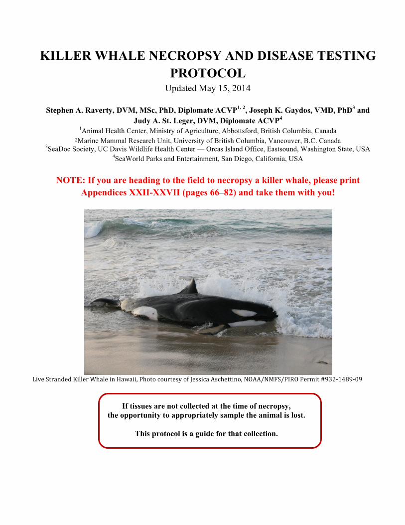
KILLER WHALE NECROPSY AND DISEASE TESTING PROTOCOL Updated May 15, 2014 Stephen A. Raverty, DVM, MSc, PhD, Diplomate ACVP 1, 2 , Joseph K. Gaydos, VMD, PhD 3 and Judy A. St. Leger, DVM, Diplomate ACVP 4 1 Animal Health Center, Ministry of Agriculture, Abbottsford, British Columbia, Canada 2 Marine Mammal Research Unit, University of British Columbia, Vancouver, B.C. Canada 3 SeaDoc Society, UC Davis Wildlife Health Center — Orcas Island Office, Eastsound, Washington State, USA 4 SeaWorld Parks and Entertainment, San Diego, California, USA NOTE: If you are heading to the field to necropsy a killer whale, please print Appendices XXII-XXVII (pages 66–82) and take them with you! Live Stranded Killer Whale in Hawaii, Photo courtesy of Jessica Aschettino, NOAA/NMFS/PIRO Permit #932148909 If tissues are not collected at the time of necropsy, the opportunity to appropriately sample the animal is lost. This protocol is a guide for that collection.
Transcript of KILLER WHALE NECROPSY AND DISEASE TESTING PROTOCOL · KILLER WHALE NECROPSY AND DISEASE TESTING...
KILLER WHALE NECROPSY AND DISEASE TESTING PROTOCOL
Updated May 15, 2014
Stephen A. Raverty, DVM, MSc, PhD, Diplomate ACVP1, 2, Joseph K. Gaydos, VMD, PhD3 and Judy A. St. Leger, DVM, Diplomate ACVP4
1Animal Health Center, Ministry of Agriculture, Abbottsford, British Columbia, Canada 2Marine Mammal Research Unit, University of British Columbia, Vancouver, B.C. Canada
3SeaDoc Society, UC Davis Wildlife Health Center — Orcas Island Office, Eastsound, Washington State, USA 4SeaWorld Parks and Entertainment, San Diego, California, USA
NOTE: If you are heading to the field to necropsy a killer whale, please print
Appendices XXII-XXVII (pages 66–82) and take them with you!
Live Stranded Killer Whale in Hawaii, Photo courtesy of Jessica Aschettino, NOAA/NMFS/PIRO Permit #932-‐1489-‐09
If tissues are not collected at the time of necropsy, the opportunity to appropriately sample the animal is lost.
This protocol is a guide for that collection.
Revised May 15, 2014
Killer Whale Necropsy Protocol -‐ 2014 Page 2
Table of Contents
SIGNIFICANT PATHOGEN ALERT: .................................................................................................... 3 CETACEAN BRUCELLA: ................................................................................................................................... 3 CETACEAN MORBILLIVIRUS: ....................................................................................................................... 4 INFLUENZA: ....................................................................................................................................................... 5 SALMONELLA: ................................................................................................................................................... 5 APICOMPLEXANS: ............................................................................................................................................ 5
PREVIOUSLY REPORTED PATHOGENS: .......................................................................................... 6
INTRODUCTION: .................................................................................................................................... 7 EQUIPMENT CHECKLIST: .................................................................................................................... 9
LOGISTICS AND NECROPSY RECOMMENDATIONS .................................................................. 10 SAFETY ............................................................................................................................................................. 10 NECROPSY TEAM ROLES ............................................................................................................................ 11 EXTERNAL EXAM AND PRE-‐DISSECTION SAMPLING ........................................................................ 11 DECOMPOSITION TABLE: ........................................................................................................................... 12 IMAGING CONSIDERATIONS ...................................................................................................................... 12 SKELETAL EXAMINATION AND PREPARATION CONSIDERATIONS ............................................. 13
ACKNOWLEDGMENTS ....................................................................................................................... 14
LITERATURE CITED: .......................................................................................................................... 14 KILLER WHALE NECROPSY PROTOCOL COMPREHENSIVE KILLER WHALE TISSUE SAMPLE CHECKLIST .......................................................................................................................... 18 APPENDIX I: Sample Priorities based on Tissue Condition ................................................. 24 HIGH PRIORITY SAMPLES: ........................................................................................................................ 24 INTERMEDIATE PRIORITY SAMPLES: .................................................................................................... 25 LOW PRIORITY SAMPLES: ......................................................................................................................... 25
APPENDIX II: Pathogens reported in killer whales ................................................................ 27 Table 1: Pathogens detected directly or via serology in killer whales ...................................... 27 Table 2: Endoparasites identified or suggested by serology in killer whales ......................... 28
APPENDIX III: The One Hour Necropsy Protocol ..................................................................... 29
APPENDIX IV: Researchers requesting killer whale tissues ................................................ 30 APPENDIX IVB: Request for marine mammal post-‐mortem samples ........................................ 34
APPENDIX V: Permitting concerns and authorities ................................................................ 35
APPENDIX VI: Pathogen and tissue sample list for Polymerase Chain Reaction Studies .................................................................................................................................................................. 36 APPENDIX VII: Marine mammal blubber sampling protocol .............................................. 38
APPENDIX VIII: Collection of samples for contaminant analysis ....................................... 40 APPENDIX IX: Collection of samples for biotoxin analysis .................................................. 41
APPENDIX X: Webpage resources for submission forms ..................................................... 43
APPENDIX XI: Photography instructions and considerations ............................................ 44
Revised May 15, 2014
Killer Whale Necropsy Protocol -‐ 2014 Page 3
APPENDIX XII: Ancillary imaging instructions ........................................................................ 45
APPENDIX XIII: Oil spill concerns and sampling ..................................................................... 46 APPENDIX XIV: Considerations and sampling for live stranded killer whales ............. 48
APPENDIX XV: Cetacean ear extraction and fixation protocol ........................................... 50 APPENDIX XVI: Barotrauma considerations and sampling protocol for gas bubbles 54
Appendix XVII: Dorsal fin measurement and collection request ...................................... 56
APPENDIX XVIII: Archiving tissue samples ............................................................................... 58 APPENDIX XIX: Lesion Description Form .................................................................................. 59
APPENDIX XX: NOAA Fisheries NW Region Killer Whale Stranding Protocol ............... 60 *For killer whale strandings in other parts of the world ..................................................... 64
Appendix XXI: Protocol for examining killer whales for signs of human interaction 65
APPENDIX XXII: Fetal examination and sample management ........................................... 66 APPENDIX XXIII: Morphometric analysis of stranded killer whales ................................ 68
APPENDIX XXIV: Gross pathology data recording form ........................................................ 69
APPENDIX XXV: Checklist -‐ tissues to collect from a Code 2 or 3 killer whale carcass .................................................................................................................................................................. 77
APPENDIX XXVI: Checklist -‐ tissues to collect from a Code 4 killer whale carcass ...... 79 APPENDIX XXVII: Checklist -‐ tissues to collect from a Code 5 killer whale carcass .... 81
Photo Identifier: ................................................................................................................................. 82
SIGNIFICANT PATHOGEN ALERT: Pathogens acquire significance because they cause harm to humans or animals. Examination of deceased animals has inherent safety concerns. Certain pathogens such as Brucella sp., influenza, and arboviruses warrant elevated vigilance and care. Likewise, rapid detection of fatal, transmissible agents that may impact killer whale population health is critical to inform management activity. Chief among these pathogen of concern are Brucella spp., cetacean morbillivirus, influenza, Salmonella spp., and apicomplexans.
CETACEAN BRUCELLA: Marine mammal associated Brucella spp. that differ from recognized named species within the genus have been increasingly detected in a number of pinnipeds and cetaceans in the United Kingdom, New Zealand, the United States and Canada (Ross et al., 1996; Foster et al., 1996; Nielsen et al., 2001; Van Bressem et al., 2001a). Antibodies to Brucella spp. have been identified in post mortem heart blood and in live captured (A73) killer whale with no attendant pathology or clinical disease (Jepson et al., 1997; Raverty et al., 2004). Infection by Brucella has resulted in placentitis and abortion in captive bottlenose dolphins and
Revised May 15, 2014
Killer Whale Necropsy Protocol -‐ 2014 Page 4
blubber abscesses and meningoencephalitis in wild striped dolphins (Ewalt et al., 1994; Gonzalez et al., 2002). There are no Brucella species-specific gross lesions. To further resolve the possible contribution of these bacteria to impaired reproductive function and microscopic lesions in stranded killer whales. Attempted Brucella species-specific culture and isolation as well as molecular screening should be routinely undertaken with each stranded animal. Tissue samples should include multiple levels of the reproductive tract, brain, lung, spleen, lymph nodes and any gross abnormalitieslesions. To ensure optimum bacterial recovery, samples obtained at the time of necropsy should be shipped overnight on wet ice to a reference laboratory or frozen at -70 C and forwarded for evaluation as soon as possible (Table 2). Brucella serology may be considered. However, there are currently no validated serologic tests for killer whales (Gall et al., 2000). If indicated by histopathology, immunohistochemistry with monoclonal or polyclonal antibodies specific to Brucella may prove a valuable adjunct to confirm infection and assess the disease processes. Zoonosis Warning: Marine Brucella sp. has infected a laboratory worker after occupational exposure (Brew et al., 1999) and neurobrucellosis with granuloma formation has been documented in two additional individuals with no known history of exposure (Sohn et al., 2003). The virulence of these strains to humans is currently unknown and appropriate public health and safety precautions at the time of necropsy are warranted. The precautions can include gloves, goggles, and a face mask when potentially aerosolizing tissue (such as when using a reciprocating saw).
CETACEAN MORBILLIVIRUS: Porpoise and dolphin morbilliviruses are antigenically and genetically similar and are now generally considered strains of the same viral species, cetacean morbillivirus (Kennedy, 1998). This virus has caused large-scale epizootics in several odontocetes species (Van Bressem et al., 1991; Duignan et al., 1995; Van Bressem et al., 2001b). Detection of antibodies in a subadult killer whale recently captured in the northwest Pacific Ocean that succumbed to bacterial pneumonia (A. Mironova, per comm.) suggests that killer whales have been exposed to cetacean morbillivirus. Although no morbillivirus antibodies or gene sequences have yet been detected in stranded cetaceans in the temperate northeastern Pacific Ocean, this virus is likely endemic in multiple small cetaceans from around the world (Van Bressem et al., 2001b). Because of the virulence of this virus and its potential to cause large-scale mortality in small populations, morbillivirus should be ruled out during all killer whale necropsies. Continued surveillance for antibodies to cetacean morbilliviruses in antemortem serum or post mortem heart blood samples by indirect enzyme linked immunosorbant assay (iELISA) or virus neutralization and attempted virus isolation are strongly recommended. Cetacean morbillivirus is pantropic (infects a variety of cell types) and potential gross necropsy findings include skin ulcerations, stomatitis, pneumonia, and generalized signs of sepsis such as edema of internal organs and accumulation of serosanguinous fluid in the pleural and peritoneal cavities (Lipscomb et al., 1994). Gross lesions are not specific of morbillivirus infection but microscopic lesions are highly characteristic (Domingo et al., 2002). Microscopic lesions commonly seen with morbillivirus infection such as syncytia and acidophilic inclusions in cytoplasm and nuclei of epithelial cells can be widespread, focal, or obscured by severe necrosis caused by opportunistic bacterial and fungal infections. Microscopic examination and laboratory testing are essential to confirm morbillivirus infection. The tests used include immunohistochemistry, reverse transcriptase polymerase chain reaction (RT-PCR), and virus isolation on Vero cells or bovine fetal lung cells (Domingo et al., 1990; Van Bressem et al.,
Revised May 15, 2014
Killer Whale Necropsy Protocol -‐ 2014 Page 5
1991; Barrett et al., 1993; Van Bressem et al., 1999; Saliki et al, 2002). Potential sequelae to cetacean morbillivirus infection include opportunistic bacterial and fungal infections, as well as toxoplasmosis (Lipscomb et al., 1994; Schulman and Lipscomb, 1999). These viruses are not likely a pathogen of concern for humans – but they represent a potentially significant health threat to killer whale populations.
INFLUENZA: The detection of an influenza virus (H3N8) in the harbor seals stranded in the Northeastern United States in 2011 has renewed interest and concern regarding the potential risk of exposure and infection of other marine mammal species, terrestrial animals, birds and humans who may come into contact with carrier animals. To date, histopathology of stranded killer whales throughout the northeastern Pacific has not detected microscopic lesions consistent with infection by influenza and no virus has been identified by reverse transcriptase polymerase chain reaction in screened cases. However, the recent report of West Nile Virus in a display killer whale (St. Leger et al., 2012) suggests that orca may be susceptible to a broader array of viral pathogens than previously appreciated. Influenza from cetaceans could present a zoonotic concern and appropriate personal protective equipment (especially respiratory protection) should be utilized to reduce the likelihood of infection.
SALMONELLA: Post mortem examination of an offshore neonate stranded in central California and an adult female killer whale in Hawaii did not reveal gross evidence of septicemia or localized bacterial infection. However, microscopic review of sampled tissues from the neonate disclosed inflammation of the umbilicus and multisystemic inflammation due to Salmonella newport. Salmonella muenchen was recovered from the adult female; the lack of associated inflammatory infiltrate within examined tissues suggested an asymptomatic carrier. There are over 2,200 recognized serovars of Salmonella. Salmonella newport is an emerging human health concern and is among the most common isolates from dairy cattle. It is important to note that these bacteria may directly infect people and can be carried on clothing, boots, or equipment to contaminate other areas. Thorough hand washing and disinfection of necropsy equipment should limit the risk of human and animal exposure.
APICOMPLEXANS: The advent of molecular screening and gene sequencing has greatly enhanced our ability to detect a variety of disease agents, including tissue cyst forming protozoal parasites, such as those of the Apicomplexa. In marine mammals these include Toxoplasma gondii, Sarcocystis neurona, Sarcocystis spp, Neospora caninum and Neospora spp (Miller, 2008; Colgrove et al., 2010; Gibson et al., 2011). Representatives of this group of protozoa are of increasing concern due to potential land to sea transmission (Miller et al., 2004, etc.). Sexual reassortment has resulted in the emergence of hypervirulent clones. Although these pathogens have been implicated in sporadic mortality in near and off shore cetaceans, significant losses have been incurred historically in pinnipeds and otters. These parasites are associated with meningoencephalitis and transplacental infections or placentitis. Individual and dual parasite infections of Toxoplasma gondii and Sarcocystis spp. have been detected in a killer whale; however, the contribution of these parasites to strandings has not yet been resolved. Efforts are ongoing to screen stranded killer whales for possible infection. Subsequent genotyping is routinely undertaken to determine a potential source of exposure.
Revised May 15, 2014
Killer Whale Necropsy Protocol -‐ 2014 Page 6
PREVIOUSLY REPORTED PATHOGENS: Reported pathogens from free-ranging or captive killer whales (Gaydos et al., 2004) are growing in number. Implementation of comprehensive necropsies and ancillary diagnostics has significantly contributed to this (Barbieri et al., 2013). Increasing knowledge of recognized pathogens and the potential contribution to clinical disease greatly enhances our understanding of killer whale morbidity and mortality. Please see Appendix II for reported infectious disease pathogens (Table 1) and endoparasites (Table 2) identified in killer whales.
Revised May 15, 2014
Killer Whale Necropsy Protocol -‐ 2014 Page 7
INTRODUCTION: This protocol was first established in 2005 with goals to:
1. Provide guidelines for more comprehensive necropsies and disease testing to improve our knowledge about diseases of killer whales (Orcinus spp.)
2. Standardize screening to facilitate retrospective natural history and disease epidemiology studies.
In the past seven years, this protocol has greatly facilitated and enhanced killer whale examinations in the Northeast Pacific region, and we hope that this revised version will reflect scientific advancements in disease screening, heighten awareness of health concerns, and increase the number of complete postmortem standardized necropsies performed on killer whales. The project was sponsored by the U.S. National Oceanic and Atmospheric Administration (NOAA Fisheries) in response to the limited information known about diseases of free-‐ranging killer whales. This information is critical to understanding how disease might impact the recovery of small declining killer whale populations, such as the southern resident killer whales. Historic estimates of this population were more than 200 whales until the mid-‐ to late-‐1800's: the most recent census indicates 80 individuals. Since the start of killer whale photo-‐identification in 1974, the population has had several periods of growth and decline, including a 17% reduction (mean annual decline rate of 2.9%) between 1996 and 2001, prompting the petition and successful listing under the US Federal Endangered Species Act. Since 2001, the population grew to a high of 90 individuals in September 2006. The population has fluctuated. AS of September 2013, it totaled 81 individuals. The Recovery Plan for Southern Resident killer whales (NMFS 2008) recommends development of protocols for responding to stranded killer whales and investigations of dead killer whales to inform recovery, including necropsies following the 2005 protocol. This updated protocol contributes to implementation of these actions and will contribute to gathering important knowledge about the health of the whales and the threats they face.
A retrospective evaluation of stranded killer whales reported an average of seven to eight dead or beach cast killer whales around the world annually, making each killer whale stranding an important opportunity to learn more about the biology and health status of these animals (Barbieri et al., 2013). We hope killer whale researchers and responders around the world will use this protocol to increase information garnered from postmortem examinations.
Revised May 15, 2014
Killer Whale Necropsy Protocol -‐ 2014 Page 8
The objectives of this revised standardized necropsy and disease testing protocol are:
• Facilitate more comprehensive and systematic killer whale post mortem examinations • Prioritize morphometrics and tissue sampling when complete necropsies are not feasible
or in cases of more advanced autolysis • Establish baseline patterns of morbidity and mortality in killer whales to facilitate
retrospective evaluation of temporal and geographic differences in killer whale health • Ascertain the contribution of contaminant and heavy metal accumulation to killer whale
health • Improve reporting of human interactions (blunt force and sharp injury trauma) • Introduce methods to investigate potential sonar or seismic related strandings • Develop protocols to conduct neonatal killer whale examinations • Enhance photo documentation of gross abnormalities or lesions • Identify resources for information regarding climatic and oceanographic factors, which
may contribute to and facilitate back tracking of environmental factors associated with strandings Provide contact information and shipping addresses for priority samples required for diagnostics and long term research efforts
• List protocols and contacts in the event of a catastrophic oil or other noxious chemical spill
• Through sampling requests, prioritize key organs to provide additional insights into the natural history and biology of wild stranded killer whales through sampling requests
A revised and expanded necropsy and sampling protocol is presented in the following text and relates specifically to North America. While the testing is focused on North American resources, the testing is universal and this protocol can be implemented globally. If resources are available, it is recommended that all killer whale necropsies follow this protocol. If your facility has appropriate tissue fixatives (formalin), a freezer and access to a microbiology laboratory, most of the listed tests should be readily accomplishable.
Revised May 15, 2014
Killer Whale Necropsy Protocol -‐ 2014 Page 9
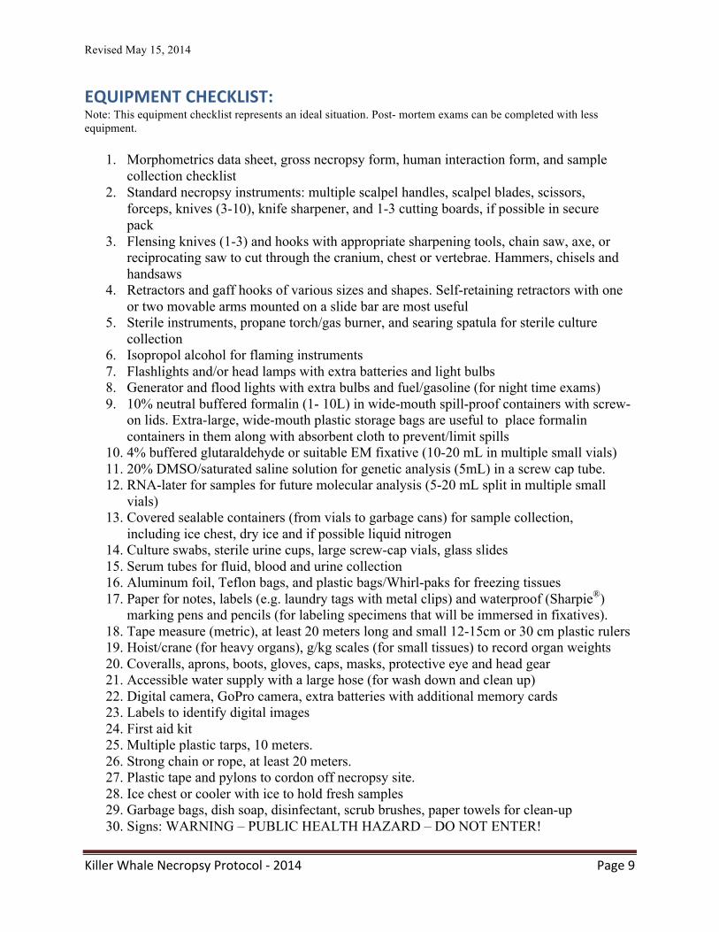
EQUIPMENT CHECKLIST: Note: This equipment checklist represents an ideal situation. Post- mortem exams can be completed with less equipment.
1. Morphometrics data sheet, gross necropsy form, human interaction form, and sample collection checklist
2. Standard necropsy instruments: multiple scalpel handles, scalpel blades, scissors, forceps, knives (3-10), knife sharpener, and 1-3 cutting boards, if possible in secure pack
3. Flensing knives (1-3) and hooks with appropriate sharpening tools, chain saw, axe, or reciprocating saw to cut through the cranium, chest or vertebrae. Hammers, chisels and handsaws
4. Retractors and gaff hooks of various sizes and shapes. Self-retaining retractors with one or two movable arms mounted on a slide bar are most useful
5. Sterile instruments, propane torch/gas burner, and searing spatula for sterile culture collection
6. Isopropol alcohol for flaming instruments 7. Flashlights and/or head lamps with extra batteries and light bulbs 8. Generator and flood lights with extra bulbs and fuel/gasoline (for night time exams) 9. 10% neutral buffered formalin (1- 10L) in wide-mouth spill-proof containers with screw-
on lids. Extra-large, wide-mouth plastic storage bags are useful to place formalin containers in them along with absorbent cloth to prevent/limit spills
10. 4% buffered glutaraldehyde or suitable EM fixative (10-20 mL in multiple small vials) 11. 20% DMSO/saturated saline solution for genetic analysis (5mL) in a screw cap tube. 12. RNA-later for samples for future molecular analysis (5-20 mL split in multiple small vials) 13. Covered sealable containers (from vials to garbage cans) for sample collection,
including ice chest, dry ice and if possible liquid nitrogen 14. Culture swabs, sterile urine cups, large screw-cap vials, glass slides 15. Serum tubes for fluid, blood and urine collection 16. Aluminum foil, Teflon bags, and plastic bags/Whirl-paks for freezing tissues 17. Paper for notes, labels (e.g. laundry tags with metal clips) and waterproof (Sharpie®) marking pens and pencils (for labeling specimens that will be immersed in fixatives). 18. Tape measure (metric), at least 20 meters long and small 12-15cm or 30 cm plastic rulers 19. Hoist/crane (for heavy organs), g/kg scales (for small tissues) to record organ weights 20. Coveralls, aprons, boots, gloves, caps, masks, protective eye and head gear 21. Accessible water supply with a large hose (for wash down and clean up) 22. Digital camera, GoPro camera, extra batteries with additional memory cards 23. Labels to identify digital images 24. First aid kit 25. Multiple plastic tarps, 10 meters. 26. Strong chain or rope, at least 20 meters. 27. Plastic tape and pylons to cordon off necropsy site. 28. Ice chest or cooler with ice to hold fresh samples 29. Garbage bags, dish soap, disinfectant, scrub brushes, paper towels for clean-up 30. Signs: WARNING – PUBLIC HEALTH HAZARD – DO NOT ENTER!
Revised May 15, 2014
Killer Whale Necropsy Protocol -‐ 2014 Page 10
LOGISTICS AND NECROPSY RECOMMENDATIONS From a logistical perspective, advanced development of contingency plans will greatly facilitate identification, reporting, communication, recovery, necropsy and disposal of stranded animals. Key individuals for a killer whale stranding response should be identified and contact information provided to responsible government agencies, regional stranding coordinator, local aquarium facilities, and whale watching representatives and stranding networks. For example the West Coast Marine Mammal Stranding Network has a protocol for initial communications and considerations for killer whale stranding response, including identifying logistics for performing necropsies (Appendix XX).
If a killer whale strands in an inaccessible or remote site, or is identified floating in offshore areas, efforts to recover the animal and relocate by boat to a more accessible site are strongly recommended. If the animal can be re-floated, this may be accomplished by a large rope or chain secured around the peduncle or immediately behind the pectoral flippers and towed by a suitable vessel. To limit drag, the two front flippers should be tied together and maintained out of the water. To facilitate the post mortem examination, the animal should be positioned in lateral recumbency and secured ashore at high tide with exposure of the carcass attained with ebb flow. As tidal changes may limit the duration of the examination, use of heavy equipment (cranes, backhoes, hoists) and flatbed trucks to transport the animal to a more secure facility or a diagnostic laboratory may be considered. These animals may weigh up to 4000-6000 pounds and an appropriate vehicle should be employed. If the carcass is moved by truck, the vehicle should be weighed at a commercial weigh scale before and after transport to obtain the body mass of the carcass.
Should the animal require euthanasia, consultation with the regional stranding coordinator and a marine mammal veterinarian is required. Ante-mortem blood samples should be collected and appropriately stored for later clinical pathology (hematology and clinical chemistry), hormone analysis, serology, archiving, immune function and ancillary diagnostic and research investigations. With a fresh dead animal (code 2), post mortem blood may be collected from the tail flukes, dorsal fin, axillary artery, or heart. Even in animals with advanced states of decomposition, efforts to harvest tissues for histopathology, contaminants, genetics, parasitology, and molecular studies should be undertaken. Skeletal remains from animals in stages of severe decomposition (code 5) can also prove invaluable to ongoing studies in killer whale natural history.
SAFETY Safety of the public and individuals involved with the post-mortem examination is a prime consideration. With any field necropsy, there is a risk of human exposure to potential zoonotic pathogens as well as interference with inappropriate public involvement. Use of face masks, protective eyewear and gloves is recommended. In areas with high public exposure, access should be restricted by pylons, tape or rope and use of law enforcement or fisheries officials may be warranted.
Revised May 15, 2014
Killer Whale Necropsy Protocol -‐ 2014 Page 11
NECROPSY TEAM ROLES To facilitate the flow of the post mortem examination, team members should be identified and assigned to specific tasks before the necropsy is initiated. A lead pathologist or prosector should be designated and individuals appointed to complete data entry, process research samples (Appendix IV), label and record diagnostic material (Appendix I), document lesions and observations with photographs, liaise with the media or undertake additional tasks as necessary. Appropriate measurements (Appendix XXIII) should be recorded by designated team members and photographs of the dorsal fin and saddle patch, eye patches, and any other potential identifying features obtained before the necropsy is initiated (Appendix XXIV). A digital still camera or GoPro® should be used to record details of the post-mortem examination. Consider forming two teams to increase data and tissue collections. One team can collect morphometrics while another team collects external photos and documents external lesions. If you have 2 lead prosectors, there can be a head and abdomen team until they meet in the middle. Consider organizing a single sampling station just away from the necropsy. ALL tissues are harvested and then sent to sampling table for subsampling. The data sheets and sampling team leader are stationed there making sure ALL protocols are filled. In this way a single block of liver harvested from the whale is delivered to the sampling table and is subsampled to fill all protocols and requests. Specific sample vials (usually fluids) are brought to the carcass to be filled before the organs are excised.
EXTERNAL EXAM AND PRE-‐DISSECTION SAMPLING In the case of live strandings, ante-mortem blood samples should be collected and appropriately stored for later clinical pathology (hematology and clinical chemistry), hormone analysis, serology, archiving, immune function and ancillary diagnostic and research investigations. With a fresh dead animal, post mortem blood may be collected from the tail flukes, dorsal fin, axillary artery, or heart. Even in animals with advanced states of decomposition, efforts to harvest tissues for histopathology, contaminants, genetics, parasitology, and molecular studies may be undertaken. Skeletal remains from animals in stages of severe decomposition can also prove invaluable to ongoing studies in killer whale natural history.
External examination and photo documentation of the eyes, mouth, blowhole, skin, mammary glands, genital slits and anus should be performed prior to cutting the animal. The dorsal fin and area immediately around the base of the fin should be examined for evidence of any prior attachment of LIMPET satellite tags (Andrews et al. 2009). Signs of human interaction should be recorded (Appendix XXI). Once the external examination and tissue sampling (swabs, cytology and tissues) has been completed and lesions documented (i.e. by photography and description), proceed with the dissection.
Revised May 15, 2014
Killer Whale Necropsy Protocol -‐ 2014 Page 12
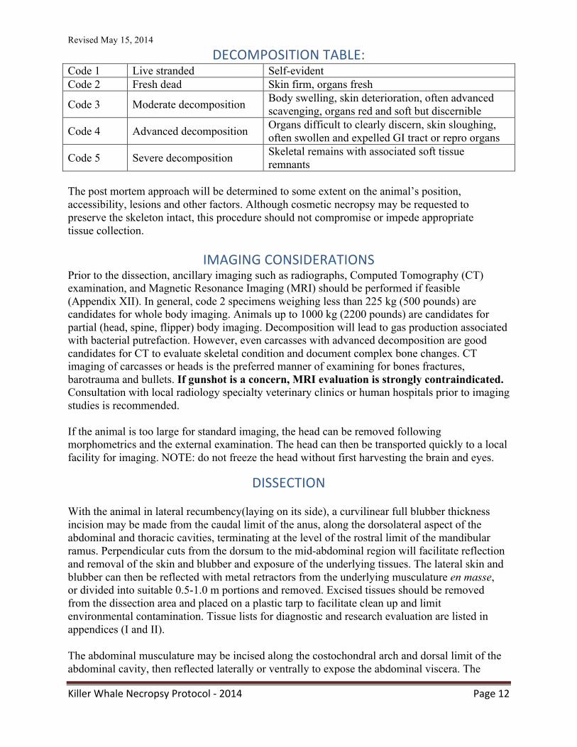
DECOMPOSITION TABLE: Code 1 Live stranded Self-evident Code 2 Fresh dead Skin firm, organs fresh
Code 3 Moderate decomposition Body swelling, skin deterioration, often advanced scavenging, organs red and soft but discernible
Code 4 Advanced decomposition Organs difficult to clearly discern, skin sloughing, often swollen and expelled GI tract or repro organs
Code 5 Severe decomposition Skeletal remains with associated soft tissue remnants
The post mortem approach will be determined to some extent on the animal’s position, accessibility, lesions and other factors. Although cosmetic necropsy may be requested to preserve the skeleton intact, this procedure should not compromise or impede appropriate tissue collection.
IMAGING CONSIDERATIONS Prior to the dissection, ancillary imaging such as radiographs, Computed Tomography (CT) examination, and Magnetic Resonance Imaging (MRI) should be performed if feasible (Appendix XII). In general, code 2 specimens weighing less than 225 kg (500 pounds) are candidates for whole body imaging. Animals up to 1000 kg (2200 pounds) are candidates for partial (head, spine, flipper) body imaging. Decomposition will lead to gas production associated with bacterial putrefaction. However, even carcasses with advanced decomposition are good candidates for CT to evaluate skeletal condition and document complex bone changes. CT imaging of carcasses or heads is the preferred manner of examining for bones fractures, barotrauma and bullets. If gunshot is a concern, MRI evaluation is strongly contraindicated. Consultation with local radiology specialty veterinary clinics or human hospitals prior to imaging studies is recommended. If the animal is too large for standard imaging, the head can be removed following morphometrics and the external examination. The head can then be transported quickly to a local facility for imaging. NOTE: do not freeze the head without first harvesting the brain and eyes.
DISSECTION With the animal in lateral recumbency(laying on its side), a curvilinear full blubber thickness incision may be made from the caudal limit of the anus, along the dorsolateral aspect of the abdominal and thoracic cavities, terminating at the level of the rostral limit of the mandibular ramus. Perpendicular cuts from the dorsum to the mid-abdominal region will facilitate reflection and removal of the skin and blubber and exposure of the underlying tissues. The lateral skin and blubber can then be reflected with metal retractors from the underlying musculature en masse, or divided into suitable 0.5-1.0 m portions and removed. Excised tissues should be removed from the dissection area and placed on a plastic tarp to facilitate clean up and limit environmental contamination. Tissue lists for diagnostic and research evaluation are listed in appendices (I and II).
The abdominal musculature may be incised along the costochondral arch and dorsal limit of the abdominal cavity, then reflected laterally or ventrally to expose the abdominal viscera. The
Revised May 15, 2014
Killer Whale Necropsy Protocol -‐ 2014 Page 13
diaphragm should be assessed, and if intact, incised and deflated to exclude pneumothorax. If a cosmetic post-mortem is requested, the ribs may be detached at the costosternal junction and reflected, or alternatively a chain or reciprocating saw may be employed to remove the thoracic wall. It is important that protective eye wear or face shields be employed by the operator and prosectors. The tongue may be exteriorized by incision of the blubber and skeletal musculature along the entire length of the medial aspect of the mandibles and then reflected ventrally. If feasible, the lung, heart, larynx, trachea and esophagus, and tongue (pluck) should be removed to a tarp for thorough evaluation. With larger animals, dissection of thoracic viscera in situ may be warranted. The head may be detached by dissection of the atlanto-occipital articulation and the skin overlying the dorsolateral aspect of the nape and cranium removed. This exposure will facilitate removal of the dorsal aspect of the skull by either chain or reciprocating saw and exposure of the brain. It is important to evaluate the entire length of the vertebral column to assess possible vertebral fractures or subluxations associated with boat strikes or other trauma; a representative portion of spinal cord should be recovered from the cervical, mid- thoracic, thoracolumbar and lumbar regions.
Due to the importance of the reproductive organs in disease screening and assessment of reproductive status, recover and completely excise the reproductive tract for evaluation. As with other organ systems, decomposition and physical characteristics will determine the best sampling plan for this system.
The mesenteric stalk should then be identified, evaluated for lesions, then transected to facilitate removal and evaluation of the abdominal viscera. The viscera should be placed on a separate tarp to that of the thoracic contents to limit cross contamination. The entire length of bowel should be detached from the mesenteric attachment and opened for visual inspection by incising along one side of the mesenteric border. The stomach should then be incised along the greater curvature and the gastric contents recovered and appropriately packaged and labeled. Samples will be partitioned for a variety of ancillary investigations (Appendix I). The remaining internal viscera should be evaluated by routine or conventional diagnostic protocols and appropriate research and diagnostic samples harvested and labeled.
With suspect sonar related strandings, arrangements should be made for CT scan of the entire head or ears and close evaluation of the larynx should be undertaken for evidence of submucosal hemorrhage. If the CT is not conducted prior to the necropsy, the head and ears can be collected and scanned at a later time. EARs can also be extracted and fixed for analysis (Appendix XV). Samples of peribullar adipose tissue should be collected into 10% neutral buffered formalin for histopathologic evaluation. Note: decomposition to code 3 can produce intravascular and parenchymal gas bubbles. These are distinguished from bubbles associated with acoustic trauma based on tissue freshness and associated lesions such as pulmonary and peribullar fat hemorrhage and damage to the ear bones. Appendix XVI provide guidance for gas bubble sampling.
SKELETAL EXAMINATION AND PREPARATION CONSIDERATIONS Postmortem investigations should involve review of both soft and hard tissues. Examination of bones for malformations, degenerative changes, fractures, inflammation and masses is critical to a thorough understanding of the health issues affecting an individual killer whale. Bones are most commonly evaluated through diagnostic imaging (radiographs and CT exams) and at the gross exam with bone exposure by flensing. Due to the large size of killer whales and the
Revised May 15, 2014
Killer Whale Necropsy Protocol -‐ 2014 Page 14
difficulty in soft tissue removal, a thorough examination typically requires either maceration of tissue, or soft tissue removal via beetles or decomposition through burying. This step is often critical to evaluate the skeleton and obtain a clear diagnosis. Bone fissures, cracks and fractures can occur ante- or postmortem. Because of this, presence or absence of associated hemorrhage, reactive change along bone margins or muscle damage in the vicinity of breaks should be specifically noted. Boat strike can occur post-mortem. Bone scrapes likewise, can occur as the direct result of trauma or on exposed bones tossed against rocks and sand postmortem. Again, ancillary findings help to determine the significance. Lastly, fracture patterning and the morphology of fractured edges, e.g. presence of blood clots may substantially contribute to the diagnosis of ante-mortem trauma. Cleaned skeletons also have value to museums, researchers, and educational institutions. Once the examination is completed, please contact Dr. Brad Hanson or Dr. John Ford (see Appendix IIB) for options for long-term curation.
APPENDICES, CONTACTS, AUTHORIZATION AND PERMITS
An equipment list is attached (page 10) and diagnostic, as well as research, tissue lists are provided in Appendices I and II. With oil spill and forensic cases, chain of custody forms should be appropriately completed and forwarded with tissue samples (Appendix XIII).When tissues samples are forwarded to a reference lab or contact individual outside the country of origin, appropriate authorization and permits from the lead agency such as US Fish and Wildlife (for CITES) and NOAA/NMFS (for MMPA) are required (see Appendix V).
ACKNOWLEDGMENTS We wish to thank the numerous people who contributed content to this document. We offer special thanks to stranding coordinators, stranding responders, and First Nations peoples who have made and continue to make the enactment of these investigations a reality. Funding for this project was provided by the U.S. Department of Commerce National Oceanic & Atmospheric Administration. In-kind support also was provided by the SeaDoc Society, a program of the Wildlife Health Center, School of Veterinary Medicine, University of California, Davis, Sea World Parks and Entertainment, and the Animal Health Center, British Columbia Ministry of Agriculture. Specific thanks are due to Lynne Barre, Kathy Burek, John Ford, Sal Frasca, Justin Greenman, Michael Grigg, Frances Gulland, Brad Hanson, Aleria Jensen, Linda Lowenstine, Bill McClelland, Michael Moore, Erika Nilson, Hendrick Nollens, Ann Pabst, Lisa Spavin, Justin Viezbicke, and Gina Ylitalo for reviewing this document prior to the 2014 release. Killer whale drawings on gross pathology recording form by Lee Harrison/©Free Morgan Foundation.
LITERATURE CITED: Barbieri, M. M., S. A. Raverty, M. B. Hanson, and S. Venn-Watson, J. K. B. Ford and J. K.
Gaydos. 2013. Spatial and temporal analysis of killer whale strandings in the North Pacific Ocean and the benefits of a coordinated stranding response protocol. Marine Mammal Science DOI: 10.1111/mms.12044, 15 pages.
Barrett, T., I. K. G., L. Mamaev, L Goatley, M-F. Van Bressem, and A.D.M.E. Osterhaus. 1993.
Revised May 15, 2014
Killer Whale Necropsy Protocol -‐ 2014 Page 15
Dolphin and porpoise morbilliviruses are genetically distinct to phocine distemper virus. Virology 200: 10101012.
Bossart, G. D., and E. Eimstad. 1988. Erysipelothrix vesicular glossitis in a killer whale (Orcinus orca). Journal of Zoo Animal Medicine 19:42-47.
Bossart, G. D., T. A. Brawner, C. Cabal, M. Kuhns, E. A. Eimstad, J. Caron, M. Trimm, and P. Bradley. 1990. Hepatitis B-like infection in a Pacific white-sided dolphin. Journal of the American Veterinary Medical Association 196:127-130.
Bossart, G. D., C. Cray, J. L. Solorazano, S. J. Decker, L. H. Cornell, and N. H. Altman. 1996. Cutaneous papillomaviral-like papillomatosis in a killer whale (Orcinus orca). Marine Mammal Science 12:274-281
Brew, S.D., L. L. Perrett, J. A. Stack, A. P. MacMillan, and N.J. Staunton. 1999. Human exposure to Brucella recovered from a sea mammal. Veterinary Record 144:483
Cloeckaert A., M. Grayon, O. Grepinet, and K.S. Boumedine. 2003. Classification of Brucella spp. Isolated from marine mammals by DNA polymorphism at the omp-2 locus. Microbes and Infections 5(7):593-602
Dailey, M. D., 2001. Parasitic Diseases. In CRC Handbook of Marine Mammal Medicine, 2nd
Edition L. A. Dierauf and F. M. D. Gulland (eds.). CRC Press, Boca Raton, Florida. Pp. 357-379.
Daily, M. D. and R. L. Brownell. 1972. A checklist of marine mammal parasites. In Mammals of the Sea, Biology and Medicine. S. Ridgway (ed.). Charles C. Thompson, Springfield, Illinois. Pp. 528-589.
Domingo, M, L. Ferrer, M. Pumarola, A. Marco, J. Plana, S. Kennedy, M. McAlisky, and B.K. Rima. 1990. Morbillivirus in dolphins. Nature 348:21.
Domingo, M., Kennedy, S. and M-F. Van Bressem. 2002. Marine Mammal Mass Mortalities. In Marine Mammals Biology and Conservation P.J.G.H. Evans and A.J. Raga (eds.). Kluwer Academic / Plenum Publishers, New York, Pp. 425-456.
Duignan, P. J., C. House, J. R. Geraci, N. Duffy, B. K. Rima, M. T. Walsh, G. Early, D. J. St Aubin, S. Sadove, H. Koopman, and H. Rhinehart. 1995. Morbillivirus infection in cetaceans of the western Atlantic. Veterinary Microbiology. 44:241-249.
Dunn, J. L., J. D. Buck, and T. R. Robeck. 2001. Bacterial Diseases of Cetaceans and Pinnipeds. In CRC Handbook of Marine Mammal Medicine, 2nd Edition. L. A. Dierauf and F. M. D. Gulland (eds.). CRC Press, Boca Raton, FL. Pp. 309-335.
Ewalt, D. R., D. R. Payeur, B. M. Martin, D. R. Cummins, and W.G. Miller. 1994. Characteristics of a Brucella species isolated from bottlenose dolphins (Tursiops truncatus). Journal of Veterinary Diagnostic Investigation 6:448-452.
Ford, J. K. B., G. M. Ellis, K. C. Balcomb. 2000. Killer whales: The natural history and geneology of Orcinus orca in British Columbia and Washington. University of British Columbia Press, Vancouver.
Foster, G., K. L. Jahans, R. J. Reid, and H. M. Ross. 1996. Isolation of Brucella species from cetaceans, seals and an otter. Veterinary Record. 138:583-586.
Gall, D, K. Nielsen, L. Forbes, D. Davis, P. Elzer, S. Olsen, S. Balsevicius, L. Kelly, P. Smith, S. Tan, and D. Joly. 2000. Validation of the fluorescent polarization assay and comparison to other serologic assays for the detection of serum antibodies to Brucella abortus in Bison. Journal of Wildlife Diseases. 36:469-476.
Gaydos, J.K., K. C. Balcomb, III, R. Osborne, and L Dierauf. 2004. Evaluating potential infectious disease threats for southern resident killer whales, Orcinus orca: a model for endangered species. Biological Conservation. 117:253-262.
Geraci, J. R, J. Harwood, and V. J. Lounsbury. 1999. Marine mammal die-offs. Causes,
Revised May 15, 2014
Killer Whale Necropsy Protocol -‐ 2014 Page 16
investigations and issues. In Conservation and Management of Marine Mammals. J. R. Twiss and R. R. Reeves (eds.). Smithsonian Institution Press, Washington, D.C. Pp. 367-396.
Gibson, D. I. and R. A. Bray. 1997. Oschmarinella albamarina (Treschev, 1968) n. comb., a liver fluke from the killer whale Orcinus orca (L.) off the British coast. Systematic Parasitology. 36:39-45.
Gibson, D.I., E. A. Harris, R. A. Bray, P. D. Jepson, T. Kuiken, J. R. Baker, and V.R. Simpson. 1998. A survey of the helminth parasites of cetaceans stranded on the coast of England and Wales during the period 1990-1994. Journal of Zoology, London. 244:563-574.
Gonzalez L, I. A. Patterson, R. J. Reid, G. Foster, M. Barberan, J. M. Blasco, S. Kennedy, F. E. Howie, J. Godfroid, A. P. MacMillan, A. Schock, and D. Buxton. 2002. Chronic meningoencephalitis associated with Brucella sp. infection in live-stranded striped dolphins (Stenella coeruleoalba). Journal of Comparative Pathology. 126:147-152.
Greenwood, A. G., and D. C. Taylor. 1985. Captive Killer Whales in Europe. Aquatic Mammals 1:10-12.
Gulland, F. M., L. A. Dierauf, and T. K. Rowles. 2001. Marine mammal stranding networks. In CRC Handbook of Marine Mammal Medicine 2
nd
Edition. L. A. Dierauf and F. M. Gulland (eds.). CRC Press, New York, New York. Pp. 45-67.
Harms, C.A., W. A. McLellan, M. J. Moore, S. G. Barco, E. O. Clarke EO, V. G. Thayer and T. K. Rowles. 2014. Low residue euthanasia of stranded mysticetes. Journal of Wildlife Diseases 50:63-73.
Heptner, V.G., K. K. Chapskii, V. A. Arsen'ev., and V. E. Sokolov. 1976. Mammals of the Soviet Union. Volume II, part 3. Pinnipedia and Odontoceti. Vysshaya Shkola Publishers, Moscow, Soview Union. (English translation, 1996, Science Publishers, Lebanon, New Hampshire).
Hicks, C. L., R. Kinoshita, and P. W. Ladds. 2000. Pathology of meliodosis in captive marine mammals. Australian Veterinary Journal 78:193-195.
Jepson, P. D., S. Brew, A. P. MacMillan, J. R. Baker, J. Barnett, J. K. Kirkwood, T. Kuiken, I. R. Robinson, and V. R. Simpson. 1997. Antibodies to Brucella in marine mammals around the coast of England and Wales. The Veterinary Record 141:513-515.
Kennedy S. Morbillivirus infection in aquatic mammals. 1998. Journal of Comparative Pathology. 119:201-225.
Lipscomb T.P., Kennedy S., Moffett D., and B.K. Ford. 1994. Morbilliviral disease in an Atlantic bottlenose dolphin (Tursiops truncatus) from the Gulf of Mexico. Journal of Wildlife Diseases. 30:572-576.
Nielsen, O., R. E. Stewart, K. Nielsen, L. Measures, and P. Duignan. 2001. Serologic survey of Brucella spp. antibodies in some marine mammals of North America. Journal of Wildlife Diseases. 37:89-100.
NMFS. 2008. Recovery plan for Southern Resident killer whales (Orcinus orca). National Marine Fisheries Service, Northwest Region, Seattle, Washington.
Raverty, S., K. Nielsen, O. Nielsen, B. Hanson, and J. K. Gaydos. 2004. Detection of Brucella spp. antibodies in post mortem heart blood and antemortem serum of killer whales (Orcinus orca) in the Pacific Northwest. Proceedings of the 35
th
International Association for Aquatic Medicine Conference, Galveston, Texas. Pp 114-115.
Reidarson, T. H., J. F. McBain, L. M. Dalton, and M. G. Rinaldi. 1999. Diagnosis and treatment of fungal infections in marine mammals. In Zoo and Wild Animal Medicine Current Therapy 4
th
Edition. M. E. Fowler and R. E Miller (eds.). W.B. Saunders Company, Philadelphia, Pennsylvania. Pp. 478-485.
Revised May 15, 2014
Killer Whale Necropsy Protocol -‐ 2014 Page 17
Ridgway, S.H. 1979. Reported causes of death of captive killer whales (Orcinus orca). Journal of Wildlife Diseases 15: 99-104. Ross, H.M., K. L. Jahans, A. P. MacMillian, R. J. Reid, P. M. Thompson, and G. Foster. 1996. Brucella species infection in North Sea seal and cetacean populations. Veterinary Record 138:647-648.
Rowles, T. K., F. Van Dolah, and A. A. Hohn. 2001. Gross necropsy and specimen collection protocols. In CRC Handbook of Marine Mammal Medicine 2
nd
Edition. L. A. Dierauf and F. M. Gulland (eds.). CRC Press, New York, New York. Pp. 449-470.
Saliki J. T., E. J. Cooper, and J. P. Gustavson. 2002. Emerging morbillivirus infections of marine mammals: development of two diagnostic approaches. Annals of the New York Academy of Science. 969:51-59.
Schulman, Y.S and T. Lipscomb. 1999. Dermatitis and invasive ciliated protozoa in dolphins that died during the 1987-1988 Atlantic bottlenose dolphin morbillivirus epizootic. Veterinary Pathology 36:171-174.
Sneizek, J.H., D. W. Coates, and E.B. Small. 1995. Kyaroikeus cetarius n.g., n. sp., a parasitic ciliate from the respiratory tract of odontocete cetacea. Journal of Eukaryotic Microbiology. 42:260-268.
Sohn, A.H., W. S. Probert, C. A. Glaser, N. Gupta, A. W. Bollen, J. D. Wong, E. M. Grace, and W.C. McDonald. 2003. Human neurobrucellosis with intracerebral granuloma caused by a marine mammal Brucella spp. Emerging Infectious Diseases. 9(4): 485-8.
St. Leger, J.A., G. Wu, M. Anderson, L. Dalton, E. Nilson, and D. Wang. West Nile Virus Infection in Killer Whale, Texas, USA, 2007. Emerging Infectious Diseases. 17:1531-1533.
Sweeney, J. C., G. Miyaki, P. M. Vainik, and R. H. Conklin. 1976. Systemic mycoses in marine mammals. Journal of the American Veterinary Medical Association 169:946-948.
US Federal Register. 2005. Endangered and threatened wildlife and planes: Endangered status for southern resident killer whales. 70(222):69903-69912.
Van Bressem, M. F., K. Van Waerebeek, and J. A. Raga.1999. A review of virus infections of cetaceans and the potential impact of morbilliviruses, poxviruses, and papillomaviruses on host population dynamics. Diseases of Aquatic Organisms 38:53-65.
Van Bressem, M. F., K. Van Waerebeek, J. A. Raga, J. Godfroid, S. D. Brew, and A. P. MacMillan, 2001a. Serological evidence of Brucella species infection in odontocetes from the south Pacific and the Mediterranean. Veterinary Record, 148:657-661.
Van Bressem, M. F., K Van Waerebeek, P. Jepson, J. A. Raga, P. J. Duignan, O. Nielsen, A. P. Di Beneditto, S. Siciliano, R. Remos, W. Kant, V. Peddemors, R Kinoshita, P. S. Ross, A. Lopez-Fernandez, K. Evans, E. Crespo, and T Barrett. 2001b. An insight into the epidemiology of dolphin morbillivirus worldwide. Veterinary Microbiology 81:287-304.
Van Bressem, M.-F., I. K. G. Visser, M. W. G. Van De Bildt, J. S. Teppema, J. A. Raga, and A. D. M. E. Osterhaus. 1991. Morbillivirus infection in Mediterranean striped dolphins (Stenella coeruleoalba). Veterinary Record. 129:471-472.
Young, S. J., R. L. Lewis, J. K. B Ford, and G. Ellis. 1997. First case report – mortality of a wild resident killer whale (Orcinus orca) from Erysiplothrix rhusopathiae. Proceedings of the International Association for Aquatic Animal Medicine, Baltimore Maryland, pp. 64-68.
Revised May 15, 2014
Killer Whale Necropsy Protocol -‐ 2014 Page 18
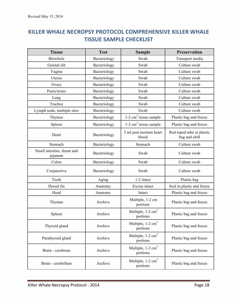
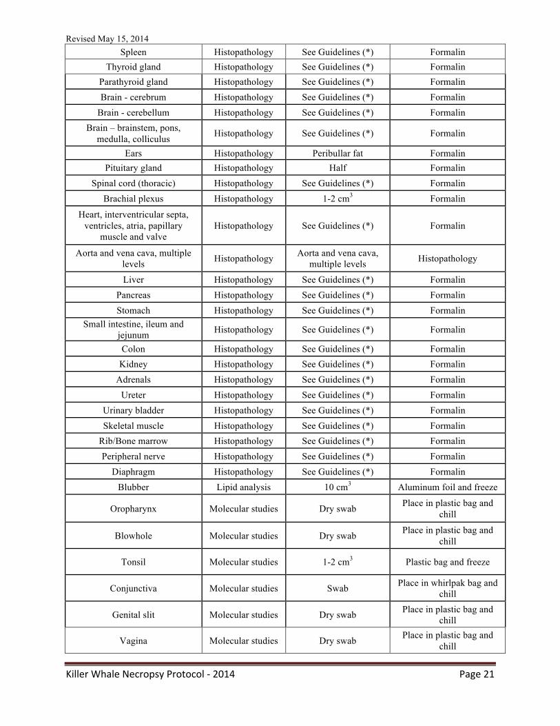
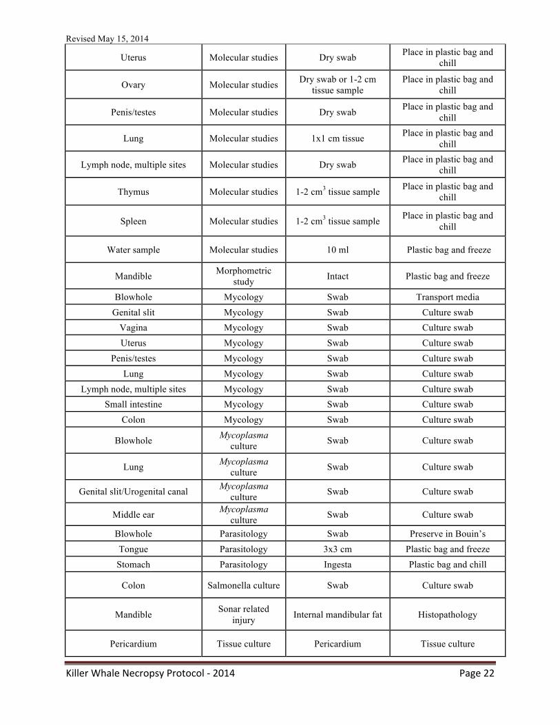
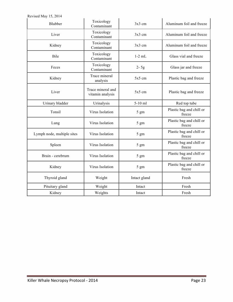
KILLER WHALE NECROPSY PROTOCOL COMPREHENSIVE KILLER WHALE TISSUE SAMPLE CHECKLIST
Tissue Test Sample Preservation
Blowhole Bacteriology Swab Transport media Genital slit Bacteriology Swab Culture swab
Vagina Bacteriology Swab Culture swab Uterus Bacteriology Swab Culture swab Ovary Bacteriology Swab Culture swab
Penis/testes Bacteriology Swab Culture swab Lung Bacteriology Swab Culture swab
Trachea Bacteriology Swab Culture swab Lymph node, multiple sites Bacteriology Swab Culture swab
Thymus Bacteriology 1-2 cm3 tissue sample Plastic bag and freeze
Spleen Bacteriology 1-2 cm3 tissue sample Plastic bag and freeze
Heart Bacteriology 5 ml post mortem heart blood
Red toped tube or plastic bag and chill
Stomach Bacteriology Stomach Culture swab Small intestine, ileum and
jejunum Bacteriology Swab Culture swab
Colon Bacteriology Swab Culture swab
Conjunctiva Bacteriology Swab Culture swab
Teeth Aging 1-2 intact Plastic bag Dorsal fin Anatomy Excise intact Seal in plastic and freeze
Head Anatomy Intact Plastic bag and freeze
Thymus Archive Multiple, 1-2 cm portions Plastic bag and freeze
Spleen Archive Multiple, 1-2 cm3 portions Plastic bag and freeze
Thyroid gland Archive Multiple, 1-2 cm3 portions Plastic bag and freeze
Parathyroid gland Archive Multiple, 1-2 cm3
portions Plastic bag and freeze
Brain - cerebrum Archive Multiple, 1-2 cm3 portions Plastic bag and freeze
Brain - cerebellum Archive Multiple, 1-2 cm3 portions Plastic bag and freeze
Revised May 15, 2014
Killer Whale Necropsy Protocol -‐ 2014 Page 19
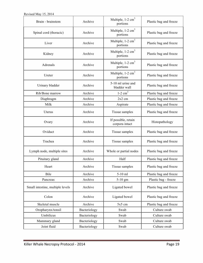
Brain - brainstem Archive Multiple, 1-2 cm3 portions Plastic bag and freeze
Spinal cord (thoracic) Archive Multiple, 1-2 cm3 portions Plastic bag and freeze
Liver Archive Multiple, 1-2 cm3
portions Plastic bag and freeze
Kidney Archive Multiple, 1-2 cm3 portions Plastic bag and freeze
Adrenals Archive Multiple, 1-2 cm3 portions Plastic bag and freeze
Ureter Archive Multiple, 1-2 cm3 portions Plastic bag and freeze
Urinary bladder Archive 5-10 ml urine and bladder wall Plastic bag and freeze
Rib/Bone marrow Archive 1-2 cm3 Plastic bag and freeze Diaphragm Archive 2x2 cm Plastic bag and freeze
Milk Archive Aspirate Plastic bag and freeze
Uterus Archive Tissue samples Plastic bag and freeze
Ovary Archive If possible, retain corpora intact Histopathology
Oviduct Archive Tissue samples Plastic bag and freeze
Trachea Archive Tissue samples Plastic bag and freeze
Lymph node, multiple sites Archive Whole or partial nodes Plastic bag and freeze
Pituitary gland Archive Half Plastic bag and freeze
Heart Archive Tissue samples Plastic bag and freeze
Bile Archive 5-10 ml Plastic bag and freeze Pancreas Archive 5-10 gm Plastic bag - freeze
Small intestine, multiple levels Archive Ligated bowel Plastic bag and freeze
Colon Archive Ligated bowel Plastic bag and freeze
Skeletal muscle Archive 5x5 cm Plastic bag and freeze Oropharynx/tonsil Bacteriology Swab Culture swab
Umbilicus Bacteriology Swab Culture swab Mammary gland Bacteriology Swab Culture swab
Joint fluid Bacteriology Swab Culture swab
Revised May 15, 2014
Killer Whale Necropsy Protocol -‐ 2014 Page 20
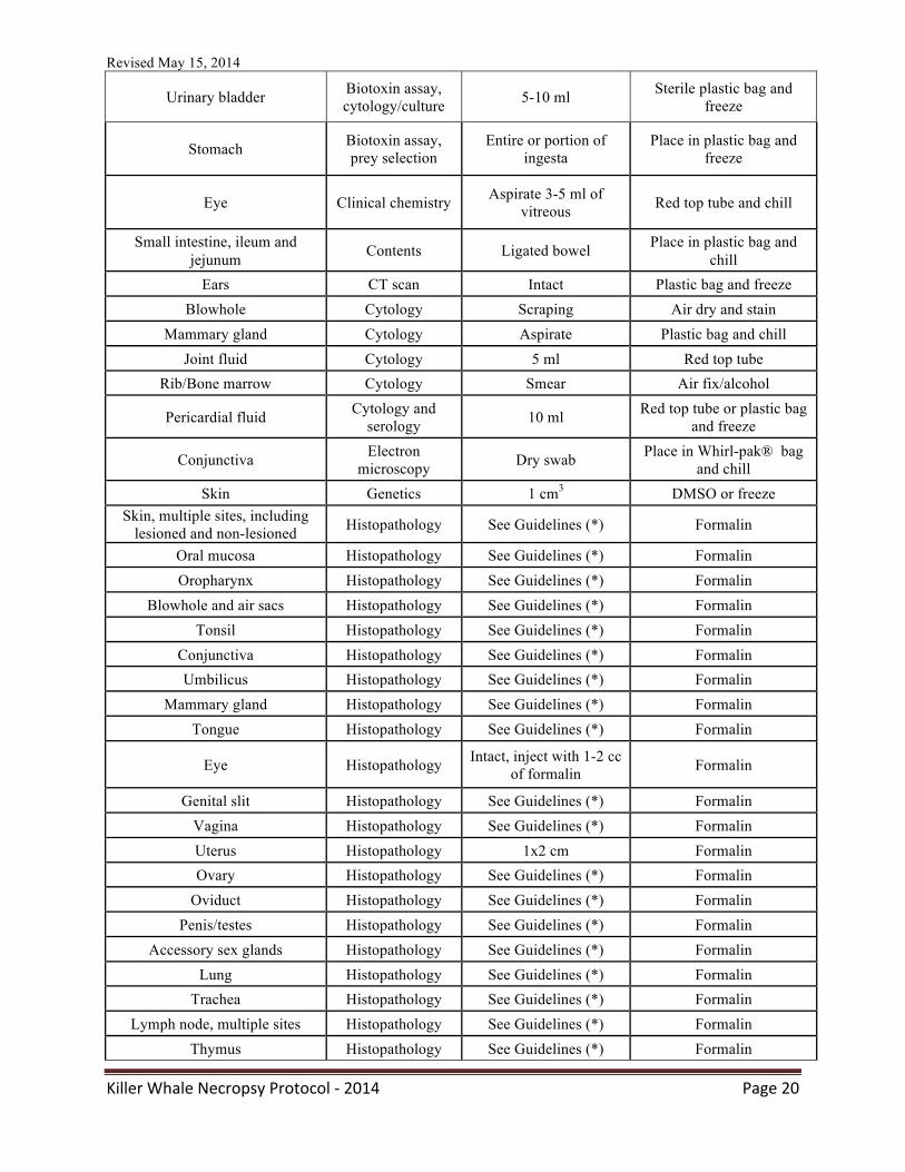
Urinary bladder Biotoxin assay, cytology/culture 5-10 ml Sterile plastic bag and
freeze
Stomach Biotoxin assay, prey selection
Entire or portion of ingesta
Place in plastic bag and freeze
Eye Clinical chemistry Aspirate 3-5 ml of vitreous Red top tube and chill
Small intestine, ileum and jejunum Contents Ligated bowel Place in plastic bag and
chill Ears CT scan Intact Plastic bag and freeze
Blowhole Cytology Scraping Air dry and stain Mammary gland Cytology Aspirate Plastic bag and chill
Joint fluid Cytology 5 ml Red top tube Rib/Bone marrow Cytology Smear Air fix/alcohol
Pericardial fluid Cytology and serology 10 ml Red top tube or plastic bag
and freeze
Conjunctiva Electron microscopy Dry swab Place in Whirl-pak® bag
and chill Skin Genetics 1 cm3 DMSO or freeze
Skin, multiple sites, including lesioned and non-lesioned Histopathology See Guidelines (*) Formalin
Oral mucosa Histopathology See Guidelines (*) Formalin Oropharynx Histopathology See Guidelines (*) Formalin
Blowhole and air sacs Histopathology See Guidelines (*) Formalin Tonsil Histopathology See Guidelines (*) Formalin
Conjunctiva Histopathology See Guidelines (*) Formalin Umbilicus Histopathology See Guidelines (*) Formalin
Mammary gland Histopathology See Guidelines (*) Formalin Tongue Histopathology See Guidelines (*) Formalin
Eye Histopathology Intact, inject with 1-2 cc of formalin Formalin
Genital slit Histopathology See Guidelines (*) Formalin Vagina Histopathology See Guidelines (*) Formalin Uterus Histopathology 1x2 cm Formalin Ovary Histopathology See Guidelines (*) Formalin
Oviduct Histopathology See Guidelines (*) Formalin Penis/testes Histopathology See Guidelines (*) Formalin
Accessory sex glands Histopathology See Guidelines (*) Formalin Lung Histopathology See Guidelines (*) Formalin
Trachea Histopathology See Guidelines (*) Formalin Lymph node, multiple sites Histopathology See Guidelines (*) Formalin
Thymus Histopathology See Guidelines (*) Formalin
Revised May 15, 2014
Killer Whale Necropsy Protocol -‐ 2014 Page 21
Spleen Histopathology See Guidelines (*) Formalin Thyroid gland Histopathology See Guidelines (*) Formalin
Parathyroid gland Histopathology See Guidelines (*) Formalin Brain - cerebrum Histopathology See Guidelines (*) Formalin
Brain - cerebellum Histopathology See Guidelines (*) Formalin Brain – brainstem, pons,
medulla, colliculus Histopathology See Guidelines (*) Formalin
Ears Histopathology Peribullar fat Formalin Pituitary gland Histopathology Half Formalin
Spinal cord (thoracic) Histopathology See Guidelines (*) Formalin Brachial plexus Histopathology 1-2 cm3 Formalin
Heart, interventricular septa, ventricles, atria, papillary
muscle and valve Histopathology See Guidelines (*) Formalin
Aorta and vena cava, multiple levels Histopathology Aorta and vena cava,
multiple levels Histopathology
Liver Histopathology See Guidelines (*) Formalin Pancreas Histopathology See Guidelines (*) Formalin Stomach Histopathology See Guidelines (*) Formalin
Small intestine, ileum and jejunum Histopathology See Guidelines (*) Formalin
Colon Histopathology See Guidelines (*) Formalin Kidney Histopathology See Guidelines (*) Formalin
Adrenals Histopathology See Guidelines (*) Formalin Ureter Histopathology See Guidelines (*) Formalin
Urinary bladder Histopathology See Guidelines (*) Formalin Skeletal muscle Histopathology See Guidelines (*) Formalin
Rib/Bone marrow Histopathology See Guidelines (*) Formalin Peripheral nerve Histopathology See Guidelines (*) Formalin
Diaphragm Histopathology See Guidelines (*) Formalin Blubber Lipid analysis 10 cm3 Aluminum foil and freeze
Oropharynx Molecular studies Dry swab Place in plastic bag and chill
Blowhole Molecular studies Dry swab Place in plastic bag and chill
Tonsil Molecular studies 1-2 cm3 Plastic bag and freeze
Conjunctiva Molecular studies Swab Place in whirlpak bag and chill
Genital slit Molecular studies Dry swab Place in plastic bag and chill
Vagina Molecular studies Dry swab Place in plastic bag and chill
Revised May 15, 2014
Killer Whale Necropsy Protocol -‐ 2014 Page 22
Uterus Molecular studies Dry swab Place in plastic bag and chill
Ovary Molecular studies Dry swab or 1-2 cm tissue sample
Place in plastic bag and chill
Penis/testes Molecular studies Dry swab Place in plastic bag and chill
Lung Molecular studies 1x1 cm tissue Place in plastic bag and chill
Lymph node, multiple sites Molecular studies Dry swab Place in plastic bag and chill
Thymus Molecular studies 1-2 cm3 tissue sample Place in plastic bag and chill
Spleen Molecular studies 1-2 cm3 tissue sample Place in plastic bag and chill
Water sample Molecular studies 10 ml Plastic bag and freeze
Mandible Morphometric study Intact Plastic bag and freeze
Blowhole Mycology Swab Transport media Genital slit Mycology Swab Culture swab
Vagina Mycology Swab Culture swab Uterus Mycology Swab Culture swab
Penis/testes Mycology Swab Culture swab Lung Mycology Swab Culture swab
Lymph node, multiple sites Mycology Swab Culture swab Small intestine Mycology Swab Culture swab
Colon Mycology Swab Culture swab
Blowhole Mycoplasma culture Swab Culture swab
Lung Mycoplasma culture Swab Culture swab
Genital slit/Urogenital canal Mycoplasma culture Swab Culture swab
Middle ear Mycoplasma culture Swab Culture swab
Blowhole Parasitology Swab Preserve in Bouin’s Tongue Parasitology 3x3 cm Plastic bag and freeze Stomach Parasitology Ingesta Plastic bag and chill
Colon Salmonella culture Swab Culture swab
Mandible Sonar related injury Internal mandibular fat Histopathology
Pericardium Tissue culture Pericardium Tissue culture
Revised May 15, 2014
Killer Whale Necropsy Protocol -‐ 2014 Page 23
Blubber Toxicology Contaminant 3x3 cm Aluminum foil and freeze
Liver Toxicology Contaminant 3x3 cm Aluminum foil and freeze
Kidney Toxicology Contaminant 3x3 cm Aluminum foil and freeze
Bile Toxicology Contaminant 1-2 mL Glass vial and freeze
Feces Toxicology Contaminant 2- 5g Glass jar and freeze
Kidney Trace mineral analysis 5x5 cm Plastic bag and freeze
Liver Trace mineral and vitamin analysis 5x5 cm Plastic bag and freeze
Urinary bladder Urinalysis 5-10 ml Red top tube
Tonsil Virus Isolation 5 gm Plastic bag and chill or freeze
Lung Virus Isolation 5 gm Plastic bag and chill or freeze
Lymph node, multiple sites Virus Isolation 5 gm Plastic bag and chill or freeze
Spleen Virus Isolation 5 gm Plastic bag and chill or freeze
Brain - cerebrum Virus Isolation 5 gm Plastic bag and chill or freeze
Kidney Virus Isolation 5 gm Plastic bag and chill or freeze
Thyroid gland Weight Intact gland Fresh
Pituitary gland Weight Intact Fresh Kidney Weights Intact Fresh
Revised May 15, 2014
Killer Whale Necropsy Protocol -‐ 2014 Page 24
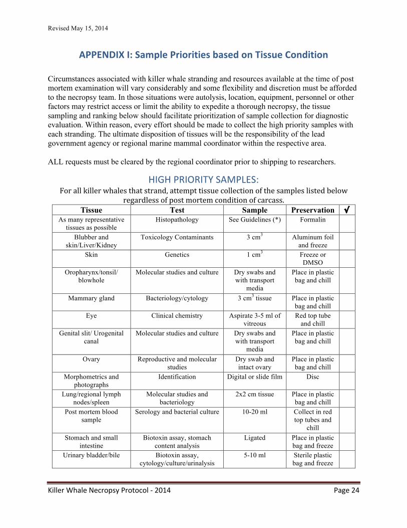
APPENDIX I: Sample Priorities based on Tissue Condition
Circumstances associated with killer whale stranding and resources available at the time of post mortem examination will vary considerably and some flexibility and discretion must be afforded to the necropsy team. In those situations were autolysis, location, equipment, personnel or other factors may restrict access or limit the ability to expedite a thorough necropsy, the tissue sampling and ranking below should facilitate prioritization of sample collection for diagnostic evaluation. Within reason, every effort should be made to collect the high priority samples with each stranding. The ultimate disposition of tissues will be the responsibility of the lead government agency or regional marine mammal coordinator within the respective area.
ALL requests must be cleared by the regional coordinator prior to shipping to researchers.
HIGH PRIORITY SAMPLES: For all killer whales that strand, attempt tissue collection of the samples listed below
regardless of post mortem condition of carcass. Tissue Test Sample Preservation √
As many representative tissues as possible
Histopathology See Guidelines (*) Formalin
Blubber and skin/Liver/Kidney
Toxicology Contaminants 3 cm3 Aluminum foil and freeze
Skin Genetics 1 cm3 Freeze or DMSO
Oropharynx/tonsil/ blowhole
Molecular studies and culture Dry swabs and with transport
media
Place in plastic bag and chill
Mammary gland Bacteriology/cytology 3 cm3 tissue Place in plastic bag and chill
Eye Clinical chemistry Aspirate 3-5 ml of vitreous
Red top tube and chill
Genital slit/ Urogenital canal
Molecular studies and culture Dry swabs and with transport
media
Place in plastic bag and chill
Ovary Reproductive and molecular studies
Dry swab and intact ovary
Place in plastic bag and chill
Morphometrics and photographs
Identification Digital or slide film Disc
Lung/regional lymph nodes/spleen
Molecular studies and bacteriology
2x2 cm tissue Place in plastic bag and chill
Post mortem blood sample
Serology and bacterial culture 10-20 ml Collect in red top tubes and
chill
Stomach and small intestine
Biotoxin assay, stomach content analysis
Ligated Place in plastic bag and freeze
Urinary bladder/bile Biotoxin assay, cytology/culture/urinalysis
5-10 ml Sterile plastic bag and freeze
Revised May 15, 2014
Killer Whale Necropsy Protocol -‐ 2014 Page 25
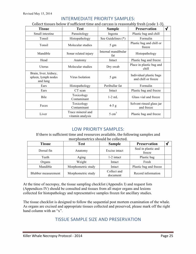
INTERMEDIATE PRIORITY SAMPLES: Collect tissues below if sufficient time and carcass is reasonably fresh (code 1-‐3).
Tissue Test Sample Preservation √ Small intestine Parasitology Ingesta Plastic bag and chill
Tonsil Histopathology See Guidelines (*) Formalin
Tonsil Molecular studies 5 gm Plastic bag and chill or freeze
Mandible Sonar related injury Internal mandibular fat Histopathology
Head Anatomy Intact Plastic bag and freeze
Uterus Molecular studies Dry swab Place in plastic bag and chill
Brain, liver, kidney, spleen, lymph nodes
and lung Virus Isolation 5 gm Individual plastic bags
and chill or freeze
Ears Histopathology Peribullar fat Formalin Ears CT scan Intact Plastic bag and freeze
Bile Toxicology Contaminant 1-2 mL Glass vial and freeze
Feces Toxicology Contaminant 4-5 g Solvent rinsed glass jar
and freeze
Liver Trace mineral and vitamin analysis 5 cm3 Plastic bag and freeze
LOW PRIORITY SAMPLES: If there is sufficient time and resources available, the following samples and
morphometrics should be collected. Tissue Test Sample Preservation √
Dorsal fin Anatomy Excise intact Seal in plastic and freeze
Teeth Aging 1-2 intact Plastic bag Organs Weight Intact Fresh
Mandible Morphometric study Intact Plastic bag and freeze
Blubber measurement Morphometric study Collect and document Record information
At the time of necropsy, the tissue sampling checklist (Appendix I) and request lists (Appendices IV) should be consulted and tissues from all major organs and lesions collected for histopathology and representative samples frozen for ancillary studies.
The tissue checklist is designed to follow the sequential post mortem examination of the whale. As organs are excised and appropriate tissues collected and preserved, please mark off the right hand column with an “x”.
TISSUE SAMPLE SIZE AND PRESERVATION
Revised May 15, 2014
Killer Whale Necropsy Protocol -‐ 2014 Page 26
*Guidelines for fixation of tissues for histopathologic evaluation: preserve all lesions and as many of the tissues listed below as possible in 10% buffered formalin. Tissue samples should be between 3-5 cm2 in area and up to 0.5 to 1.0 cm in width and immersed in a ratio of 1 part tissue to 10-15 parts formalin. If electron microscopy (EM) fixative such as glutaraldehyde is available, preserve minced (1-2 mm3) pieces of kidney, liver, spleen and lung.
Representative 3-5 cm blocks of tissue from lesions and major organs (e.g., lung, liver, kidney, spleen) should be placed in individually labeled small (preferably Whirl-pak®
) plastic bags and placed on dry or wet ice for initial storage and transportation. Also, collect post-mortem serum (from heart blood), urine, eye fluid, bile, ingesta, and any abnormal fluid accumulations. Heart blood should be spun as soon as possible to limit the degree of hemolysis. Upon arrival to a diagnostic or reference laboratory, samples should be frozen at -70 degrees Celsius. If this is unavailable, temporary storage in conventional freezer without automatic defrost cycle is acceptable. A 1-2 cm block of skin, muscle or flipper for genetic analysis should be excised and foil-wrapped and frozen. The sample can be placed in DMSO/saline solution if there is likelihood that the samples cannot remain frozen until they reach their final destination, but freezing without preservative is preferred.
For each lesion, up to 2-3 swabs may be obtained and samples should be chilled for transport to a diagnostic facility. In addition to routine TSA and blood agar cultures, special media for isolation of halophilic bacteria should also be inoculated.
Revised May 15, 2014
Killer Whale Necropsy Protocol -‐ 2014 Page 27
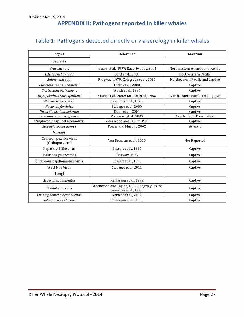
APPENDIX II: Pathogens reported in killer whales
Table 1: Pathogens detected directly or via serology in killer whales
Agent Reference Location
Bacteria
Brucella spp. Jepson et al., 1997; Raverty et al., 2004 Northeastern Atlantic and Pacific Edwardsiella tarda Ford et al., 2000 Northeastern Pacific Salmonella spp. Ridgway, 1979; Colegrove et al., 2010 Northeastern Pacific and captive
Burkholderia pseudomallei Hicks et al., 2000 Captive Clostridium perfringens Walsh et al., 1994 Captive
Erysipelothrix rhusiopathiae Young et al., 2002; Bossart et al., 1988 Northeastern Pacific and Captive Nocardia asteroides Sweeney et al., 1976 Captive Nocardia farcinica St. Leger et al, 2009 Captive
Nocardia otitidiscaviarum Dunn et al., 2001 Captive Pseudomonas aeruginosa Rozanova et al., 2003 Avacha Gulf (Kamchatka)
Streptococcus sp., beta-‐hemolytic Greenwood and Taylor, 1985 Captive Staphylococcus aureus Power and Murphy 2002 Atlantic
Viruses
Cetacean pox like virus (Orthopoxvirus) Van Bressem et al., 1999 Not Reported
Hepatitis-‐B like virus Bossart et al., 1990 Captive
Influenza (suspected) Ridgway, 1979 Captive
Cutaneous papilloma-‐like virus Bossart et al., 1996 Captive
West Nile Virus St. Leger et al, 2011 Captive
Fungi
Aspergillus fumigatus Reidarson et al., 1999 Captive
Candida albicans Greenwood and Taylor, 1985; Ridgway, 1979; Sweeney et al., 1976 Captive
Cunninghamella bertholletiae Kakizoe et al., 2012 Captive Saksenaea vasiformis Reidarson et al., 1999 Captive
Revised May 15, 2014
Killer Whale Necropsy Protocol -‐ 2014 Page 28
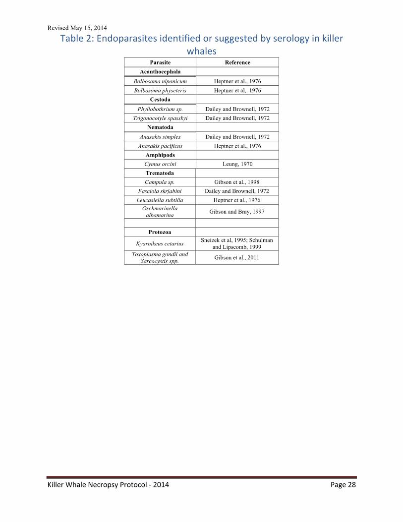
Table 2: Endoparasites identified or suggested by serology in killer whales
Parasite Reference Acanthocephala
Bolbosoma niponicum Heptner et al., 1976 Bolbosoma physeteris Heptner et al,. 1976
Cestoda
Phyllobothrium sp. Dailey and Brownell, 1972 Trigonocotyle spasskyi Dailey and Brownell, 1972
Nematoda
Anasakis simplex Dailey and Brownell, 1972 Anasakis pacificus Heptner et al., 1976
Amphipods Cymus orcini Leung, 1970 Trematoda Campula sp. Gibson et al., 1998
Fasciola skrjabini Dailey and Brownell, 1972 Leucasiella subtilla Heptner et al., 1976
Oschmarinella albamarina Gibson and Bray, 1997
Protozoa
Kyaroikeus cetarius Sneizek et al, 1995; Schulman and Lipscomb, 1999
Toxoplasma gondii and Sarcocystis spp. Gibson et al., 2011
Revised May 15, 2014
Killer Whale Necropsy Protocol -‐ 2014 Page 29
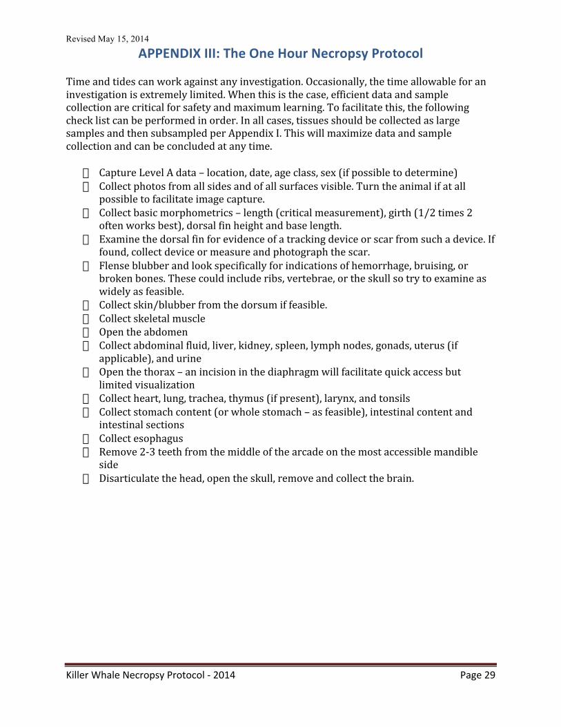
APPENDIX III: The One Hour Necropsy Protocol Time and tides can work against any investigation. Occasionally, the time allowable for an investigation is extremely limited. When this is the case, efficient data and sample collection are critical for safety and maximum learning. To facilitate this, the following check list can be performed in order. In all cases, tissues should be collected as large samples and then subsampled per Appendix I. This will maximize data and sample collection and can be concluded at any time.
� Capture Level A data – location, date, age class, sex (if possible to determine) � Collect photos from all sides and of all surfaces visible. Turn the animal if at all
possible to facilitate image capture. � Collect basic morphometrics – length (critical measurement), girth (1/2 times 2
often works best), dorsal fin height and base length. � Examine the dorsal fin for evidence of a tracking device or scar from such a device. If
found, collect device or measure and photograph the scar. � Flense blubber and look specifically for indications of hemorrhage, bruising, or
broken bones. These could include ribs, vertebrae, or the skull so try to examine as widely as feasible.
� Collect skin/blubber from the dorsum if feasible. � Collect skeletal muscle � Open the abdomen � Collect abdominal fluid, liver, kidney, spleen, lymph nodes, gonads, uterus (if
applicable), and urine � Open the thorax – an incision in the diaphragm will facilitate quick access but
limited visualization � Collect heart, lung, trachea, thymus (if present), larynx, and tonsils � Collect stomach content (or whole stomach – as feasible), intestinal content and
intestinal sections � Collect esophagus � Remove 2-‐3 teeth from the middle of the arcade on the most accessible mandible
side � Disarticulate the head, open the skull, remove and collect the brain.
Revised May 15, 2014
Killer Whale Necropsy Protocol -‐ 2014 Page 30
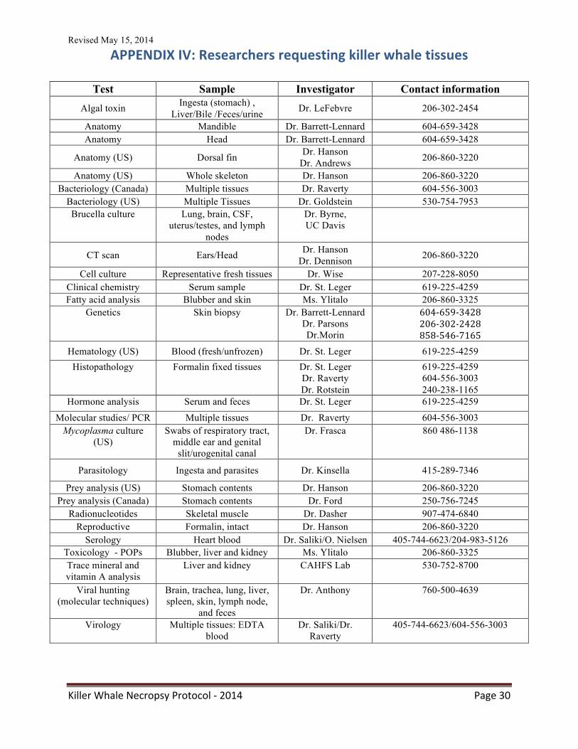
APPENDIX IV: Researchers requesting killer whale tissues
Test Sample Investigator Contact information Algal toxin Ingesta (stomach) ,
Liver/Bile /Feces/urine Dr. LeFebvre 206-302-2454
Anatomy Mandible Dr. Barrett-Lennard 604-659-3428 Anatomy Head Dr. Barrett-Lennard 604-659-3428
Anatomy (US) Dorsal fin Dr. Hanson Dr. Andrews 206-860-3220
Anatomy (US) Whole skeleton Dr. Hanson 206-860-3220 Bacteriology (Canada) Multiple tissues Dr. Raverty 604-556-3003
Bacteriology (US) Multiple Tissues Dr. Goldstein 530-754-7953 Brucella culture Lung, brain, CSF,
uterus/testes, and lymph nodes
Dr. Byrne, UC Davis
CT scan Ears/Head Dr. Hanson Dr. Dennison 206-860-3220
Cell culture Representative fresh tissues Dr. Wise 207-228-8050 Clinical chemistry Serum sample Dr. St. Leger 619-225-4259 Fatty acid analysis Blubber and skin Ms. Ylitalo 206-860-3325
Genetics Skin biopsy Dr. Barrett-Lennard Dr. Parsons Dr.Morin
604-‐659-‐3428 206-‐302-‐2428 858-‐546-‐7165
Hematology (US) Blood (fresh/unfrozen) Dr. St. Leger 619-225-4259 Histopathology Formalin fixed tissues Dr. St. Leger
Dr. Raverty Dr. Rotstein
619-225-4259 604-556-3003 240-238-1165
Hormone analysis Serum and feces Dr. St. Leger 619-225-4259
Molecular studies/ PCR Multiple tissues Dr. Raverty 604-556-3003 Mycoplasma culture
(US) Swabs of respiratory tract,
middle ear and genital slit/urogenital canal
Dr. Frasca 860 486-1138
Parasitology Ingesta and parasites Dr. Kinsella 415-289-7346
Prey analysis (US) Stomach contents Dr. Hanson 206-860-3220 Prey analysis (Canada) Stomach contents Dr. Ford 250-756-7245
Radionucleotides Skeletal muscle Dr. Dasher 907-474-6840 Reproductive Formalin, intact Dr. Hanson 206-860-3220
Serology Heart blood Dr. Saliki/O. Nielsen 405-744-6623/204-983-5126 Toxicology - POPs Blubber, liver and kidney Ms. Ylitalo 206-860-3325 Trace mineral and vitamin A analysis
Liver and kidney CAHFS Lab 530-752-8700
Viral hunting (molecular techniques)
Brain, trachea, lung, liver, spleen, skin, lymph node,
and feces
Dr. Anthony 760-500-4639
Virology Multiple tissues: EDTA blood
Dr. Saliki/Dr. Raverty
405-744-6623/604-556-3003
Revised May 15, 2014
Killer Whale Necropsy Protocol -‐ 2014 Page 31
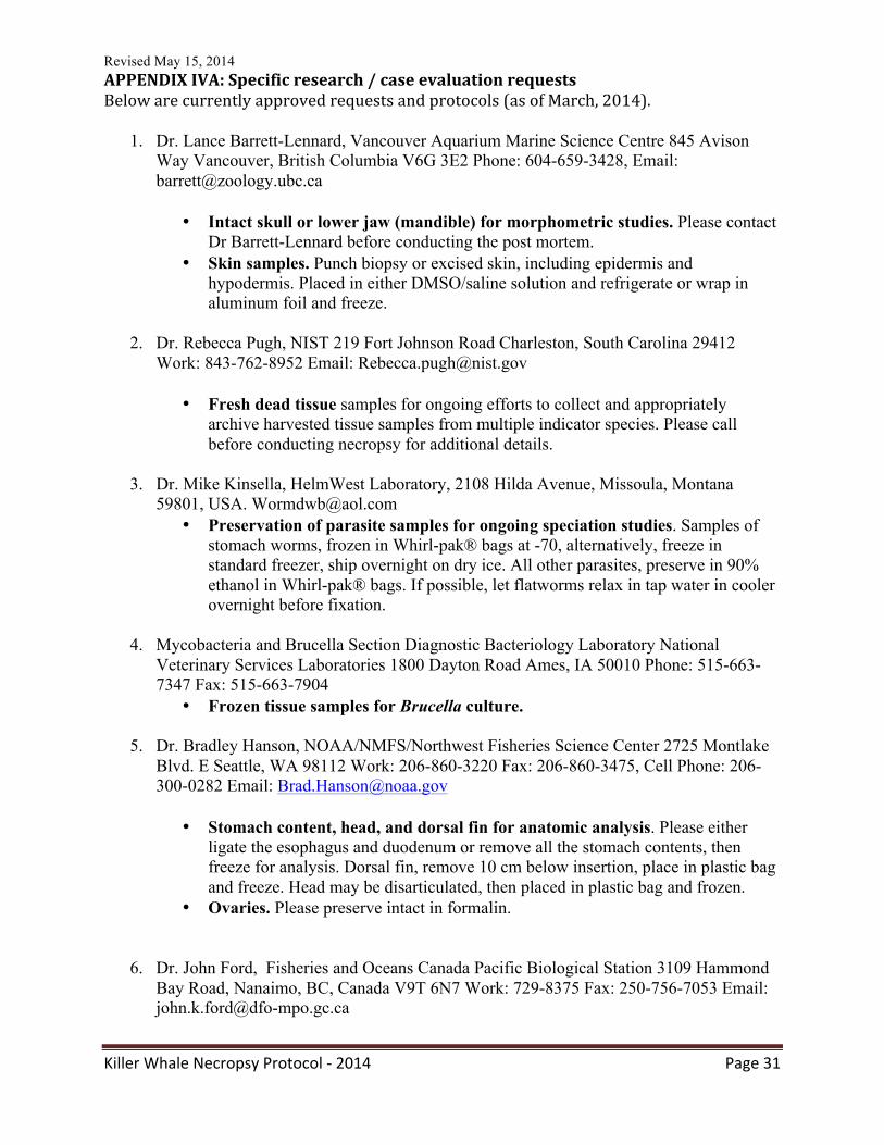
APPENDIX IVA: Specific research / case evaluation requests Below are currently approved requests and protocols (as of March, 2014).
1. Dr. Lance Barrett-Lennard, Vancouver Aquarium Marine Science Centre 845 Avison Way Vancouver, British Columbia V6G 3E2 Phone: 604-659-3428, Email: [email protected]
• Intact skull or lower jaw (mandible) for morphometric studies. Please contact Dr Barrett-Lennard before conducting the post mortem.
• Skin samples. Punch biopsy or excised skin, including epidermis and hypodermis. Placed in either DMSO/saline solution and refrigerate or wrap in aluminum foil and freeze.
2. Dr. Rebecca Pugh, NIST 219 Fort Johnson Road Charleston, South Carolina 29412
Work: 843-762-8952 Email: [email protected]
• Fresh dead tissue samples for ongoing efforts to collect and appropriately archive harvested tissue samples from multiple indicator species. Please call before conducting necropsy for additional details.
3. Dr. Mike Kinsella, HelmWest Laboratory, 2108 Hilda Avenue, Missoula, Montana
59801, USA. [email protected] • Preservation of parasite samples for ongoing speciation studies. Samples of
stomach worms, frozen in Whirl-pak® bags at -70, alternatively, freeze in standard freezer, ship overnight on dry ice. All other parasites, preserve in 90% ethanol in Whirl-pak® bags. If possible, let flatworms relax in tap water in cooler overnight before fixation.
4. Mycobacteria and Brucella Section Diagnostic Bacteriology Laboratory National
Veterinary Services Laboratories 1800 Dayton Road Ames, IA 50010 Phone: 515-663-7347 Fax: 515-663-7904
• Frozen tissue samples for Brucella culture.
5. Dr. Bradley Hanson, NOAA/NMFS/Northwest Fisheries Science Center 2725 Montlake Blvd. E Seattle, WA 98112 Work: 206-860-3220 Fax: 206-860-3475, Cell Phone: 206-300-0282 Email: [email protected]
• Stomach content, head, and dorsal fin for anatomic analysis. Please either ligate the esophagus and duodenum or remove all the stomach contents, then freeze for analysis. Dorsal fin, remove 10 cm below insertion, place in plastic bag and freeze. Head may be disarticulated, then placed in plastic bag and frozen.
• Ovaries. Please preserve intact in formalin.
6. Dr. John Ford, Fisheries and Oceans Canada Pacific Biological Station 3109 Hammond Bay Road, Nanaimo, BC, Canada V9T 6N7 Work: 729-8375 Fax: 250-756-7053 Email: [email protected]
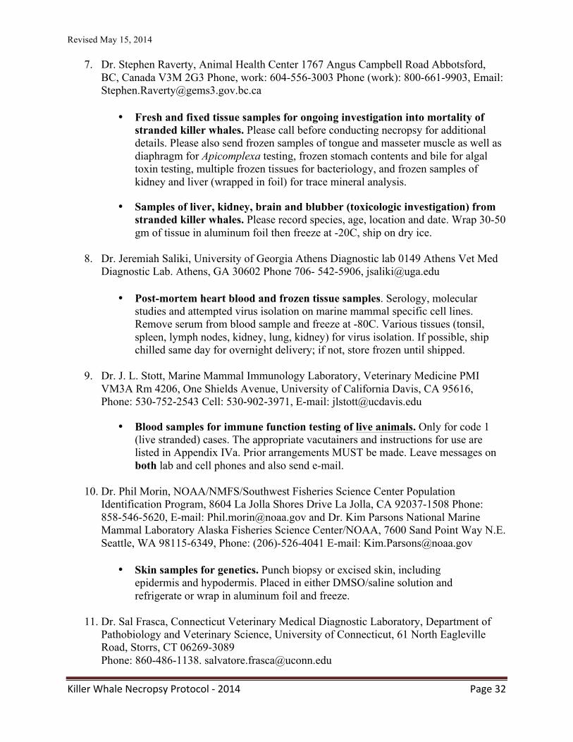
Revised May 15, 2014
Killer Whale Necropsy Protocol -‐ 2014 Page 32
7. Dr. Stephen Raverty, Animal Health Center 1767 Angus Campbell Road Abbotsford,
BC, Canada V3M 2G3 Phone, work: 604-556-3003 Phone (work): 800-661-9903, Email: [email protected]
• Fresh and fixed tissue samples for ongoing investigation into mortality of stranded killer whales. Please call before conducting necropsy for additional details. Please also send frozen samples of tongue and masseter muscle as well as diaphragm for Apicomplexa testing, frozen stomach contents and bile for algal toxin testing, multiple frozen tissues for bacteriology, and frozen samples of kidney and liver (wrapped in foil) for trace mineral analysis.
• Samples of liver, kidney, brain and blubber (toxicologic investigation) from stranded killer whales. Please record species, age, location and date. Wrap 30-50 gm of tissue in aluminum foil then freeze at -20C, ship on dry ice.
8. Dr. Jeremiah Saliki, University of Georgia Athens Diagnostic lab 0149 Athens Vet Med
Diagnostic Lab. Athens, GA 30602 Phone 706- 542-5906, [email protected]
• Post-mortem heart blood and frozen tissue samples. Serology, molecular studies and attempted virus isolation on marine mammal specific cell lines. Remove serum from blood sample and freeze at -80C. Various tissues (tonsil, spleen, lymph nodes, kidney, lung, kidney) for virus isolation. If possible, ship chilled same day for overnight delivery; if not, store frozen until shipped.
9. Dr. J. L. Stott, Marine Mammal Immunology Laboratory, Veterinary Medicine PMI
VM3A Rm 4206, One Shields Avenue, University of California Davis, CA 95616, Phone: 530-752-2543 Cell: 530-902-3971, E-mail: [email protected]
• Blood samples for immune function testing of live animals. Only for code 1 (live stranded) cases. The appropriate vacutainers and instructions for use are listed in Appendix IVa. Prior arrangements MUST be made. Leave messages on both lab and cell phones and also send e-mail.
10. Dr. Phil Morin, NOAA/NMFS/Southwest Fisheries Science Center Population
Identification Program, 8604 La Jolla Shores Drive La Jolla, CA 92037-1508 Phone: 858-546-5620, E-mail: [email protected] and Dr. Kim Parsons National Marine Mammal Laboratory Alaska Fisheries Science Center/NOAA, 7600 Sand Point Way N.E. Seattle, WA 98115-6349, Phone: (206)-526-4041 E-mail: [email protected]
• Skin samples for genetics. Punch biopsy or excised skin, including epidermis and hypodermis. Placed in either DMSO/saline solution and refrigerate or wrap in aluminum foil and freeze.
11. Dr. Sal Frasca, Connecticut Veterinary Medical Diagnostic Laboratory, Department of
Pathobiology and Veterinary Science, University of Connecticut, 61 North Eagleville Road, Storrs, CT 06269-3089 Phone: 860-486-1138. [email protected]
Revised May 15, 2014
Killer Whale Necropsy Protocol -‐ 2014 Page 33
• Tissue samples for attempted Mycoplasma spp isolation. Swabs or fresh tissue should be aseptically collected from representative levels of the respiratory system, including the blowhole, larynx, trachea, tracheal bifurcation, lungs, mediastinal lymph nodes, middle ear, and genital slit/urogenital tract. Swabs should be chilled and forwarded by courier.
12. John Pierce Wise, Sr., Ph.D. Director, Center for Integrated and Applied Environmental
Toxicology Associate Professor of Toxicology and Molecular Epidemiology Bioscience Research Institute University of Southern Maine 178 Science Building 96 Falmouth Street Portland, ME 04103 Phone: 207-228-8047 Email: [email protected]
• Fresh tissue samples to be cultured and stabilized (“immortalized”) for
subsequent toxicological studies and for placement in the marine mammal cell repository for other permitted researchers. Please collect skin (w/dermis), kidney, liver, bronchus, testes/ovaries, brain from all young animals and call for storage and shipping recommendations.
13. Ms. Gina Ylitalo, NOAA Fisheries / Northwest Science Center 2725 Montlake
Boulevard East Seattle, WA 98112 Phone: 206-860-3325, E-mail: [email protected]
• Samples of liver, kidney blubber with skin, skeletal muscle, bile, and feces.
(Contaminant toxicologic investigation). Please record species, age, location and date. Wrap 30-50 gm of tissue in aluminum foil or place in appropriate glass containers, then freeze at -20C, ship on dry ice (Appendix IX).
14. Dr. Doug Dasher, University of Alaska Fairbanks 905 N. Koyukuk 245 O'Neill Building
P.O. Box 757220 Fairbanks, AK 99775-7220 Phone: 907-474-6840, E-mail: [email protected]
• Samples of skeletal muscle for detection of Cs 137 and Cs 134
radionucleotides. Please collect 1kg of dorsal skeletal muscular and freeze (-20 is fine) in a plastic Ziploc or Whirl-pak® bag. Use clean gloves and keep sand or sediments off of the sample. Ship frozen for testing.
15. Dr. Dawn Noren, Research Fishery Biologist, NOAA NMFS Northwest Fisheries Science Center, Seattle, WA Phones: Office: 206-302-2439/ Cell: 206-423-0215, E-mail: [email protected]
• Samples of skeletal muscle to measure muscle myoglobin content and acid buffering capacity (to assess variability in muscle biochemistry and diving capability with development and across ecotypes). Collect muscle samples from all age carcasses that are in fresh condition (Code 2). Samples are to be collected from the mid-belly of the primary locomotor muscle (m. longissimus dorsi). The location of the sampling site is below the anterior insertion site of the dorsal fin. Samples (3X3 inch block) should be completely wrapped in foil, placed in a Ziploc bag, and frozen immediately after collection.
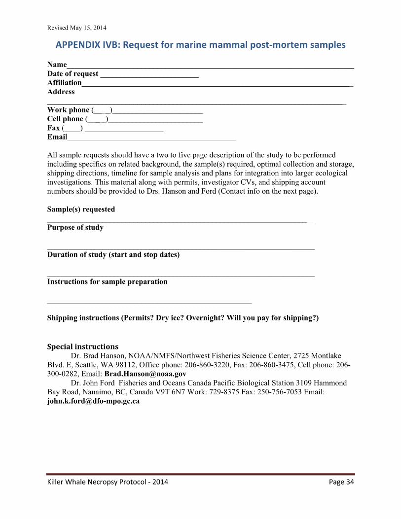
Revised May 15, 2014
Killer Whale Necropsy Protocol -‐ 2014 Page 34
APPENDIX IVB: Request for marine mammal post-‐mortem samples Name_________________________________________________________________________ Date of request _________________________ Affiliation_____________________________________________________________________ Address ____________________________________________________________________________ Work phone (__ _)_______________________ Cell phone (___ _)________________________ Fax (____) ____________________ Email All sample requests should have a two to five page description of the study to be performed including specifics on related background, the sample(s) required, optimal collection and storage, shipping directions, timeline for sample analysis and plans for integration into larger ecological investigations. This material along with permits, investigator CVs, and shipping account numbers should be provided to Drs. Hanson and Ford (Contact info on the next page). Sample(s) requested __________________________________________________________________ Purpose of study ____________________________________________________________________ Duration of study (start and stop dates) ____________________________________________________________________ Instructions for sample preparation ____________________________________________________ Shipping instructions (Permits? Dry ice? Overnight? Will you pay for shipping?)
Special instructions Dr. Brad Hanson, NOAA/NMFS/Northwest Fisheries Science Center, 2725 Montlake
Blvd. E, Seattle, WA 98112, Office phone: 206-860-3220, Fax: 206-860-3475, Cell phone: 206-300-0282, Email: [email protected]
Dr. John Ford Fisheries and Oceans Canada Pacific Biological Station 3109 Hammond Bay Road, Nanaimo, BC, Canada V9T 6N7 Work: 729-8375 Fax: 250-756-7053 Email: [email protected]
Revised May 15, 2014
Killer Whale Necropsy Protocol -‐ 2014 Page 35
APPENDIX V: Permitting concerns and authorities Marine mammals are protected internationally under a variety of treaties and acts. All stranding response should begin with contacting the proper authorities. In the USA, marine mammals are protected under the Marine Mammal Protection Act and the Endangered Species Act. In Canada, killer whales are covered by the Species at Risk Act (SARA). International shipment of samples and materials must comply with the Convention on International Trade of Endangered Species (CITES) as well as US Department of Agriculture/ Animal and Plant Health Inspection Service (USDA/APHIS). Permits are required for international exchanges. USA: NOAA Office of Protected Resources (OPR) http://www.nmfs.noaa.gov/pr/ Dr. Teri Rowles. [email protected] (301) 713-‐2322 x-‐178 Stranding network hotlines: http://www.nmfs.noaa.gov/pr/health/networks.htm CITES: http://www.cites.org/common/cop/15/doc/E15-‐30-‐01T.pdf USDA/APHIS: http://www.aphis.usda.gov/ US Fish and Wildlife Service: http://www.fws.gov/ CANADA: Fisheries and Oceans Canada, http://www.pac.dfo-‐mpo.gc.ca/fm-‐gp/species-‐especes/mammals-‐mammiferes/index-‐eng.html Paul Cottrell, DFO Marine Mammal Coordinator (604) 666-‐9965 SARA: http://www.sararegistry.gc.ca/default_e.cfm CITES : http://www.dfo-‐mpo.gc.ca/acts-‐lois/cites-‐eng.htm DFO Marine Mammal Response Hotline (British Columbia): (800) 465-‐4336
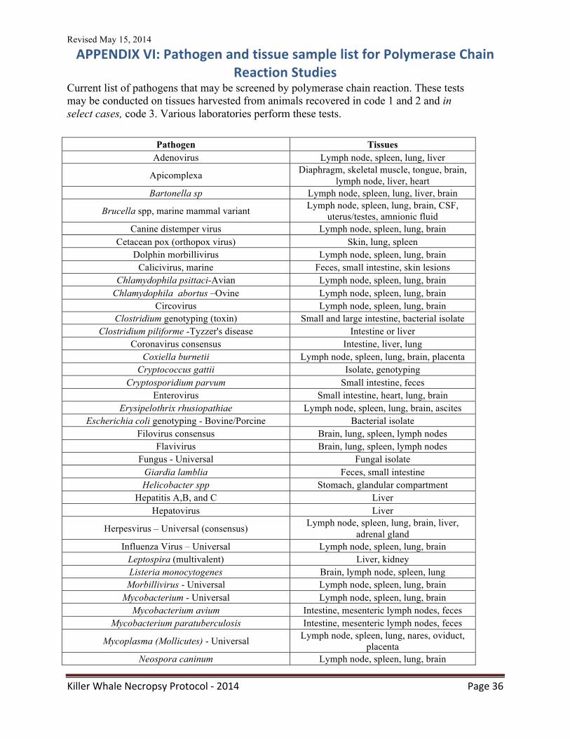
Revised May 15, 2014
Killer Whale Necropsy Protocol -‐ 2014 Page 36
APPENDIX VI: Pathogen and tissue sample list for Polymerase Chain Reaction Studies
Current list of pathogens that may be screened by polymerase chain reaction. These tests may be conducted on tissues harvested from animals recovered in code 1 and 2 and in select cases, code 3. Various laboratories perform these tests.
Pathogen Tissues Adenovirus Lymph node, spleen, lung, liver
Apicomplexa Diaphragm, skeletal muscle, tongue, brain, lymph node, liver, heart
Bartonella sp Lymph node, spleen, lung, liver, brain
Brucella spp, marine mammal variant Lymph node, spleen, lung, brain, CSF, uterus/testes, amnionic fluid
Canine distemper virus Lymph node, spleen, lung, brain Cetacean pox (orthopox virus) Skin, lung, spleen
Dolphin morbillivirus Lymph node, spleen, lung, brain Calicivirus, marine Feces, small intestine, skin lesions
Chlamydophila psittaci-Avian Lymph node, spleen, lung, brain Chlamydophila abortus –Ovine Lymph node, spleen, lung, brain
Circovirus Lymph node, spleen, lung, brain Clostridium genotyping (toxin) Small and large intestine, bacterial isolate
Clostridium piliforme -Tyzzer's disease Intestine or liver Coronavirus consensus Intestine, liver, lung
Coxiella burnetii Lymph node, spleen, lung, brain, placenta Cryptococcus gattii Isolate, genotyping
Cryptosporidium parvum Small intestine, feces Enterovirus Small intestine, heart, lung, brain
Erysipelothrix rhusiopathiae Lymph node, spleen, lung, brain, ascites Escherichia coli genotyping - Bovine/Porcine Bacterial isolate
Filovirus consensus Brain, lung, spleen, lymph nodes Flavivirus Brain, lung, spleen, lymph nodes
Fungus - Universal Fungal isolate Giardia lamblia Feces, small intestine Helicobacter spp Stomach, glandular compartment
Hepatitis A,B, and C Liver Hepatovirus Liver
Herpesvirus – Universal (consensus) Lymph node, spleen, lung, brain, liver, adrenal gland
Influenza Virus – Universal Lymph node, spleen, lung, brain Leptospira (multivalent) Liver, kidney Listeria monocytogenes Brain, lymph node, spleen, lung
Morbillivirus - Universal Lymph node, spleen, lung, brain Mycobacterium - Universal Lymph node, spleen, lung, brain
Mycobacterium avium Intestine, mesenteric lymph nodes, feces Mycobacterium paratuberculosis Intestine, mesenteric lymph nodes, feces
Mycoplasma (Mollicutes) - Universal Lymph node, spleen, lung, nares, oviduct, placenta
Neospora caninum Lymph node, spleen, lung, brain
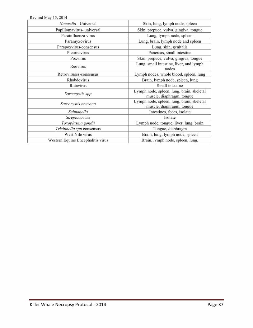
Revised May 15, 2014
Killer Whale Necropsy Protocol -‐ 2014 Page 37
Nocardia - Universal Skin, lung, lymph node, spleen Papillomavirus- universal Skin, prepuce, vulva, gingiva, tongue
Parainfluenza virus Lung, lymph node, spleen Paramyxovirus Lung, brain, lymph node and spleen
Parapoxvirus-consensus Lung, skin, genitalia Picornavirus Pancreas, small intestine
Poxvirus Skin, prepuce, vulva, gingiva, tongue
Reovirus Lung, small intestine, liver, and lymph nodes
Retroviruses-consensus Lymph nodes, whole blood, spleen, lung Rhabdovirus Brain, lymph node, spleen, lung
Rotavirus Small intestine
Sarcocystis spp Lymph node, spleen, lung, brain, skeletal muscle, diaphragm, tongue
Sarcocystis neurona Lymph node, spleen, lung, brain, skeletal muscle, diaphragm, tongue
Salmonella Intestines, feces, isolate Streptococcus Isolate
Toxoplasma gondii Lymph node, tongue, liver, lung, brain Trichinella spp consensus Tongue, diaphragm
West Nile virus Brain, lung, lymph node, spleen Western Equine Encephalitis virus Brain, lymph node, spleen, lung,
Revised May 15, 2014
Killer Whale Necropsy Protocol -‐ 2014 Page 38
APPENDIX VII: Marine mammal blubber sampling protocol
Tissue Sampling for Chemical Contaminant Analyses
Supplies for sampling will include: • Four 12" x 13" solvent-‐rinsed Teflon sheets, solvent-‐rinsed 17-‐mL Teflon screw top
vials for blood, and 4-‐mL amber vials for bile. • Four 18 oz. Whirl-‐pak® bags (4.5" x 8.5") or Zip-‐Lock bags • Ballpoint and marking pens
Sampling Protocol:
• Priority for collection of samples is: full-‐thickness blubber with skin, liver, muscle, blood (when possible), and bile.
Blubber collection procedures: It is important to use standardized sampling procedures so that, even when there are low levels of contaminants present, the differences may be attributed to biological processes and contaminant exposure and not to variation in the collection process. The following procedures are essential to prevent cross-‐contamination of tissues within an animal and ensure uniformity of samples among animals.
1. Collect full-‐thickness blubber with skin attached, if possible. This reduces variation caused by possible composition differences within tissues of the same animal. It also provides us with uniform samples and information from all participating organizations which can be directly compared based on the demographics of the animals. Sample size: blubber 10 – 20 g. NOTE: please collect full thickness blubber from the dorsal region (behind the dorsal fin). 2. If possible, rinse all instruments with isopropyl alcohol before each blubber sample is sampled. This will minimize cross-‐contamination of tissues. 3. Keep samples as cold as possible after collection. Some of the organic contaminants are volatile or are degraded by compounds released during cell death. In addition, lipids may be lost (e.g., leaching may occur) if the samples are not kept as cold as possible. To decrease changes in contaminant levels and lipid due to these processes, keep the samples on ice following the necropsy and freeze as soon as possible in a –20°C freezer or colder freezer (e.g., –80°C freezer). For fatty acid analyses, the samples should be stored in a –80°C freezer.
For tissue collection, use a stainless steel knife and clean and rinse the knife with alcohol between necropsies of each animal. Wrap each tissue sample in a pre-‐rinsed Teflon sheet
Revised May 15, 2014
Killer Whale Necropsy Protocol -‐ 2014 Page 39
or in a pre-‐rinsed vial and then place sample into a labeled Whirl-‐pak® or a Ziploc bag. Label each bag with:
-‐ Animal ID Number -‐ Species -‐ Tissue Type -‐ Date Collected
Remove as much of the air as possible from the bag before it is sealed. Place samples on ice. As soon as possible, freeze at lowest temperature available. Please provide copy of full necropsy report. Shipment of Samples:
Ship frozen samples on blue ice or ~5 lbs dry ice, early in the week via FedEx overnight to: Gina Ylitalo/Jennie Bolton, NWFSC, ECD, 2725 Montlake Blvd. E., Seattle, WA 98112-‐2097. Call Gina (206-‐860-‐3325) or Jennie (206-‐860-‐3359) the day the samples are shipped with the invoice number for tracking, if necessary: Marine Mammal Tissue Contaminant Analyses Environmental Conservation Division,
Northwest Fisheries Science Center National Marine Fisheries Service, National Oceanic and Atmospheric Administration
2725 Montlake Blvd. East, Seattle, WA 98112-‐2097 Phone: (206) 860-‐3325, FAX (206) 860-‐3335

Revised May 15, 2014
Killer Whale Necropsy Protocol -‐ 2014 Page 40
APPENDIX VIII: Collection of samples for contaminant analysis OBJECTIVES: To obtain relatively fresh tissues to determine concentrations of various contaminants. APPROPRIATE SAMPLE SOURCES All code 2 (fresh dead) animals and necropsy material. SAMPLING PROTOCOL 1. Obtain 50-‐ 75 grams (an absolute minimum of 10 grams is required for the basic
analysis) of the following tissues: Skin Kidney Heart Blubber Liver Brain Muscle Lung Testes/Ovaries
2. For metals analysis: Place in plastic collection bags, Whirl-pak® bags or conical tubes
(NO ALUMINUM FOIL CONTACT). 3. For organics analysis: Place in brown amber hexane washed vials (NO PLASTICS
CONTACT) 4. Label with animal ID and tissue type. 5. Samples should be frozen immediately (-‐80°C if possible).
Additional sampling For genetic analysis: place about 2 g of tissue in a 2 ml plastic vial. Label appropriately. Freeze at -‐20 deg.
SHIPPING PROTOCOL 1. Ship with dry ice (preferred method) in a Styrofoam box as soon as possible via the
fastest method, overnight is best. Please call as soon as possible to let us know that samples are on their way. (See contact info.) We will accept weekend deliveries; however call to get the weekend address.
2. Enclose a copy of official documentation (i.e. NMFS Level A in the case of stranders, Subsistence Harvest Data Forms, or other appropriate official documentation) detailing collector, location, circumstances, and animal information. (Our NMFS permit requires that we keep track of the origins of all of our tissues and document who obtained samples for us.)
3. If using FedEx, our Account number is 2546-‐3232-‐5. 4. Send samples to: Wise Lab, 476 Science Bldg,96 Falmouth St, Portland, ME 04103
CONTACT INFO See our website at http://www.usm.maine.edu/toxicology/research/nmcl.html Sandy Wise Phone: 207-‐228-‐8047 E-‐mail: [email protected]
Revised May 15, 2014
Killer Whale Necropsy Protocol -‐ 2014 Page 41
APPENDIX IX: Collection of samples for biotoxin analysis Supplies
* Normal sized samples: 50-‐mL plastic centrifuge tubes or other plastic tubes * Large samples: sealable/Ziploc plastic bags or bottles
Prey Fish If possible, the species should be identified before freezing. Small fish should be collected and frozen, then shipped whole. For large species, stomach contents (whole stomach), liver and flesh should be sampled and stored separately. Minimum of 5 g (up to 50 g) flesh should be obtained. All tissues can be stored frozen (-‐20°C) in Ziploc bags until shipment on dry ice. Mammals Sampling of code 1 or 2 animals is preferred although code 3 animals and later are still useful for toxin analysis. The most useful tissues/fluids for confirming biotoxin exposure are generally feces, urine liver, and stomach contents. However, samples from additional compartments (intestinal contents, kidney, lung, brain, whole blood, serum) are also valuable depending on the toxins of interest, and are useful for metabolism and body burden studies. All samples should be immediately placed in a cooler on ice and frozen (-‐20°C or -‐80°C) as soon as possible after collection. Samples should be shipped on dry ice to the laboratory for analysis. Prior to shipping samples, please contact receiving laboratory to ensure proper receipt of the samples and sample data. All sample containers must be labeled with the animal ID and sample type in indelible ink (include date and species if space permits), such that labels remain legible when wet. When this is not possible, a small tag containing sample information inserted inside the sample container may be useful. A copy of the NOAA level A datasheet for each animal must accompany each shipment. If this datasheet is not available, please include the following data with the sample shipment: species and common name, stranding date (typically date of initial observation), stranding location (latitude/longitude in decimal degrees), animal length, weight, condition code, sex, and any additional relevant information. In addition, also send a digital version of data sheets and sample logs to your contact at the laboratory. Please include alternate animal IDs when multiple field ID numbers exist. Animal IDs should be consistent with those submitted to the national stranding database. Sample containers and volumes listed below are recommended but not required.
Urine -‐ Collect a minimum of 1 ml urine, more if available (up to 50 ml). Store frozen (-‐20°C) in capped plastic centrifuge tubes. Feces – Collect a minimum of 5 g (up to 50 g). Store frozen (-‐20°C) in capped plastic centrifuge tubes or other container suitable for freezer storage. Intestinal contents -‐ Collect a minimum of 5 g (up to 50 g). Store frozen (-‐20°C) in capped plastic centrifuge tubes or other container suitable for freezer storage.
Revised May 15, 2014
Killer Whale Necropsy Protocol -‐ 2014 Page 42
Indicate which portion of the intestine was sampled (e.g. upper, mid-‐, lower intestine). Bile is also useful for analysis of lipophilic toxins. Stomach contents – Collect a minimum of 5 g (up to 50 g) of solid or semi-‐solid contents if available. Store frozen (-‐20°C) in capped plastic centrifuge tubes or other container suitable for freezer storage. If stomach fluid only is available, collect at least 5ml in a plastic tube or vial. Indicate which portion of the stomach was sampled if applicable (e.g. pyloric, fundic, etc.). If stomachs contain undigested or partially digested prey or food items, please collect separately from gastric fluid. Any indication of prey species or identification of contents are very valuable to interpretation of analyses. Liver, kidney, lung, spleen, brain – collect a minimum of 5 g (up to 50 g). Store frozen (-‐20°C) in plastic tubes, Ziploc bags or other leak-‐proof containers. Serum – obtain serum by centrifugation (1500-‐3000 x g; 5 minutes) of whole, heparinized blood. The top layer is the serum. Collect >0.5 ml of serum and store frozen (-‐20°C) in a plastic tube. Whole blood -‐ Heparinized whole blood can be spotted directly onto blood collection cards and stored at room temperature in the presence of desiccant pouches. Blood cards with detailed instructions can be obtained from your contact at the Marine Biotoxins Program laboratory. If blood cards are not available, liquid whole blood may still be useful. *Please note, if samples are to be analyzed for multiple algal toxins, a larger amount of sample is needed in order to perform multiple toxin extractions.
Revised May 15, 2014
Killer Whale Necropsy Protocol -‐ 2014 Page 43
APPENDIX X: Webpage resources for submission forms NVSL: http://www.aphis.usda.gov/library/forms/pdf/VS_Form10_4.pdf CAHFS Lab: http://cahfs.ucdavis.edu/local-‐assets/pdfs/StandardSubmissionForm_6-‐13.pdf UC Davis Marine Mammal Diagnostic Lab: http://www.vetmed.ucdavis.edu/whc/mehds/ Connecticut Diagnostic Lab: http://cvmdl.uconn.edu/forms/CVMDL%20Submission%20Form.pdf
Revised May 15, 2014
Killer Whale Necropsy Protocol -‐ 2014 Page 44
APPENDIX XI: Photography instructions and considerations Capturing gross images allows the prosector to share what they saw after the fact. Once the carcass is left or cut, the images from the examination are lost unless captured for further review. Digital images are an easy way to quickly document lesions and changes for later discussion and consultation. In a worse-‐case scenario, almost everyone has access to a phone with camera abilities. The preferred method of image capture is a dedicated camera with 1-‐3 memory cards to facilitate capture of numerous large image files. If all else fails, use your cell phone! General rules:
1. Photograph the dorsal fin, saddle patch, eye patch, and any other identifiable features (scars, coloration, etc) so the animal can be traced back to life history images.
2. Be sure to start with a case identifier in at least the first images so that the case can later be associated with the images
3. Take a series of external images to note carcass condition and location prior to initiating the examination
4. Put in items for scale – preferably a ruler but a pen, coin, or even a gloved hand will do to demonstrate how big something is.
5. Put scale items to the side rather than the center of the image – they are for scale. They should not obstruct the lesion.
6. Take overall (wide angle) and close-‐up images so that the lesions are easy to see and can be put into context.
7. Take images before collecting samples – but if you forget, getting an image after collecting samples is better than not getting an image.
8. Remove blood/sand/debris as best as possible to obtain a clean, distraction free photo
9. Make sure the image is in focus before moving on 10. Take images of the external surfaces, the mouth, the genitalia, the thorax, the
abdomen, gastric contents, and anything that appears unusual or abnormal. 11. Download and label the images within 24 hours of the completion of the gross
necropsy examination. Do not consider the exam completed until this is done. 12. Photo adjustments to consider post exam:
• Adjusting brightness or contrast • Cropping the image • Modify background colors • Remove excess glare
Note: A photo identifier is attached at the end of this protocol for you to cut out and use as a scale and to identify the samples. You may want to print and laminate this or place in a plastic bag to facilitate cleaning.
Revised May 15, 2014
Killer Whale Necropsy Protocol -‐ 2014 Page 45
APPENDIX XII: Ancillary imaging instructions CT imaging CT imaging of dead animals is indicated to evaluate for evidence of bone trauma, for evidence of thoracic injury or abnormal gas accumulations, and to a lesser degree to evaluate the soft tissues. The code of the cadaver is important and any evidence of autolysis/decomposition (including histological) means caution must be applied to any gas accumulations observed. CT gantries have limitations. Table weight limitations are typically around 300lb, although modified tables are available at some veterinary schools that can accommodate far greater weight. The gantry size will also be a potential limitation. Gantry diameter for CT is typically 80-‐90cm in width (although some larger gantries do exist) but height is less than width because of the table, which can reduce size to 60cm in some cases. The cadaver cannot touch the gantry during scanning (there must be a small amount of air between the cadaver and the gantry for successful scanning). Knowledge of weight and size limitations prior to organizing scanning is strongly recommended. In smaller cadavers removal of the dorsal fin prior to scanning may permit whole body evaluation. In larger animals decapitation may be required in order to scan the head. Imaging protocol multislice scanner:
• 0.5mm slice thickness through each individual ear, axial scan mode, bone reconstruction algorithm only
• 3mm slice thickness through the head and 3-‐5mm slice thickness through the thorax and abdomen using soft tissue and bone reconstruction algorithms. Helical scan mode can be used (pitch equivalent to single slice pitch of 1.4-‐1.7)
Imaging protocol single slice scanner:
• 1mm slice thickness through each individual ear (if possible) or both ears simultaneously, bone reconstruction algorithm only
• 3mm slice thickness through the head and 3-‐5mm slice thickness through the thorax and abdomen using soft tissue and bone reconstruction algorithms. Helical scan mode can be used with pitch 1.4-‐1.7
Revised May 15, 2014
Killer Whale Necropsy Protocol -‐ 2014 Page 46
APPENDIX XIII: Oil spill concerns and sampling Oil spill events can involve a variety of marine species. Killer whales are no exception. While killer whales have the ability to swim away from specific areas, oil events with wide dispersal can impact both water and prey quality and condition. The duration of effects may be long-‐lasting. Thus, if killer whales strand in areas where oil contamination is, was, or could be a concern specific determinations for the effects of oil are indicated. Oil spill response is generally overseen by specialists focused in this arena. Event reporting as well as response assistance is available 24/7 via:
http://www.vetmed.ucdavis.edu/owcn/
This web site provides information on contact personnel as well as forms for data collection and chain of custody reporting. It is an invaluable resource for oil spill events in the United States. IMPORTANT: DO NOT enter an oiled area without proper training and personal protection Oil spill response is considered an investigative action and all efforts should be conducted:
1. By personnel with knowledge of safety concerns and actions relative to oil and oil spills
2. By persons with knowledge and abilities relative to evidence collection 3. As a team effort with folks interested in investigating oiling events, the effects of oil,
and the general health of killer whales in a specific area.
Measuring the effects of oil and petroleum-‐based products as well as dispersants may involve measuring for metabolic as well as toxic products. When such conditions are a consideration, collection of numerous tissues in Teflon-‐coated bags or within aluminum foil as dictated by the analysis to be performed will facilitate further evaluations. Photo documentation of lesions and conditions is especially important. Chain of custody (COC) rules are an important element in oil spill sample management. Before logging any samples be sure you have discussed this effort with the event manager. While they are still in draft form, NOAA’s marine mammal oil spill response guidelines, which include an evidence collection protocol and a petroleum hydrocarbon tissue sampling protocol among other things, are available at:
http://www.nmfs.noaa.gov/pr/pdfs/health/eis_appendixl.pdf
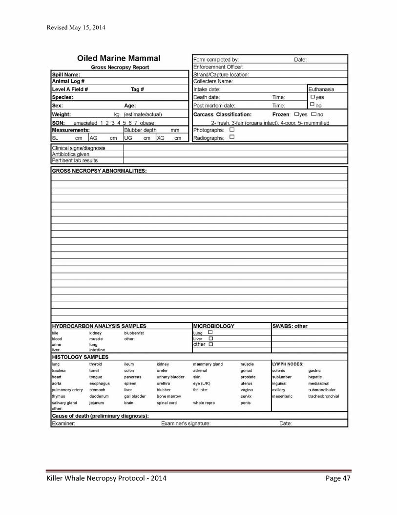
Revised May 15, 2014
Killer Whale Necropsy Protocol -‐ 2014 Page 47
Revised May 15, 2014
Killer Whale Necropsy Protocol -‐ 2014 Page 48
APPENDIX XIV: Considerations and sampling for live stranded killer whales
Killer whales can and do strand live. When this happens, the events can be single strandings or associated with mass stranding events. In either situation, steps should be taken to assure:
1. Safety of humans in the area 2. Safety and humane care for the whales 3. Collection of scientific samples/data as feasible 4. Protection of wild populations
Management of live stranded animals includes efforts such as cooling, wetting, providing shade and even assuring proper orientation of the animal on its ventrum to facilitate breathing. The response efforts for killer whales are of a great magnitude owing simply to the size of many killer whales. The first step in managing such an event is to contact the local authorities that work with marine mammals. A team approach is generally the best first step to assure a positive result. Overall management of live strandings is beyond the scope of this work. However, general information is available through the Geraci and Lousbury’s 2005 book Marine Mammals Ashore. Two main considerations for sample collection
1. Safety – killer whales are large and strong. Even when beached, these animals have the ability to swing wildly and rapidly. This should always be considered first and foremost
2. Reasonability – If the animal is about to die or will be euthanized, many of these samples can be collected post-‐mortem from the fresh carcass.
Depending on the animal’s physical condition, data and samples that can be collected include:
1. Morphometrics – specifically total length, dorsal fin height, pectoral fin characteristics
3. Image capture (Photos) including evidence of human interaction – nets, lines, ship strike injuries, prop wounds
4. Skin sample collection via scrape for genetics 5. Blood collection – ideal samples are approximately 50mLs and include 3 mLs for a
CBC and the rest of the blood is allowed to clot and separated to collect serum. Serum should evaluated for blood chemistries and hormones and the remaining material should be banked for additional evaluations.
6. If possible, fresh whole blood should be collected for immunology studies – see research requests – Jeff Stott’s lab.
7. Lesion sampling – scrapes, swabs for microbiology and molecular analysis A note on euthanasia: Unfortunately, circumstances arise where euthanasia is necessary for stranded animals. These include:
• Disabling injuries such as boat strike or penetrating wounds
Revised May 15, 2014
Killer Whale Necropsy Protocol -‐ 2014 Page 49
• Significant hemorrhage • Unresponsive hyperthermia • Massive skin blistering and/or sloughing • Loss of reflexes or muscle tone
Important considerations for euthanasia are that the process be quick and complete with minimal discomfort for the animal in question and safe for others in the area. Authorities overseeing stranding response may have a specific protocol for euthanasia and should be contacted. In some cases, allowing the animal to die naturally may be more humane than intervening. Final considerations are that the effort minimally impact scientific specimens and that the process be completed in such a manner as to appear professional to all public onlookers. Ballistics may be used by trained individuals to avoid drug residue problems but are not recommended for larger killer whale. If such a manner of euthanasia is selected as best, the animal should first be curtained off to prevent public viewing. The site of the bullet entry is critical (see Harms et al., 2014 – below). Routine euthanasia with barbiturate overdose is feasible for killer whales. Drug volume requirements should be carefully determined for animal size. Newborn killer whales are approximately 300-‐400 pounds. Adults can weigh 10,000-‐16,000 pounds. Drug residues including impacts on scavengers and long half-‐life in the aquatic environment are a consideration. Carcass disposal options may make barbiturate administration an unreasonable manner of euthanasia. If barbiturates are used, the preferred manner of administration is via cardiac stick following premedication with a sedative (Geraci and Lounsbury, 2005). Attempts to use the tail or peripheral veins may be thwarted by vascular shunting or shock. Without the use of a premedicating sedative, the animal may begin to rhythmically raise and lower the tail in a swimming motion. This motion, “flurrying” can propel the animal from its original position. Given the large size of killer whales, methods developed for baleen whales may be more appropriate for this species. Alternate methods of euthanasia employ tranquilization with drugs such as midazolam, acepromazine, and xylazine followed by saturated KCl solution (Harms et al., 2014). Harms, C.A., W. A. McLellan, M. J. Moore, S. G. Barco, E. O. Clarke EO, V. G. Thayer and T. K.
Rowles. 2014. Low residue euthanasia of stranded mysticetes. Journal of Wildlife Diseases 50:63-‐73.
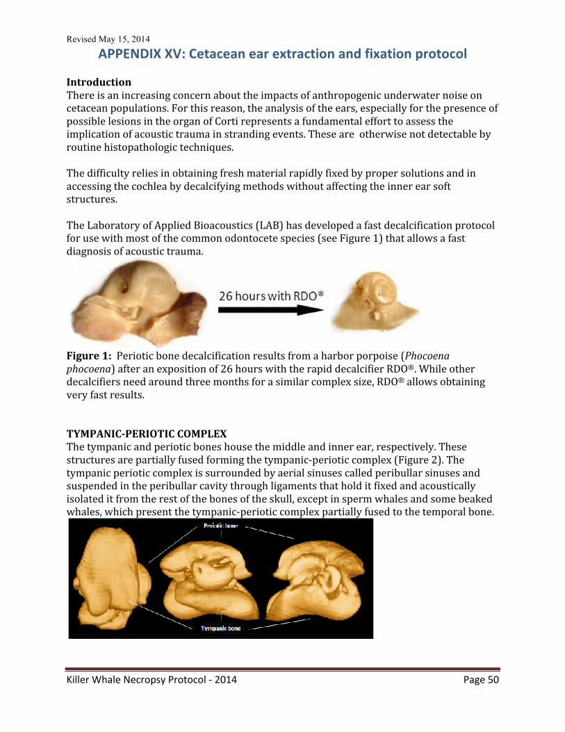
Revised May 15, 2014
Killer Whale Necropsy Protocol -‐ 2014 Page 50
APPENDIX XV: Cetacean ear extraction and fixation protocol Introduction There is an increasing concern about the impacts of anthropogenic underwater noise on cetacean populations. For this reason, the analysis of the ears, especially for the presence of possible lesions in the organ of Corti represents a fundamental effort to assess the implication of acoustic trauma in stranding events. These are otherwise not detectable by routine histopathologic techniques. The difficulty relies in obtaining fresh material rapidly fixed by proper solutions and in accessing the cochlea by decalcifying methods without affecting the inner ear soft structures. The Laboratory of Applied Bioacoustics (LAB) has developed a fast decalcification protocol for use with most of the common odontocete species (see Figure 1) that allows a fast diagnosis of acoustic trauma.
Figure 1: Periotic bone decalcification results from a harbor porpoise (Phocoena phocoena) after an exposition of 26 hours with the rapid decalcifier RDO®. While other decalcifiers need around three months for a similar complex size, RDO® allows obtaining very fast results. TYMPANIC-‐PERIOTIC COMPLEX The tympanic and periotic bones house the middle and inner ear, respectively. These structures are partially fused forming the tympanic-‐periotic complex (Figure 2). The tympanic periotic complex is surrounded by aerial sinuses called peribullar sinuses and suspended in the peribullar cavity through ligaments that hold it fixed and acoustically isolated it from the rest of the bones of the skull, except in sperm whales and some beaked whales, which present the tympanic-‐periotic complex partially fused to the temporal bone.
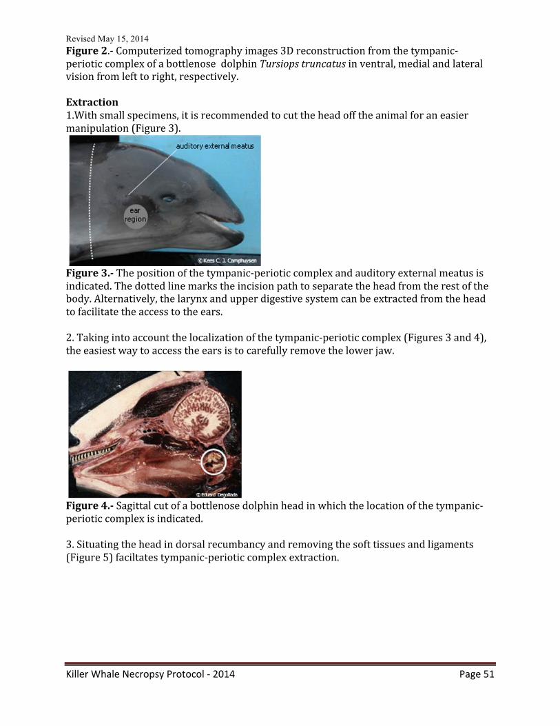
Revised May 15, 2014
Killer Whale Necropsy Protocol -‐ 2014 Page 51
Figure 2.-‐ Computerized tomography images 3D reconstruction from the tympanic-‐periotic complex of a bottlenose dolphin Tursiops truncatus in ventral, medial and lateral vision from left to right, respectively. Extraction 1.With small specimens, it is recommended to cut the head off the animal for an easier manipulation (Figure 3).
Figure 3.-‐ The position of the tympanic-‐periotic complex and auditory external meatus is indicated. The dotted line marks the incision path to separate the head from the rest of the body. Alternatively, the larynx and upper digestive system can be extracted from the head to facilitate the access to the ears. 2. Taking into account the localization of the tympanic-‐periotic complex (Figures 3 and 4), the easiest way to access the ears is to carefully remove the lower jaw.
Figure 4.-‐ Sagittal cut of a bottlenose dolphin head in which the location of the tympanic-‐periotic complex is indicated. 3. Situating the head in dorsal recumbancy and removing the soft tissues and ligaments (Figure 5) faciltates tympanic-‐periotic complex extraction.
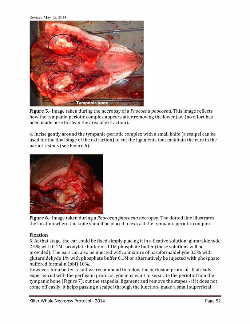
Revised May 15, 2014
Killer Whale Necropsy Protocol -‐ 2014 Page 52
Figure 5.-‐ Image taken during the necropsy of a Phocoena phocoena. This image reflects how the tympanic-‐periotic complex appears after removing the lower jaw (no effort has been made here to clean the area of extraction). 4. Incise gently around the tympanic-‐periotic complex with a small knife (a scalpel can be used for the final stage of the extraction) to cut the ligaments that maintain the ears in the paraotic sinus (see Figure 6).
Figure 6.-‐ Image taken during a Phocoena phocoena necropsy. The dotted line illustrates the location where the knife should be placed to extract the tympanic-‐periotic complex. Fixation 5. At that stage, the ear could be fixed simply placing it in a fixative solution: glutaraldehyde 2.5% with 0.1M cacodylate buffer or 0.1M phosphate buffer (these solutions will be provided). The ears can also be injected with a mixture of paraformaldehyde 0.5% with glutaraldehyde 1% with phosphate buffer 0.1M or alternatively be injected with phosphate buffered formalin (pbf) 10%. However, for a better result we recommend to follow the perfusion protocol.. If already experienced with the perfusion protocol, you may want to separate the periotic from the tympanic bone (Figure 7); cut the stapedial ligament and remove the stapes -‐ if it does not come off easily, it helps passing a scalpel through the junction-‐ make a small superficial
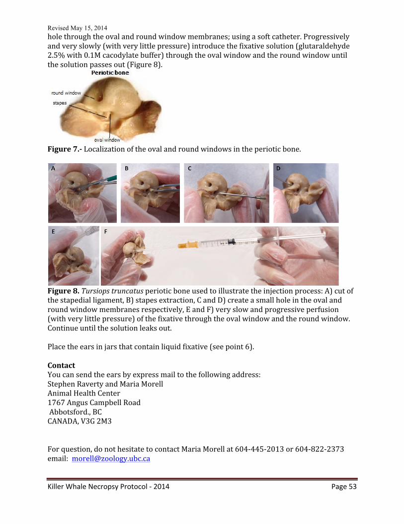
Revised May 15, 2014
Killer Whale Necropsy Protocol -‐ 2014 Page 53
hole through the oval and round window membranes; using a soft catheter. Progressively and very slowly (with very little pressure) introduce the fixative solution (glutaraldehyde 2.5% with 0.1M cacodylate buffer) through the oval window and the round window until the solution passes out (Figure 8).
Figure 7.-‐ Localization of the oval and round windows in the periotic bone.
Figure 8. Tursiops truncatus periotic bone used to illustrate the injection process: A) cut of the stapedial ligament, B) stapes extraction, C and D) create a small hole in the oval and round window membranes respectively, E and F) very slow and progressive perfusion (with very little pressure) of the fixative through the oval window and the round window. Continue until the solution leaks out. Place the ears in jars that contain liquid fixative (see point 6). Contact You can send the ears by express mail to the following address: Stephen Raverty and Maria Morell Animal Health Center 1767 Angus Campbell Road Abbotsford., BC CANADA, V3G 2M3 For question, do not hesitate to contact Maria Morell at 604-‐445-‐2013 or 604-‐822-‐2373 email: [email protected]
Revised May 15, 2014
Killer Whale Necropsy Protocol -‐ 2014 Page 54
APPENDIX XVI: Barotrauma considerations and sampling protocol for gas bubbles
These instructions are a summary of the “protocol for gas sampling and analysis in marine mammals”. For further information please visit the link to this article: http://www.nature.com/protocolexchange/protocols/2299 Material you need:
-‐ 2-‐mL additive free glass tube (Kendall Monoject™ blood collection tube, ref: 301116 ) -‐ BD Vacutainer® one use holder (ref: 364815) -‐ Double pointed needle with a rubber barrier on the tube puncture side (BD
Vacutainer® eclipse™ blood collection needle, ref: 368607). -‐ Disposable insulin syringes (BD Plastipak U-‐100 insulin ref: 329651).
Dissection
1. Carefully remove the skin and blubber minimizing damage to the major subcutaneous veins.
2. Examine the visible and larger subcutaneous veins for bubbles. 3. Take photos of veins with bubbles. 4. Sample bubbles*1. CRITICAL STEP: If pneumothorax is suspected, gas sampling could be done by using the Vacutainer® inserting the double pointed needle in between the ribs*2. Do not open thoracic cavity! 5. Open first the abdominal cavity carefully (try not to cut medium to large size
vessels). 6. Examine the mesenteric and renal veins as well as the lumbo-‐caudal venous plexus
for bubbles. 7. Take photos of bubbles within vessels. 8. Sample bubble’s content “in situ” using the insulin syringes*1. 9. Look for renal subcapsular emphysema. 10. Sample the subcapsular (gas) emphysema in situ using the Vacutainer® 2 . 11. Sample intestinal gases using the Vacutainer® 2. Preferably take at least three
samples from different locations. 12. Open thoracic cavity. If desired, ribs could be disarticulated except the first 3 or 4
cranial ones. These ribs should be cut at 1/3 from the vertebral articulation. 13. Examine the coronary vessels. 14. Take photos of vessels and bubbles. 15. Sample bubbles*1. 16. Follow up with routine necropsy protocol. CRITICAL STEP: do not cut any systemic vein or sample organs until this step is reached. 17. Separate the head from the body. 18. You might disarticulate the mandible to have a better access to the pteryoid sacs. 19. Sample pterygoid sacs using the Vacutainer® *2.
CRITICAL STEP: do not open the sinuses before gas sampling. *1Gas sampling from bubbles in veins
Revised May 15, 2014
Killer Whale Necropsy Protocol -‐ 2014 Page 55
CRITICAL STEP: place the vein under water whenever possible to avoid atmospheric air contamination. 1. Sample each bubble with a new dispensable insulin syringe (BD Plastipak U-‐100
insulin) 2. Inject the content immediately into a new Vacutainer® each time. 3. Label the Vacutainer® with volume recovered and location of the bubble. CRITICAL STEP: Use one new syringe and one new Vacutainer® for each bubble.
*2 Gas sampling from cavities (intestine, pterigoyd air sacs) and gas associated lesions (pneumothorax and subcapsular emphysema)
1. Couple the Vacutainer® plastic holder to the double pointed needle 2. Insert the needle into the cavity 3. Push the Vacutainer® against the other end of the needle 4. Leave for a few seconds 5. Remove the Vacutainer® 6. Remove the needle CRITICAL STEP: If any of these steps is not done following this sequence, atmospheric air contamination will occur.
CRITICAL STEP: If steps from 3-‐13 are not done carefully following this sequence, air contamination will occur.
Storage and transport
1. Store the samples at room temperature and atmospheric pressure. 2. Store blank tubes with the samples; one blank per sample or a minimum of 3 blanks
per animal. 3. If samples need to be transported in a plane, they should travel within the passenger
cabin to prevent dramatic changes in atmospheric pressure that might alter the vacuum tubes, or use a plastic housing resistant to negative pressures (PREVCO™ subsea housing).
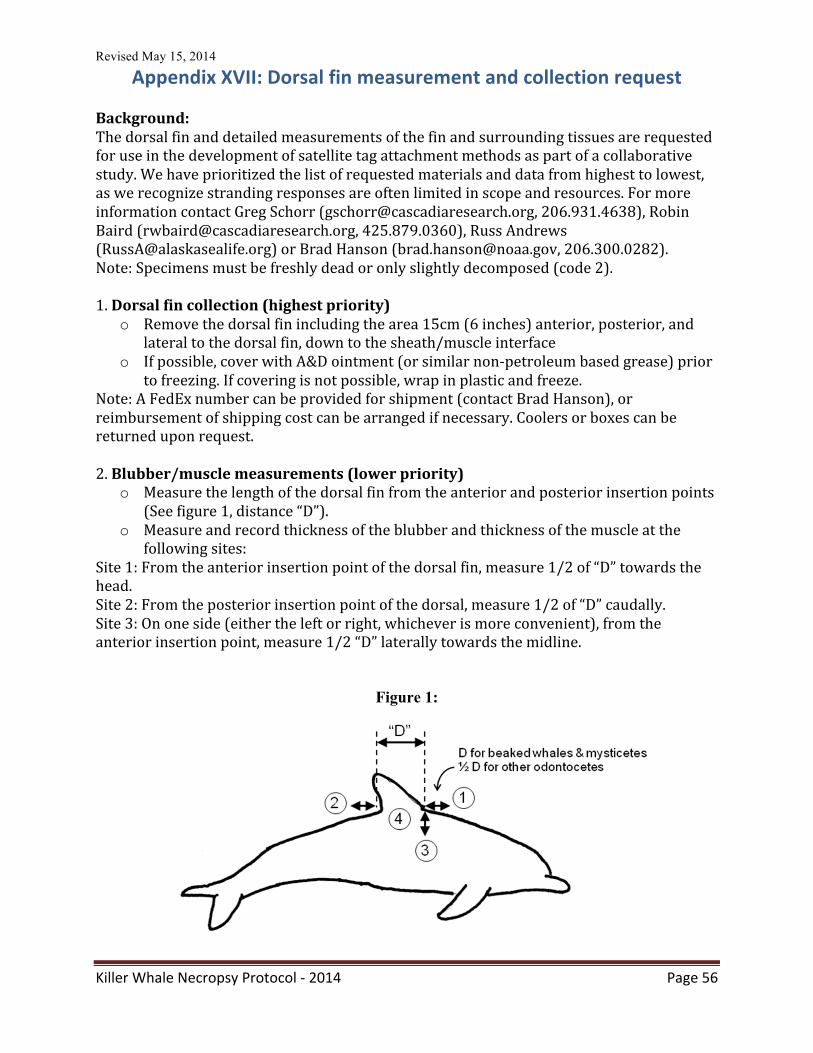
Revised May 15, 2014
Killer Whale Necropsy Protocol -‐ 2014 Page 56
Appendix XVII: Dorsal fin measurement and collection request Background: The dorsal fin and detailed measurements of the fin and surrounding tissues are requested for use in the development of satellite tag attachment methods as part of a collaborative study. We have prioritized the list of requested materials and data from highest to lowest, as we recognize stranding responses are often limited in scope and resources. For more information contact Greg Schorr ([email protected], 206.931.4638), Robin Baird ([email protected], 425.879.0360), Russ Andrews ([email protected]) or Brad Hanson ([email protected], 206.300.0282). Note: Specimens must be freshly dead or only slightly decomposed (code 2). 1. Dorsal fin collection (highest priority)
o Remove the dorsal fin including the area 15cm (6 inches) anterior, posterior, and lateral to the dorsal fin, down to the sheath/muscle interface
o If possible, cover with A&D ointment (or similar non-‐petroleum based grease) prior to freezing. If covering is not possible, wrap in plastic and freeze.
Note: A FedEx number can be provided for shipment (contact Brad Hanson), or reimbursement of shipping cost can be arranged if necessary. Coolers or boxes can be returned upon request. 2. Blubber/muscle measurements (lower priority)
o Measure the length of the dorsal fin from the anterior and posterior insertion points (See figure 1, distance “D”).
o Measure and record thickness of the blubber and thickness of the muscle at the following sites:
Site 1: From the anterior insertion point of the dorsal fin, measure 1/2 of “D” towards the head. Site 2: From the posterior insertion point of the dorsal, measure 1/2 of “D” caudally. Site 3: On one side (either the left or right, whichever is more convenient), from the anterior insertion point, measure 1/2 “D” laterally towards the midline.
Figure 1:
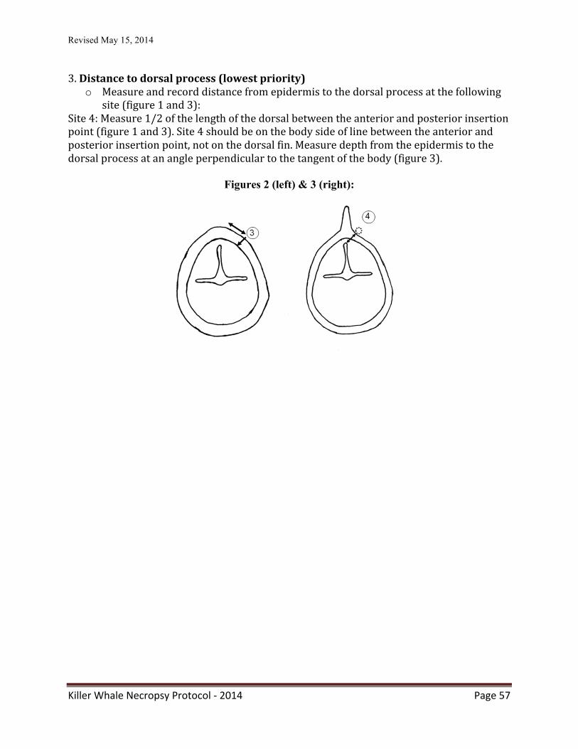
Revised May 15, 2014
Killer Whale Necropsy Protocol -‐ 2014 Page 57
3. Distance to dorsal process (lowest priority)
o Measure and record distance from epidermis to the dorsal process at the following site (figure 1 and 3):
Site 4: Measure 1/2 of the length of the dorsal between the anterior and posterior insertion point (figure 1 and 3). Site 4 should be on the body side of line between the anterior and posterior insertion point, not on the dorsal fin. Measure depth from the epidermis to the dorsal process at an angle perpendicular to the tangent of the body (figure 3).
Figures 2 (left) & 3 (right):
Revised May 15, 2014
Killer Whale Necropsy Protocol -‐ 2014 Page 58
APPENDIX XVIII: Archiving tissue samples Archiving tissues is critical. Despite the attempt to include all desired sampling in this necropsy protocol, inevitably, there will be requests for further testing. Efforts have been made to set up killer whale tissue respositories in the United States and Canada. Both contain -‐80C freezers with back-‐up generators and recording of daily freezer temperatures. Researchers that have collected tissues for archiving and would like to deposit them in a killer whale tissue bank should contact: In the United States: Dr. Brad Hanson, NOAA/NMFS/Northwest Fisheries Science Center, 2725 Montlake Blvd. E, Seattle, WA 98112, Office phone: 206-‐860-‐3220, Fax: 206-‐860-‐3475, Cell Phone 206-‐300-‐0282, Email: [email protected] In Canada: Dr. Stephen Raverty, Animal Health Center 1767 Angus Campbell Road, Abbotsford, BC, Canada V3M 2G3 Phone, work: 604-‐556-‐3003 Phone, work: 800-‐661-‐9903 Email: [email protected]
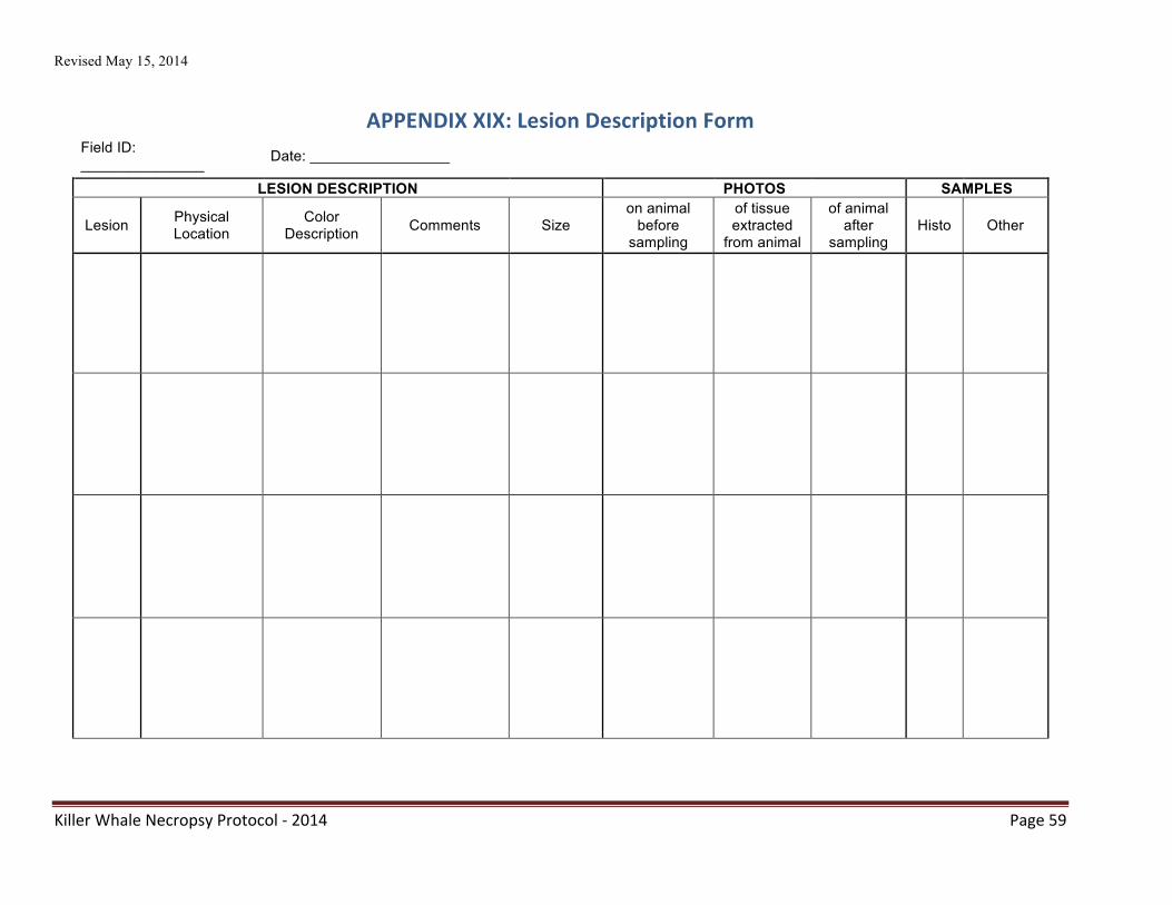
Revised May 15, 2014
Killer Whale Necropsy Protocol -‐ 2014 Page 59
APPENDIX XIX: Lesion Description Form Field ID: _______________ Date: _________________
LESION DESCRIPTION PHOTOS SAMPLES
Lesion Physical Location
Color Description Comments Size
on animal before
sampling
of tissue extracted
from animal
of animal after
sampling Histo Other
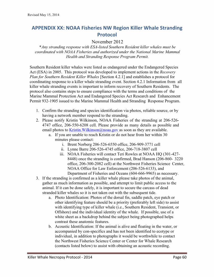
Revised May 15, 2014
Killer Whale Necropsy Protocol -‐ 2014 Page 60
APPENDIX XX: NOAA Fisheries NW Region Killer Whale Stranding Protocol
November 2012 *Any stranding response with ESA-listed Southern Resident killer whales must be
coordinated with NOAA Fisheries and authorized under the National Marine Mammal Health and Stranding Response Program Permit.
Southern Resident killer whales were listed as endangered under the Endangered Species Act (ESA) in 2005. This protocol was developed to implement actions in the Recovery Plan for Southern Resident Killer Whales [Section 4.2.1] and establishes a protocol for coordinating response to a killer whale stranding event. Section 4.2.1 Information from all killer whale stranding events is important to inform recovery of Southern Residents. The protocol also contains steps to ensure compliance with the terms and conditions of the Marine Mammal Protection Act and Endangered Species Act Research and Enhancement Permit 932-1905 issued to the Marine Mammal Health and Stranding Response Program.
1. Confirm the stranding and species identification via photos, reliable source, or by having a network member respond to the stranding.
2. Please notify Kristin Wilkinson, NOAA Fisheries of the stranding at 206-526- 4747 office, 206-550-6208 cell. Please provide as many details as possible and email photos to [email protected] as soon as they are available.
a. If you are unable to reach Kristin or do not hear from her within 30 minutes please contact:
i. Brent Norberg 206-526-6550 office, 206-909-3771 cell ii. Lynne Barre 206-526-4745 office, 206-718-3807 cell
iii. NOAA Fisheries will contact Teri Rowles at NOAA HQ (301-427- 8448) once the stranding is confirmed, Brad Hanson (206-860- 3220 office, 206-300-2082 cell) at the Northwest Fisheries Science Center, NOAA Office for Law Enforcement (206-526-6133), and Department of Fisheries and Oceans (604-666-9965) as necessary.
3. If the stranding is confirmed as a killer whale please take photos of the animal, gather as much information as possible, and attempt to limit public access to the animal. If it can be done safely, it is important to secure the carcass of dead stranded killer whales so it is not taken out with the subsequent tide.
a. Photo Identification: Photos of the dorsal fin, saddle patch, eye patch or other identifying feature should be a priority (preferably left side) to assist with identifying type of killer whale (i.e., Southern Resident, Transient, or Offshore) and the individual identity of the whale. If possible, use of a white sheet as a backdrop behind the subject being photographed helps contrast these anatomic features.
b. Acoustic Identification: If the animal is alive and floating in the water, or accompanied by con-specifics and has not been identified to ecotype or individual, in addition to photographs it would be worthwhile to contact the Northwest Fisheries Science Center or Center for Whale Research (contacts listed below) to assist with obtaining an acoustic recording.
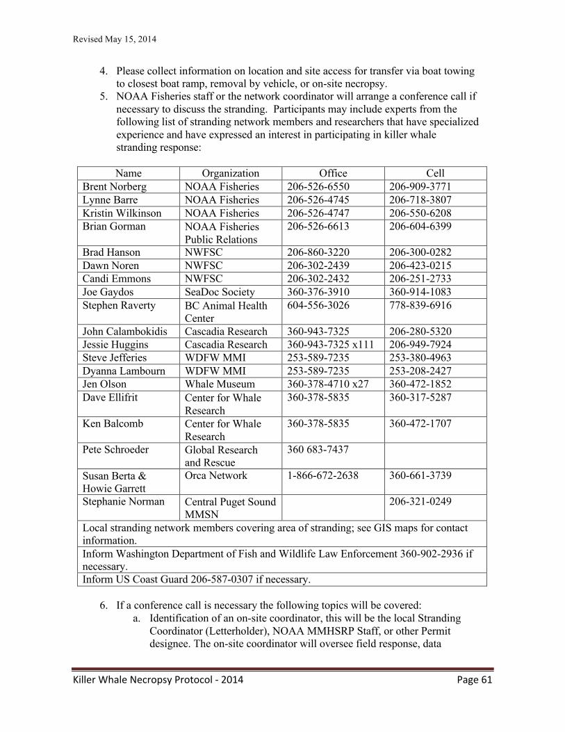
Revised May 15, 2014
Killer Whale Necropsy Protocol -‐ 2014 Page 61
4. Please collect information on location and site access for transfer via boat towing to closest boat ramp, removal by vehicle, or on-site necropsy.
5. NOAA Fisheries staff or the network coordinator will arrange a conference call if necessary to discuss the stranding. Participants may include experts from the following list of stranding network members and researchers that have specialized experience and have expressed an interest in participating in killer whale stranding response:
Name Organization Office Cell
Brent Norberg NOAA Fisheries 206-526-6550 206-909-3771 Lynne Barre NOAA Fisheries 206-526-4745 206-718-3807 Kristin Wilkinson NOAA Fisheries 206-526-4747 206-550-6208 Brian Gorman NOAA Fisheries
Public Relations 206-526-6613 206-604-6399
Brad Hanson NWFSC 206-860-3220 206-300-0282 Dawn Noren NWFSC 206-302-2439 206-423-0215 Candi Emmons NWFSC 206-302-2432 206-251-2733 Joe Gaydos SeaDoc Society 360-376-3910 360-914-1083 Stephen Raverty BC Animal Health
Center 604-556-3026 778-839-6916
John Calambokidis Cascadia Research 360-943-7325 206-280-5320 Jessie Huggins Cascadia Research 360-943-7325 x111 206-949-7924 Steve Jefferies WDFW MMI 253-589-7235 253-380-4963 Dyanna Lambourn WDFW MMI 253-589-7235 253-208-2427 Jen Olson Whale Museum 360-378-4710 x27 360-472-1852 Dave Ellifrit Center for Whale
Research 360-378-5835 360-317-5287
Ken Balcomb Center for Whale Research
360-378-5835 360-472-1707
Pete Schroeder Global Research and Rescue
360 683-7437
Susan Berta & Howie Garrett
Orca Network 1-866-672-2638 360-661-3739
Stephanie Norman Central Puget Sound MMSN
206-321-0249
Local stranding network members covering area of stranding; see GIS maps for contact information. Inform Washington Department of Fish and Wildlife Law Enforcement 360-902-2936 if necessary. Inform US Coast Guard 206-587-0307 if necessary.
6. If a conference call is necessary the following topics will be covered: a. Identification of an on-site coordinator, this will be the local Stranding
Coordinator (Letterholder), NOAA MMHSRP Staff, or other Permit designee. The on-site coordinator will oversee field response, data
Revised May 15, 2014
Killer Whale Necropsy Protocol -‐ 2014 Page 62
collection, and specimen disposition in consultation with the Permit Holder or designated Co-investigator.
i. The On-site coordinator will be responsible for identifying key staff which would include:
1. Lead Veterinarian – Responsible for conducting a health assessment on the animal and overall care during the networks response.
2. Field Logistics – Responsible for responding to the stranding, managing volunteers and their roles at the site, and communicating relevant information to NOAA Fisheries.
3. Data Manager – tracking specimen disposition and samples. Managing all data that is collected during the response and sharing with the appropriate parties.
4. Necropsy Lead (if necessary) – lead and conduct the necropsy of the animal. Work with the data manager on specimen disposition and where samples will be sent for analysis.
b. Identify next steps for response (see Field Response below) c. Communication with interested parties and preparation for media inquiries
7. Field Response a. For a dead animal:
i. A necropsy team will be formed to conduct a full necropsy. The necropsy lead should determine where the necropsy will take place (lab or field), the condition code (fresh, moderate, advanced), and what samples will be collected (including duplicates, samples to be archived, and samples for other researchers). The Killer Whale Necropsy and Disease Testing Protocol should be followed.
1. The protocol can be found at: http://www.vetmed.ucdavis.edu/whc/pdfs/orcanecropsyprot ocol.pdf
2. The protocol provides an equipment checklist, logistics and necropsy recommendations, resources, disease information, etc.
ii. Previous information on dead stranded killer whales is listed in the table below. This information may be requested by the media.
b. For a live animal:
i. Live animal response is logistically complex, potentially dangerous for personnel and the animal, and is expensive. Review the below considerations, determine the level of intervention necessary, and consider what resources your network has to offer for response.
1. Considerations for assessing a live stranded cetacean: a. Human safety is paramount and access to the animal
should be restricted to qualified and authorized individuals.
Revised May 15, 2014
Killer Whale Necropsy Protocol -‐ 2014 Page 63
b. Can the animal be moved from the beach into deeper water on the next high tide? When is the next high tide?
c. Is there any vessel support available to assist? d. Is there a harness available to place the animal in to
tow it out to deeper water? i. Contact at Point Defiance Zoo and
Aquarium is Dr. Karen Wolf at 253-404- 3639 (Office) or 253-381-3115 (Cell)
ii. If a harness is not available one can be made out of Sampson line and some floats. See Figure 6.10 in Marine Mammals Ashore, A Field Guide for Strandings. Geraci & Lounsbury, 2005.
e. If the animal is going to stay on the beach for quite some time, keep the skin wet and if necessary, apply a salve to keep the skin from drying out.
i. An application of zinc oxide will protect skin from sun and windburn and help prevent dehydration. Skin already damaged should be kept moist, shaded, and protected with zinc oxide, antibiotic ointment, or petroleum jelly. (Geraci & Lounsbury, 2005). These products can be purchased at any drug store.
ii. In cold weather provide shelter from wind and precipitation, cover the extremities with a cloth dampened with mineral or vegetable oil. (Geraci & Lounsbury, 2005)
iii. In warm weather or sunny conditions, pay particular attention to keeping the appendages wet to facilitate heat loss (when safe and accessible.)
iv. Collect a blood sample and conduct hematology and clinical chemistry as soon as possible. Viral and bacterial swabs from the blowhole, mouth, rectum, and any lesions are also helpful. Gas analysis and an ultrasound may also be considered. Also consider taking a biopsy sample for genetics analysis.
2. Is there a tracking device available to monitor the animal after release?
3. Network members must coordinate with NMFS/NWR to develop and implement a media plan for disseminating investigation information.
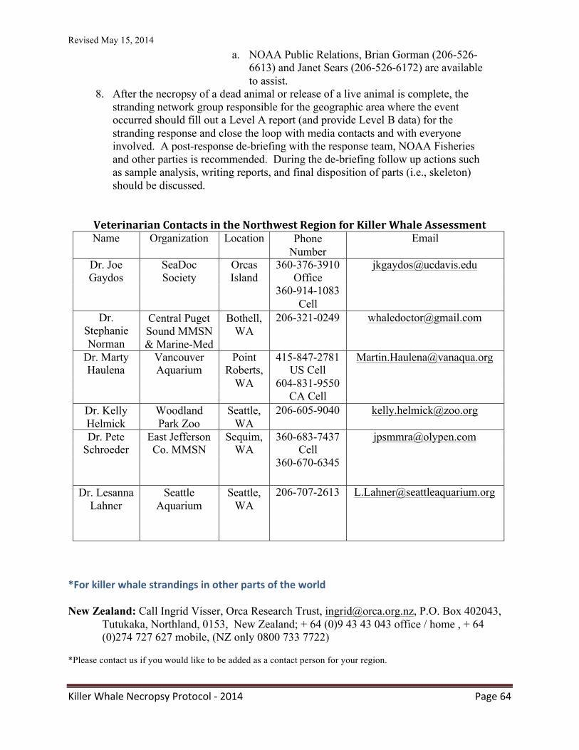
Revised May 15, 2014
Killer Whale Necropsy Protocol -‐ 2014 Page 64
a. NOAA Public Relations, Brian Gorman (206-526- 6613) and Janet Sears (206-526-6172) are available to assist.
8. After the necropsy of a dead animal or release of a live animal is complete, the stranding network group responsible for the geographic area where the event occurred should fill out a Level A report (and provide Level B data) for the stranding response and close the loop with media contacts and with everyone involved. A post-response de-briefing with the response team, NOAA Fisheries and other parties is recommended. During the de-briefing follow up actions such as sample analysis, writing reports, and final disposition of parts (i.e., skeleton) should be discussed.
Veterinarian Contacts in the Northwest Region for Killer Whale Assessment Name Organization Location Phone
Number Email
Dr. Joe SeaDoc Orcas 360-376-3910 [email protected] Gaydos Society Island Office
360-914-1083 Cell
Dr. Stephanie Norman
Central Puget Sound MMSN & Marine-Med
Bothell, WA
206-321-0249 [email protected]
Dr. Marty Vancouver Point 415-847-2781 [email protected] Haulena Aquarium Roberts, US Cell
WA 604-831-9550 CA Cell
Dr. Kelly Helmick
Woodland Park Zoo
Seattle, WA
206-605-9040 [email protected]
Dr. Pete East Jefferson Sequim, 360-683-7437 [email protected] Schroeder Co. MMSN WA Cell
360-670-6345
Dr. Lesanna Lahner
Seattle Aquarium
Seattle, WA
206-707-2613 [email protected]
*For killer whale strandings in other parts of the world New Zealand: Call Ingrid Visser, Orca Research Trust, [email protected], P.O. Box 402043,
Tutukaka, Northland, 0153, New Zealand; + 64 (0)9 43 43 043 office / home , + 64 (0)274 727 627 mobile, (NZ only 0800 733 7722)
*Please contact us if you would like to be added as a contact person for your region.
Revised May 15, 2014
Killer Whale Necropsy Protocol -‐ 2014 Page 65
Appendix XXI: Protocol for examining killer whales for signs of human interaction The following form should be used to examine killer whales for signs of human interaction. If you would like more information on the identification of human interaction in marine mammals or specific details regarding filling this form out, please see the complete paper from which this form originates: Moore, K. T. and S. G. Barco. 2013. Handbook for recognizing, evaluating and documenting
human interaction in stranded cetaceans and pinnipeds. NOAA Technical Memorandum, NOAA-‐TM-‐NMFS-‐SWFSC-‐510, 102 pp.
Available on-‐line at: http://swfsc.noaa.gov/publications/TM/SWFSC/NOAA-‐TM-‐NMFS-‐SWFSC-‐510.pdf
Revised May 15, 2014
Killer Whale Necropsy Protocol -‐ 2014 Page 66
APPENDIX XXII: Fetal examination and sample management
Killer whale fetuses can be encountered as gestational fetuses within dead stranded pregnant females or as abortions or still births. Killer whale pregnancy can last 15-‐18 months. Determining if a small-‐sized animal is a neonate or a fetus can be difficult. Fetal decomposition can occur in utero or after expulsion. A fully developed, full sized animal can be delivered as a stillborn due to fetal death just prior to or at parturition. Because of these complexities, a thorough examination and sample collection is critical for fetal review. A killer whale can be identified as fetus if:
• The developing fetus is present within a gravid uterus or birth canal or the abdomen of an adult female with an internal uterine rupture
• The fetal development is incomplete Findings that suggest but do not confirm the animal as a fetus include:
• The body is less than 2.5m (8.2 feet) in length or 182 kg (400 pounds) in mass. • Gastric content includes amnionic fluid and no milk • Fetal folds are present
Taking a history on a fetus: 1. Maternal information including age, overall health, past pregnancies and their out comes as known 2. Environmental situation including weather and conspecific events – is aggression/trauma a concern? 3. Paternal identity or possibilities 4. Any observed situation related to abortion – time/character of anorexia, contractions, straining, delivery complications, additional cases Fetal/Tissue Examination: 1. Collect placenta, fetus, and amniotic fluid 2. Measure fetal length, weight, the distance between the eyes, note developmental features, estimate degree of decomposition (fresh, mild, moderate, severely deteriorated, mummified) 3. Examine the skin for meconium (green or orange) staining. Likewise, look for tan discoloration of the trachea and bronchi suggesting meconium inhalation. 4. Examine the placenta for completeness of expulsion. If the placenta is presented in sections, try to piece together to form a complete membrane. Weigh, measure the length and # twists in the umbilical cord. Extra support can be found at : http://placentation.ucsd.edu/killerwhalefs.htm Specific points of examination: Note: any abnormalities, photograph and sample for formalin 1. Observe for any malformations or organ abiotrophies (when something does not form)
Revised May 15, 2014
Killer Whale Necropsy Protocol -‐ 2014 Page 67
2. Observe for any swelling/edema or hemorrhage – especially in the head/neck region that might suggest dystocia 3. Specifically observe the umbilicus for swelling/hemorrhage 4. Observe ribs and skull for swelling/fractures/irregularities 5. Observe organs for irregularity, firmness, necrosis, meconium staining, or abscesses 6. Examine brain for completeness; rule out hydrocephalus 7. Observe placenta for thickening, thinning, or discoloration 8. Specifically note the character of gastric contents – amnionic fluid versus curdled milk Collection of Fetal samples for freezing/ancillary testing: Abdominal/thoracic fluid (5 ml) Blood (3-‐5ml) Brain Gastric fluid (5-‐10 ml) Kidney Liver Lung Pericardial fluid (3ml)
Spleen Thymus Tissue pool (liver, spleen, lung, brain) in viral transport media Tissue pool (liver, spleen, lung, brain) in RNA later Umbilical cord/placenta
Fetal samples for culture: Stomach content – aerobic, anaerobic, fungal, and Campylobacter sp. cultures Liver – aerobic culture Lung -‐ aerobic culture Umbilicus – aerobic Other cultures as indicated by gross findings Fetal samples for 10% neutral buffered formalin: brain bladder colon/rectum gonads esophagus heart intestines kidney larynx liver lung pituitary gland lymph nodes
parathyroid placenta (see exam details below) skeletal muscle skin spleen stomach trachea thymus thyroid tonsil umbilicus
Placental exam: 1. Save sections of placenta, amnionic sac, and umbilicus in 10% neutral buffered formalin 2. Save two 10 x 10cm sections of placenta frozen in whirl paks 3. Save small section (1cm x 1cm – cut into fragments) of placenta in RNA later and freeze 4. Save small section in EM fixative (Gluteraldehyde or Karnovski’s solution)
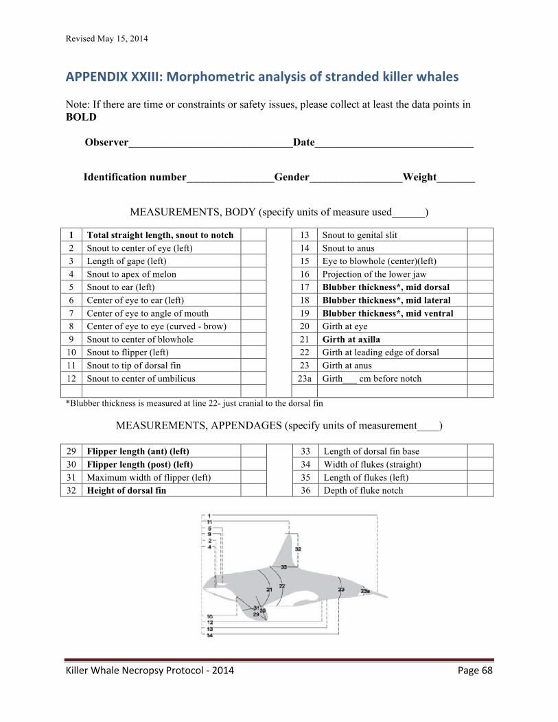
Revised May 15, 2014
Killer Whale Necropsy Protocol -‐ 2014 Page 68
APPENDIX XXIII: Morphometric analysis of stranded killer whales Note: If there are time or constraints or safety issues, please collect at least the data points in BOLD
Observer______________________________Date_____________________________
Identification number________________Gender_________________Weight_______
MEASUREMENTS, BODY (specify units of measure used______)
1 Total straight length, snout to notch 13 Snout to genital slit 2 Snout to center of eye (left) 14 Snout to anus 3 Length of gape (left) 15 Eye to blowhole (center)(left) 4 Snout to apex of melon 16 Projection of the lower jaw 5 Snout to ear (left) 17 Blubber thickness*, mid dorsal 6 Center of eye to ear (left) 18 Blubber thickness*, mid lateral 7 Center of eye to angle of mouth 19 Blubber thickness*, mid ventral 8 Center of eye to eye (curved - brow) 20 Girth at eye 9 Snout to center of blowhole 21 Girth at axilla
10 Snout to flipper (left) 22 Girth at leading edge of dorsal 11 Snout to tip of dorsal fin 23 Girth at anus 12 Snout to center of umbilicus 23a Girth___ cm before notch
*Blubber thickness is measured at line 22- just cranial to the dorsal fin
MEASUREMENTS, APPENDAGES (specify units of measurement____)
29 Flipper length (ant) (left) 33 Length of dorsal fin base 30 Flipper length (post) (left) 34 Width of flukes (straight) 31 Maximum width of flipper (left) 35 Length of flukes (left) 32 Height of dorsal fin 36 Depth of fluke notch
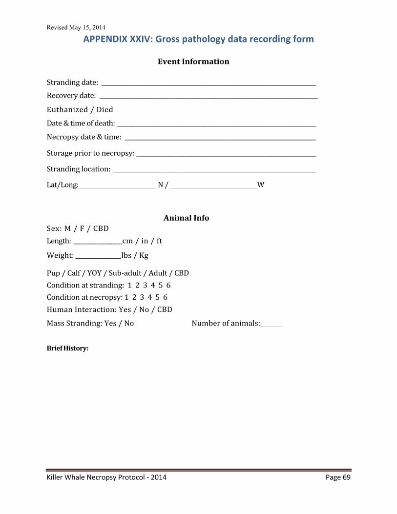
Revised May 15, 2014
Killer Whale Necropsy Protocol -‐ 2014 Page 69
APPENDIX XXIV: Gross pathology data recording form
Event Information
Stranding date: __________________________________________________________________________
Recovery date: _____________________________________________________________________________
Euthanized / Died
Date & time of death: ________________________________________________________________________
Necropsy date & time: __________________________________________________________________ Storage prior to necropsy: ______________________________________________________________ Stranding location: ______________________________________________________________________ Lat/Long: N / W
Animal Info
Sex: M / F / CBD Length: __________________ cm / in / ft
Weight: _______________ lbs / Kg Pup / Calf / YOY / Sub-‐adult / Adult / CBD
Condition at stranding: 1 2 3 4 5 6
Condition at necropsy: 1 2 3 4 5 6 Human Interaction: Yes / No / CBD
Mass Stranding: Yes / No Number of animals:
Brief History:
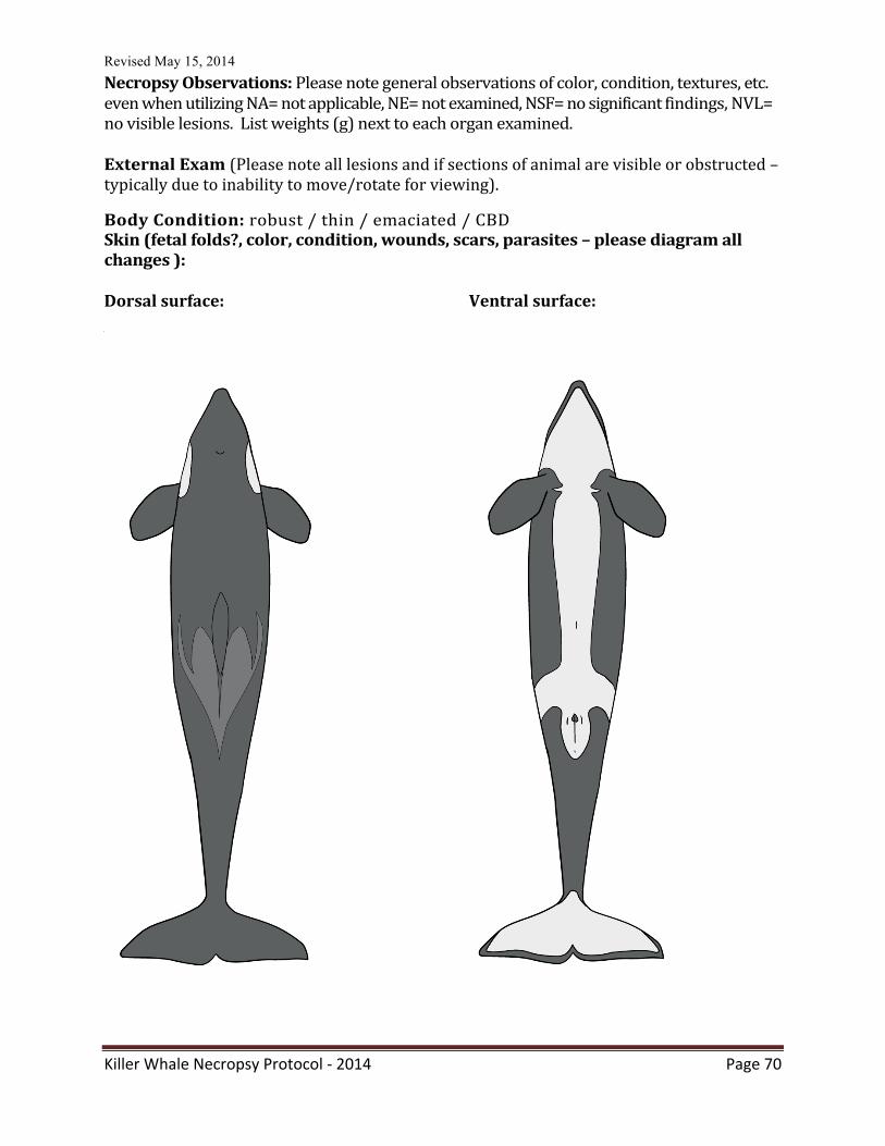
Revised May 15, 2014
Killer Whale Necropsy Protocol -‐ 2014 Page 70
Necropsy Observations: Please note general observations of color, condition, textures, etc. even when utilizing NA= not applicable, NE= not examined, NSF= no significant findings, NVL= no visible lesions. List weights (g) next to each organ examined. External Exam (Please note all lesions and if sections of animal are visible or obstructed – typically due to inability to move/rotate for viewing).
Body Condition: robust / thin / emaciated / CBD Skin (fetal folds?, color, condition, wounds, scars, parasites – please diagram all changes ): Dorsal surface: Ventral surface:
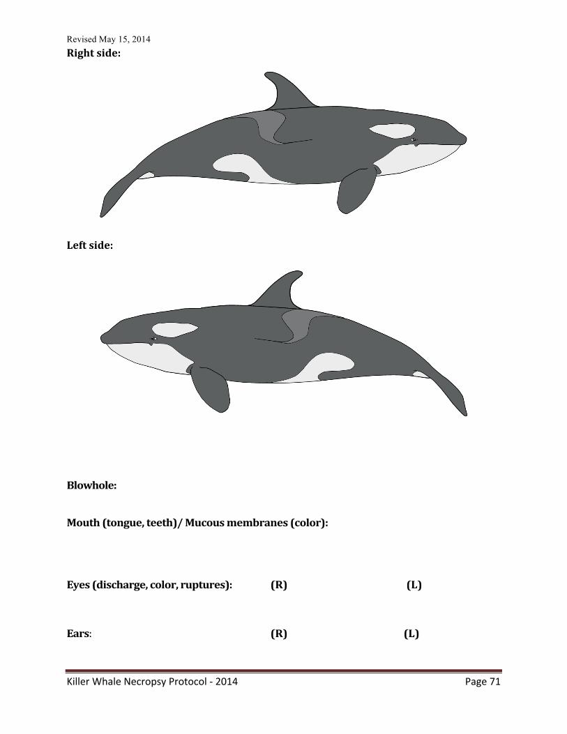
Revised May 15, 2014
Killer Whale Necropsy Protocol -‐ 2014 Page 71
Right side:
Left side:
Blowhole:
Mouth (tongue, teeth)/ Mucous membranes (color):
Eyes (discharge, color, ruptures): (R) (L)
Ears: (R) (L)
Revised May 15, 2014
Killer Whale Necropsy Protocol -‐ 2014 Page 72
Genital slit/anus/mammary openings/umbilicus:
Musculoskeletal system (bones, joints, muscles) Blubber:
Diaphragm:
Skeletal:
Circulatory System Pericardium:
Heart: (weight -‐ kg)
Vessels: Pulmonary System Larynx:
Trachea:
Bronchi:
Revised May 15, 2014
Killer Whale Necropsy Protocol -‐ 2014 Page 73
Lungs (color, condition, edema, congestion, consolidation, granulomas, emphysema, lesions): (R)(Weight kg)
(L)(Weight kg)
Tracheobronchial Lymph: Gastrointestinal System Esophagus: Stomach (contents, ulcers, mucosa, parasites): Small Intestine: Large Intestine/colon/anus: Peritoneum, mesentery, omentum: Hepatic / Pancreatic Liver (weight kg, color, congestion, lesions, size): Bile Duct / Pancreaticoduodenal duct (color, amount):
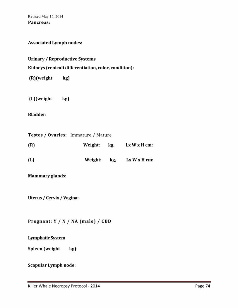
Revised May 15, 2014
Killer Whale Necropsy Protocol -‐ 2014 Page 74
Pancreas:
Associated Lymph nodes:
Urinary / Reproductive Systems Kidneys (reniculi differentiation, color, condition):
(R)(weight kg)
(L)(weight kg)
Bladder:
Testes / Ovaries: Immature / Mature
(R) Weight: kg, Lx W x H cm:
(L) Weight: kg, Lx W x H cm: Mammary glands: Uterus / Cervix / Vagina: Pregnant: Y / N / NA (male) / CBD Lymphatic System Spleen (weight kg): Scapular Lymph node:
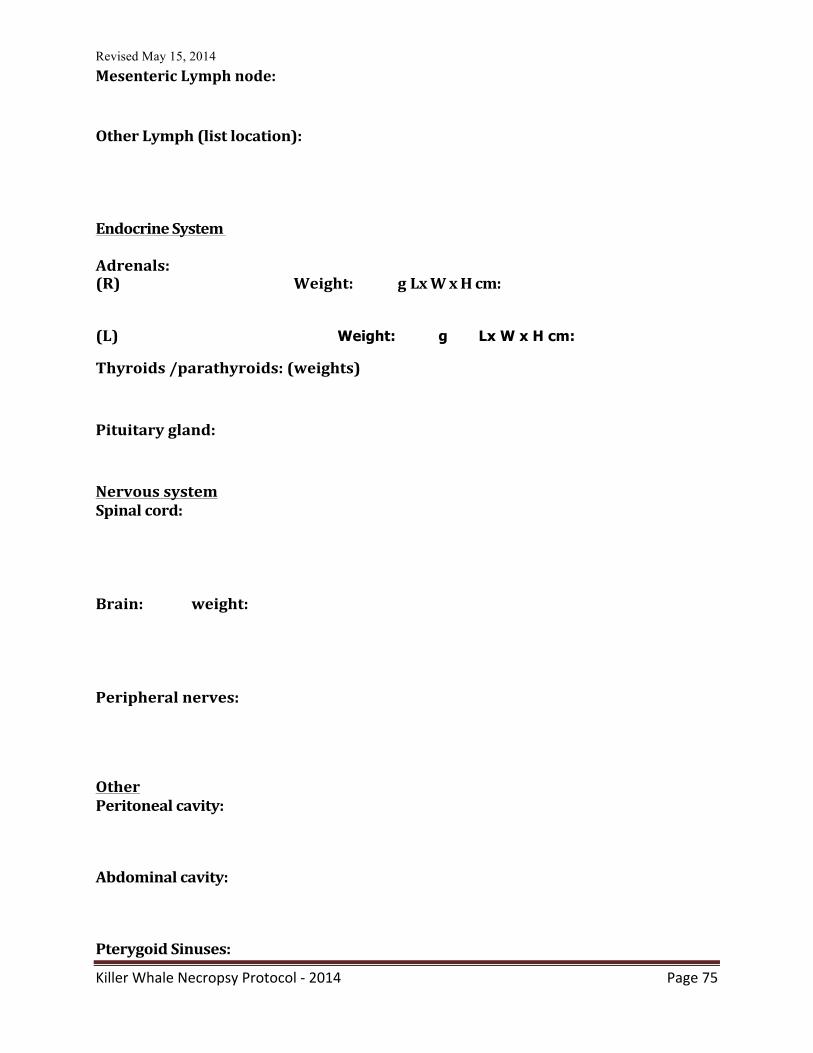
Revised May 15, 2014
Killer Whale Necropsy Protocol -‐ 2014 Page 75
Mesenteric Lymph node:
Other Lymph (list location): Endocrine System
Adrenals: (R) Weight: g Lx W x H cm:
(L) Weight: g Lx W x H cm:
Thyroids /parathyroids: (weights)
Pituitary gland:
Nervous system Spinal cord: Brain: weight: Peripheral nerves: Other Peritoneal cavity: Abdominal cavity: Pterygoid Sinuses:
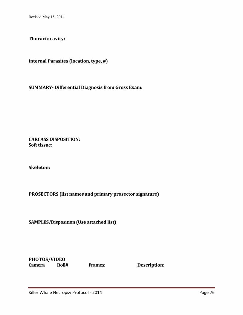
Revised May 15, 2014
Killer Whale Necropsy Protocol -‐ 2014 Page 76
Thoracic cavity: Internal Parasites (location, type, #)
SUMMARY-‐ Differential Diagnosis from Gross Exam: CARCASS DISPOSITION: Soft tissue: Skeleton: PROSECTORS (list names and primary prosector signature) SAMPLES/Disposition (Use attached list)
PHOTOS/VIDEO Camera Roll# Frames: Description:
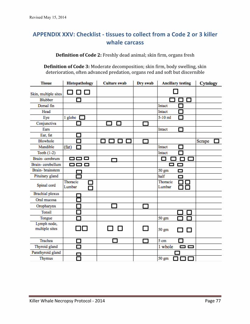
Revised May 15, 2014
Killer Whale Necropsy Protocol -‐ 2014 Page 77
APPENDIX XXV: Checklist -‐ tissues to collect from a Code 2 or 3 killer whale carcass
Definition of Code 2: Freshly dead animal; skin firm, organs fresh
Definition of Code 3: Moderate decomposition; skin firm, body swelling, skin deterioration, often advanced predation, organs red and soft but discernible
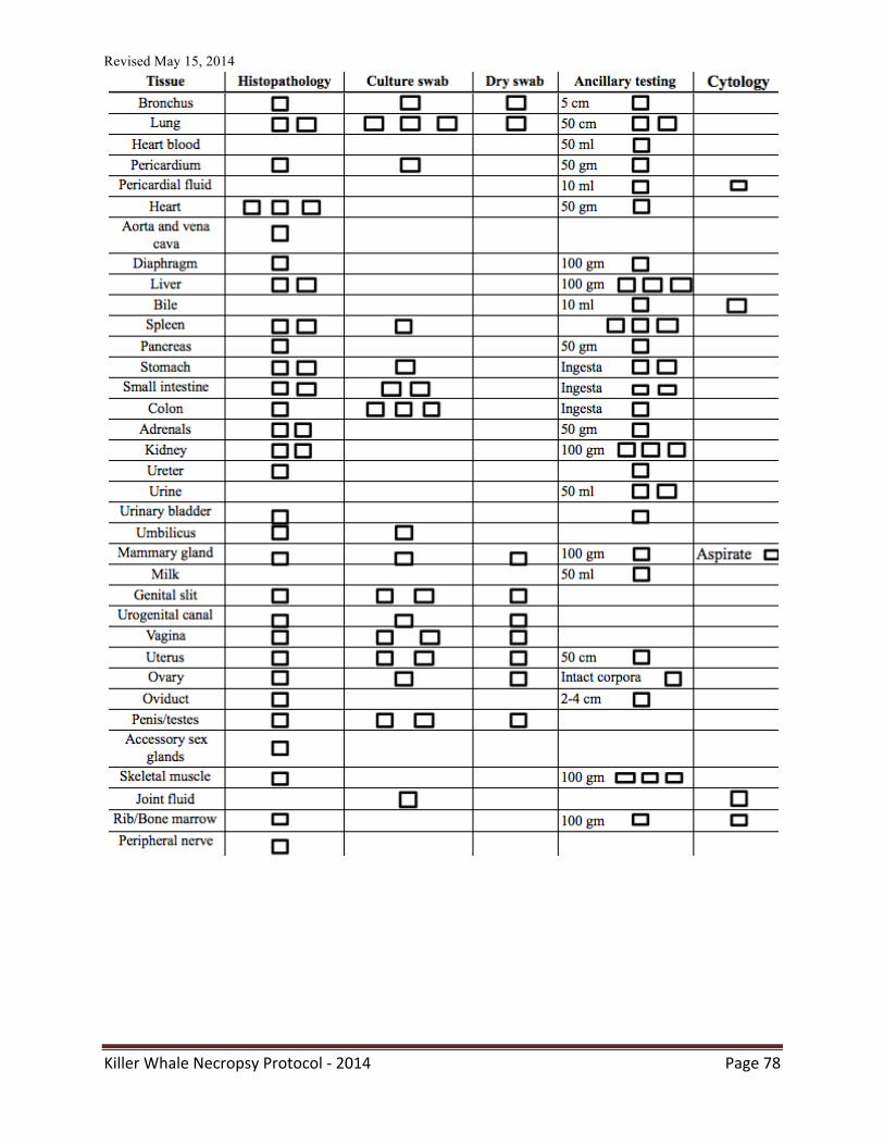
Revised May 15, 2014
Killer Whale Necropsy Protocol -‐ 2014 Page 78
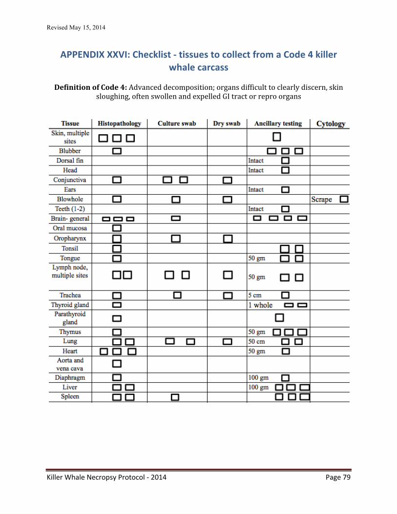
Revised May 15, 2014
Killer Whale Necropsy Protocol -‐ 2014 Page 79
APPENDIX XXVI: Checklist -‐ tissues to collect from a Code 4 killer whale carcass
Definition of Code 4: Advanced decomposition; organs difficult to clearly discern, skin
sloughing, often swollen and expelled GI tract or repro organs
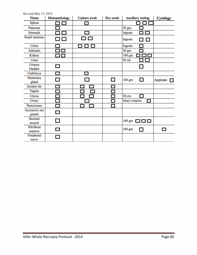
Revised May 15, 2014
Killer Whale Necropsy Protocol -‐ 2014 Page 80
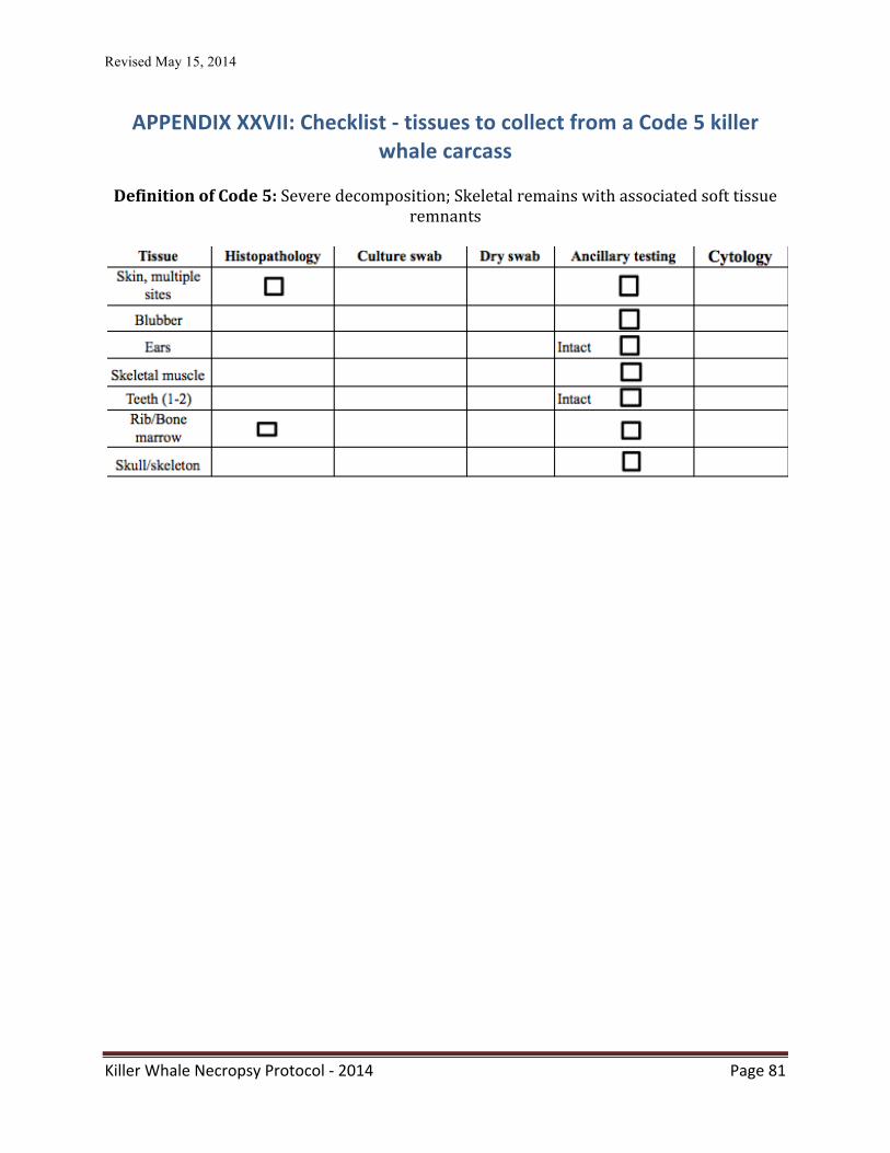
Revised May 15, 2014
Killer Whale Necropsy Protocol -‐ 2014 Page 81
APPENDIX XXVII: Checklist -‐ tissues to collect from a Code 5 killer whale carcass
Definition of Code 5: Severe decomposition; Skeletal remains with associated soft tissue
remnants
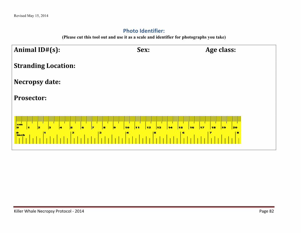
Revised May 15, 2014
Killer Whale Necropsy Protocol -‐ 2014 Page 82
Photo Identifier: (Please cut this tool out and use it as a scale and identifier for photographs you take)
Animal ID#(s): Sex: Age class: Stranding Location: Necropsy date: Prosector:


![Killer Whale [Save the World]](https://static.fdocuments.net/doc/165x107/55aaa5551a28ab783e8b47de/killer-whale-save-the-world.jpg)