Kidney Transplantation Techniques - cdn - InTech
18
Chapter 7 Kidney Transplantation Techniques Farzad Kakaei, Saman Nikeghbalian and Seyed Ali Malekhosseini Additional information is available at the end of the chapter http://dx.doi.org/10.5772/54829 1. Introduction First successful kidney transplantation was done over 60 years ago and now because of major advances in immunosuppressive medicine, this represents the treatment of choice for patients with end-stage renal disease (ESRD). The kidney was the first organ to be transplanted regularly, and it remains the most common organ transplanted today but the surgical technique has changed very little from the original pelvic operation during this long period. In most cases kidney is placed retroperitoneally and the iliac arteries and veins are used for perfusion of this organ and the ureter is transplanted directly to the bladder. But the sophis‐ ticated intensive care units and advanced perioperative anesthetic techniques lead to the use of more marginal donors for more complicated recipients. Now using a kidney graft from a donor after cardiac death or proceeding to kidney transplantation as a part of multivisceral or other abdominal organ transplantation is a routine procedure in the major transplant centers of the world. In such conditions the kidney grafts are not harvested in an optimized preoper‐ ative planning and may be damaged during the surgery. Then we may confront with a graft with 2 or more delicate or very short arteries or veins, ruptured capsule or transected ureter. We may use grafts with congenital anomalies such as horseshoe kidneys or duplicated ureteral system. Also the recipient procedure may be her or his second, third or more transplantation surgery and no more iliac vessels remained for anastomosis and the bladder may be so damaged that makes the anastomosis of the ureter to the bladder impossible. The transplant surgeon should always be ready to conquer such challenges. Using an intraperitoneal space, using the aorta or inferior vena cava or other major arteries and veins such as splenic vessels, and the native ureters for reconstruction of the urine outflow should be an in-hand procedure for every transplant surgeon. © 2013 Kakaei et al.; licensee InTech. This is an open access article distributed under the terms of the Creative Commons Attribution License (http://creativecommons.org/licenses/by/3.0), which permits unrestricted use, distribution, and reproduction in any medium, provided the original work is properly cited.
Transcript of Kidney Transplantation Techniques - cdn - InTech

Chapter 7
Kidney Transplantation Techniques
Farzad Kakaei, Saman Nikeghbalian andSeyed Ali Malekhosseini
Additional information is available at the end of the chapter
http://dx.doi.org/10.5772/54829
1. Introduction
First successful kidney transplantation was done over 60 years ago and now because ofmajor advances in immunosuppressive medicine, this represents the treatment of choicefor patients with end-stage renal disease (ESRD). The kidney was the first organ to betransplanted regularly, and it remains the most common organ transplanted today butthe surgical technique has changed very little from the original pelvic operation duringthis long period.
In most cases kidney is placed retroperitoneally and the iliac arteries and veins are used forperfusion of this organ and the ureter is transplanted directly to the bladder. But the sophis‐ticated intensive care units and advanced perioperative anesthetic techniques lead to the useof more marginal donors for more complicated recipients. Now using a kidney graft from adonor after cardiac death or proceeding to kidney transplantation as a part of multivisceral orother abdominal organ transplantation is a routine procedure in the major transplant centersof the world. In such conditions the kidney grafts are not harvested in an optimized preoper‐ative planning and may be damaged during the surgery. Then we may confront with a graftwith 2 or more delicate or very short arteries or veins, ruptured capsule or transected ureter.We may use grafts with congenital anomalies such as horseshoe kidneys or duplicated ureteralsystem. Also the recipient procedure may be her or his second, third or more transplantationsurgery and no more iliac vessels remained for anastomosis and the bladder may be sodamaged that makes the anastomosis of the ureter to the bladder impossible. The transplantsurgeon should always be ready to conquer such challenges. Using an intraperitoneal space,using the aorta or inferior vena cava or other major arteries and veins such as splenic vessels,and the native ureters for reconstruction of the urine outflow should be an in-hand procedurefor every transplant surgeon.
© 2013 Kakaei et al.; licensee InTech. This is an open access article distributed under the terms of the CreativeCommons Attribution License (http://creativecommons.org/licenses/by/3.0), which permits unrestricted use,distribution, and reproduction in any medium, provided the original work is properly cited.

In this chapter we will review basic steps of the standard approach to recipient’s procedurefrom preparing the graft, then the skin incision till the skin closure with special attention tobasic vascular and urinary tract re-establishment techniques and also intraoperative care ofthe patient. Then we proceed to the special and unusual situations including: complex vascularand ureteral reconstruction techniques, using kidneys with congenital and other anatomicalanomalies, en bloc double kidney transplantation, using other vasculature for transplantingthe kidney in different intraperitoneal spaces, and kidney transplantation conjoint with otherabdominal organs.
2. Graft preparation
Preservation of the viability of the graft during the time between explantation and implantationis vital for early and late graft function after transplantation. Most kidney transplant teamsconsist of at least two separate groups. One group prepares the donor and the other team isdoing the recipient operation at the same time or with some delay depending on the durationneeds for transferring the graft from the donor operating room to the recipient operationtheatre. In many countries such as the United States or in the Euro Zone the kidney grafts fromthe deceased donors are transferred between hospitals, cities or even countries according tothe Human Leukocyte Antigen (HLA) matching or other important criteria for attributing thegraft to a preferred recipient. In such conditions it’s better to use every effort to improve thegraft longevity. Using better preservation solutions or automatic machine perfusion systemsare among the routine measurements in such conditions which are discussed in other chaptersof this book. The surgeons and coordinators should shorten the ischemic time of the graft aslong as possible and during all of this period the temperature of the graft should be maintainedbetween 1-4° centigrade to decrease the injury to the graft.
Simple hypothermia is not enough for preserving the viability of the graft and evacuation of thegraft blood and replacing it with a preservation solution is a mandatory step in the graft prepa‐ration. Graft cold irrigation in the deceased donors is done during the harvesting operation byirrigation of the clamped aorta and the solution used for this irrigation may be any of the pre-prepared solutions such as Belzer University of Wisconsin’s (UW), Histidine-Tryptophan-Ke‐toglutarate (HTK, Bretschneider or Custodiol), Euro-Collins, Celsior or other newer solutionssuch as Biolasol® (Dolińska B, et al, 2012)[1]. Table 1 shows the compositions of some of thesesolutions. All of the blood should be evacuated from the graft during this phase. In the living do‐nor, all of the irrigation is done after removing the graft the donor body in an iced cold basin. Inthe countries that the living donor still forms over 75% of the donor pool such as China or India,irrigation of the living donor graft is done by more simple solutions such as lactated Ringer’ssolution and many studies shows that when the total ischemic time is less than 60 minutes (as inmost living donor programs) the long-term graft survival is not impacted significantly by usingthese simple solutions comparing with more complex solutions (Prasad GS, et al, 2007)[2]. Inour center we add lidocaine (100 mg/liter), sodium bicarbonate (10 meq/liter) and heparin (5000IU/liter) to this simple solution. Also, we use intravenous Mannitol and Furosemide in the do‐nor just before the arterial clamping for better diuresis before nephrectomy.
Current Issues and Future Direction in Kidney Transplantation168

Name Composition Claimed advantages
Belzer UW solution (Viaspan®)
Potassium lactobionate: 100 mmol/lKH2PO4: 25 mmol/lMgSO4: 5 mmol/lRaffinose: 30 mmol/lAdenosine: 5 mmol/lGlutathione: 3 mmol/lAllopurinol: 1 mmol/lHydroxyethyl starch: 50 g/l
Allows for kidney preservation time up to 48 hoursAllows for liver preservation time up to 24 hoursAllows for pancreas preservation time up to 24 hoursProvides enough time to admit patients from distant locationsProvides enough time to improve recipient matchingProvides enough time to operate in a semi-elective situation
Histidine-Tryptophan-Ketoglutarate (Custodiol®)
Sodium chloride: 15 mmol/lPotassium chloride: 9 mmol/lPotassium hydrogen 2-Ketoglutarate: 1mmol/lMagnesium chloride: 4 mmol/lHistidine · HCl:18.0 mmol/lHistidine: 180 mmol/lTryptophan: 2 mmol/lMannitol: 30 mmol/lCalcium chloride: 0.015 mmol/l
Rapid homogenous cooling due to low viscositySuperior recovery of functionExcellent ischemic toleranceVirtual absence of side effectsSimple perfusion technique (ready-to-use, no additives or preparation)
Celsior Mannitol 60 mmol/lLactobionic Acid 80 mmol/lGlutamtic Acid 20 mmol/lHistidine 30 mmol/lCalcium Chloride 0.25 mmol/lPotassium Chloride 15 mmol/lMagnesium Chloride 13 mmol/lSodium Hydroxide 100 mmol/lReduced Glutathione 3 mmol/l
low potassium comparing with UWprevention of tissue edemaprevention of free radical damageprevention of calcium overload with adequate bufferBetter for heart and lung transplantation (as depicted by its manufacturer, Genzyme)
Euro-Collins (Renograf®)
Potassium phosphate 42.5 mmol.lPotassium chloride 15 mmol/lSodium bicarbonate 10 mmol/lAnhydrous glucose 35 g/lMannitol 31.7 mmol/lRaffinose 3.5 mmol/l
Preserves the kidney up to 48 hoursAn out of date solution in most US and European centers
Table 1. Composition of the more common organ preservation solutions.
When possible, the donor team should report the detailed graft anatomy (including numberof arteries, veins and ureters and any anatomical anomaly or inadvertent injury to the graftduring the donor operation) to the recipient team, especially when the graft is transferred fromanother hospital locally or regionally. It is very important to prevent any more injury to thegraft and its capsule, vessels or ureter during the back table procedure, especially in case ofdeceased donor grafts which usually accompanied with other abdominal organs or at leastcovered by the peritoneum or peri-renal fats or other non-important tissues. Direct contact ofthe ice with the graft should be prevented by inserting the graft in a separate basin or organbag filled with a cold solution and then inserting this bag in another iced filled basin.
First of all, for irrigation of the living donor graft, the surgeon should find the artery andcanulate it with an atraumatic olive-headed heparin irrigation needle as shown in figure 1.Using other devices such as Angiocath©, Baranule© or any types of intravenous needles for
Kidney Transplantation Techniqueshttp://dx.doi.org/10.5772/54829
169

irrigation should be discouraged because of risk of intimal injury induced by such cannulas.In many cases, it may be difficult to find the artery first because it is hidden by other hilartissues or retracted to the deeper hilar areas of the graft. In such conditions the irrigation maybe started by canulation the more accessible renal vein, till the surgeon finds the artery. All thedissections should better be done after complete irrigation. At this point all of the renalparenchyma will appear in yellow-pink color. All of the dissections should be done delicatelyby using atraumatic or microvascular instruments, without any more injury to the vesselsintima or their major branches and any more unusual traction of the vessel wall.
Figure 1. Special olive-headed needles for irrigation (Courtesy of GEISTER Medizintechnik GmbH, Tuttlingen/ Germany)
When using the left kidney of the living donor the adrenal and gonadal vein should be on thegraft in order to have a longer vein for future anastomosis. In both right or left kidneys or livingdonor or deceased donor grafts, the surgeon should make every effort to preserve the con‐nective tissues between the ureter and the gonadal vein to prevent ischemic injury to thedelicate collateral vessels of the ureter. Always the ureter should be accompanied by at leastone centimeter of the peri-ureteral tissues and also the hilar inferior triangle (e.g. the windowbetween the inferior pole of the graft and the ureteral origin from the renal pelvis) should bemaintained intact. Removing peri-renal fat or other tissues should be postponed till completerenal revascularization. These tissues are protective for handling of the graft and might be usedfor graft covering or anchoring during or after revascularization.
The window between the renal artery and vein in the renal hilum is full of accessory branchesand lymphatic vessel. All of the major arterial branches especially of the inferior pole should bemaintained intact. Any injury to this branches leads to regional ischemia or necrosis of the kid‐ney or ureter which may lead to future graft dysfunction or ischemia – induced hypertension inthe donor or ureteral necrosis, ureteral anastomosis disruption or urine leakage. Some surgeons
Current Issues and Future Direction in Kidney Transplantation170

suggest that all of the major lymphatic vessels should be ligated to prevent future lymphocele,however, the most important measurement for preventing the lymphocele is avoiding exces‐sive dissections around the iliac artery during the preparing the implantation site.
The best approach for prevention of arterial branch injury is to start with dissection of the renalvein and follow its wall through the hilum until sufficient length is achieved by ligating theminor veins. We suture-ligate the accessory minor vein branches and also the major lumbarveins by 6-0 Prolene suture for prevention of postoperative bleeding from hilar vessels.
If the graft has more than one artery, vein or ureter, the surgeon should decide which type of re‐construction is suitable according to the condition of the graft and the recipient. In the deceaseddonor it’s better to use a Carrel patch of aorta and inferior vena cava in line with the graft ves‐sels. But this has two major impacts on future graft implantation. First, this results in a longerthan usual artery (especially in the right side) or vein (especially in the left side) which may beresults in kinking (and future thrombosis or hypertension) after the anastomosis. And second, itwill results in a large Carrel patch in some cases. The surgeon has to remove a large patch fromthe recipient’s vessels for a good anastomosis. If complicated by graft non-function, then futureremoval of the graft will result in a large defect of the recipient vessels which will be dangerousor even limb life threatening. Also, the Carrel patch of the aorta may be severely atheroscleroticand could not be used for a safe anastomosis. Any reconstruction will elongate the total ische‐mic time of the graft, and we should do every effort to prevent this by postponing unnecessarydissections and reconstructions to the time after at least partial reperfusion of the graft.
According to these important issues, when possible, we prefer to use no reconstruction priorto implantation to decrease the ischemic time. Every transplant surgeon should be fully trainedand familiar with microvascular techniques in such conditions. Every arterial branch shouldbe anastomosed separately. The major artery is anastomosed first usually to the internal iliacartery, which provides a longer arterial conduit and allow more free movements of the graftfor venous anastomosis. Smaller arteries are anastomosed after reperfusion of the graft to theexternal iliac artery or even to the smaller arteries such as inferior epigastric artery (El-SherbinyM, et al, 2008)[3]. When all arterial branches have the same size, then reperfusion is postponedtill the end of anastomosis of all of the arterial branches usually to the external iliac artery butif the kidney has a large artery and some other smaller arteries then reperfusion is started aftercompletion of the large artery anastomosis. Arteries less than 1 mm could be ligated speciallyin the upper pole. Also ligation of the arteries with resultant ischemic area of less than 15% ofthe upper or middle pole is acceptable and by reducing the total operation duration will reducethe complications in the recipient comparing with adding a long microvascular anastomosisto the operation. Arteries larger than 1 mm in the lower pole should be reperfused by anasto‐mosis if possible to prevent ischemia of the ureter.
If the surgeon decides to reconstruct the arteries before implantation then multiple varietiesof techniques could be used: side to side anastomosis of the same size arteries or end to sideanastomosis of a small artery to a larger artery. Using microvascular techniques with a goodillumination and at least 4.5X magnification and 7-0 or 8-0 Prolene sutures, all of the ties shouldbe placed out of the intimal surface and the lumen should be protected by a smooth metalprobe to prevent inadvertent back-wall suturing. In the deceased donor, the surgeon can use
Kidney Transplantation Techniqueshttp://dx.doi.org/10.5772/54829
171

freely every small bifurcated or trifurcated donor artery (such as the celiac artery) for thesedelicate reconstructions. In such complex situations such as severe atherosclerosis of the renalartery orifice when eversion endarterectomy is not possible (Nghiem DD, Choi SS, 1992) [4]or results in a damaged artery, the best approach for salvage of the graft is transecting thediseased part of the renal artery and using a small branch of the donor artery such as the leftgastric or splenic artery as an elongation conduit of the renal artery. In the case of living donors,a short segment of the recipient saphenous vein may be a good choice for this purpose but ithas a real risk of future aneurismal transformation in the future (Sharma A, et al, 2010) [5].Sometimes we could use a combination of these techniques. For example when the graft has2 large-size and 1 small-size artery, the best option is to perform an anastomosis between thesmall-size artery and one of the larger size branches and then perform two separate anasto‐moses in the recipient. This action will reduce the total operative time of the recipient.
Approach to the vein branches is a little different because of intra-parenchymal communica‐tions between the vein branches. We could ligate non-major venous branches, but when thevein branches are in the same size we should reconstruct them before venous anastomosis.Some surgeons prefer to mobilize the external iliac vein by ligating the internal iliac vein orsuperior gluteal vein or other side branches of this vein, but usually these maneuvers are futilein providing better window for venous anastomosis especially when we use the right kidneyfrom a living donor. In such conditions we prefer to perform the venous anastomosis first orplacing the graft in an upside down direction (ureter in the upper part) (Webb J et al, 2003) [6].In the deceased donor, using a part of the donor external iliac, internal jugular or inferior venacava as an extension graft is more preferable for adding the length of the vein graft. Suchreconstructions should be done in the back table prior to implantation.
In our opinion, ureteral reconstruction also should be discouraged in case of multiple graftureters. When the ureters have insufficient length, or denuded in their entire length, mobili‐zation of the recipient bladder or using of the recipient ureter is preferred.
At the end of graft preparation some authors suggest that the graft should be wrapped in icedor cold saline soaked surgical gauzes or cloth stockinet or surgical glove to remain coldthroughout the implantation procedure. In our opinion this is a time consuming and fruitlessmaneuver when the surgeons could do the anastomoses rapidly. Also using the ice packets inthe site of implantation is not necessary.
3. Implantation site
So many factors impact the surgeon’s decision on which site he could implant the kidney graft(table 2). These factors include: the graft size comparing with the recipient, the size, length andnumber of graft arteries, veins and/or ureters, previous surgeries (for example previous failedkidney transplantation, previous pelvic exploration for bladder reconstruction or anti-refluxsurgeries), associated abdominal organ (liver, pancreas or small bowel) transplantation, lateral‐ity of the donor kidney (left or right), anomalies of the donor graft (horseshoe kidney, doublepelvis, double ureter, etc.), and at last the number of kidney grafts (double kidney from a pedia‐
Current Issues and Future Direction in Kidney Transplantation172

tric or old age or marginal donor). Traditionally the right iliac fossa is the standard fossa for akidney transplantation procedure and the left iliac fossa is the preferred site for simultaneouskidney-pancreas transplantation. In the pediatric recipient when the graft is larger than usualwe should use the main abdominal fossa for implantation. The most important limiting factorfor each of these procedures is the length of the renal vein and also the length of the donor ureterand mobility of the recipient urinary bladder. In most instances when the recipient internal iliacartery is used as the arterial inflow, it provides a good length for mobilization and would not bea limiting factor. The right iliac fossa is the preferred site because of the more superficial posi‐tion of the external iliac vein. The deep branches of the iliac vein can be suture ligated and cut ifmore superficialization is needed. If the recipient ureter is not diseased it can be used for urinaryoutflow reconstruction if the donor ureter is short.
Factor Preferred Site Rationale
Graft size comparing with the recipient Abdominal fossa if the graft is verylarge
Prevention of kidney compartmentsyndrome
The size, length and numberof graft arteries and veins
Iliac fossa is preferred Prevention of entering to the abdominalcavity and postoperative ileus
The size, length and number of ureters Iliac fossa is preferred if the recipientureter is not diseased.Retrovesical area if the ureters areshort but vessels are long enough
Prevention of urine leakage or ureteralstricture
Previous surgeries Opposite iliac fossa Prevention of vessel or visceral injury,prevention of lymphocele, shorteroperative time
Associated abdominal organ transplantation Left iliac fossa and in theretroperitoneal spaceAbdominal cavity for en bloc orcomposite grafts
Prevention of adding the complicationsof each graft on the other graft
Laterality ofthe donor kidney (left or right)
It’s better to use right iliac fossa More superficial position of iliac veinSome authors use the opposite sidebecause of position of the transplantedgraft for future percutaneousinterventions on the urinary system
The number of kidney grafts Retroperitoneal space of right iliacfossa
If the iliac arteries are not large enoughit’s better to use the abdominal aortaand inferior vena cava
Anomalies of the donor graft Abdominal cavity if the graft is large,if the graft is small iliac fossa is better.
Enough space for the graft and enoughstations for vascular anastomosis
Table 2. Factors influencing the choice of implantation site
4. Skin preparation and incision
Skin preparation and drape is not so different from other clean abdominal operations. Thepatient should bathe before entering the operation theatre. Hair removal is better done with
Kidney Transplantation Techniqueshttp://dx.doi.org/10.5772/54829
173

hair clippers immediately before surgery. We use scrub povidone iodine or any types ofalcoholic or polyethylene glycol type solutions (e.g. Decocept®) for initial washing and thennormal povidone iodine for 2 times for the final preparation. Also we use a sterile (Opsite®)drape for complete covering of the incision region. The standard skin incision is the traditionalhockey-stick Gibson incision or an oblique Rutherford Morison in the right iliac fossa. Gibsonincision starts at the tubercule of pubis and continued laterally transverse to inguinal ligamentand then upward in a curvilinear manner in the lateral border of the rectus abdominis muscletill 1-2 cm above the level of umbilicus. In larger adults extension till the anterior superior iliacspine may be enough. The epigastric vessels and the round ligament in females usually needto be ligated and transected, but the spermatic cord simply retracted medially by releasing theborder of inguinal canal. The surgeon should avoid entering the peritoneal space and anydefect in the peritoneum should be repaired before continuing the incision.
All the dissections should be accompanied by strict hemostasis and avoiding extreme injuryto the abdominal wall muscles to simplify the future abdominal wall repair at the end of theprocedure. All the bleeding sites should be completely hemostatized during this time becauseat the end of the procedure hemostasis will be very difficult. Also most renal failure patientshas bleeding tendency due to platelet dysfunction specially in the first 2 hours after thehemodialysis or in those patient who underwent preemptive renal transplantation. If hemo‐stasis is not complete wound or peri-graft hematoma is inevitable which will lead to the othercomplications such as infection, dehiscence, hydronephrosis or kidney compartment syn‐drome due to compression to the graft.
After entering the retroperitoneal space and revealing the anatomy of the iliac vessels and theirsuitability for transplantation, the iliac vein should be prepared first by ligating all lymphaticsaround it. It’s better to avoid the first major deep iliac lymph node (Cloquet’s node). Dissectionsaround the external iliac artery should be limited and if the internal iliac artery has a good con‐tour and length, it’s better to use it as the arterial inflow. If this artery has atherosclerotic plaquesan endarterectomy could be done. We use the external iliac artery only when the internal iliac ar‐tery of the other side is used previously, or when a large size discrepancy is revealed or severeatherosclerosis reduce the arterial flow to a very low and crucial level. Using the internal iliac ar‐tery slightly increases the postoperative lymphocele because of more dissections needed for itsreleasing, but if the surgeon ligate all the lymphatics it would not be a major problem.
Without a good exposure, transplantation is a very difficult procedure and using a, Denis-Browne (Figure 2), Kirschner(Figure 3) or Bookwalter-type (Figure 4) self retaining retractor is acritical step in the implantation procedure. Many manufacturers have invented more powerfulretractors. Some of them like Thompson® retractor, although are very useful and unique for liv‐er or kidney-pancreas transplantation, but their use for kidney transplantation alone is timeconsuming and is best limited to super-obese recipients. Some of them such as Henley or Dar‐ling or Gosset abdominal retractor only are useful in pediatric or thin patients with a shallowpelvis. Balfour and Balfour-Baby, Collin and Baby Collin, Ricard and Sullivan- O’Connor havethe same problem. Some of them such as Omni-Flex® (Omni-Tract® surgical, Minnesota Scien‐tific, MN, USA) or SynFrame® retractor systems (Synthes® Spine Inc., PA, USA) are modifica‐tions to the original Thompson retractor but their use may be more sophisticated.
Current Issues and Future Direction in Kidney Transplantation174

Figure 2. Denis-Browne retractor
Figure 3. Kirschner retractor
Figure 4. Bookwalter retractor
Kidney Transplantation Techniqueshttp://dx.doi.org/10.5772/54829
175

5. Vascular anastomosis
After preparation of the place of the implantation, the surgeon should transfer the graftto its position transiently for better evaluation of the anastomoses sites. Some authorssuggest that slush ice should put in the bed of the graft in the recipient, but we abso‐lutely disagree with this opinion, because the total vascular reconstruction time is usual‐ly less than 20 minutes and adding ice only increase the risk of local hypothermic injury.The surgeon should do his best efforts to reduce the total arterial and venous clampingtime. First the site of each anastomosis and the position of the graft should be specifiedaccurately according to the size and length of the vessels and also the length of the ure‐ter and position of the recipient bladder or ureter and the final position of the implantedkidney. As described previously, we prefer to use the internal iliac artery and externaliliac vein for vascular anastomoses. For reducing the vein clamping time (with subse‐quent risk of deep vein thrombosis), we perform the arterial anastomosis first. But whenthe vein is shorter than usual or when the left iliac fossa is used for implantation, orwhen the abdominal cavity and aorta and inferior vena cava or the external iliac arteryare used for implantation, it’s better to perform the venous anastomosis first.
The principles of vascular anastomosis are not different from any standard vascular surgery.The best suture size is usually 5-0 and 6-0 Prolene® sutures for venous and arterial anasto‐mosis. The size of the needles depends of the location of the anastomosis but in most cases theneedle should be taper-point or taper-cutting-tip round-bodied 3/8 circle with 11 – 13 mmlength for better performance. For smaller arteries 7-0 or 8-0, 1/2 circle, 7-9.3 mm needles maybe more suitable. For severe atherosclerotic arteries use of special visible Ethicon Visi-Black®Everpoint®, or Tapercut® needles with spatulated heads which is more firm and crash-resistant is needed.
After confirming the exact length and position of the anastomosis site to prevent kinking orrotation, vascular clamps are applied to the first vessel. We prefer to use Bulldog clamps tothe internal iliac artery and iliac veins and Satinsky clamps for side-clamping of external iliacand common iliac artery or aorta or inferior vena cava. We discourage systemic heparinizationbefore clamping because of bleeding tendency in chronic renal failure patients, but otherauthors recommend this. Heparinized saline is enough for irrigation of the vessels during theanastomosis.
For end-to-side anastomoses a patch from the vessel should be removed for preventing futureconstriction. This patch is removed from the arteries by No. 3, 4 or 5 aortic punches dependingon the arterial size and by special Metzenbaum or Potts scissors from the veins. Also we shouldavoid the venous valve site in the external iliac vein, if possible. The wall of the vein is verythin proximal to the venous valves (sinuses of Valsalva) and may be ruptured during theanastomosis.
For end-to side anastomosis of a renal artery to the external iliac or common iliac or aorta, thegraft artery should be spatulated in the direction of its lower corner. For end-to-end anasto‐mosis of the renal artery to the internal iliac artery, the renal artery should be spatulated from
Current Issues and Future Direction in Kidney Transplantation176
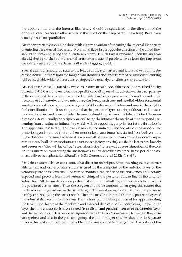
the upper corner and the internal iliac artery should be spatulated in the direction of theopposite lower corner (in other words in the direction the deep part of the artery). Renal veinusually needs no spatulation.
An endarterectomy should be done with extreme caution after cutting the internal iliac arteryor entering the external iliac artery. No intimal flaps in the opposite direction of the blood flowshould be remained at the end of endarterectomy. If such flap is remained, then the surgeonshould decide to change the arterial anastomosis site, if possible, or at least the flap mustcompletely secured to the arterial wall with a tagging U-stitch.
Special attention should be paid to the length of the right artery and left renal vein of the de‐ceased donor. They are both too long for anastomosis and if not trimmed or shortened, kinkingwill be inevitable which will result in postoperative renal dysfunction and hypertension.
Arterial anastomosis is started by two corner stitch in each side of the vessel as described first byCarrel in 1902. Care is taken to include equal bites of all layers of the arterial wall in each passageof the needle and the adventitia remained outside. For this purpose we perform a 1 mm adventi‐tiectomy of both arteries and use microvascular forceps, scissors and needle holders for arterialanastomosis and also recommend using a 4.5-6X loop for magnification and surgical headlightsfor better illumination. It’s so important that the posterior layer suturing of the arterial anasto‐mosis is done first and from outside. The needle should move from inside to outside of the morediseased artery (usually the recipient artery) to tag the intima to the media of the artery and pre‐venting from creating an intimal flap which will be a good trigger point for future thrombosis.The upper suture is tied but the lower is maintained untied till the end of the anastomosis. Theposterior layer is sutured first and then anterior layer anastomosis is started from both corners.In the children or for small arteries at least one half of the anastomosis should be done by sepa‐rate sutures. In all other continuous anastomoses (artery or vein), we tie the last suture looselyand preserve a “Growth factor” or “expansion factor” to prevent purse-string effect of the con‐tinuous suture on constricting the anastomosis as first described by Starzl in the portal anasto‐mosis of liver transplantation (Starzl TE, 1984, Zomorrodi, et al, 2012) [7, 8]) [7].
For vein anastomosis we use a somewhat different technique. After inserting the two cornerstitches, an anchoring or stay suture is used in the midpoint of the anterior layer of thevenotomy site of the external iliac vein to maintain the orifice of the anastomosis site totallyexposed and prevent from inadvertent catching of the posterior suture line in the anteriorsuture line. All the anastomosis is performed circumferentially by a single stitch that used asthe proximal corner stitch. Then the surgeon should be cautious when tying this suture thatthe two remaining part are in the same length. The anastomosis is started from the proximalpart by entering tying the corner stitch. Then the needle is entered from the posterior layer ofthe internal iliac vein into its lumen. Then a four-point technique is used for approximatingthe two intimal layers of the renal vein and external iliac vein. After completing the posteriorlayer then the anastomosis is continued from distal and proximal corner to the anterior layerand the anchoring stitch is removed. Again a “Growth factor” is necessary to prevent the pursestring effect and also in the pediatric group, the anterior layer stitches should be in separatemanner for make future growth possible. If the venotomy site is larger than the orifice of the
Kidney Transplantation Techniqueshttp://dx.doi.org/10.5772/54829
177

renal vein, then after completing the posterior layer, the excessive part should be repairedbefore starting the anterior layer, preferably by another suture line.
6. Unusual situations
In case of thrombosed or fibrotic external iliac vein (due to multiple previous femoral veincanulations or previous DVT) or severe atherosclerotic iliac arteries, the best approach is touse the abdominal major vasculature for renal transplantation. The surgeon may decide to usethe common iliac artery or vein if spared from the disease or close the wound and explore theopposite iliac fossa if preoperative Investigations or intraoperative sonographywere negativefor the same complication. In extreme cases when the IVC is also thrombosed or fibrotic, orwhen the infrarenal aorta also is atretic or severely atherosclerotic, using the splenic or nativerenal vein and artery may be an option, provided that the native ureters has a normal functionand anatomy.
Another unusual case is the horseshoe kidney. Anomalous vasculature is the rule in thesecases. Crossed fused or non-fused ectopic kidneys have the same problem. One option forapproaching this type of anomaly is to incise the ismusth between the two conjoined kidneysand use each kidney for a separate recipient. The major problem is the resultant two graftswith so many arterial and venous branches and also short and multiple ureters. Because ofshortage of donor organs most centers prefer this approach. But sometimes dividing thehorseshoe kidney is so difficult and may results in damaging both kidneys. In these cases it’sbetter to use the anomalous kidney as an individual graft and use the aorta and IVC as thearterial inflow and venous outflow of the graft. Such large size graft often could not be placedretroperitoneally and should be implanted in an intraperitoneal space. The same principle isapplied to double kidney grafts from a pediatric or old age or more marginal donors such asdonation after cardiac death (DCD) donors: transplanting each unit separately or using theaorta and IVC as the vascular conduits of the graft. Circumaortic or retroaortic renal veins areother problematic vascular anomalies that make the transplantation procedure more difficult.In experienced hands, these anomalies per se are not contraindication for donation even fromthe living donors
When a suspicious lesion is found on the kidney graft, it should be incised or excised and sentfor frozen section pathologic investigation. Hemostasis could be done by sutures or argonbeam coagulators, following the principles of any standard partial nephrectomy. Benignlesions should be removed completely and grafts with any non-benign pathology should bediscarded. Solitary cysts are very common and if small, needs no investigation. There are manycase reports in the literature about transplanting kidneys from deceased donors with adultpolycystic kidney disease, without any short-term complications. These grafts should only beused when the donor kidney function is good and the recipient is fully aware of the donordisease. These cases are best suitable for sedentary recipients with a short life expectancy,provided that no other contraindication such as HLA mismatch is found.
Current Issues and Future Direction in Kidney Transplantation178

Kidney transplantation may be accompanied by pancreas, liver (Nadim MK, et al, 2012)[9],heart (Florman S, Kim-Schluger L.,2012) [10], lung (Rana RK, et al, 2011) [11] or multiorgantransplantation. In such situations usually the more important transplantation (heart, lung,liver, pancreas or small bowel) is done first. And after stability of the recipient, kidneytransplantation is performed. Even when the abdomen is entered during the first procedure,it’s better to use the retroperitoneal iliac fossa for the second transplant by the same incision.This will reduce the complications associated with urine leakage. In case of simultaneouskidney –pancreas transplantation the kidney transplant is done first in the left iliac fossa andduring the time of this procedure, the other team prepares the pancreas graft by ex vivo surgeryfor the second transplantation which is usually use the right common or external iliac arteryas the inflow. The kidney transplantation combined with multivisceral transplantation isusually is an en-bloc transplantation. This means that the kidney is not separated from thedonor aorta and inferior vena cava (IVC). All major vascular anastomoses are done by aortaas the inflow artery and IVC and/or portal vein as the venous outflow. The urinary recon‐struction is performed after complete reperfusion of all abdominal organs.
7. Declamping and reperfusion
After completing the vascular anastomoses, the opposite corner stay sutures remained untieduntil reperfusion. The recipient systolic blood pressure should be at least 120 mmHg and thecentral venous pressure between 10 to 14 cm H2O. The use of vasopressors such as dopaminefor increasing the blood pressure is controversial. Immunosuppressant is best infused beforedeclamping according to the protocols of each transplant ward. Some authors suggests someover-hydration, infusing Furosemide and Mannitol and correction of acid-base imbalanceaccording to the last arterial blood gas base deficit before declamping to prevent the so called“reperfusion syndrome”. Unlike liver or small bowel transplantation, in most cases reperfu‐sion syndrome will not be a problematic issue, because the kidney graft is relatively small,except when using an adult kidney for a pediatric recipient or in cases of a long implantationtime with complete aortic or common or external iliac artery clamping time. In such cases thecause of “reperfusion syndrome” is transient ischemia of the lower limbs. The anesthesiologistshould prepare sodium bicarbonate, calcium gluconate, and insulin with 50% Glucose beforedeclamping for managing this complication and obtain an arterial blood gas before and afterthe declamping for estimating the severity of acidosis and monitor the electrocardiogram fordiagnosis of hyperkalemia.
Arterial declamping is done first and after complete filling of the graft, veins are also opened.In this phase brisk bleeding is a rule, especially when we applied “growth factors” to the lastties. Most of the bleeding will be stopped spontaneously after complete dilatation of theanastomotic lines. Small bleeding sites may be covered by small parts of any hemostatic agentsuch as Surgicel®, N-butyl cyanoacrylate glues, Tachosil® or similar agents (Sageshima J, etal, 2011) [13]. All the other larger bleeding sites should be transligated or repaired by fineProlene® sutures especially near the hilum, but extreme caution should be paid not to includethe delicate hilar arterial branches in the sutures.
Kidney Transplantation Techniqueshttp://dx.doi.org/10.5772/54829
179

The kidney should be firm and well-perfused after 1-2 minutes and urine flow usually startsafter that. If the graft is flaccid and the patient’s blood pressure is good, arterial kinking is thefirst differential diagnosis. This usually is resolved by repositioning of the graft. Also thesurgeon could transiently clamp the renal vein or the distal part of the external iliac artery. Ifnot, thrombosis must be considered and ruled out as soon as possible.
8. Urinary reconstruction
After completing the reperfusion stage usually the urine flow is started. Sometimes, especiallyin case of deceased donors or when the nephrectomy has been performed with difficulty inthe living donors, the urine flow will be delayed. If the color and contour of the graft look goodand the arterial and venous flow is good with a well-palpable thrill in the hilum, the surgeonshould proceed to urinary reconstruction.
First of all the urinary bladder should be filled with sterile normal saline serum throughpreviously installed urinary catheter. Some surgeons add 10ml/lit povidone iodine and 80 mg/lit Gentamicin or 500 mg/lit Amikacin to the irrigation fluid for better sterility of the bladder(Salehipour M, et al, 2010) [13] but its effect is controversial. The kidney should be positionedin its final expected place to prevent the tension on the remained ureter before cutting theexcess length of the ureter. It’s better to use the smallest possible length of the ureter to reducefuture ischemic complications. If this step is forgotten the final length of ureter may be shorterthan expected and this will result in kinking of the vasculature and changing the location ofthe kidney from its ideal position.
The surgeon has many options for urinary reconstruction: ureteroneocystostomy, ureter‐oureterostomy, pyeloureterostomy, and pyelocystostomy or even ureteroenterostomy toan ileal conduit or Koch (Manassero F, et al, 2011) [14] or pyelopyelostomy in case of or‐thotopic kidney transplantation or complicated case (Wagner M, et al, 1994) [15]. Thetype of reconstruction depends on the position of the graft, the length, condition andnumber of the donor ureter(s), the condition of the recipient’s bladder or bladder substi‐tute (including its capacity and continence), previous operations on the recipient bladderor ureter (and its antireflux condition). The anastomosis should be done by absorbablesutures, usually polydioxanone sutures. Because of the risk of infection, use of any typesof stents, such as double J stents or newer antireflux stents are controversial (ParapiboonW, et al, 2012) [16], but we use it in our center and remove it after 3 weeks. At least 4techniques and their modifications are discussed in the literature for ureteroneocystosto‐my (Kayler L, et al, 2010) [17]. Prevention of leakage, stricture and reflux is the final goalof all of these techniques. The two most common types are transvesical or Leadbetter-Po‐litano (LP) technique and the extravesical or modified Lich-Gregoir (LG) technique. Weuse and recommend the second technique because it needs fewer dissections and use on‐ly one small cystostomy incision (comparing with 2 large cystostomy incision needs forLP technique) with comparable antireflux characteristics and fewer complications. TheLG technique can be performed in a very shorter time. After distending the bladder, the
Current Issues and Future Direction in Kidney Transplantation180

detrusor muscle dissected bluntly in the dome of the bladder approximately for a lengthof 3 cm till the mucosa bulges out. The ureter shortened to its ideal length and spatulat‐ed for a length of 2 cm in its anti-mesoureteral direction and then the bladder mucosaincised. Anastomosis is started near the heel of the spatulated ureter 2-3 mm in the op‐posite direction of the corner of the ureter. In this manner, the tie is placed outside andwith some distance from the corner. The mucosa of the bladder is then sutured to theureteral end with simple continuous sutures. After completing the anastomosis, an ab‐sorbable suture is used for approximating the detrusor muscle to close over the anasto‐mosis and creating a small submucosal tunnel for its antireflux mechanism. The LPtechniques and the two other extravesical techniques are better described in the literature(Kayler L, et al, 2010) [17]. In the LP technique, a large anterior cystostomy is done forvisualization of the bladder interior and the ureter is transferred through another smallposterior cystostomy and then through the mucosa and after anchoring the distal end tothe mucosa, the bladder is closed in 2 layers with absorbable sutures. Another extravesi‐cal technique is the single or double U-stitch technique. In these techniques after open‐ing the submucosal tunnel by creating by dissection of detrusor muscle and incising thebladder mucosa only 1 U-stitch (Shanfield, 1972) [18] at the toe or 2 U-stitch (MacKinnonet al, 1968) [19] at the toe and heel of the trimmed ureter is used for anchoring the ure‐ter to bladder mucosa and then the detrussor muscle closed as the same manner of theLG technique.
Another extravesical technique uses two parallel incisions in the detrusor muscle, firstposterior for transferring the ureter in a submucosal tunnel and the second incision foranastomosis of the ureter to the ureteral mucosa (Barry JM, 1983) [20]. In the last technique,the ureter is anastomosed to the bladder full-thickness wall without any antireflux mechanism(Starzl, et al, 1989) [21]. In our opinion, the surgeon should be familiar with all of these methodsand use them as needed, but we have the most experience with the modified LG techniquewithout any major urologic complication (Davari HR, et al, 2006) [22].
When the graft ureter is short, ischemic, or denuded, the surgeon should use the native uretersfor ureteroureterostomy or pyeloureterostomy if they are completely in a healthy condition(no stricture, no infection, no dilation or no reflux) or decide to perform a pyeloneocystostomy.This should be done with extreme caution to prevent kinking or pressure on the graft vascu‐lature or repositioning of the graft. A Boari flap or psoas hitch is often necessary in all cases.
In case of previous bladder surgery such as antireflux surgeries or cystoplasty or bladderaugmentation, it’s very important that the site of final urinary reconstruction is fully depictedbefore proceeding with vascular anastomosis, or even before proceeding with nephrectomyin the living donor. Also the blood supply of the tissues used for augmentation should beconsidered. Creating a submucosal flap in the augmented bladder may results in ischemia ofthe tissues used for augmentation and if possible it’s better to use the native bladder area forureteral anastomosis.
In case of double or multiple ureters (such as horseshoe kidneys or en bloc transplantation oftwo kidneys), the ureters can be anastomosed separately to the bladder, or one to the bladderand the shorter ones to the native ureter. Another option is anastomosis of the ureters to each
Kidney Transplantation Techniqueshttp://dx.doi.org/10.5772/54829
181

other and then anastomosis of the conjoined ureter to the bladder. In our opinion usingseparate anastomoses (if possible) reduces the future complications.
9. Wound closure
Wound closure is the final step of the procedure. Closing is done by 2-layer repair of theabdominal muscles (first transverse and internal oblique as one layer and then the externaloblique muscle), by a No. 0 loop Nylon suture. Using any drain before closure is controversialbut if used it should be a closed suction drain such as a Jackson-Pratt drain and every effortshould be used that the drain has no compression effect on the renal vasculature and the ureter.The exit site also should be assessed for bleeding. Every bleeding site should be assessed andrepaired before closure to prevent postoperative hematoma. Diffuse oozing at the end ofoperation may be the result of platelet dysfunction or heparin overdose and should bemanaged accordingly by desmopressin and protamine sulfate, respectively. Excess perirenalfat should be removed, and the graft should be placed in a retroperitoneally created pouchparallel with the psoas muscle, to prevent compression of the kidney between the abdominalwall and the pelvic bones. If the kidney volume is greater than this space, or the renal vascu‐lature or ureter is shorter than usual, then “compartment syndrome” is inevitable is theabdominal muscles repaired in the usual manner. In such situation, the renal artery inflow isgood but the outflow will be disturbed because of pressure of the abdominal wall on the renalvein. Renal venous pressure increase and then the graft will be congested and the urine flowwill decreased. If remained unmanaged, this will eventually lead to decreasing renal arteryflow and finally to renal artery thrombosis and graft loss. If the surgeon could not repositionthe graft in to the supravesical area and anchor it to the abdominal wall without vascularkinking, many other options should be tried. One option is to incise the rectus sheath afterclosing the muscles. Another option is to close the abdominal wall from distal and proximaland let the part which is covering the kidney remains unclosed or closed by an artificial meshwhich is used for hernia repair. The last option is to let abdominal musculature remainedcompletely opened and only covered by the skin. The resultant incisional hernia will berepaired in the future, usually 3 months after the transplantation. The best treatment of suchconditions is “prevention” by matching the size of the donor and recipient and special attentionto the length of the graft vasculature and ureter and also creating the pouch as the first stepduring the procedure.
Author details
Farzad Kakaei1, Saman Nikeghbalian2 and Seyed Ali Malekhosseini2
1 Tabriz University of Medical Sciences, Tabriz, Iran
2 Shiraz University of Medical Sciences, Shiraz, Iran
Current Issues and Future Direction in Kidney Transplantation182

References
[1] Dolińska B, Ostróżka-Cieślik A, Caban A, Cierpka L, Ryszka F. Comparing the effectof Biolasol® and HTK solutions on maintaining proper homeostasis, indicating thekidney storage efficiency prior to transplantation. Ann Transplant. 2012 Jun 29;17(2):74-8.
[2] Prasad GS, Ninan CN, Devasia A, Gnanaraj L, Kekre NS, Gopalakrishnan G.: Is Euro-Collins better than ringer lactate in live related donor renal transplantation? Indian JUrol. 2007 Jul;23(3):265-9.
[3] El-Sherbiny M, Abou-Elela A, Morsy A, Salah M, Foda A. The use of the inferiorepigastric artery for accessory lower polar artery revascularization in live donor renaltransplantation. Int Urol Nephrol. 2008;40(2):283-7.
[4] Nghiem DD, Choi SS. Eversion endarterectomy of the cadaver donor renal artery: amethod to increase the use of elderly donor kidney allografts. J Urol. 1992 Mar;147(3):653-5.
[5] Sharma A, King AL, Lee HM, Posner MP. Saphenous vein graft aneurysm after renaltransplantation: a case report. Transplantation. 2010 May 15;89(9):1162-3.
[6] Webb J, Soomro N, Jaques B, Manas D, Talbot D. The upside down transplant kidney.Clin Transplant. 2003 Oct;17(5):484.
[7] Starzl TE, Iwatsuki S, Shaw BW Jr. A growth factor in fine vascular anastomoses. SurgGynecol Obstet. 1984 Aug;159(2):164-5.
[8] Zomorrodi A, Kakei F, Farshi A, Zomorrodi S (2012) Is Placing an Expansion Space atthe Anastomosing Site of the Vessel for Prevention of Pursiness, Safe? J TransplantTechnol Res 2:113. doi:10.4172/2161-0991.1000113
[9] Nadim MK, Sung RS, Davis CL, Andreoni KA, Biggins SW, Danovitch GM, Feng S,Friedewald JJ, Hong JC, Kellum JA, Kim WR, Lake JR, Melton LB, Pomfret EA, Saab S,Genyk YS Simultaneous Liver-Kidney Transplantation Summit: Current State andFuture Directions. Am J Transplant. 2012 Jul 23.
[10] Florman S, Kim-Schluger L. Organ transplantation update, part II: heart and kidney.Mt Sinai J Med. 2012 May-Jun;79(3):303-4.
[11] Rana RK, Ghandehari S, Falk JA, Simsir SA, Ghaly AS, Cheng W, Cohen JL, Peng A,Czer LS, Schwarz ER, Chaux GE. Successful combined heart-bilateral lung-kidneytransplantation from a same donor to treat severe hypertrophic cardiomyopathy withsecondary pulmonary hypertension and renal failure: case report and review of theliterature. Transplant Proc. 2011 Sep;43(7):2820-6. Review.
[12] Sageshima J, Ciancio G, Uchida K, Romano A, Acun Z, Chen L, Burke GW 3rd.Absorbable cyanoacrylate surgical sealant in kidney transplantation. Transplant Proc.2011 Sep;43(7):2584-6.
Kidney Transplantation Techniqueshttp://dx.doi.org/10.5772/54829
183

[13] Salehipour M, Salahi H, Fathikalajahi A, Mohammadian R, Emadmarvasti V, BahadorA, Nikeghbalian S, Kazemi K, Dehghani M, Malek-Hosseini SA. Is perioperativeintravesically applied antibiotic solution effective in the prophylaxis of urinary tractinfections after renal transplantation? Urol Int. 2010;85(1):66-9. Epub 2010 Mar 17
[14] Manassero F, Di Paola G, Mogorovich A, Giannarini G, Boggi U, Selli C. Orthotopicbladder substitute in renal transplant recipients: experience with Studer technique andliterature review. Transpl Int. 2011 Sep;24(9):943-8. Epub 2011 Jul 1. Review.
[15] Wagner M, Dieckmann KP, Klän R, Fielder U, Offermann G. Rescue of renal transplantswith distal ureteral complications bypyelo-pyelostomy. J Urol. 1994 Mar;151(3):578-81.
[16] Parapiboon W, Ingsathit A, Disthabanchong S, Nongnuch A, Jearanaipreprem A,Charoenthanakit C, Jirasiritham S, Sumethkul V. Impact of early ureteric stent removaland cost-benefit analysis in kidney transplant recipients: results of a randomizedcontrolled study. Transplant Proc. 2012 Apr;44(3):737-9.
[17] Kayler L, Kang D, Molmenti E, Howard R: Kidney transplant ureteroneocystostomytechniques and complications: review of the literature. Transplant Proc. 2010 Jun;42(5):1413-20.
[18] Schanfield I: New experimental methods for implantation of the ureter in bladder andconduit. Transplant Proc 4:637, 1972
[19] MacKinnon KJ, Oliver JA, Morehouse DD, et al: Cadaver renal transplantation:emphasis on urological aspects. J Urol 99:486, 1968
[20] Barry JM: Unstented extravesical ureteroneocystostomy in kidney transplantation. JUrol 129:918, 1983.
[21] Starzl TE, Shapiro R, Tzakis A, et al: A new technique of extravesical ureteroneocys‐tostomy for renal transplantation. Transplant Proc 21:3856, 1989
[22] Davari HR, Yarmohammadi H, Malek-hosseini SA, Salahi H, Bahador A, SalehipourM. Urological complications in 980 consecutive patients with renal transplantation. IntJ Urol. 2006 Oct;13(10):1271-5.
Current Issues and Future Direction in Kidney Transplantation184