Kidney myxosporean parasites in red drum (Sciaenidae) from Florida
8
Vol. 17: 9-16. 1993 DISEASES OF AQUATIC ORGANISMS Dis. aquat. Org. Published October 21 . - - - Kidney myxosporean parasites in red drum Sciaenops ocellatus (Sciaenidae) from Florida, USA, with a description of Parvicapsula renalis n. sp. Jan H. Landsberg Florida Marine Research Institute, State of Florida Department of Natural Resources, 100 Eighth Avenue Southeast. St. Petersburg, Florida 33701-5095. USA ABSTRACT: Two species of myxosporean parasites were found In red drum Sciacnops ocellatus [L.) from Florida, USA. These species were located in the posterior kidney and are confirmed as Henneguya ocellata Iversen & Yokel 1963 and Parvicapsula renalis n. sp. Intraluminal developmental stages and mature spores of P renalis n. sp. were observed. In front view, mature spores were elongate (9.5 X 3.55 to 4.7 pm), asymmetrical and slightly curved, with one valve slightly larger than the other. The pyriform polar capsules (3.0 X 2.0 pm) pointed in towards each other and were situated in a raised 'head' region that appeared to be separated from the spore cavity by a 'membrane' The polar filaments had 5 to 8 evenly spaced coils KEY WORDS: Kidney . Myxosporean . Parasite . Parvicapsula renalis n, sp. . Red drum Sciaenops ocella tus INTRODUCTION The Florida Department of Natural Resources' (FDNR) Florida Marine Research Institute (FMRI) is conducting a long-term research program to deter- mine the feasibility of increasing depleted feral stocks of red drum Sciaenops ocellatus (L.) (Sciaenidae) by releasing, into the wild, fish cultured at the Stock Enhancement Research Facility (SERF) at Palmetto, Florida, USA. In this program, cultured, cultured and tagged-released, and feral fish are routinely screened for parasites so that baseline information can be devel- oped on the differences and similarities in the charac- teristic parasite fauna of the different groups. Some of these parasites could become pathogenic in maricul- ture systems. This study was undertaken as part of the parasite-classification survey and discusses 2 species, Henneguya ocellata Iversen & Yokel 1963 and Parvicapsula renalis n. sp., found in the kidneys of feral red drum. MATERIALS AND METHODS Cultured fish were obtained from SERF during November 1991 and during January, May and July 1992. Cultured, tagged-released fish and feral fish were obtained from Bishops Harbor (BH), Manatee County. Florida, in March, April and July to September 1992 and were obtained monthly from Murray Creek, Volusia County (VC), Florida, from November 1991 to November 1992. Necropsies were performed on 117 fish as part of the parasite-classification survey. Tissue portions of 5 to 10 mm were taken from gill, pseudo- branch, posterior kidney, swim bladder, gall bladder, liver, urinary bladder, stomach, ceca, anterior and pos- terior intestine, spleen and musculature. Fresh squash preparations of these tissues, as well as of skin and fin scrapings and of blood, were examined by light microscopy for myxosporeans. Parallel evaluations of tissues were also made from stained smears or histo- logical preparations. Smears were air-dried, methanol- 0 Inter-Research 1993
Transcript of Kidney myxosporean parasites in red drum (Sciaenidae) from Florida

Vol. 17: 9-16. 1993 DISEASES OF AQUATIC ORGANISMS
Dis. aquat. Org. Published October 21
. - - -
Kidney myxosporean parasites in red drum Sciaenops ocellatus (Sciaenidae) from Florida,
USA, with a description of Parvicapsula renalis n. sp.
Jan H. Landsberg
Florida Marine Research Institute, State of Florida Department of Natural Resources, 100 Eighth Avenue Southeast. St. Petersburg, Florida 33701-5095. USA
ABSTRACT: Two species of myxosporean parasites were found In red drum Sciacnops ocellatus [L . ) from Florida, USA. These species were located in the posterior kidney and are confirmed as Henneguya ocellata Iversen & Yokel 1963 and Parvicapsula renalis n. sp. Intraluminal developmental stages and mature spores of P renalis n. sp. were observed. In front view, mature spores were elongate (9.5 X 3.55 to 4.7 pm), asymmetrical and slightly curved, with one valve slightly larger than the other. The pyriform polar capsules (3.0 X 2.0 pm) pointed in towards each other and were situated in a raised 'head' region that appeared to be separated from the spore cavity by a 'membrane' The polar filaments had 5 to 8 evenly spaced coils
KEY WORDS: Kidney . Myxosporean . Parasite . Parvicapsula renalis n, sp. . Red drum Sciaenops ocella tus
INTRODUCTION
The Florida Department of Natural Resources' (FDNR) Florida Marine Research Institute (FMRI) is conducting a long-term research program to deter- mine the feasibility of increasing depleted feral stocks of red drum Sciaenops ocellatus (L.) (Sciaenidae) by releasing, into the wild, fish cultured a t the Stock Enhancement Research Facility (SERF) at Palmetto, Florida, USA. In this program, cultured, cultured and tagged-released, and feral fish are routinely screened for parasites so that baseline information can be devel- oped on the differences and similarities in the charac- teristic parasite fauna of the different groups. Some of these parasites could become pathogenic in maricul- ture systems. This study was undertaken as part of the parasite-classification survey and discusses 2 species, Henneguya ocellata Iversen & Yokel 1963 and Parvicapsula renalis n. sp., found in the kidneys of feral red drum.
MATERIALS AND METHODS
Cultured fish were obtained from SERF during November 1991 and during January, May and July 1992. Cultured, tagged-released fish and feral fish were obtained from Bishops Harbor (BH), Manatee County. Florida, in March, April and July to September 1992 and were obtained monthly from Murray Creek, Volusia County (VC), Florida, from November 1991 to November 1992. Necropsies were performed on 117 fish as part of the parasite-classification survey. Tissue portions of 5 to 10 mm were taken from gill, pseudo- branch, posterior kidney, swim bladder, gall bladder, liver, urinary bladder, stomach, ceca, anterior and pos- terior intestine, spleen and musculature. Fresh squash preparations of these tissues, a s well as of skin and fin scrapings and of blood, were examined by light microscopy for myxosporeans. Parallel evaluations of tissues were also made from stained smears or histo- logical preparations. Smears were air-dried, methanol-
0 Inter-Research 1993

10 Dis aquat. Org. 17 9-16, 1993
fixed, and stained with Giemsa: 0.1 M phosphate buf- fer (1:1, pH 7.4) for 20 min. Stained slides were rinsed ~71th 0.1 P1 phosphate buffer and air-dried for later evaluation.
Histological slides were made from tissues that were fixed in 5 %paraformaldehyde in 0.1 M phosphate buf- fer, dehydrated in a graded ethanol series, and embed- ded in JB-4 glycol methacrylate resin. Sections were cut to 3.5 pm on an LKB 2218 Historange microtome and were stained in Weigert's hematoxylin and eosin (H&E), in periodic acid Schiff's hematoxylin metanil yellow (Quintero-Hunter et al. 1991), or in thionin stain adapted to glycol methacrylate (P. Nagle & I. Quintero- Hunter, FMRI, unpubl.). Representative samples were A B deposited in the U.S. National Museum (USNM) and in the FDNR Marine Invertebrate Collection at FMRI Fig. 1 Parvicapsula renalis n, sp. Line drawing of mature (FSBC I). spores. (A) Front view; (B) side view. Scale bar = 5.0 pm
5.6 ,pm, ranqe = 3.0 to 10.0 pm) and found only m 1 examined range-(cm) H. ocelldta P renalis n. sp. I
Fresh specimens of Parvicapsula renalis n. sp. were viewed and photomicrographs made with differential interference contrast optics. Measurements of fixed Spore characteristics: Mature.spores were elongate, spores in histological section, taken a t a magnification 9.5 pm long (range 9.0 to 10.0 pm , n = 15), asymmetri- of X1000 and given in Fm, are reported as the mean cal, and slightly curved. One valve was slightly larger and range (in parentheses) for both Henneguya ocel- than the other. The posterior spore was wider (4.7 pm; Iata and P renalis n. sp. Fish standard lengths are range 4.0 to 5.0 pm, n = 11) than the anterior region given in cm. (3.55 pm; range 3.0 to 4.0 pm, n = 11) measured at the
point parallel with the base of the polar capsules. Suture line extended through the anterior pole to-
Description of Parvicapsula renalis n. sp. (Fig. 1) wards, but not necessarily bisecting, the posterior pole. Pyriform polar capsules (3.0 X 2.0 pm) pointed in to-
Host: Red drum Sciaenops ocellatus. wards each other and situated in a raised 'head' region Locality: Atlantic Ocean at Murray Creek, Volusia that appeared to be separated from the spore cavity by
County, Florida. USA (29' 08' N, 80' 53' W) . Salinity a 'membrane'. The polar filaments had 5 to 8 evenly was 22 ppt. spaced coils.
Site of infection: Lumen of posterior kidney proximal tubules. RESULTS
Type slides: Syntype slides of histological sections of posterior kidney stained in hematoxylin and eosin, to- Prevalence levels of Parvicaps~~la renalis n. sp, in red gether with photographic samples, have been depos- drum are reported in Table 1 Salinities in these areas ited in the USNM, Beltsville, Maryland, USA (number ranged from 8.5 to 27.0 ppt. Both developmental stages 82820). Two other syntype histological slides "?lined in thion'n have been deposited in the Table 1 Prevalence and geographic distnbution of red drum FDNR Marine Invertebrate Collection at FMRI, St. kidney myxosporeans Abbreviations as ~n the text Petersburg, Flonda (FSBC l 50409).
the lumen of the kidney tubules. These stages in- I SERF 3 1 3.1-8 8 0.0 cluded primary cells with 1, 2 or 4 nuclei; uni-
0.0 1 Developmental stages: Extrasporogonic stages
were round to oval (n = 22, mean diameter = 5.4 X
nucleate primary cells with 1, 2 or 4 daughter cells; binucleate primary cells with 1, 2, 4 , 6 or 8 daughter cells.
Sporogonic stages: These were oval and con- tained 12 cells wlthin a uninucleate or binucleate pseudoplasrnod~um in the lumen of the kidney
Site No. of Standard Prevalence ("G) red drum length
B H Feral 5 8.2-21.0 0.0 0.0 Tagged 4 11.5-26.0 25.0 0.0
VC Feral 31 2.8-23 5 0.0 6.5 Tagged 46 5.3-17 8 0.0 13.0
tubules.

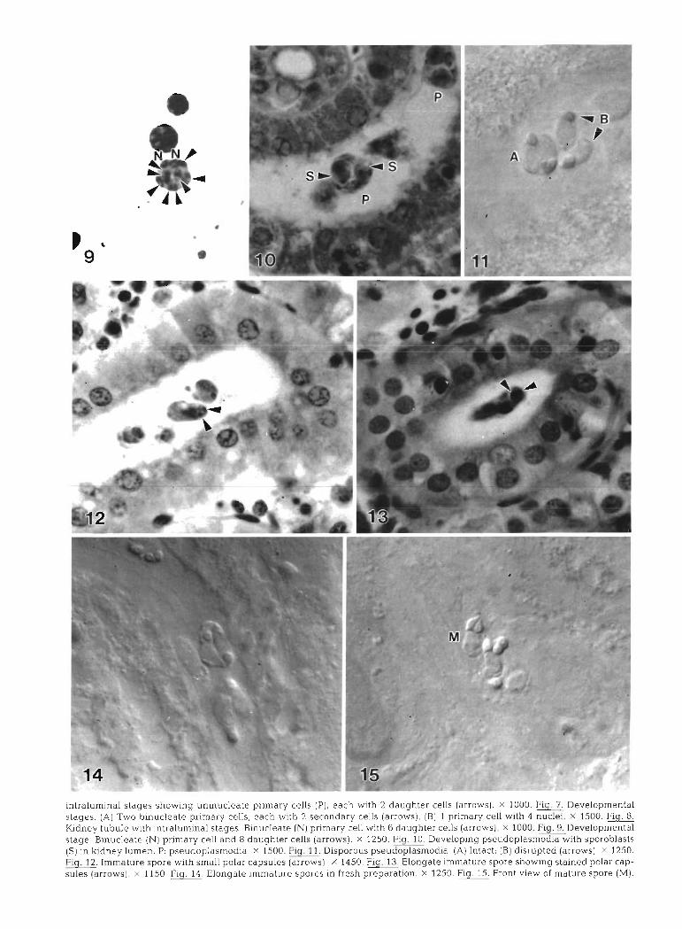
.(M) a~ods ainleur jo Ma!A luoi.~ .md .OSZL X ,uol~eredald qsalj U! salods arn1euIur.r aleBuo.13 rfjrj 'OSI l X .Is~olle) salns -des le~od pau!els but~oqs aiods ainleuu! ale6uola mj .OSP~ x .(s~ol~e) sa~nsdes ieiod [[PUS yl!hz alods alnleurlul .m -OSZ~ X .(s~oi~e) paldn~slp (g) !lseluI (V) .e!potuse1dopnasd snolods~r~ .!d~~~] X .e!pourseldopnasd :d .uamnI daup~y U! (S) slse~qolods ql!~ e!pouseldopnasd 6u1do1a~aa WJ -0~~1 X .(s~olre) SI~J lalgbnep 8 pue IIas Lleurpd (N) aleaIsnu!g .aBels leluaudolanaa.6~ .0001 .(smoi~e) sllas ~alqbnep g qlpz IIas L.~euud (N) alea~snu~g .saBels ~eu~urnle~~u! ~I!M alnqnl Aaupx
.OOSI X '!alsnu P ~IIM nas Aieur!ld I [g) !(SMOJJ~) sI[as Aiepuosas z ql!~ qsea 'sIlas Alemud alealsnujq OML (v) .saliels 1eluaurdolahaa 'L'Ij!g .0001 X .(~h%~ll~) sIlas ~alq6nep z ql!~ qsea '(d) sI1as d~eurud aleaIsnu!un ~UIMO~S sabels Ieu!urnIellu!

14 Dis. aquat. Org 17: 9-16, 1993
and mature spores of P renalis n. sp. were found dur- ing March, June and August, whereas only develop- mental stages were found during September and October. Other samples were negative.
Histological sections of the posterior kidney showed developmental stages and spores of Parvicapsula re- nalis n . sp. in the proximal tubules. In 1 case, kidney hypertrophy was macroscopically detectable, and the kidney was about 3 times the normal size. No parasites were detected in the glomeruli, haematopoietic tissue or blood. In some fish, kidneys infected with intralurni- nal stages showed light to heavy vacuolation in the tubular epithelium (Fig. 2). In some cases, hyaline droplets were predominant in the tubular epithelium (Fig. 3), and in others, large numbers of rodlet cells were apparent.
Extrasporogonic stages
Early parasite stages were identified in impression smears and histological sections of posterior kidney. The frequencies of different types of developing stages identified from 1 impression smear stained in Giemsa are given in Table 2. Stages included primary cells
Table 2. Parvicapsula renaljs n. sp. Frequency and type of developmental stages of P. renalis n. sp. from 1 impression
smear from red drum posterior kidney
Primary cell Daughter cells No. of cells (no. of nuclei)
1 7 2 - 14
I ' Daughter cell released I
were present but were not as common as binucleate primary cells with 1, 2 (Figs. 4 & ?), 4, 6 (Fig. 8) or 8 (either 2 X 4 or 8) (Fig. 9) daughter cells
Sporogonic stages
In later stages of development of the 'pseudo- plasmodia' (primary cell and developing sporoblasts), 2 groups of 6 sporogonic (formerly daughter) cells formed separate sporoblasts (Fig. 10). Pseudoplas- modia usually had a primary cell with 1 or 2 primary nuclei and were disporous (Fig. 11). Immature spores appeared to have small polar capsules (Fig. 12) and were extremely elongate (Figs. 13 & 14).
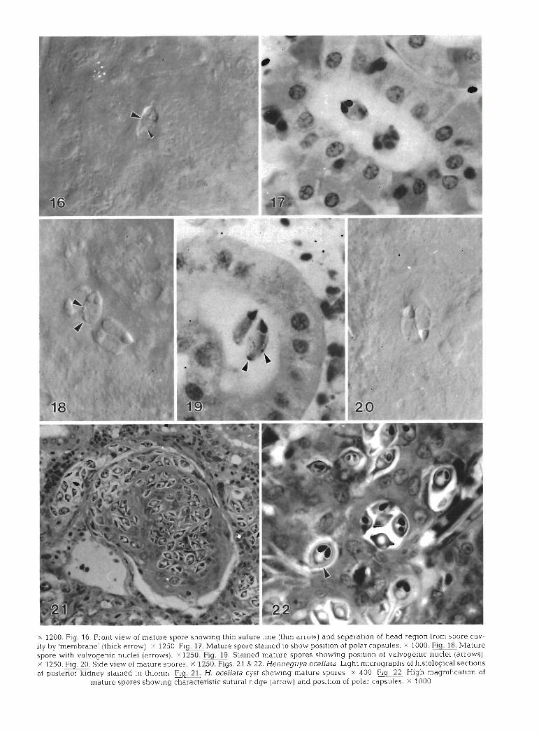
In front view, mature spores were elongate, 9.5 pm long (range 9.0 to 10.0 pm, n = 15), asymmetrical, and slightly curved (Fig. 15). One valve was slightly larger than Lhe olher. The posterior spore was wider (4.7 pm; range 4.0 to 5.0 pm, n = 11) than the anterior region (3.55 vm; range 3.0 to 4.0 km, n = 11) measured at the point parallel with the base of the polar capsules. The barely noticeable suture line extended through the anterior pole towards, but not necessarily bisecting, the posterior pole. In some cases the line appeared to run through the polar capsules (Fig. 16). The polar cap- sules were situated in a raised 'head' region and appeared to be separated from the spore cavity by a 'membrane' (length of cavity to anterior tip = 3.3 ,pm; range 3.0 to 4.0 pm, n = 15) (Fig. 1.6). The pyriform polar capsules had an average length of 3.0 pm (range 2.5 to 3.0 pm, n = 15) and width of 2.0 pm (range 1.5 to 2.0 pm, n = 15) and pointed in towards each other (Fig. 17). The polar filaments appeared to have 5 to 8 evenly spaced coils. The valves were uneven and their nuclei projected into the spore cavity in fresh specimens (Fig. 18). Valvogenic nuclei were also q u ~ t e distinct in thionin-stained spores (Fig. 19). In side view, mature spores were conical, with the posterior end wider (4.25 ,pm; range 4.0 to 5.0 pm, n = 4) than the anterior region (3.0 pm; no range, n = 4) measured at the point parallel wlth the base of the polar capsule (Fig. 20). The sporo- plasm was binucleate.
Henneguya ocellata Iversen & Yokel. 1963
In 1 tagged red drum (SL = 19.0 cm) sampled from with 1 (Fig. 4), 2 or 4 (Fig. 7, needs confirmation by Bishop's Harbor, several histozoic myxosporean cysts electron microscopy) light-mauve nuclei, a Light-blue containing mature spores (Table 1) were confirmed as cytoplasm, but no daughter (future sporogonic) cells. Henneguya ocellata (Iversen & Yokel 19fi3). The cysts Within prlmary cells in other stages, d~stlnct daughter were polysporous and were located in the posterior cells with a deep-blue cytoplasm and a darkly staining, kidney (Fig. 21). Spore measurements were as follows: mauve nucleus were present. Uninucleate primary total length = 25.0 pm (range 19.0 to 29.0 pm, n = 8). cells contain.ing 1 (Fig. 5), 2 (Fig 6) or 4 daughter cells breadth = 7.3 pm (range 6.0 to 8.0 pm, n = 32), tbick-
Landsberg: K~dney parasites in red drum 15
ness = 5.0 pm (range 4.0 to 6.0 ,pm, n = 13), polar cap- sule length = 3.2 pm (range 3.0 to 4.0 pm, n = 12), polar capsule width = 2.0 pm (no range, n = 12). The distinct, heavy sutural ridge noted by Iversen & Yokel (1963) was also confirmed (Fig. 22) .
DISCUSSION
The only previously recorded myxosporean from red drum is Henneguya ocellata, which was present in the intestine (Iversen & Yokel 1963). The cysts found in the kidney in the present study were confirmed as H. ocel- lata and represent localization in a new tissue site.
Four species of Parvicapsula have previously been described from the lumen of the unnary bladder of marine fish. These are P. asymn~etrica (Shulman 1953) in Cyclopterus lumpus; P. lobata (= Ceratomyxa lobata Evdokimova 1977) (Gaevskaya et al. 1982) in Austro- atherina incisa; P, schulmani (Kovaleva & Gaevskaya 1981) in Beryx splendens; and P. unicornls (Kabata 1962) in Callionymus lyra, Lin~anda l jn~anda and Lepidorhombus whiffiagonis. In addition, Parvicapsula sp. was reported from the kidney of Oncorhynchus kisutch (Hoffman 1981, Johnstone 1984). This latter species caused chronic proliferative nephritis and, apparently, significant mortality in cultured fish. Developmental stages and mature spores were histozoic in the renal tubule epithelium, and mature spores were also found in the tubular lumen. Unlike P. renalis n. sp., both P. unicornis and P, lobata have a distinct extension of the spore at the acapsular pole. The ratio of polar capsule length to spore length is much greater in P. asymmetrica (1:6.6 to 10.0), P. schulmani (1:6.0) and Parvjcapsula sp. (1:?.0 to 10.0) than in P. renalis n. sp. (1:3.2), which has proportion- ately larger polar capsules. In Parvicapsula sp. , the only other species recorded from the kidney, develop- ment was histozoic and disporoblastic and/or panspo- roblastic (Johnstone 1984). In P. renalisn. sp., develop- ment was coelozoic and disporoblastic. P renalis n , sp. thus appears, with respect to spore morphology, geo- graphical area, site location, development and host specificity, to be a different species than those previ- ously described.
Pathological changes similar to those seen here in renal tissue have been described in Sphaerospora spp. Dykova & Lom (1982) noted hyaline droplet degenera- tion and vacuolisation of renal epithelial cells asso- ciated with S. renicola which, in severe cases, led to nephrosis and necrosis of the tubular epithelium. Desser et al. (1983) and Supamattaya et al. (1991) also noted vacuolation of renal tubule epithelial cells in fish infected with S. angulata and S. epinepheli, respec- tively.
Based on the types and the relative frequencies of developmental stages found in the kidney, it is postu- lated that the life cycle of Parvicapsula renalis n. sp. may have the following sequence. Primary cells with 1 or 2 nuclei give rise, either by endogeny or binary fis- sion, to 1, 2 or 4 daughter cells. Division continues in a synchronous or an asynchronous manner until, in ad- vanced stages, binucleate primary cells include 6 or 8 daughter (sporogonic) cells, which eventually form 2 groups (sporoblasts) of 6 cells. The 6 sporogonic cells in each sporoblast develop into the 2 valvogenic, 2 capsulogenic and 2 sporoplasm cells that constitute a spore. Each enveloping (formerly primary) cell sur- rounding the 2 spores of the pseudoplasmodium usu- ally has 2 nuclei, although 1 nucleus may also be pos- sible. This hypothetical life cycle needs confirmation through more detailed study.
The sequential development of Parvicapsula renalis n. sp. in the kidney of red drum appears to be similar to that of Sphaerospora spp. in the posterior kidney of other fish. Sphaerospora spp. usually form a pseudo- plasmodium with a uninucleate primary cell and a se- ries of daughter cells, which eventually transform into sporoblasts. Development may be monosporous, a s in S. ictaluri (Hedrick et al. 1990); disporous, as in S. reni- cola (Dykova & Lom 1982) and S. angulata (Desser et al. 1983); or both, as in S. epinepheli (Supamattaya et al. 1991). The difference between the development of Sphaerospora spp. and P. renalis n. sp. as described here appears to be the presence of 2 nuclei in the pri- mary (envelope) cell of the pseudoplasmodiun1 in P. re- nalis n , sp. In Sphaerospora spp. , only 1 nucleus is present in the primary cell (Lom et al. 1982). In Parvicapsula sp. , both disporoblasts and pansporo- blasts developed in renal tubule epithelia, but the number of nuclei in the disporoblast is unclear (Johnstone 1984). Developmental stages of other Parvicapsula spp. have not been well described.
It is interesting to note some similarities between the developnlental stages of Parvicapsula renalis n. sp. and those of PKX, the enigmatic myxosporean that causes proliferative kidney disease, primarily in salmonids. Mature spores of PKX have never been isolated, al- though 2 studies have shown evidence of immature spores (Kent & Hedrick 1986, Clifton-Hadley & Feist 1989). The immature spores pictured in these latter 2 studies bear a remarkable likeness to the immature spores of P , renalis n. sp. (Figs. 12 to 14), and appear to be more elongate than Sphaerospora spp. spores, to which they are often compared. However, possible re- lationships to Parvicapsula spp. have been recognised (Kent & Hedrick 1986, Clifton-Hadley & Feist 1989). The PKX forms intraluminally in kidney tubules as pseudoplasmodia with primary, secondary and tertiary cells (Kent & Hedrick 1986). Unlike Sphaerospora spp. ,
16 Dis. aquat. Org. 17: 9-16, 1993
in which the pseudoplasmodium has uninucleate enveloping cells (Lom et al. 1982), and like P. renalis n. sp. , described here with hinucleate enveloping cells, PKX usually has 2 or 3 nuclei in the envelope cell of the pseudoplasmodium (Kent & Hedrick 1986). Ultra- structural study is required to confirm the life-cycle sequence and relationship of P. renalis n. sp, to other kidney Parvicapsula and Sphaerospora species and to Pm.
Acknowledgements. The research described in this study was supported by funding from the Department of the Interior U.S. Fish and Wildlife Service, Federal Aid for Sportfish Restoration, Project Number F-44. I thank Kathy Childress for prov~ding fish; Noretta Perry and Barbara Blakesley for histo- logical and photographic support; Ruth Reese, Judy Leiby, William Lyons and David Camp for their comments on the manuscript; and Llyn French for drawings.
LITERATURE CITED
Clifton-Hadley. R. S., Feist, S. W. (1989). Proliferative kidney disease in brown trout Salmo trutta: further evidence of a myxosporean etiology Dis. aquat. Org. 6: 99-103
Desser, S. S., Molnar, K., Horvath, I. (1983). An ultrastructural study of the myxosporeans Sphaerospora angulata and Sphaerospora carassii in the common carp, Cyprinus car- plo. J. Protozool. 30: 415-422
Dykova, I., Lom, J . (1982). Sphaerospora renicola n. sp., a myxosporean from carp kidney, and its pathogenicity. Z Parasitkd. 68: 259-269
Evdokimova, E. B. (1977). Myxosporidia of Patagonian shelf teleosts (Atlantic shore of Argentina). Parazitologiya 11: 166-178
Gaevskaya, A. V., Koveleva, A. A., Shulman, S. S. (1982). Neoparvicapsula gen. n. and the positlon of the family
Responsible Subject Editor: W Korting, Hannover, Germany
Parvicapsulidae in the system of the Myxosporidia. Zool. Zh. 61: 774-777
Hedrick, R. P., McDoweU, T., Groff, J . M. (1990) Sphaerospora ~ctaluri n. sp. (Myxosporea Sphaero- sporidae] observed in the kidney of channel catflsh. Ictalurus punctatus Rafinesque. J. Protozool. 37: 101-112
Hoffman, G. L. (1981). The fish pathogens, Parvicapsula sp. and Mitraspora cypnni Myxosporea, new to North Amenca. In: Olah, J., Molnar, K., Jeney, Z. (eds.) Fish, pathogens and environment in European polyculture. Fisheries Research Institute, Szarvas, p. 184-197
Iversen, E. S., Yokel, B. (1963). A myxosporidian (sporozoan) paras~te in the red drum. Sciaenops ocellatus. Bull. mar. Sci. Gulf Caribb. 13: 449-453
Johnstone, A. K (1984). Pathogenesis and life cycle of the myxozoan Parvicapsula sp. infecting marine cultured coho salmon. Ph.D. thesis, University of Washington, Seattle
Kabata, Z. (1962). Five new species of Myxosporidia from manne fishes. Parasitology 52: 177-186
Kent, M. L., Hednck, R . P. (1986). Development of the PKX myxosporean in rainbow trout Salrno gairdnen. Dis. aquat. Org. 1.169-182
Kovaleva, A. A., Gaevskaya, A. V. (1981). On new records of myxosporidians of the genus Parvicapsula from Atlantic fishes. Zool. Zh. 60: 771-773
Lom, J . , Dykova, I., Lhotakova, S. (1982). Fine structure of Sphaerospora renicola Dykova and Lom, 1982, a myxo- sporean from carp kidney and comments on the origin of pansporoblasts. Protistologica 18: 489-502
Quintero-Hunter, I., Grier, H., Muscato, M. (1991). Enhancement of histological detail using metanil yellow as a counterstain in periodic acid/Schiff's hematoxylin stalning of glycol methacrylate tissue sections. Biotech. Histochem. 66: 169-172
Shulrnan, S. S. (1953). New and little known Sporozoa of the Whlte Sea. Zool. Zh. 32: 1481-1497
Supamattaya, K., Fischer-Scherl, T., Hoffmann, R. W.. Boonyaratpalin. S. (1991). Sphaerospora epinepheli n. sp. (Myxosporea: Sphaerosporidae) observed in grouper (Epinephelus malabaricus). J . Protozool. 38: 448-454
Manuscript first received: March 22, 1993 Revised version accepted: July 6, 1993