
Kidney Introduction Pathogenesis of glomerular diseases
53
Kidney Introduction Pathogenesis of glomerular diseases
-
Upload
dolan-macias -
Category
Documents
-
view
77 -
download
6
description
Kidney Introduction Pathogenesis of glomerular diseases. Objectives. By the end of this session the student should be able to: List the major clinical presentation of disorders of the kidney. Describe the anatomical components of the kidney and list the major diseases of each. - PowerPoint PPT Presentation
Transcript of Kidney Introduction Pathogenesis of glomerular diseases

KidneyIntroduction
Pathogenesis of glomerular diseases

Objectives
• By the end of this session the student should be able to:– List the major clinical presentation of disorders
of the kidney.– Describe the anatomical components of the
kidney and list the major diseases of each.– List and describe the types of immune
mechanisms in glomerular diseases.
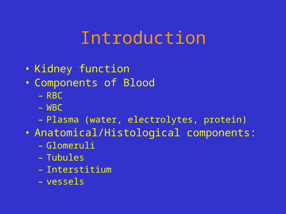
Introduction
• Kidney function• Components of Blood
– RBC– WBC– Plasma (water, electrolytes, protein)
• Anatomical/Histological components:– Glomeruli– Tubules– Interstitium– vessels

Diseases of the kidneyGlomeruli
GlomerulonephritisPrimarySecondaryChronic
TubulointerstitiumAcute tubular necrosisPyelonephritis
Acute chronic
VesselsNephrosclerosis
BenignMalignant
Urinary obstruction
– Stones
– Hydronephrosis
Cystic diseases of the kidney
Tumors
Renal dysfunction
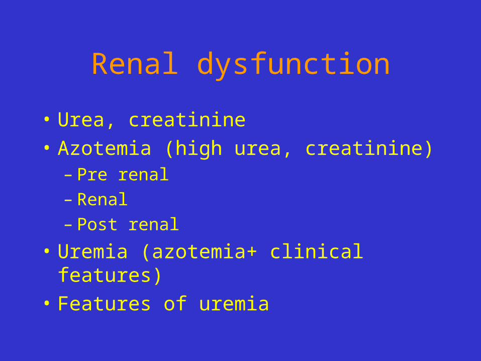
• Urea, creatinine
• Azotemia (high urea, creatinine)– Pre renal– Renal– Post renal
• Uremia (azotemia+ clinical features)
• Features of uremia

Major clinical presentations
• Acute nephritic syndrome– Gross hematuria, mild-moderate proteinuria,
hypertension, azotemia, edema
• Nephrotic syndrome– Heavy proteinuria (>3.5g/day),
hypoalbuminemia, edema, hyperlipidemia, lipiduria
• Asyptomatic hematuria or proteinuria

Major clinical presentations
• Rapidly progressive glomerulonephritis
• Acute renal failure
• Chronic renal failure
• UTI urinary tract infection
• Renal colic (stones)
• Obstruction
• Mass lesion
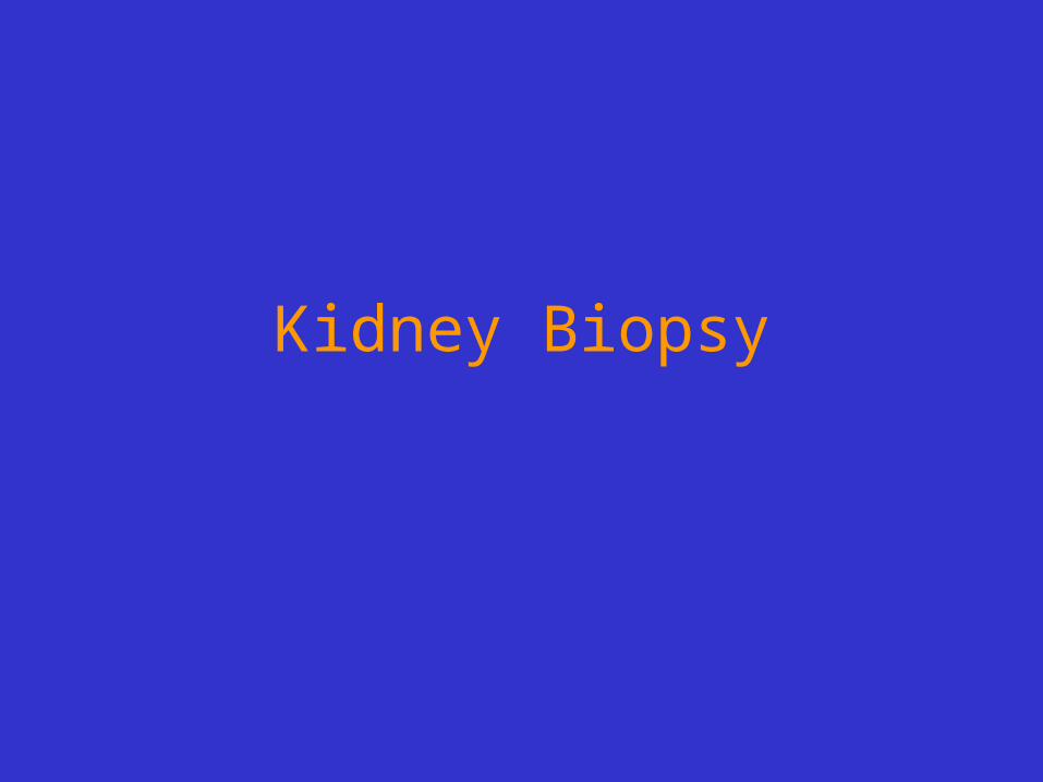
Kidney Biopsy

Kidney Biopsy
• Send fresh
• Routine processing– Immunoflourescence (IF)– Electron microscopy (EM)– Light microscopy (formaline fixed)

Case Presentation

• A 47-year-old black male truck driver presents to the emergency room with intractable nausea and vomiting, dyspnea on exertion, and dizziness. The nausea began about two weeks prior to admission; vomiting has occurred within the last few days. The chest pain has been present for only 2-3 days and is described as retrosternal, burning, and worse on inspiration. There is no history of medication or toxin exposure. His past medical history is positive for hypertension diagnosed 14 years ago with no follow-up. He has smoked 1 ppd for 27 years.

• On physical examination, he is a thin, black man in moderate distress. His blood pressure is 160/120, temperature 36.7°C, pulse 100 (nl 60-100/min). His skin is pale with numerous areas of bruising. Lung exam reveals bilateral rales to the mid lung fields, and cardiac exam reveals muffled heart sounds, a friction rub, and a I/VI systolic ejection murmur. Chest x-ray shows moderate cardiomegaly with increased pulmonary vascular markings and hazy obliteration of the lower lung bases. Abdominal ultrasound examination shows a right kidney size of 7 cm (nl approx. 10 cm) and a left kidney size of 6.8 cm without evidence of pelvicalyceal dilation.

• Urinalysis:– protein - 1+– blood - 1+– glucose - neg– casts - neg– bacteria - neg

• WBC: 6,700/mm3
• Platelets: 250,000/mm3
• Hematocrit: 26%• Creatinine: 200 mmol/L (high, normal <120)
• BUN: 215 mg/dL (high, normal <25)• Calcium: 6.2 mg/dL (low, normal 8-10mg/dl)
• Uric Acid: 16.5 mg/dL (high, normal 5-7)
• Cardiac markers of MI: negative

• An echocardiogram reveals a moderate amount of fluid around the heart. A pericardiocentesis is performed, and 250 cc of serosanguineous fluid is removed. An urgent request for hemodialysis is made, and the patient is dialyzed with some relief in his breathing and chest pain. However, the next morning, the patient complains of abdominal pain and passes several melanotic stools, followed by gross blood. Hypotension and arrhythmias follow, and death supervenes. An autopsy is performed.
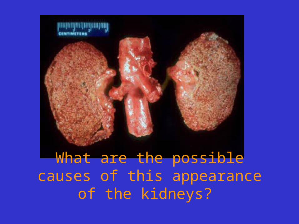
What are the possible causes of this appearance of the kidneys?

Hypertension, D.M., Chronic glomerulonephritis

Describe the four compartments (glomeruli, tubules, interstitium, and vasculature)

Describe the abnormality

What is the abnormality?

What is the abnormality?
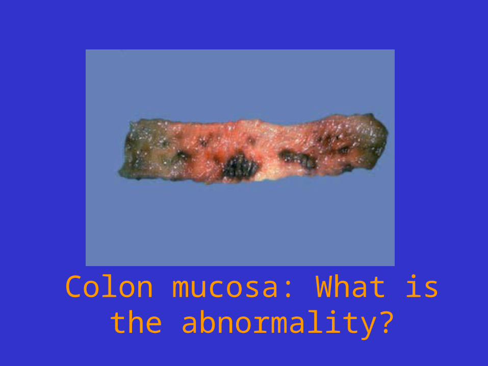
Colon mucosa: What is the abnormality?

Four parathyroid glands

Discussion
• Hypertension
• Chronic Renal Failure
• Uremia
• Urinalysis
• Blood work
• Kidney findings
• Secondary hyper parathyroidism

Glomerular diseases

Glomerulus
• Structure• Filtering membrane
– 1. Endothelial cells– 2. GBM glomerular basement membrane– 3. Visceral epithelium
• GFR: glomerular filtration rate• Mesangium• Permeablility
– Water, albumin– Size, charge


• Glomerular disease
–Primary
–Seconday
–Hereditary

Glomerular disease– Primary
• Minimal change GN• Membranous GN• Focal segmental GS• Membranoproliferative GN• Diffuse proliferative GN• Crescentic GN
– Seconday• SLE, DM, Amyloidosis, Goodpasture, vasculitis
– Hereditary• Albort syndrome
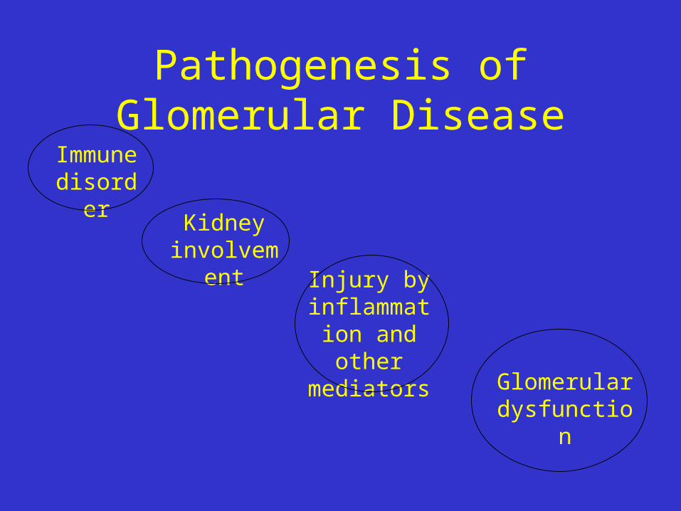
Pathogenesis of Glomerular Disease
Immune disorder
Kidney involvement
Injury by inflammation
and other mediators
Glomerular dysfunction

Pathogenesis of Glomerular Disease
Immune disorder
Glomerular dysfunction
1. Circulating immune complex
2. Immune complex formation
3. Cell-mediated

Pathogenesis
• 1. Circulating Immune complex nephritis (type III hypersensitivity)– Antigen is not glomerular origin
– Intrinsic- SLE
– Extrinsic- Poststreptococcal GN, HepB, Malaria
– Ag-Ab complex is trapped in glomeruli
– Complement activation
– injury
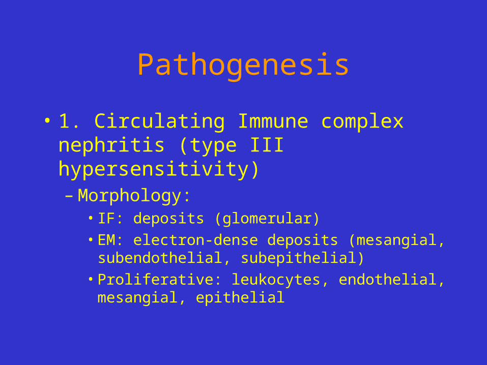
Pathogenesis
• 1. Circulating Immune complex nephritis (type III hypersensitivity)– Morphology:
• IF: deposits (glomerular)
• EM: electron-dense deposits (mesangial, subendothelial, subepithelial)
• Proliferative: leukocytes, endothelial, mesangial, epithelial

Pathogenesis
• 1. Circulating Immune complex nephritis (type III hypersensitivity)– What happen
• Short lived Ag-Ab complex---- Recovery
• Repeated Ag-Ab complex------- chronic GN

Pathogenesis of Glomerular Disease
Immune disorder
Glomerular dysfunction
1. Circulating immune complex
2. In-situ Immune complex formation
3. Cell-mediated

Pathogenesis
• 2. In-situ Immune complex nephritis– In-situ
• Intrinsic
• Extrinsic/planted
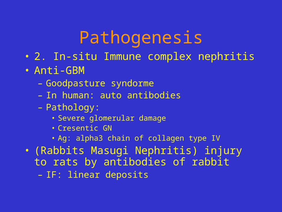
Pathogenesis• 2. In-situ Immune complex nephritis• Anti-GBM
– Goodpasture syndorme– In human: auto antibodies– Pathology:
• Severe glomerular damage• Cresentic GN• Ag: alpha3 chain of collagen type IV
• (Rabbits Masugi Nephritis) injury to rats by antibodies of rabbit– IF: linear deposits

Pathogenesis
• 2. In-situ Immune complex nephritis– Haymann Nephritis:
• Immunizing rats to proximal tubular brush border
• IF: granular deposits of Ig and complement along the GBM
• Ag (megalin) on visceral epithelial cells
• Result in membranous GN
Pathogenesis
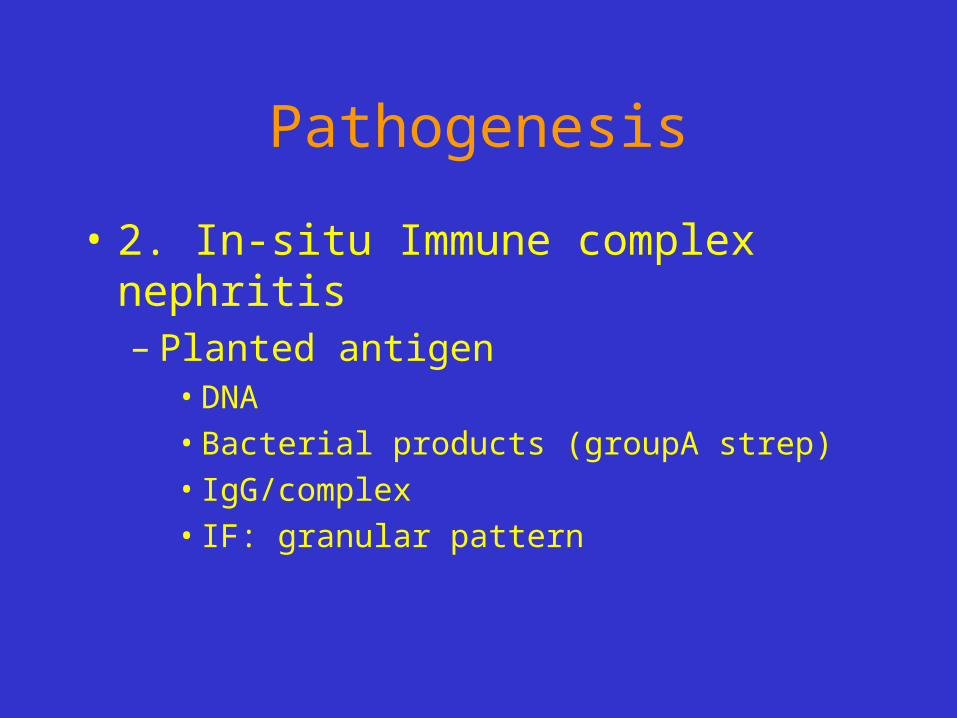
• 2. In-situ Immune complex nephritis– Planted antigen
• DNA
• Bacterial products (groupA strep)
• IgG/complex
• IF: granular pattern

Pathogenesis of Glomerular Disease
Immune disorder
Glomerular dysfunction
1. Circulating immune complex
2. In-situ Immune complex formation
3. Cell-mediated
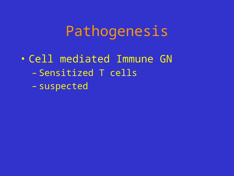
Pathogenesis
• Cell mediated Immune GN– Sensitized T cells– suspected

Pathogenesis of Glomerular Disease
Immune disorder
Glomerular dysfunction
1. Circulating immune complex
2. In situ Immune complex formation
3. Cell-mediated

Pathogenesis of Glomerular Disease
Immune disorder
Kidney involvement
Injury by inflammation
and other mediators
Glomerular dysfunction
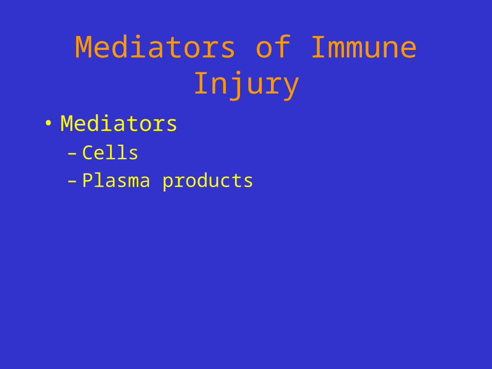
Mediators of Immune Injury
• Mediators– Cells– Plasma products
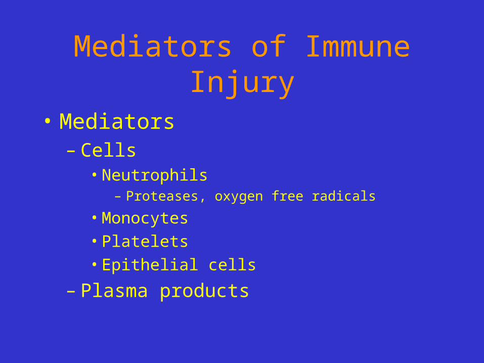
Mediators of Immune Injury
• Mediators– Cells
• Neutrophils– Proteases, oxygen free radicals
• Monocytes
• Platelets
• Epithelial cells
– Plasma products

Mediators of Immune Injury
• Mediators– Cells– Plasma products
• Direct cytotoxicity by Ab
• Fibrin related products
• Complement activiation
• C5-C9 membrane attack complex

Epithelial cell injury
• Ab to visceral epithelium
• Toxins
• Cytokins
• Loss of foot processes, vacuolization, detachment
• proteinuria

Pathogenesis of Glomerular Disease
Immune disorder
Kidney involvement
Injury by inflammation
and other mediators
Glomerular dysfunction

Renal ablation glomerulopathy
• Any disease resulting in decrease GFR to 30-50%
• Progress to end-stage renal failure
• glomerulosclerosis

Renal ablation glomerulopathy
• Glomerulosclerosis—hypertrophy—increase in single nephron GRF—increase blood flow—capillary hypertension—endothelial/epithelial inury—protein/fibrin/lipid deposition—capillary collapse—lyaline degeneration—proliferation of mesangial cells—increase mesangial matrix—sclerosis.

Objectives
• By the end of this session the student should be able to:– List the major clinical presentation of disorders
of the kidney.– Describe the anatomical components of the
kidney and list the major diseases of each.– List and describe the types of immune
mechanisms in glomerular diseases.


![Up-regulation of extracellular matrix proteoglycans and ... · The pathogenesis of crescentic glomerulonephritis diseases ofthe kidney [6].In normal adulthuman kidney, (CGN) and the](https://static.fdocuments.net/doc/165x107/5f0ffdf67e708231d446e768/up-regulation-of-extracellular-matrix-proteoglycans-and-the-pathogenesis-of.jpg)


