Khalil Allah. Nazem Feb 2013. In the face of an increasing prevalence of TKA, intensified efforts...
34
The infected T.K.R Khalil Allah. Nazem Feb 2013
-
Upload
lambert-morrison -
Category
Documents
-
view
218 -
download
2
Transcript of Khalil Allah. Nazem Feb 2013. In the face of an increasing prevalence of TKA, intensified efforts...

The infected T.K.R
Khalil Allah. NazemFeb 2013
In the face of an increasing prevalence of TKA, intensified efforts at infection prevention seem logical to reduce the overall burden of PJI.
As an over view, prevention of PJI relies upon Augmentation of the host response Optimization of the wound
environment Reduction of bacterial contamination
in the pre-intra and post operation times

In addition to prevention, a thorough understanding of the principle of diagnosis and treatment is essential

Although the incidence rates of infection following TKR appear to have fallen over the past several decades, the reported incidence varies in many studies

Many inherent patient risk factors are known to predispose toward post operative deep infection. Host factors include a diagnosis of RA, skin ulcers, D.M ,history of malignancy, obesity, smoking, renal or liver transplantation, HIV positive, prior open knee surgery or periarticular Fx, prior septic arthritis or osteomyelitis.

Risk factors related to surgeon:
Increased INR after operation hematoma require reoperation early wound healing complications
Recent intra articular injection of corticosteroide .
Prolonged operative time
Proper use of AnB prophylaxis represents the single most effective method of reducing infection in TKR.
Optimization of surgical environment. Use low dose ABLC in high risk patients
and revision surgery. Frequent irrigation. Carful surgical technique. Excellent wound closure is important
variables under the surgeon control
Risk factors related to surgeon:

Hematogenous infection of TKR in early postoperative period or many years after operation is often influenced by the surgeon through education efforts made with arthroplasty patients. (Oral- genitourinary- gastrointestinal)
In general invasive procedures that potentially cause bacteremia should simply avoided in the first 3-6 months.(Dental procedures greater than 75 minute in DM and RA are at higher risk). AAOS no longer has published guidelines for the use of prophylactic AnB for high risk patients (Antibiotic prophylaxis for bacteremia in patients with joint replacement.)
Diagnosis
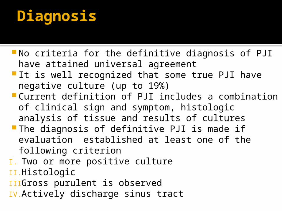
No criteria for the definitive diagnosis of PJI have attained universal agreement
It is well recognized that some true PJI have negative culture (up to 19%)
Current definition of PJI includes a combination of clinical sign and symptom, histologic analysis of tissue and results of cultures
The diagnosis of definitive PJI is made if evaluation established at least one of the following criterion
I. Two or more positive cultureII. HistologicIII. Gross purulent is observed IV. Actively discharge sinus tract

It seems reasonable to identify offending organism and enacting directed treatment strategies

In most series gram positive , may polymicrobial (9%), in current era many resistant organism
MRSA and MRSE have emerged as common nosocomial pathogens often requiring complex AnBs and potentially inferior treatment outcome
Resistant Infections definitely need two staged operations
Fungal PJI are rare and needs two staged treatment

Identification and diagnosis of biofilm organisms is difficult
I. Culture independent molecular method (detection of 16s ribosomal deoxy ribonucleic acid)
II. Culture samples obtain by sonication of prosthesis
Timing of the clinical presentation is a critical factor in diagnosis
These various clinical presentation is critical factor and classified as a useful guide to selecting the most appropriate treatment option
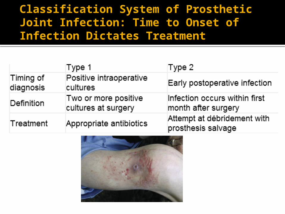
Classification System of Prosthetic Joint Infection: Time to Onset of Infection Dictates Treatment

Classification System of Prosthetic Joint Infection: Time to Onset of Infection Dictates Treatment


Treatment
Variables that must consider before treatment include:
I. Deep or superficialII. Duration from T.K.AIII. Host factorsIV. Soft tissues (extensor mechanism)V. Implant is loose or fixedVI. Pathogens responsibleVII. Ability of surgeonVIII.Patient's expectations

Treatment goals
Eradication of infection, alleviation of pain, maintenance of function
When confronted with an infected T.K.A, the treating physician should start by considering the question prosthesis retain or removal

Treatment methods where the prosthesis is retained
I. Antibiotic suppression:This method alone will not eliminate deep infection but can be used as suppression treatment when the following criteria are met
I. Prosthesis removal is not feasibleII. Microorganism has low virulence and susceptible to an
oral antibioticIII. The antibiotic can be tolerated without serious
toxicityIV. The prosthesis is not looseV. Other prosthesis or cardiac valvular prosthesis are not
present Success rates are 16-24% Rifampin with a quinolone has been reported to be more
successful

Treatment methods where the prosthesis is retainedII. Debridement with prosthesis retentionThis method indicated only in infections in early post operative
period or acute hematogenous with fixed and functional prosthesis and patient has this criterion
I. Short duration of symptomsII. Susceptible gram-positive organism III. Absence of prolonged postoperative drainage or sinus tractIV. No prosthesis loosing V. No other arthroplasty or cardio vascular prosthesis
Success rates are 19-32% Factors that worsen the results are
I. Post operative drainage longer then 2weeksII. Existence of sinus tractIII. Hinged prosthesisIV. Immune compromised hosts
Arthroscopy is not suitable surgical method for debridement and retentions
Treatment methods where by the prosthesis is removed
Resection arthroplasty:The ideal candidate is a patient with
polyarticular RA with limited ambulatory demands, which allow the patient to sit more readily than is feasible with a knee arthrodesis. The primary disadvantage is mobility and pain during transfer or ambulation

Treatment methods where by the prosthesis is removed
Advantages: excellent potential for resolving infections alleviating pain, providing stable knee
Disadvantage: elimination of knee motion Indications:
I. high functional demandsII. Single joint diseaseIII. Young patientsIV. Extensor mechanism disruption V. Poor soft tissueVI. Systemic immunocompromiseVII. Bad micro organism
Arthrodesis:
Relative contraindication:I. Bilateral knee diseaseII. Ipsilateral hip or ankle
diseaseIII. Over segmental bone lossIV. Contralateral limb
amputation

Method of Arthrodesis:
I. IM nailingII. External fixationIII. Dual plate fixation IM nailing appears to show a higher
trend toward success union but has a higher risk of recurrent infection compare to ext fixation
Amputation
Is rarely indicated except in cases if life threatening systemic sepsis or persistent local infection associated with massive bone loss.
Factors must commonly leading to amputation include multiple revisions sever bone loss, intractable pain
Reimplantation
Is currently the primary accepted method of treatment for infected T.K.A
contraindications:I. persisted or recalcitrant infectionII. medical conditionsIII. extensor mechanism disruptionsIV. Poor soft tissue envelope
Reimplantation can be performed as a direct exchange technique or two stages
Factors associated with successful direct exchange:
I. Infection by gram positiveII. Absence of sinus formationIII. Use of antibiotic cementedIV. A prolonged 12week course of AB
therapy This method indicated only in
groups of patients highly selected by arthroplasty surgeons familiar with the treatment of prosthetic infection

Two stage reimplantation
This protocol consist of soft tissue debridement and removal of infected prosthesis and cement, followed by 6weeks IV antibiotics and subsequent reimplantation
The success rate is 85-95% Use of adjunctive antibiotic delivery
provided by the ABLC gradually lead to decrease AB duration and shorter time delays prior to reimplantation
Antibiotic cement spacers Low dose ABLC (<2g per 40g) should be used for
prophylaxis in reimplantation or primary T.K.As in high risk patients or for prosthetic fixation at reimplantation
The higher dose rations should be reserved for treatment of active infection with spacer
AB elution increase by prosity of cement and mixing with another AB 3.6 gr of tobramycin + 1gr vancomycin in 40gr cement are good
The primary function of block spacer include delivery of local AnB and maintenance of collateral ligament length
Potential disadvantages include presence of a foreign body and bone loss incurred.

Different types of block spacers:
I. Simple tibiofemoral blockII. Molded arthrodesis blockIII. Articulating mobile spacersIV. Medullary dowels
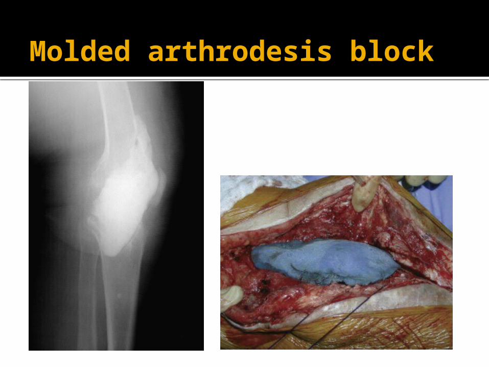
Molded arthrodesis block

Time period between resection and reimplantation
Usually 4-6 weeks One of the most important issues is
the determination of when it is safe and appropriate to proceed with reimplantation.
Guidelines:
C.R.P, ESR Open biopsy or aspiration It is preferable to utilize intraoprative
decision making based on the appearance of the knee joint supplemented by analysis of frozen section
If concern arises about the presence of persistent infection, it is prudent to perform other debridement inserted new ABLC spacers, closed the wound, and await the results of culture and sensitivity testing.