Jurnal Teknologi Full paper -...
6
65:1 (2013) 9–14 | www.jurnalteknologi.utm.my | eISSN 2180–3722 | ISSN 0127–9696 Full paper Jurnal Teknologi Influence of Iron Bacteria on the Corrosion Behavior of Carbon Steel: SEM Study E. Hamzah a* , C. L. Khohr a , A. Abdolahi a , Z. Ibrahim b a Faculty of Mechanical Engineering, Universiti Teknologi Malaysia b Faculty of Bioscience and Bioengineering, Universiti Tekhnologi Malaysia *Corresponding author: [email protected] Article history Received :2 January 2013 Received in revised form : 1 October 2013 Accepted :15 October 2013 Graphical abstract Abstract In this work, the iron bacteria were cultured and inoculated into the cooling water before immersion, and low carbon steel coupons were immersed for one month. Then, microbially influenced corrosion (MIC) of carbon steel in the presence of these bacteria was investigated using scanning electron microscopy (SEM), x-ray diffraction spectroscopy (XRD) and weight loss methods. SEM results showed that large amounts of corrosion products and heterogeneous biofilm layer were formed on the coupon surface. SEM also revealed the uniform-pitting corrosion on the steel surface due to bacteria colonization. XRD results show that the main constituents present in corrosion product are composed of iron oxides and iron hydroxides. Keywords: Microbially influenced corrosion, carbon steel, iron bacteria, SEM, XRD Abstrak Dalam penyelidikan ini, bakteria besi dibiakkan dan di inokulasi ke dalam air pendinginan sebelum rendaman, dan kupon keluli karbon rendah telah direndam selama satu bulan. Kemudian, kupon keluli karbon yang mengalami kakisan dipengaruhi mikrob (MIC) dengan kehadiran bakteria-bakteria tersebut dikaji dengan menggunakan kaedah misroskopi elektron imbasan (SEM), spektroskopi pembelauan sinar- x (XRD) dan kaedah kehilangan berat. Keputusan SEM menunjukkan terdapat jumlah yang besar produk kakisan dan lapisan biofilem heterogen terbentuk di atas permukaan kupon. Keputusan SEM juga menunjukkan kakisan bopeng seragam berlaku diatas permukaan keluli disebabkan penjajahan bakteria. Keputusan XRD menunjukkan juzuk utama terdapat didalam produk kakisan terdiri daripada oksida besi dan hidroksida besi. Kata kunci: Kakisan dipengaruhi mikrob; keluli karbon; bakteria besi; SEM; XRD © 2013 Penerbit UTM Press. All rights reserved. 1.0 INTRODUCTION Microbially influenced corrosion or MIC is known as one of the destructive types of corrosion caused by activities of micro- organisms such as bacteria. The bacteria colonization and biofilm formation on the metal surface is recognized as the first stage of microbially influenced corrosion [1-3]. The appearance of metal surface due to MIC is mostly in the form of localized corrosion such as pit or crevice [4, 5]. Bridges, water cooling systems, ships and many types of equipments in an aquatic environment are some examples, which are in danger of failure due to MIC [6-9]. Carbon steel owes to its good mechanical properties, and low cost belongs to the most applicable metals in industry. However, the main limitation to use it in an aquatic environment is its less resistivity to corrosion. Carbon steel can corrode in bacteria inoculated environment such as a cooling water system as a result of bacteria colonization [10, 11]. One type of bacteria which could induce the corrosion process on the carbon steel is iron bacteria. This type of bacteria tends to oxidize the insoluble ferrous ions to soluble ferric ions, thereafter led to precipitation of Fe (OH)3 on the steel surface. Thus, iron bacteria in the biofilm state could produce large amounts of corrosion products composed of iron oxide, and hydroxide precipitates on the steel surface [11, 12]. Due to the importance of iron bacteria in corrosion of metals, the present study is carried out to achieve a better understanding on MIC behavior of carbon steel in the presence of iron bacteria. In this work, SEM and XRD techniques were used to characterize the biofilm layer, corrosion products and study pitting corrosion on the steel surface.
Transcript of Jurnal Teknologi Full paper -...

65:1 (2013) 9–14 | www.jurnalteknologi.utm.my | eISSN 2180–3722 | ISSN 0127–9696
Full paper Jurnal
Teknologi
Influence of Iron Bacteria on the Corrosion Behavior of Carbon Steel: SEM Study E. Hamzaha*, C. L. Khohra, A. Abdolahia, Z. Ibrahimb
aFaculty of Mechanical Engineering, Universiti Teknologi Malaysia bFaculty of Bioscience and Bioengineering, Universiti Tekhnologi Malaysia *Corresponding author: [email protected]
Article history
Received :2 January 2013
Received in revised form :
1 October 2013 Accepted :15 October 2013
Graphical abstract
Abstract
In this work, the iron bacteria were cultured and inoculated into the cooling water before immersion, and
low carbon steel coupons were immersed for one month. Then, microbially influenced corrosion (MIC) of
carbon steel in the presence of these bacteria was investigated using scanning electron microscopy (SEM), x-ray diffraction spectroscopy (XRD) and weight loss methods. SEM results showed that large
amounts of corrosion products and heterogeneous biofilm layer were formed on the coupon surface. SEM
also revealed the uniform-pitting corrosion on the steel surface due to bacteria colonization. XRD results show that the main constituents present in corrosion product are composed of iron oxides and iron
hydroxides.
Keywords: Microbially influenced corrosion, carbon steel, iron bacteria, SEM, XRD
Abstrak
Dalam penyelidikan ini, bakteria besi dibiakkan dan di inokulasi ke dalam air pendinginan sebelum rendaman, dan kupon keluli karbon rendah telah direndam selama satu bulan. Kemudian, kupon keluli
karbon yang mengalami kakisan dipengaruhi mikrob (MIC) dengan kehadiran bakteria-bakteria tersebut
dikaji dengan menggunakan kaedah misroskopi elektron imbasan (SEM), spektroskopi pembelauan sinar-x (XRD) dan kaedah kehilangan berat. Keputusan SEM menunjukkan terdapat jumlah yang besar produk
kakisan dan lapisan biofilem heterogen terbentuk di atas permukaan kupon. Keputusan SEM juga
menunjukkan kakisan bopeng seragam berlaku diatas permukaan keluli disebabkan penjajahan bakteria. Keputusan XRD menunjukkan juzuk utama terdapat didalam produk kakisan terdiri daripada oksida besi
dan hidroksida besi.
Kata kunci: Kakisan dipengaruhi mikrob; keluli karbon; bakteria besi; SEM; XRD
© 2013 Penerbit UTM Press. All rights reserved.
1.0 INTRODUCTION
Microbially influenced corrosion or MIC is known as one of the
destructive types of corrosion caused by activities of micro-
organisms such as bacteria. The bacteria colonization and biofilm
formation on the metal surface is recognized as the first stage of
microbially influenced corrosion [1-3]. The appearance of metal
surface due to MIC is mostly in the form of localized corrosion
such as pit or crevice [4, 5]. Bridges, water cooling systems, ships
and many types of equipments in an aquatic environment are
some examples, which are in danger of failure due to MIC [6-9].
Carbon steel owes to its good mechanical properties, and low
cost belongs to the most applicable metals in industry. However,
the main limitation to use it in an aquatic environment is its less
resistivity to corrosion. Carbon steel can corrode in bacteria
inoculated environment such as a cooling water system as a result
of bacteria colonization [10, 11]. One type of bacteria which
could induce the corrosion process on the carbon steel is iron
bacteria. This type of bacteria tends to oxidize the insoluble
ferrous ions to soluble ferric ions, thereafter led to precipitation of
Fe (OH)3 on the steel surface. Thus, iron bacteria in the biofilm
state could produce large amounts of corrosion products
composed of iron oxide, and hydroxide precipitates on the steel
surface [11, 12]. Due to the importance of iron bacteria in
corrosion of metals, the present study is carried out to achieve a
better understanding on MIC behavior of carbon steel in the
presence of iron bacteria. In this work, SEM and XRD techniques
were used to characterize the biofilm layer, corrosion products
and study pitting corrosion on the steel surface.

10 E. Hamzah et al. / Jurnal Teknologi (Sciences & Engineering) 65:1 (2013), 9–14
2.0 MATERIALS AND METHODS
2.1 Preparation of Coupons
Low carbon steel (99.6% of Fe, 0.028% of carbon, 0.030 of
phosphorus, 0.035% of sulphur and 0.30% of copper.) coupons
with a diameter of 20 mm and thickness of 4 mm were used as
test material. The surfaces of the coupons were sequentially
ground with a series of grit SiC papers (180, 500, 800, and 1200)
and polished using 0.3 µm alumina powders. Coupons were
rinsed with deionized water, followed by degreasing with acetone,
and dried in at room temperature. The prepared coupons were
then immersed in the bacteria inoculated medium.
2.2 Growth of Bacteria
Iron bacteria were cultured on the agar plate by serial dilution
technique. In this method, eight small test tubes and agar plates
were prepared. These tubes were filled up with water. In the first
tube, 1 ml of the cooling water was diluted with 9 ml of distilled
water. The mixture was shaken in the shaker for 24 hours with
speed of 150 rpm at 30oC temperature. From the first tube, 1 ml of
the water dilution was transferred into the second tube contains 9
ml of distilled water. The second tube was also shaken in the
shaker for 24 hours with speed of 150 rpm at 30oC temperature.
This similarly procedure was repeated for the 6 remaining tubes
and spread plates. The optical density (OD) values were recorded
for each tube. Finally, when the OD reached to the higher value,
then 0.1 ml of the dilution from the considered tube was spread
over the agar plate. Thereafter, the agar plate was incubated in the
incubator oven at a temperature of 30 °C for 24 hours. Iron
bacteria were transferred from the inoculated agar plate to the
cooling water to assess the MIC behavior of carbon steel in the
presence of bacteria. Total eight spread plates (with different
dilution factor) were sealed with paraflim to prevent mixing up
with other impurities present in the air. These bacteria were taken
and inoculated in the cooling water before immersion of the steel
coupons.
2.3 Immersion Test
The immersion test had been carried out by exposing the coupons
in the bacteria inoculated medium for one month. For comparison
purposes, the coupons were immersed in two types of cooling
water in the presence of iron bacteria (biotic coupon) and (b)
cooling water without the presence of iron bacteria (control
coupon). At the end of immersion time the coupons were dried
followed by mechanical and chemical cleaning in accordance to
the ASTM standard (Designation: G1-03) for weight loss
measurement.
2.4 SEM Analysis
Biofilm layer and corrosion products formed on coupons were
analyzed by scanning electron microscopy. The dried samples
were coated with a thin gold layer and observed under the field
emission electron microscope. Furthermore, the pitting corrosion
appeared on the steel surface is observed using scanning electron
microscope.
2.5 XRD Characterization
An X-Ray Diffraction method was carried out to detect the
chemical composition of the corrosion products formed on the
steel surface due to bacterial colonization.
2.6 Corrosion Rate Measurement
Coupons were cleaned and weighed after drying. The weight loss
was determined based on the difference between their initial and
final weight of the coupon after an immersion test. The corrosion
rate were calculated based on Equation (1).
Corrosion rates = (K× W) / (A × T × D) (1)
Where: K = 2.40×10⁶ D, T = time of exposure (hr), A = exposed
surface area (cm2), W = mass loss (g), D = density (g/cm3)
3.0 RESULTS AND DISCUSSION
3.1 Material Characterisation
The microstructure of carbon steel coupons observed by optical
image analyser is shown in Figure 1. The microstructure is
composed of ferrite phase (white region) and pearlite phase (dark
region).
Figure 1 Optical micrograph of carbon steel (magnification ×100)
Generally, ferrite (α) is pure iron, and the pearlite is a fine
mixture of ferrite and cementite (Fe3C) in the lamellar form. In
fact, the composition of the steel surface is an ideal surface for
bacterial colonization and biofilm development. The bacteria
nucleated and formed colonies on the coupon surface due to the
presence of iron element that was required the bacterial growth.
3.2 Detection of Bacteria
In order to ensure that bacteria was present in the cooling system
the presence of iron bacteria in water taken from the cooling
system was identified using IRB BART kits. Figure 2(a) shows
the control test tube containing filter sterilized cooling water.
Figure 2(b) and (c) show the tubes contain the mixture of cooling
water and iron bacteria.
11 E. Hamzah et al. / Jurnal Teknologi (Sciences & Engineering) 65:1 (2013), 9–14
Figure 2 IRB-BART Kits (a) Control set. (b) and (c) Bacterial samples
Figure 2(b) and (c) also showed that the color of water
samples changed from yellowish to orange, which is due to the
formation of ferric compounds by iron bacteria. Thus, the
presence of iron bacteria in the cooling water system was
confirmed.
3.3 Surface Analysis
Figure 3 shows the surface of as-received sample before
immersion in the solution. The surface is clean without any trace
of corrosion product.
Figure 3 As-received carbon steel coupon
Figure 4, however, shows the sample corroded after
immersion in the bacteria-containing solution and in the control
medium. It was found that the corrosion product is brownish in
colour (Figure 4 (a) and (c)) which is typical colour of iron rust.
When the corrosion products were removed, it appears that the
coupon has lost some of its material, which is indicated by a
rough surface.
Figure 4 (a) Corroded biotic coupon (before cleaning) (b) Corroded
control coupon (before cleaning) (c) Corroded biotic coupon (After cleaning) (d) Corroded control coupon (After cleaning) after 1 month
exposure time
Based on visual inspection, the biotic coupon has more corrosion
products compared with the control sample, and thus it can be
deduced that the presence of bacteria has increased the corrosion
rate of the coupon and the weight loss. Iron bacteria tend to
oxidize insoluble ferrous ions to soluble ferric ions that lead to
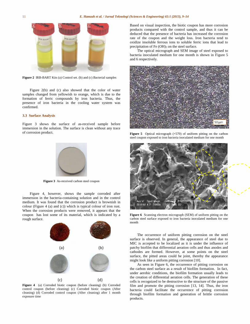
precipitation of Fe (OH)3 on the steel surface. The optical micrograph and SEM image of steel exposed to
bacteria inoculated medium for one month is shown in Figure 5
and 6 respectively.
Figure 5 Optical micrograph (×570) of uniform pitting on the carbon
steel coupon exposed to iron bacteria inoculated medium for one month
Figure 6 Scanning electron micrograph (SEM) of uniform pitting on the
carbon steel surface exposed to iron bacteria inoculated medium for one month
The occurrence of uniform pitting corrosion on the steel
surface is observed. In general, the appearance of steel due to
MIC is accepted to be localized as it is under the influence of
patchy biofilm that differential aeration cells and thus anodes and
cathodes are formed. However, at some points on the steel
surface, the pitted areas could be joint, thereby the appearance
might look like a uniform pitting corrosion [10].
As seen in Figure 6, the occurrence of pitting corrosion on
the carbon steel surface as a result of biofilm formation. In fact,
under aerobic conditions, the biofilm formation usually leads to
the creation of differential aeration cells. The generation of these
cells is recognized to be destructive to the structure of the passive
film and promote the pitting corrosion [13, 14]. Thus, the iron
bacteria could facilitate the occurrence of pitting corrosion
through biofilm formation and generation of brittle corrosion
products.

12 E. Hamzah et al. / Jurnal Teknologi (Sciences & Engineering) 65:1 (2013), 9–14
Generally, it is impossible to make a relationship between the pit
morphology and the type of bacteria. It has been found that the pit
morphology could be formed by factors other than bacteria
colonization. Furthermore, the pit morphology is more related to
biofilm layer formed by bacteria not any specific bacterial types.
Thus, the biofilm layer formed by different types of bacteria could
result in formation of a pit of similar appearance [15, 16].
3.4 Biofilm and Corrosion Product Analysis
Figure 7 shows the SEM image of carbon steel exposed for one
month in iron bacteria inoculated cooling water.
Figure 7 Scanning electron micrograph showing the hold fast structure by which the iron bacteria filaments attach to the metal surface and colonize
The whole surface of steel surface is covered with a
heterogeneous biofilm layer and corrosion products. Biofilm layer
is encrusted with corrosion products, which means that the
formation rate of corrosion products is higher than the formation
rate of biofilm layer. In view of the energetic involved large
amounts of iron must be oxidized to supply the energy
requirements for bacteria growth. Thus, the corrosion products are
usually in larger amounts compared with bacterial biomass.
Figure 7 also shows the mode of attachment of iron bacteria
filaments to the corrosion products; indicate that the oxide
corrosion products are suitable places for growing of bacteria.
Actually, the bacteria develop at the edge of the tubercle where
oxygen and iron are readily available, since iron bacteria require
oxygen for their growth [11].
Generally, as the first attachment of bacteria cells to the steel
surface, they tend to form a biofilm layer to survive and prolong
their lives. Biofilm secretes polymeric substances known as EPS
to facilitate its growth. Extracellular polymeric substances are
comprised of macromolecules such as proteins, polysaccharides,
nucleic acids and lipids. EPS tend to attach to the corrosion
products, to gain elements for bacterial growth. The activity and
growth of bacteria could be increased through feeding from the
corrosion products, which are enriched of oxygen [2, 17].
Figure 8 shows SEM images of some corrosion products
formed on the carbon steel surface that was exposed to medium
containing the iron bacteria for one month.
Figure 8 SEM micrographs showing: (a) fine plates (‘‘flowery’’ structures) typical of lepidocrocite, (b) globular (‘‘cotton balls’’) structures typical of
goethite
The corrosion products are mainly iron oxides and
hydroxides. The morphological structures of corrosion products
are flowery structures of lepidocrocite and the typical cotton ball
structure of goethite [14]. Normal corrosion products formed on
carbon steel in aerobic condition are called tubercles. The
formation of tubercles can be described as follows;
Fe Fe+2 + 2e- (anode) (2)
O2 + 2 H2O +4 e- 4 OH- (cathode) (3)
2 Fe+2 + ½ O2 + 5H2O 2Fe (OH) 3 + 4H+
(tubercle) (4)
Generally, iron can exist in three oxidation states (Fe0, Fe2+
and Fe3+). In presence of oxygen, the metallic iron (Fe0)
oxidises to form the stable Fe2+. Subsequent oxidation of Fe2+ to
Fe3+ takes place at pH >5 could produce energy for growth and
activity of iron bacteria. Since the amount of energy extracted
from this reaction is quite small for iron bacteria,
(approximately −31 kJ), large quantities of Fe2+ have to be
oxidized to Fe3+ to generate enough energy for bacteria growth
and biofilm formation [18]. Thus, a large amount of corrosion
products could be formed on the carbon steel due to presence of
iron bacteria.
As shown in Figure 9, the XRD analysis was also carried
out to confirm that the major constituents of the corrosion
products in presence of iron bacteria are: goethite (α-FeOOH), wustite (FeO), magnetite (Fe
3O
4), hematite (Fe
2O
3) and
Lepidocrocite (γ-FeOOH).

13 E. Hamzah et al. / Jurnal Teknologi (Sciences & Engineering) 65:1 (2013), 9–14
Figure 9 X-Ray Diffraction analysis of corrosion products in presence of iron bacteria
3.5 Corrosion Rate Measurement
The corrosion rate of the biotic and control coupons was
determined based on Equation (1) using the weight loss method
to evaluate the corrosion damage on the coupons in one month.
Figure 10 shows the corrosion rate of biotic and control
coupons.
Figure 10 Corrosion rates of the biotic and control coupons in one month immersion time
Figure 10 shows that the corrosion rate of biotic coupons is
higher than the corrosion rate of control coupons. Thus, it be
concluded that iron bacteria accelerate the corrosion rate of
biotic coupons. Large amounts of iron must be oxidized to
supply the energy requirements for growth of iron bacteria. This
phenomenon accelerates the corrosion rate of biotic coupons.
However differential aeration cell could also accelerate the
corrosion rate of biotic coupons. In differential aeration cell, the

14 E. Hamzah et al. / Jurnal Teknologi (Sciences & Engineering) 65:1 (2013), 9–14
area beneath the biofilm is lacked of an oxygen act as an anode
and the metal surface which is enriched of the oxygen act as a
cathode. Thus, the corrosion process on the surface of biotic
coupons was enhanced [18, 19].
4.0 CONCLUSION
1. Carbon steel immersed in solution contains iron bacteria
(biotic coupon) has more corrosion product carbon steel
immersed in solution without presence of iron bacteria (control
coupon). Thus, iron bacteria are responsible for accelerating the
corrosion rate and formation of more corrosion products on the
biotic coupon surface.
2. The corrosion process on the carbon steel surface was
affected by the presence of iron bacteria. The bacteria in the
form of biofilm lead to the formation of differential aeration and
concentration cells, causing uniform pitting corrosion on the
steel surface.
3. SEM results showed large amounts of corrosion products, and
heterogeneous biofilm layer formed on the coupon surface in
presence of iron bacteria.
4. XRD analysis showed that major constitute of corrosion
products are: iron oxide hydroxide goethite (α-FeOOH), wustite (FeO), magnetite (Fe
3O
4), hematite (Fe
2O
3) and Lepidocrocite
(γ-FeOOH).
References
[1] Stott, J. F. D. 1993. What Progress in the Understanding of Microbially
Induced Corrosion Has Been Made in the Last 25 Years? A Personal
View Point. Corrosion Science. 35: 667–73.
[2] Beech, I. B. and Sunner, J. 2004. Biocorrosion: Towards
Understanding Interactions Between Biofilms and Metals. Current
Opinion in Biotechnology. 15: 181–86.
[3] Geiser, M., Avci, R. and Lewandowski, Z. 2002. Microbially Initiated
Pitting on 316L Stainless Steel. International Biodeterioration & Biodegradation. 49: 235–43.
[4] Yusuke Tsutsumi, Y., Nishikata,A. and Tsura, T. 2007. Pitting
Corrosion Mechanism of Type 304 Stainless Steel Under a Droplet of
Chloride Solutions. Corrosion Science. 49: 1394–407.
[5] Shi, X., Avci, R., Geiser, M. and Lewandowski, Z. 2003. Comparative
Study in Chemistry of Microbially and Electrochemically Induced
Pitting of 316L Stainless Steel. Corrosion Science. 45: 2577–95.
[6] Beech, I. B. 2004. Corrosion of Technical Materials in the Presence of
Biofilms-current Understanding and State-of-the Art Methods of
Study. International Biodeterioration & Biodegradation. 53: 177–83.
[7] Guezennec, J. G. 1994. Cathodic Protection and Microbially induced
Corrosion. International Biodeterioration & Biodegradation. 34: 275–84.
[8] Mansfeld, F. 2007. The Interaction of Bacteria and Metal Surfaces.
Electrochimica Acta. 52: 7670–80.
[9] Sungur, E. I. and Cotuk, A. 2010. Microbial Corrosion of Galvanized
Steel in a Simulated Recirculating Cooling Tower System. Corrosion
Science. 52: 161–71.
[10] Javaherdashti, R. 2009. A Brief Review of General Patterns of MIC of
Carbon Steel and Biodegradation of Concrete. IUFS Journal of Biology. 68: 65–73.
[11] Rao, T. S., Sairam, T. N., Viswanathan, B. and Nair, K. V. K. 2000.
Carbon Steel Corrosion by Iron Oxidising and Sulphate Reducing
Bacteria in a Freshwater Cooling System. Corrosion Science.42: 1417–
31.
[12] Xu, X., Zhang, Y., Cheng, G. and Zhu, W. 2007. Localized Corrosion
Behavior of 316L Stainless Steel in the Presence of Sulfate-reducing and Iron-oxidizing Bacteria. Materials Science and Engineering: A. 14:
829–34.
[13] Yuan, S. J., Choong, A .M. F. and Pehkonen, S. O. 2007. The Influence
of the Marine Aerobic Pseudomonas Strain on the Corrosion of 70/30
Cu–Ni alloy. Corrosion Science. 49: 4352–85.
[14] Sorkhabi, H. A., Haghighi, M, Zarrini, G and Javaherdashti, R. 2011.
Corrosion Behavior of Carbon Steel in the Presence of Two Novel
Iron-Oxidizing Bacteria Isolated From Sewage Treatment Plants. Biodegradation. 23: 69–79.
[15] R. E Tatnall, D. H. Pope. 1993 Identification of MIC. Ch. 8, In: a
Practical Manual on Microbiologically Influenced Corrosion. Kobrin,
G. (ed), NACE, Houston, Texas USA.
[16] R. Javaherdashti. 2008. Microbiologically Influenced Corrosion: An
Engineering Insight. Springer-Verlag.
[17] Vu, B., Chen, M., Crawford, R. J. and Ivanova, E. P. 2009. Bacterial
Extracellular Polysaccharides Involved in Biofilm Formation. Molecules. 14: 2535–54.
[18] T. S. Rao. 2000. Carbon Steel Corrosion by Iron Oxidising and
Sulphate Reducing Bacteria in a Freshwater Cooling System.
Corrosion Science. 42: 1417–1431.
[19] XU, Congmin, 2006. Corrosion and Electrochemical Behavior of 316L
Stainless Steel in Sulfate-reducing and Iron-oxidizing Bacteria
Solutions. Chinese J. Chem. Eng. 14(6): 829–834.