JOURNAL Printed in U.S.A. Fermentation of Glucose, …jb.asm.org/content/96/2/472.full.pdf ·...
7
JOURNAL OF BACTERIOLOGY, Aug. 1968, p. 472-478 Copyright © 1968 American Society for Microbiology Fermentation of Glucose, Lactose, Galactose, Mannitol, and Xylose by Bifidobacteria WYTSKE DE VRIES AND A. H. STOUTHAMER Botanical Laboratory, Microbiology Department, Free University, Amsterdam, The Netherlands Received for publication 13 May 1968 For six strains of Bifidobacterium bifidum (Lactobacillus bifidus), fermentation bal- ances of glucose, lactose, galactose, mannitol, and xylose were determined. Prod- ucts formed were acetate, L( +)-lactate, ethyl alcohol, and formate. L( +)-Lactate de- hydrogenase of all strains studied was found to have an absolute requirement for fructose-1 , 6-diphosphate. The phosphoroclastic enzyme could not be demonstrated in cell-free extracts. Cell suspensions fermented pyruvate to equimolar amounts of acetate and formate. Alcohol dehydrogenase was shown in cell-free extracts. Possible explanations have been suggested for the differences in fermentation balances found for different strains and carbon sources. By enzyme determinations, it was shown that bifidobacteria convert mannitol to fructose-6-phosphate by an inducible polyol dehydrogenase and fructokinase. For one strain of B. bifidum, molar growth yields of glucose, lactose, galactose, and mannitol were determined. The mean value of Y (ATP), calculated from molar growth yields and fermentation balances, was 11.3. In previous papers (21, 29, 30), it was shown that bifidobacteria ferment glucose via the fructose-6-phosphate phosphoketolase route. By enzyme determinations, the presence of the glyco- lytic system and hexose monophosphate pathway was ruled out (30). For one strain of Bifido- bacterium (Lactobacillus bifidus), the fermenta- tion pathway of glucose was confirmed by the determination of the fermentation balance of glucose in resting-cell suspensions. The fermenta- tion balance found [glucose -- L(+)-lactate + 1.5 acetate] could be explained by the operation of the fructose-6-phosphate phosphoketolase route and reduction of pyruvate to lactate. It was shown that lactate dehydrogenase (EC 1 .1 .1 . 27) of this strain had an absolute require- ment for fructose-I, 6-diphosphate. This ex- plained the presence of small amounts of phos- phofructokinase (EC 2.7.1.11), an enzyme otherwise not involved in the fermentation path- way, in cell-free extracts (29). The present work investigated the pathways of pyruvate breakdown in other strains of Bifido- bacterium. For that purpose, fermentation bal- ances of glucose were determined in growing cultures of five strains of Bifidobacterium. In addition, fermentation balances of mannitol, lactose, xylose, and galactose were determined for some strains of Bifidobacterium. Acetate, lactate, ethyl alcohol, and formate proved to be the main fermentation products. Apparently, pyruvate, formed as an intermediate in the fructose-6-phosphate phosphoketolase route, was partly reduced to lactate by lactate dehydrogenase and partly split to acetyl phosphate and formate by the phosphoroclastic enzyme. Attempts were made to show alcohol dehydrogenase (EC 1.1.1.1) and the phosphoroclastic enzyme in cell-free extracts of one strain of Bifidobacterium. By enzyme studies, the way by which bifido- bacteria convert mannitol to fructose-6-phos- phate was established. Finally, molar growth yields of glucose, man- nitol, lactose, and galactose are recorded in this paper. From molar growth yields and fermenta- tion balances, Y (ATP) was calculated. MATERIALS AND METHODS Organisms and growth conditions. Of the stock collection of bifidobacteria, strains of different types were selected. The following strains were used: L. bifidus (S 128) obtained from K. C. Winkler (Labora- tory of Microbiology, State University, Utrecht, The Netherlands); B. bifidum (S 200) obtained from J. de Waart (Central Institute of Nutrition and Food Re- search, Zeist, Holland); B. bifidum B5 (S 324) and B. bifidum B420 (S 327) obtained from H. Beerens (In- stitut Pasteur de Lille, France); B. lactentis 659 (S 332) and B. liberorum 76 (S 337) obtained from G. Reuter (Institut fur Lebensmittelhygiene der Freien Universitiit, Berlin, Germany). Numbers in parenthe- ses are the collection numbers of the strains men- 472 Vol. 96, No. 2 Printed in U.S.A. on June 15, 2018 by guest http://jb.asm.org/ Downloaded from
Transcript of JOURNAL Printed in U.S.A. Fermentation of Glucose, …jb.asm.org/content/96/2/472.full.pdf ·...

JOURNAL OF BACTERIOLOGY, Aug. 1968, p. 472-478Copyright © 1968 American Society for Microbiology
Fermentation of Glucose, Lactose, Galactose,Mannitol, and Xylose by Bifidobacteria
WYTSKE DE VRIES AND A. H. STOUTHAMERBotanical Laboratory, Microbiology Department, Free University, Amsterdam, The Netherlands
Received for publication 13 May 1968
For six strains of Bifidobacterium bifidum (Lactobacillus bifidus), fermentation bal-ances of glucose, lactose, galactose, mannitol, and xylose were determined. Prod-ucts formed were acetate, L( +)-lactate, ethyl alcohol, and formate. L( +)-Lactate de-hydrogenase of all strains studied was found to have an absolute requirement forfructose-1 , 6-diphosphate. The phosphoroclastic enzyme could not be demonstratedin cell-free extracts. Cell suspensions fermented pyruvate to equimolar amountsof acetate and formate. Alcohol dehydrogenase was shown in cell-free extracts.Possible explanations have been suggested for the differences in fermentationbalances found for different strains and carbon sources. By enzyme determinations,it was shown that bifidobacteria convert mannitol to fructose-6-phosphate by aninducible polyol dehydrogenase and fructokinase. For one strain of B. bifidum,molar growth yields of glucose, lactose, galactose, and mannitol were determined.The mean value of Y (ATP), calculated from molar growth yields and fermentationbalances, was 11.3.
In previous papers (21, 29, 30), it was shownthat bifidobacteria ferment glucose via thefructose-6-phosphate phosphoketolase route. Byenzyme determinations, the presence of the glyco-lytic system and hexose monophosphate pathwaywas ruled out (30). For one strain of Bifido-bacterium (Lactobacillus bifidus), the fermenta-tion pathway of glucose was confirmed by thedetermination of the fermentation balance ofglucose in resting-cell suspensions. The fermenta-tion balance found [glucose -- L(+)-lactate +1.5 acetate] could be explained by the operationof the fructose-6-phosphate phosphoketolaseroute and reduction of pyruvate to lactate. Itwas shown that lactate dehydrogenase (EC1 .1 .1 .27) of this strain had an absolute require-ment for fructose-I, 6-diphosphate. This ex-plained the presence of small amounts of phos-phofructokinase (EC 2.7.1.11), an enzymeotherwise not involved in the fermentation path-way, in cell-free extracts (29).The present work investigated the pathways of
pyruvate breakdown in other strains of Bifido-bacterium. For that purpose, fermentation bal-ances of glucose were determined in growingcultures of five strains of Bifidobacterium. Inaddition, fermentation balances of mannitol,lactose, xylose, and galactose were determinedfor some strains of Bifidobacterium. Acetate,lactate, ethyl alcohol, and formate proved to be
the main fermentation products. Apparently,pyruvate, formed as an intermediate in thefructose-6-phosphate phosphoketolase route, waspartly reduced to lactate by lactate dehydrogenaseand partly split to acetyl phosphate and formateby the phosphoroclastic enzyme. Attempts weremade to show alcohol dehydrogenase (EC1.1.1.1) and the phosphoroclastic enzyme incell-free extracts of one strain of Bifidobacterium.By enzyme studies, the way by which bifido-
bacteria convert mannitol to fructose-6-phos-phate was established.
Finally, molar growth yields of glucose, man-nitol, lactose, and galactose are recorded in thispaper. From molar growth yields and fermenta-tion balances, Y (ATP) was calculated.
MATERIALS AND METHODSOrganisms and growth conditions. Of the stock
collection of bifidobacteria, strains of different typeswere selected. The following strains were used: L.bifidus (S 128) obtained from K. C. Winkler (Labora-tory of Microbiology, State University, Utrecht, TheNetherlands); B. bifidum (S 200) obtained from J. deWaart (Central Institute of Nutrition and Food Re-search, Zeist, Holland); B. bifidum B5 (S 324) and B.bifidum B420 (S 327) obtained from H. Beerens (In-stitut Pasteur de Lille, France); B. lactentis 659(S 332) and B. liberorum 76 (S 337) obtained from G.Reuter (Institut fur Lebensmittelhygiene der FreienUniversitiit, Berlin, Germany). Numbers in parenthe-ses are the collection numbers of the strains men-
472
Vol. 96, No. 2Printed in U.S.A.
on June 15, 2018 by guesthttp://jb.asm
.org/D
ownloaded from

VOL. 96, 1968 CARBOHYDRATE FERMENTATION BY BIFIDOBACTERIA
tioned. The strains were maintained as stab culturesin tomato-agar (Oxoid) containing 2% glucose and0.2% cysteine. Mass cultures were grown in themedium previously described (30) to which 2% glu-cose was added. A McIntosh anaerobic jar was usedto obtain anaerobic conditions. The gas phase wasN2 plus C02 (95:5). Cultures were incubated at 37 C.
Determination of fermentation balances. The me-dium used had the following composition (per liter ofwater): tryptone (Oxoid), 10 g; yeast extract (Oxoid),5 g; NaCl, 5 g; KH2PO4, 2 g; and K2HPO4, 3 g; thepH of the medium was 6.8. Exactly 10 Amoles ofglucose, galactose, mannitol, or xylose, or 5 Mmoles oflactose was added per ml of the medium. These con-centrations were growth-limiting. When maximaloptical density was reached, fermentation was as-sumed to be complete, and the culture was centri-fuged. In the supernatant fluid, acetate, L(+)-lactate,ethyl alcohol, formate, pyruvate, and acetoin weredetermined. Except for glucose, residual substrate wasnot determined. Acetate was determined by means ofthe enzymatic method of Rose (20), which is based onthe colorimetric determination of acetyl phosphateaccording to Lipmann and Tuttle (15). L(+)-Lactate,ethyl alcohol, and glucose were determined enzy-matically by means of a Biochemica Test Combination(C. F. Boehringer and Soehne, GmbH, Mannheim,Germany). Formate was measured with formatedehydrogenase (EC 1. 2.1.2) as described by Johnsonet al. (11). Pyruvate was determined according toFriedemann and Haugen (8). Acetoin was determinedaccording to Westerfeld (31). In all determinations,the medium was used as a blank. By adding knownamounts of acetate, lactate, ethyl alcohol, formate,pyruvate, or glucose to the fermentation liquid, itwas found that the medium did not influence the de-termination of these compounds. Intracellular carbo-hydrate was determined by the anthrone method (28)after the polysaccharides had been isolated from thecells as described by L. P. T. M. Zevenhuizen (Thesis,University of Amsterdam, Amsterdam, The Nether-lands, 1966).Enzyme assays. Cell-free extracts were prepared in
0.065 M potassium phosphate buffer (pH 6.8) to which0.009 M reduced gluthathione was added, as describedpreviously (30). Protein was determined according toLowry et al. (17).
Spectrophotometric assays were performed at 25 Cin quartz cuvettes (1-cm light path) with an UnicamSp 820 constant-wavelength scanner. The decrease orincrease in absorbance was followed at 340 miA in theappropriate system.The specific activity of lactate dehydrogenase and
the influence of the concentration of fructose-1,6-diphosphate on the specific activity were determinedas described previously (29). Mannitol kinase, man-nitol-l-phosphate dehydrogenase (EC 1.1.1.17), andmannitol dehydrogenase (EC 1. 1. 1. 67) were meas-ured in a system containing, per 3 ml: potassiumphosphate (pH 7.5), 190 jumoles; MgCl2, 20 ,umoles;nicotinamide adenine dinucleotide (NAD) or nicotin-amide adenine dinucleotide phosphate (NADP), 0.6,umole; adenosine triphosphate (ATP), 3 ,umoles;mannitol or sorbitol, 5 ,umoles; and cell-free extract,
approximately 0.5 mg of protein. For the determina-tion of mannitol dehydrogenase, ATP was omittedfrom the reaction mixture. Mannitol dehydrogenaseand mannitol-l-phosphate dehydrogenase were alsodetermined with fructose and fructose-6-phosphate,respectively, as substrates. In these instances, reducedNAD (NADH) was added instead of NAD. Controlsto correct for NADH oxidase were included. Hexo-kinase (EC 2.7.1.1) and fructokinase (EC 2.7.1.4)were measured by the conventional spectrophoto-metric method (23). Alcohol dehydrogenase was de-termined in a reaction mixture containing, per 3 ml:Na4P207. 10H20, 180 ,moles; semicarbazide hydro-chloride, 180 ,Amoles; glycine, 52 ,umoles; NaOH, 160/Amoles; NAD, 0.6 ,umole; ethyl alcohol, 300 MAmoles;cell-free extract, 0.5 mg of protein (pH 8.7). Severalattempts were made to show the phosphoroclasticenzyme in cell-free extracts by measuring the forma-tion of acetyl phosphate, acetate, and formate. Theincubation mixture was the same as that described byKnappe et al. (14). However, S-adenosyl-L-methi-onine was not tested as an activator. Instead, mixturesof L-methionine and ATP were used. In some experi-ments, fluoride and phosphate acetyltransferase(EC 2.3.1 .8) were omitted from the reaction mixture.
Determination of fructose-1, 6-diphosphate. Fruc-tose-I,6-diphosphate was determined in cell suspen-sions of strain S 324 fermenting glucose or lactose. Toa cell suspension [approximately 12 mg (dry weight)of bacteria per ml of 0.067 M sodium potassium phos-phate buffer, pH 6.8] 50 ,umoles of glucose or 25;smoles of lactose was added per ml. Fermentationtook place at 37 C under N2 plus CO2 (95:5). Whenthe pH had lowered to 6.0, fermentation was stoppedby keeping the reaction vessel in a water bath at 80 Cfor 5 min. Cells were extracted with perchloric acid(final concentration, 0.5 M) at 20 C for 30 min. Aftercentrifugation, the supernatant fluid was neutralizedwith 5 M K2CO3. The precipitated KCl04 was re-moved by centrifugation (0 C). In the supernatantfluid, fructose-1,6-diphosphate was determined en-zymatically with aldolase (EC 4.1.2.13), triosephosphate isomerase (EC 5.3.1.1), and glycerol-3-phosphate dehydrogenase (EC 1. 1.1. 8) (3). Positivecontrols were run in parallel.
Determination of molar growth yields. Growthyields were determined in the medium described above,to which growth-limiting concentrations of glucose,lactose, galactose, or mannitol were added. Growthyields were measured by filtration, with the use of aStefi filter apparatus and membrane filters (MF 30) ofconstant weight (Sartorius Membranfilters GmbH,Gottingen, Germany). When growth was complete(maximal optical density), the culture was centrifuged.The supernatant fluid was passed through the filterwhich had been previously dried and weighed. Thenthe sediment was transferred to the filter quantita-tively. After washing with water, the filter was driedto constant weight at 105 C.
Chemicals. NAD, NADP, NADH, ATP, and en-zymes used in the determination of fructose-1, 6-diphosphate and hexokinase, were obtained fromC. F. Boehringer and Soehne GmbH, Mannheim,Germany.
473
on June 15, 2018 by guesthttp://jb.asm
.org/D
ownloaded from

DE VRIES AND STOUTHAMER
REsULTsFermentation balances. In Table 1, fermenta-
tion balances of strains of Bifidobacterium be-longing to different types are shown. Productswere acetate, L( +)-lactate, ethyl alcohol, andformate. The proportions of the fermentationproducts varied with the strain used and with thesubstrate used with a given strain. Acetoin andpyruvate were not detected. D(-) -Lactate wasnot determined because in previous experimentswith resting cell suspensions it was shown thatthe amount of L(+)-lactate formed equaled thetotal amount of lactate formed (unpublished data).Previously, it was shown that bifidobacteria didnot form CO2 from sugars (29; unpublished data).Breakdown of glucose. From Table 1, it can be
seen that products formed from glucose wereacetate, L(+)-lactate, ethyl alcohol, and formate.Therefore, it seems that pyruvate formed fromglucose via the fructose-6-phosphate phos-phoketolase route was partly reduced to lactateand was partly split to acetyl phosphate andformate. In the latter case, half of the amount ofacetyl phosphate, formed from pyruvate, mustbe reduced to ethyl alcohol to regenerate NADfrom NADH formed by glyceraldehyde-3-phos-phate dehydrogenase (EC 1.2.1.12). If thepyruvate was converted completely into acetate,ethyl alcohol, and formate, the fermentationbalance would be
glucose -- 2 acetate + 0.5 ethyl alcohol + formate
Assuming x mole of formate is formed per moleof glucose, the theoretical balance should be
glucose -* (1.5 + 0.5x) acetate + (1 - x) lactate
+ O.5x) ethyl alcohol + x formate
The balance of strain S 324, which was the meanof seven determinations, fitted the equationrather well. The fermentation balance found ingrowing cultures of strain S 128 agreed with thefermentation balance previously found (29) inresting-cell suspensions. Only small amounts ofethyl alcohol and formate were formed. Inaccordance with the theoretical balance, morelactate and less acetate was formed. It is verypeculiar that strain S 327 formed no lactate at allfrom glucose.
Attempts to show the phosphoroclastic enzymein cell-free extracts of glucose-grown cells ofstrain S 324 failed. In a resting-cell suspension ofthis strain, the phosphoroclastic split was demon-strated clearly. The fermentation balance ofpyruvate was as follows:
pyruvate -- 0.88 acetate + 0.79 formateThe C recovery was 85%.
Breakdown of mannitol. The fermentationbalances of mannitol, determined for strainS 324, S 200, and S 332, agreed with each other.There were only differences in the amount oflactate formed. As expected, more ethyl alcoholwas formed from mannitol than from glucose.The theoretical balance, which is based on theformation of x mole of formate per mole ofmannitol, is as follows:mannitol -) (1 + 0.5x) acetate + (1 - x) lactate
+ (0.5 + 0.5x) ethyl alcohol + x formate
Comparing the balances found with the theoret-ical balances, it can be seen that the amount ofethyl alcohol formed was too low. Possibly,evaporation of ethyl alcohol took place duringthe incubation period, which lasted 2 days.
Further investigation on the pathway of break-down of mannitol was accomplished by enzymedeterminations. In Table 2, the specific activitiesof polyol dehydrogenase, glucokinase, fructo-kinase, and alcohol dehydrogenase in cell-freeextracts of glucose- and mannitol-grown cells ofstrain S 324 are shown. NAD-specific polyol de-hydrogenase and fructokinase were demonstratedin mannitol-grown cells. Because the specificactivity of NADH oxidase was very small com-pared with that of polyol dehydrogenase, nocorrection was made for NADH oxidase. Thepolyol dehydrogenase showed no activity withNADP. The specific activity of polyol dehydro-genase was about the same for mannitol andfructose. The specific activity for sorbitol wassomewhat less. The specific activity of fructo-kinase was much higher in mannitol-grown cellsthan in glucose-grown cells. Mannitol kinase andmannitol-l-phosphate dehydrogenase were ab-sent from strain S 324. It can be concluded thatmannitol was converted to fructose-6-phosphateby an inducible mannitol dehydrogenase andfructokinase. The same was found for two otherstrains of Bifidobacterium.The specific activity of alcohol dehydrogenase
was higher in mannitol-grown cells than inglucose-grown cells. The induction of alcoholdehydrogenase seems to be dependent on theamount of NADH generated from the growthsubstrate.
Fermentation of lactose, galactose, and xylose.Fermentation balances of lactose were deter-mined for strains S 324, S 200, and S 327 (Table1). The fermentation of lactose was differentfrom that of glucose. Strain S 324 formed onlytrace amounts of ethyl alcohol and formate fromlactose. Instead, much more L(+)-lactate wasformed from 1 mole of lactose than from 2 molesof glucose, galactose, or mannitol. Strain S 200
474 J. BAcTERIOL.
on June 15, 2018 by guesthttp://jb.asm
.org/D
ownloaded from

VOL. 96, 1968 CARBOHYDRATE FERMENTATION BY BIFIDOBACTERIA
TABLE 1. Fermentation balances in growing cultures of B. bifiduma
Biochemical Productscno n according to Substrateb recovery
Dehnert (6) Acetate L(+)-lactate Ethyl alcohol Formate
S 324 3 Glucose (7) 1.85(0.05) 0.21(0.02) 0.27(0.02) 0.66(0.03) 92Lactose (4) 3.10(0.01) 1.54(0.02) 0.04(0.01) 0.07(0.01) 92Galactose 1.82 0.30 0.26 0.52 93Mannitol (5) 1.35(0.03) 0.33 (0.02) 0.62(0.03) 0.55(0.04) 91
S 200 5 Glucose 1.87 0.24 0.14 0.62 89Lactose 3.50 1.11 0.14 0.56 93Mannitol 1.40 0.05 0.69 0.77 85Xylose 1.58 0.09 0.38 0.66 97
S 128 5 Glucose 1.52 0.62 0.10 0.21 89Xylose 1.23 0.40 0.18 0.48 90
S 327 1 or 2 Glucose 2.04 0 0.33 0.52 88Lactose (6) 3.15(0.04) 0.63 (0.07) 0.38(0.04) 0.79(0.01) 81
S 337 4 Glucose 1.50 0.35 0.26 0.38 83
S 332 4 Mannitol 1.39 0.15 0.69 0.50 85
a Except where otherwise indicated, two determinations were made with different fermentation super-natant fluids, and the values represent the mean, which was very close to the individual values.bNumber of determinations is given in parentheses.¢ Standard error of the mean is given in parentheses.
and S 327 also formed more lactate from lactosethan from glucose.The fermentation balance of galactose deter-
mined for strain S 324 agreed with that of glucose.Strain S 128 formed more lactate and less for-
mate and ethyl alcohol from xylose than strainS 200. Unlike growing cultures, resting-cellsuspensions of strain S 128 formed no ethylalcohol or formate from xylose; instead, theyformed acetate and lactate exclusively (29).
Determination of lactate dehydrogendse, fruc-tose-1 ,6-diphosphate, and pyruvate. It was foundthat lactate dehydrogenase of 17 Bifidobacteriumstrains studied had an absolute requirement forfructose-1 ,6-diphosphate. Specific activities, ex-pressed as micromoles of NADH converted permilligram of protein per minute, varied from 1 to5. It must be emphasized that all determinationswere carried out once and that no attention waspaid to the growth phase in which the cells wereharvested. Therefore, it is uncertain whether thedifferences in specific activities found reflectedreal differences between the strains or were influ-enced by the growth phase in which the cellshappened to be harvested. For strain S 324, it wasfound that fructose-1 , 6-diphosphate at a concen-
tration of 4 x 10⁻³ umole per ml of the reactionmixture caused 50% of maximal activity.
TABLE 2. Specific activity ofsome enzymes in cell-free extracts ofB. bifidum S 324 grown on glucose
or mannitol
Specific activity
SubstrateEnzyme used in the Glu- Man-
enzyme assay cose- nitol-grown growncells cells
Polyol dehydrogenase.. Mannitol 0 0.09Polyol dehydrogenasee.. Sorbitol 0 0.03Polyol dehydrogenaseb.. Fructose 0 0.08Kinase...... .... Fructose 0.06 0.44KinasecG.l...Glucose 0.12 0.09Alcohol dehydrogenasea Ethanol 0.03 0.10
a Specific activity expressed as micromoles ofNADH formed per milligram of protein per min-ute.
b Specific activity expressed as micromoles ofNADH converted per milligram of protein perminute.
c Specific activity expressed as micromoles ofreduced nicotinamide dinucleotide phosphateformed per milligram of protein per minute.
It might be possible that the differences betweenthe fermentation balances of glucose and lactosecould be correlated with the concentration offructose-1 ,6-diphosphate and pyruvate in the
475
on June 15, 2018 by guesthttp://jb.asm
.org/D
ownloaded from
DE VRIES AND STOUTHAMER
cells. Attempts to show fructose-i , 6-diphosphatein strain S 324, growing with an excess of glucoseor lactose, were unsuccessful. Probably traceamounts of fructose-i ,6-diphosphate, present inthe cells, were metabolized during the centrifuga-tion procedure required to concentrate the cellsinto a small volume. In cell suspensions in whichglucose or lactose were being fermented, traceamounts of fructose-i , 6-diphosphate were pres-ent [0.0005 to 0.003 Amole per mg of bacteria(dry weight)]. Differences in fructose-i , 6-diphos-phate content between glucose- or lactose-fermenting cells could not be detected. However,it must be emphasized that the determinationwas not very accurate, because of the smallamounts present. Pyruvate was determined in thesupernatant fluids of cultures growing with anexcess of glucose or lactose. The concentrations,expressed as micromoles of pyruvate per milli-liter of supematant fluid, were 0.60 zi- 0.08 (10determinations) or 0.29 ± 0.05 (8 determina-tions), respectively.
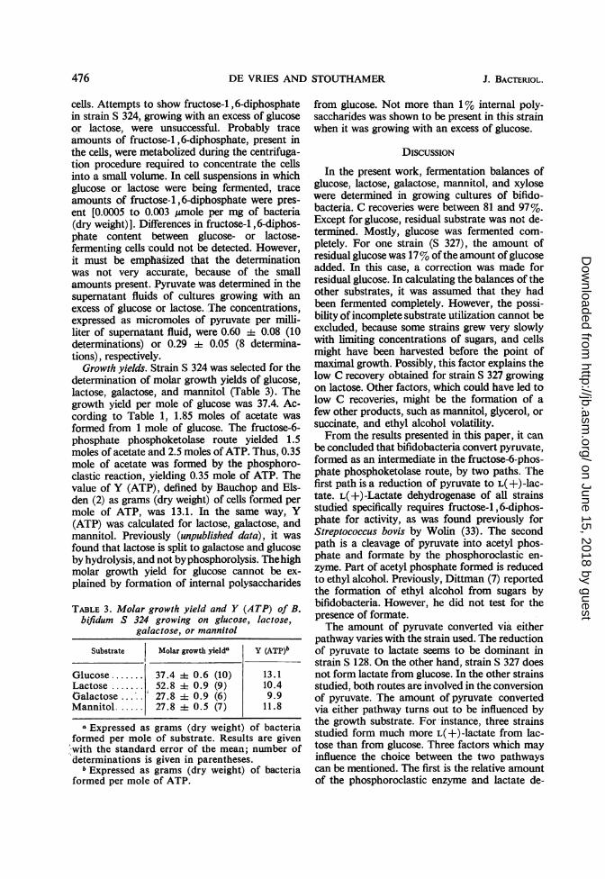
Growth yields. Strain S 324 was selected for thedetermination of molar growth yields of glucose,lactose, galactose, and mannitol (Table 3). Thegrowth yield per mole of glucose was 37.4. Ac-cording to Table 1, 1.85 moles of acetate wasformed from 1 mole of glucose. The fructose-6-phosphate phosphoketolase route yielded 1.5moles of acetate and 2.5 moles of ATP. Thus, 0.35mole of acetate was formed by the phosphoro-clastic reaction, yielding 0.35 mole of ATP. Thevalue of Y (ATP), defined by Bauchop and Els-den (2) as grams (dry weight) of cells formed permole of ATP, was 13.1. In the same way, Y(ATP) was calculated for lactose, galactose, andmannitol. Previously (unpublished data), it wasfound that lactose is split to galactose and glucoseby hydrolysis, and not byphosphorolysis. Thehighmolar growth yield for glucose cannot be ex-plained by formation of internal polysaccharides
TABLE 3. Molar growth yield and Y (ATP) of B.bifidum S 324 growing on glucose, lactose,
galactose, or mannitol
Substrate Molar growth yielda Y (ATP)b
Glucose ....... 37.4 l 0.6 (10) 13.1Lactose ....... 52.8 i 0.9 (9) 10.4Galactose 27.8 i 0.9 (6) 9.9Mannitol...... 27.8 4 0.5 (7) 11.8
a Expressed as grams (dry weight) of bacteriaformed per mole of substrate. Results are given.with the standard error of the mean; number ofdeterminations is given in parentheses.
b Expressed as grams (dry weight) of bacteriaformed per mole of ATP.
from glucose. Not more than 1% internal poly-saccharides was shown to be present in this strainwhen it was growing with an excess of glucose.
DIscussioN
In the present work, fermentation balances ofglucose, lactose, galactose, mannitol, and xylosewere determined in growing cultures of bifido-bacteria. C recoveries were between 81 and 97%.Except for glucose, residual substrate was not de-termined. Mostly, glucose was fermented com-pletely. For one strain (S 327), the amount ofresidual glucose was 17% of the amount of glucoseadded. In this case, a correction was made forresidual glucose. In calculating the balances of theother substrates, it was assumed that they hadbeen fermented completely. However, the possi-bility of incomplete substrate utilization cannot beexcluded, because some strains grew very slowlywith limiting concentrations of sugars, and cellsmight have been harvested before the point ofmaximal growth. Possibly, this factor explains thelow C recovery obtained for strain S 327 growingon lactose. Other factors, which could have led tolow C recoveries, might be the formation of afew other products, such as mannitol, glycerol, orsuccinate, and ethyl alcohol volatility.From the results presented in this paper, it can
be concluded that bifidobacteria convert pyruvate,formed as an intermediate in the fructose-6-phos-phate phosphoketolase route, by two paths. Thefirst path is a reduction of pyruvate to L( +)-lac-tate. L(+)-Lactate dehydrogenase of all strainsstudied specifically requires fructose-1,6-diphos-phate for activity, as was found previously forStreptococcus bovis by Wolin (33). The secondpath is a cleavage of pyruvate into acetyl phos-phate and formate by the phosphoroclastic en-zyme. Part of acetyl phosphate formed is reducedto ethyl alcohol. Previously, Dittman (7) reportedthe formation of ethyl alcohol from sugars bybifidobacteria. However, he did not test for thepresence of formate.The amount of pyruvate converted via either
pathway varies with the strain used. The reductionof pyruvate to lactate seems to be dominant instrain S 128. On the other hand, strain S 327 doesnot form lactate from glucose. In the other strainsstudied, both routes are involved in the conversionof pyruvate. The amount of pyruvate convertedvia either pathway turns out to be influenced bythe growth substrate. For instance, three strainsstudied form much more L(+)-lactate from lac-tose than from glucose. Three factors which mayinfluence the choice between the two pathwayscan be mentioned. The first is the relative amountof the phosphoroclastic enzyme and lactate de-
476 J. BACTERIOL.
on June 15, 2018 by guesthttp://jb.asm
.org/D
ownloaded from

VOL. 96, 1968 CARBOHYDRATE FERMENTATION BY BIFIDOBACTERIA
hydrogenase. This factor cannot be tested, be-cause all attempts to show the phosphoroclasticenzyme in cell-free extracts failed. Most proba--bly, this enzyme is very unstable and is inactivatedduring the preparation of the cell-free extract. Asecond factor, the amount of fructose-i , 6-diphos-phate present in the cell, might explain why strainS 327 does not form lactate from glucose. Thisstrain grows very slowly on glucose, and the in-tracellular concentrations of fructose-6-phos-phate and ATP may remain too low to form theconcentration of fructose-i ,6-diphosphate re-quired to activate lactate dehydrogenase. Forstrain S 324, no difference in fructose-I , 6-diphos-phate content between lactose- and glucose-fer-menting cell suspensions was found, althoughmore lactate was formed from lactose than fromglucose. In both cell suspensions, trace amounts offructose-i , 6-diphosphate were present. Assumingthat the water content of bifidobacteria is 80%and the density is 1.3, the amounts of fructose-1 ,6-diphosphate vary from 0.1 to 0.5 ,umole perml of undried bacteria. Comparison of this valuewith the amount of fructose-i ,6-diphosphate(4 X 10⁻³ umole per ml of reaction mixture) re-quired to yield 50% of maximal activity of lactatedehydrogenase makes it evident that the low con-centration of fructose-I , 6-diphosphate found inthe cells is sufficient to account for activation oflactate dehydrogenase. A third factor which mightregulate the conversion of pyruvate could be adifferent affinity of lactate dehydrogenase and thephosphoroclastic enzyme toward pyruvate. Incultures of strain S 324 growing with glucose, thestationary concentration of pyruvate was abouttwice as large as that in cultures growing withlactose. The difference between the fermentationbalances of glucose and lactose found for thisstrain might be explained by supposing that thephosphoroclastic enzyme has a lower affinity forpyruvate than does lactate dehydrogenase.Two pathways for converting mannitol are
known for bacteria. Escherichia coli, Aerobacteraerogenes, Bacillus subtilis, Staphylococcus aureus,and L. plantarum convert mannitol to fructose-6-phosphate by means of mannitol phosphotrans-ferase, utilizing ATP or phosphoenolpyruvate as aphosphate donor, and mannitol-l-phosphate de-hydrogenase (4, 5, 10, 13, 16, 26, 27, 32). Theconversion of mannitol to fructose by a dehydro-genase has been described for L. brevis (19),Gluconobacter oxydans (12, 24), Acetobactersuboxydans (1), Azotobacter agilis (18), andPseudomonas fluorescens (22). Mostly, the de-hydrogenase is a polyol dehydrogenase which con-verts both mannitol and sorbitol. In this paper,it is shown that the first steps in the degradationof mannitol by bifidobacteria are dehydrogena-
tion to fructose by an inducible, NAD-specificpolyol dehydrogenase and subsequent phos-phorylation of fructose to fructose-6-phosphateby an inducible fructokinase.Bauchop and Elsden (2) defined Y (ATP) as
grams (dry weight) of cells formed per mole ofATP. They found Y (ATP) = 10 for Strepto-coccus faecalis and Saccharomyces cerevisiae.Other investigators (9, 25) found about the samevalue for several other bacteria. It was suggestedthat Y (ATP) is a constant for different bacteria.In the present investigation, molar growth yieldsof glucose, lactose, galactose, and mannitol weredetermined for one strain of Bifidobacterium.From the molar growth yields and fermentationbalances, Y (ATP) was calculated. The values ofY (ATP) were 13.1, 10.4, 9.9, and 11.8 for glu-cose, lactose, galactose, and mannitol, respec-tively. The mean value of Y (ATP) is 11.3 ±+i 0.7(4x). This value agrees with the value of 10 ini-tially reported by Bauchop and Elsden (2) andsince found for several other bacteria.
ACKNOWLEDGMENTSThanks are due to K. C. Winkler, J. de Waart, H.
Beerens, and G. Reuter for supplying strains of B.bifidum. We are grateful to J. Knappe (University ofHeidelberg) for supplying a culture of Pseudomonasoxalaticus, and for his advice on how to carry out theisolation of formate dehydrogenase from this strain.We also wish to thank J. H. van de Kamer (Uni-versity of Utrecht) for the gas chromatographic analy-sis of one of our samples. The technical assistance ofM. G. G. Schotanus is gratefully acknowledged.
LITERATURE CITED
1. Arcus, A. C., and N. L. Edson. 1956. Polyoldehydrogenase. 2. The polyol dehydrogenase ofAcetobacter suboxydans and Candida utilis.Biochem. J. 64:385-394.
2. Bauchop, T., and S. R. Elsden. 1960. The growthof micro-organisms in relation to their energysupply. J. Gen. Microbiol. 23:457-469.
3. Bucher, T., and H. J. Hohorst. 1962. Dihydroxy-acetonphosphat, D-Fructose-1 , 6-diphosphatund Glycerinaldehyd-3-phosphat, p. 246-252.In H. U. Bergmeyer (ed.), Methoden derenzymatischen Analyse. Verlag Chemie, GmbH,Weinheim.
4. Chakravorty, M. 1964. Metabolism of mannitoland induction of mannitol 1-phosphate dehy-drogenase in Lactobacillus plantarum. J.Bacteriol. 87:1246-1248.
5. Chakravorty, M. 1965. Mannitol-l-phosphatedehydrogenase from the extracts of Lactobacil-lus plantarum. Biochim. Biophys. Acta 105:374-377.
6. Dehnert, J. 1957. Untersuchung uber die gram-positive Stuhlflora der Brustmilchkindes. II.Mitteilung. Zentr. Bacteriol. Parasitenk. Abt.I Orig. 169:66-83.
477
on June 15, 2018 by guesthttp://jb.asm
.org/D
ownloaded from
DE VRIES AND STOUTHAMER
7. Dittmann, J., J. B. Mayer, and P. Huber. 1967.Zum Stoffwechsel des Bacterium bifidum. X.Bildung van Athanol. Z. Kinderheilk. 99:115-118.
8. Friedemann, T. E., and G. E. Haugen. 1943.Pyruvic acid. II. The determination of ketoacids in blood and urine. J. Biol. Chem.147:415-442.
9. Hadjipetrou, L. P., J. P. Gerrits, F. A. G. Teu-lings, and A. H. Stouthamer. 1964. Relationbetween energy production and growth ofAerobacter aerogenes. J. Gen. Microbiol.36:139-150.
10. Horwitz, S. B., and N. 0. Kaplan. 1964. Hexitoldehydrogenase of Bacillus subtilis. J. Biol.Chem. 239:830-838.
11. Johnson, P. A., M. C. Jones-Mortimer, and J. R.Quayle. 1964. Use of a purified bacterial for-mate dehydrogenase for the micro-estimation offornate. Biochim. Biophys. Acta 89:351-353.
12. Kersters, K., W. A. Wood, and J. de Ley. 1965.Polyol dehydrogenases of Gluconobacter oxy-dans. J. Biol. Chem. 240:965-974.
13. Klungs0yr, L. 1966. Mannitol kinase in cell-freeextracts of Escherichia coli. Biochim. Biophys.Acta 122:361-364.
14. Knappe, J., E. Bohnert, and W. Briummer. 1965.S-Adenosyl-L-methionine, a component of theclastic dissimilation of pyruvate in Escherichiacoli. Biochim. Biophys. Acta 107:603-605.
15. Lipmann, F., and L. C. Tuttle. 1945. A specificmicromethod for the determination of acyl-phosphates. J. Biol. Chem. 159:21-28.
16. Liss, M., S. B. Horwitz, and N. 0. Kaplan. 1962.D-mannitol-l-phosphate dehydrogenase andD-sorbitol-6-phosphate dehydrogenase in Aero-bacter aerogenes. J. Biol. Chem. 237:1342-1350.
17. Lowry, 0. H., N. J. Rosebrough, A. L. Fan, andR. J. Randall. 1951. Protein measurement withthe Folin phenol reagent. J. Biol. Chem.193:265-275.
18. Marcus, L., and A. G. Marr. 1961. Polyol dehy-drogenases of Azotobacter agilis. J. Bacteriol.82:224-232.
19. Martinez, G., H. A. Barker, and B. L. Horecker.1963. A specific mannitol dehydrogenase fromLactobacillus brevis. J. Biol. Chem. 238:1598-1603.
20. Rose, I. A., M. Grunberg-Manago, S. R. Korey,and S. Ochoa. 1954. Enzymatic phosphoryla-tion of acetate. J. Biol. Chem. 211:737-756.
21. Scardovi, V. 1965. The fructose-6-phosphateshunt as a peculiar pattern of hexose degrada-tion in the genus Bifidobacterium. Ann. Micro-biol. Enzimol. 15:19-28.
22. Sebek, 0. K., and C. I. Randles. 1952. The oxida-tive dissimilation of mannitol and sorbitol byPseudomonas fluorescens. J. Bacteriol. 63:693-700.
23. Slein, M. W., G. T. Cori, and C. F. Cori. 1950.A comparative study of hexokinase from yeastand animal tissues. J. Biol. Chem. 186:763-790.
24. Stouthamer, A. H. 1961. Glucose and galactosemetabolism in Gluconobacter liquefaciens.Biochim. Biophys. Acta 48:484-500.
25. Stouthamer, A. H. 1961. Energy production inGluconobacter liquefaciens. Biochim. Biophys.Acta 56:19-32.
26. Strasters, K. C. 1965. Dissimilation of mannitolby Staphylococcus aureus. Antonie van Leeu-wenhoek J. Microbiol. Serol. 31:113-123.
27. Tanaka, S., S. A. Lerner, and E. C. C. Lin. 1967.Replacement of a phosphoenolpyruvate-de-pendent phosphotransferase by a nicotinamideadenine dinucleotide-linked dehydrogenase forthe utilization of mannitol. J. Bacteriol. 93:642-648.
28. Trevelyan, W. E., and J. S. Harrison. 1952. Studieson yeast metabolism. I. Fractionating andmicrodetermination of cell carbohydrates.Biochem. J. 50:298-303.
29. Vries, W. de, S. J. Gerbrandy, and A. H. Stout-hamer. 1967. Carbohydrate metabolism inBifidobacterium bifidum. Biochim. Biophys.Acta 136:415-425.
30. Vries, W. de, and A. H. Stoutbamer. 1967. Path-way of glucose fermentation in relation to thetaxonomy of bifidobacteria. J. Bacteriol.93:574-576.
31. Westerfeld, W. W. 1952. Colorimetric determina-tion of acetoin plus diacetyl. p. 39. In A. C.Neish (ed.), Analytical methods for bacterialfermentations. National Research Council ofCanada, Saskatoon.
32. Wolff, J. B., and N. 0. Kaplan. 1956. D-Manni-tol-l-phosphate dehydrogenase from Escher-ichia coli. J. Biol. Chem. 218:849-869.
33. Wolin, M. J. 1964. Fructose-1,6-diphosphaterequirement of streptococcal lactic dehydro-genases. Science 146:775-777.
478 J. BACTERIOL.
on June 15, 2018 by guesthttp://jb.asm
.org/D
ownloaded from