Journal of Radiology Research and Practice fileResearch Article Single Coronary Artery Authors...
25
Transcript of Journal of Radiology Research and Practice fileResearch Article Single Coronary Artery Authors...


Journal of Radiology Research and Practice Vol. 2015 (2015), Article ID 312482, 25 minipages.
DOI:10.5171/2015.312482
www.ibimapublishing.com
Copyright © 2015. Jonszta Tomas, Pleva Leos, Krivankova
Katerina and Pelikan Anton. Distributed under Creative Commons
CC-BY 4.0

Research Article
Single Coronary Artery
Authors
Jonszta Tomas Institute of Radiology, Ostrava University Hospital, Czech Republic
Medical Faculty, University of Ostrava, Czech Republic
Pleva Leos Cardiovascular Department, Ostrava University Hospital, Czech Republic
Medical Faculty, University of Ostrava, Czech Republic
Krivankova Katerina Institute of Radiology, Ostrava University Hospital, Czech Republic
Pelikan Anton Department of Surgery, Ostrava University Hospital, Czech Republic

Received date: 12 January 2014;
Accepted date: 9 March 2015;
Published date: 1 June 2015
Academic Editor: George D. Giannoglou
Cite this Article as: Jonszta Tomas, Pleva Leos, Krivankova Katerina and
Pelikan Anton (2015), "Single Coronary Artery," Journal of Radiology
Research and Practice, Vol. 2015 (2015), Article ID 312482,
DOI: 10.5171/2015.312482

Abstract
Single coronary artery is a rare congenital coronary artery
anomaly. It can be associated with myocardial ischemia,
increased risk of sudden cardiac death or other congenital
cardiac anomalies. The authors present a case of isolated single
coronary artery with origin of the right coronary artery (RCA)
from the left circumflex artery (LCx) with benign course, found
on coronary angiography and computed tomography coronary
angiography. A 79-years old female patient presenting with
shortness of breath and atypical chest pain was examined. No
significant stenosis was detected, and the patient was dismissed
from the hospital with adjustment of her medication only. The
contribution of conventional coronary angiography, and cardiac

computed tomography for meticulous anatomic depiction of
coronary arteries is discussed.
Keywords: Coronary anomaly, computed tomography coronary
angiography, extreme left coronary dominance

Introduction
Isolated coronary artery anomalies are found in approximately
1% of patients on coronary arteriograms and 0,3% of patients at
autopsy. 1-3 In as little as 0,024% of people, a single coronary
artery is formed.4,5 This variation is usually benign, but may be
associated with congenital heart disease such as tetralogy of
Fallot, transposition of the great arteries, truncus arteriosus, and
coronary artery fistula.6 We present a patient with single left
coronary artery detected by coronary angiography and further
depicted by cardiac CT.

Case Description
A 79-years old female with progressive shortness of breath,
occasional palpitations, atypical chest pain and diminishing
tolerance of exertion was scheduled for diagnostic coronary
angiography. On physical examination, the blood pressure was
130/90 mmHg, and her pulse rate was 68. The electrocardiogram
displayed atrial fibrillation, slight ST depression and T-wave
inversion in V4-6. The transthoracic echocardiographic
examination proved normal systolic function of the heart with
minor mitral and tricuspid valves regurgitation. She has also
been treated for hypertension for a long time, and had pacemaker
implanted in 2006 and repositioned in 2009. She suffered from
chronic atrial fibrillation and dyslipidemia. She was on

anticoagulation therapy with Warfarin. Previous chest X-ray
revealed atherosclerotic plaques in the aortic walls.
The course of the cardiac catheterization was uneventful with no
signs of significant coronary arteries stenosing process. The right
coronary artery origin was not detected during the procedure.
Both attempts of selective catheterization and aortography using
power-injector were negative for right coronary artery ostium
detection. Instead, a large single left coronary artery was
discovered with large calibre left circumflex artery continuing
distally, crossing the crux cordis and following the right
atrioventricular groove, supplying the right coronary artery
territory branches. (Figure 1, 2) This retrocardiac course of the
artery is considered a benign variant. Therefore, subsequent
coronary computed tomography angiography was ordered to
depict thoroughly the cardiac anatomy, and exclude any
coincidental anomaly of the chest arteries. The examination was
performed without prior beta blockers administration on the
single source 128- slice CT, with standard cardiac CT scanning
mode and retrospective gating protocol. The CT device employed
was Siemens Definition AS+, Erlangen, Germany and the datasets
were post-processed using Siemens Circulation and Aquarius
Terarecon software. The quality of the images was good and
allowed complete depiction of this variation. No accompanying
great vessels anomalies were detected. (Figures 3-5) The V/Q
lung scan was negative for pulmonary embolization. No
correlation between angiographic findings and the patient´s
symptoms was found. The patient was treated conservatively;
minor changes in her antihypertensive medication were only
done, and the patient´s clinical status has been satisfactory so far.

Figure: 1 Conventional Coronary Angiography: Large-
Calibre, Long Left Circumflex Artery Supplying the Right
Ventricular and Atrial Branches, Right Coronary Artery Originating as a Distal Continuation of the LCx.
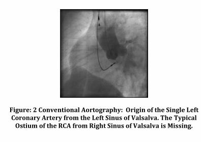
Figure: 2 Conventional Aortography: Origin of the Single Left Coronary Artery from the Left Sinus of Valsalva. The Typical
Ostium of the RCA from Right Sinus of Valsalva is Missing.

Figure: 3 Coronary CT Angiography- VRT Reconstruction:
Origin of the Left Coronary Artery from Left Sinus of Valsalva. Right Coronary Artery Doesn´t Originate from the
Right Sinus of Valsalva.

Figure: 4 Coronary CT Angiography- MIP Curved
Reconstruction along the Long Axis of the LCx-RCA: Atherosclerotic Plaques without Significant Artery Stenosis.

Figure: 5 Coronary CT Angiography- Subtracted VRT Reconstruction: Demonstrating the Long Left Circumflex
Artery Supplying the Right Ventricular and Atrial Branches.

Discussion
Coronary artery variations or anomalies are relatively often
encountered in cardiac patients. The incidence is about 1 in 100
patients undergoing cardiac catheterization. Coronary arteries
variations are classified as anomalies with or without shunt or
congenital aneurysms.7,8
In our patient, a single left coronary artery was detected, which is
considered a very rare finding. According to the scientific
literature, a single coronary artery occurs in 0,024%-0,066% of
people.4,5 In the large group of 8235 patients examined by
coronary angiography published by Donaldson et Al. (1982),
there was the right coronary artery entirely replaced by a large
circumflex artery only in 4 patients.9 Single coronary artery is

defined as only one coronary artery originating from sinus of
Valsalva, supplying the entire heart, regardless of its distribution
pattern. Depending on the course of the RCA, this finding may be
benign or may place the patient at increased cardiac risk. If the
anomalous artery traverses within the aortic wall or between the
aorta and pulmonary artery, it can be associated with myocardial
ischemia and increased risk for sudden cardiac death.
In our patient, the RCA was present as the distal continuation of
the LCx behind the atrioventricular (AV) valves, that is called the
posterior (to the AV valves) course of an ectopically originating
RCA from the left coronary artery. This variant is considered to
be benign and classified as Lipton (1979) L1 anomaly. According
to a newer classification by Yamanaka et Al. (1990), the L1 group
of single coronary artery anomalies usually has a benign clinical

course.1 This configuration can also be called extreme left
dominance. Normally, approximately 85% of the general
population is right-dominant, 8% are left-dominant and 7% are
co-dominant.10
Traditionally, most radiographic descriptions of the coronary
arteries have been based on the cine coronary angiograms
performed by coronary ostia catheterization, and direct contrast
media injection. In the last decade, the cardiac CT as a
complementary imaging modality has brought new dimensions in
coronary vessels imaging. It provides detailed anatomic
information on coronary origin, course and branching and allows
evaluation of all great mediastinal vessels. Various
postprocessing methods enable also vessel walls evaluation.11, 12

The combination of the two above mentioned methods brings
most comprehensive evaluation of the coronary anatomy.
However, rarely the single coronary artery with origination of the
RCA from the distal LCx is, there can be found a handful of case
reports in the recent scientific literature, that demonstrate the
same or similar findings, that we encountered in our patient, as
well as very useful comments on classification and nomenclature. 13-17
Conclusion
Coronary artery anomalies should be regarded as a diverse group
of congenital disorders, whose manifestations and
pathophysiological mechanisms are highly variable. Some of the

anomalies like myocardial bridges are surely encountered in
more than 1% of patients, and probably should be considered
normal variant. Some other may have life-threatening
consequences or may be encountered with other cardiac
anomalies. The cardiac CT plays a key role in diagnostic
algorithm of coronary artery variations imaging, and allows
perfect anatomic depiction of the coronary artery course.
References
1. Yamanaka, O. & Hobbs, R. E. (1990). “Coronary Artery
Anomalies in 126,595 Patients Undergoing Coronary
Arteriography,” Catheterization and Cardiovascular Diagnosis, 21
28-40.

2. Alexander, R. W. & Griffith, G. C. (1956). “Anomalies of the
Coronary Arteries and Their Significance,” Circulation, 14 800-
805.
3. Angelini, P., Velasco, J. A. & Flamm, S. (2002). “Coronary
Anomalies: Incidence, Pathophysiology, and Clinical Relevance,”
Circulation, 105 2449-2454.
4. Lipton, M. J., Barry, W. H., Obrez, I. et Al. (1979). “Isolated
Single Coronary Artery: Diagnosis, Angiographic Classification,
and Clinical Significance,” Radiology, 130 39-47
5. Desmet, W., Vanhaecke, J., Vrolix, M. et al. (1992). “Isolated
Single Coronary Artery: A Review of 50,000 Consecutive
Coronary Angiographies,” European Heart Journal, 13 1637-
1640.

6. Memisoglu, E., Hobikoglu, G., Tepe, M. S. et al. (2005).
“Congenital Coronary Anomalies in Adults: Comparison of
Anatomic Course Visualized by Catheter Angiography and
Electron Beam CT,” Catheterization and Cardiovascular
Interventions, 66 34-42.
7. Fiss, D. M. (2007). “Normal Coronary Anatomy and Anatomic
Variations,” Applied Radiology, vol. 36 (1) 14-26.
8. Morettin, L. B. (1976). “Coronary Arteriography: Uncommon
Observations,” Radiologic Clinics of North America, 14 189-208.
9. Donaldson, R. M. & Raphael, M. J. (1982). “Missing Coronary
Artery- Review of Technical Problems in Coronary Arography

Resulting from Anatomical Variants,” British Heart Journal, 47 62-
70.
10. Gensini, G. G. (1975). 'Coronary Arteriography,' Mount Kisco,
NY: Futura Publishing Co, 260–274.
11. O’Brien, J. P., Srichai, M. B., Hecht, E. M. et Al. (2007).
“Anatomy of the Heart at Multidetector CT: What the Radiologist
Needs to Know,” RadioGraphics, 27 1569-1582.
12. Rajiah, P., Halliburton, S. S., Scott, F. D. et Al. (2012).
“Strategies for Dose Reduction in Cardiovascular Computed
Tomography,” Applied Radiology, 41 (7-8) 10-15.

13. Desai, A. V., Parikh, P. & La Barbera, M. (2013). “Anomalous
Right Coronary Artery Originating from Left Circumflex Artery:
An Unusual Single Coronary Artery Anomaly,” Cath Lab Digest 21
(2) 22.
14. Chung, S. K., Lee, S. J., Park, S. H. et Al. (2010). “An Extremely
Rare Variety of Anomalous Coronary Artery: Right Coronary
Artery Originating from the Distal Left Circumflex Artery,” Korean
Circulation Journal 2010; 40(9):465-7.
15. Andreou, A. Y., Tryfonos, A., Christodoulou, C. et Al. (2012).
“Isolated single Coronary Artery with Dual Right Coronary Artery
Distribution. A Rare Anatomical Variation,” Herz. 2012;
37(4):432-5.

16. Angelini, P. (2007). “Coronary Artery Anomalies: An Entity in
Search of an Identity,” Circulation. 2007 Mar 13; 115(10):1296-
305.
17. Angelini, P. (2008). "An Illustrative Case of a Frequently-Held
Misconception: The "Absent" RCA," Journal of Invasive Cardiology
2008; 20(7):384.