05815 Isay Weinfeld - Midrash Cultural Center, Rio de Janeiro RJ, Brazil
Journal of Ethnopharmacology 175 (2015) 490–498
Contents lists available at ScienceDirect
Journal of Ethnopharmacology
http://d0378-87
n CorrE-m1 Pr
de Jane
journal homepage: www.elsevier.com/locate/jep
Anti-inflammatory effect of Schinus terebinthifolius Raddihydroalcoholic extract on neutrophil migration in zymosan-inducedarthritis
Elaine Cruz Rosas a,b,n, Luana Barbosa Correa a,b, Tatiana de Almeida Pádua a,b,Thadeu Estevam Moreira Maramaldo Costa a,b, José Luiz Mazzei d, Alan Patrick Heringer c,Carlos Alberto Bizarro a,1, Maria Auxiliadora Coelho Kaplan e, Maria Raquel Figueiredo c,Maria G Henriques a,b,n
a Laboratory of Applied Pharmacology, Farmanguinhos, Oswaldo Cruz Foundation, Rio de Janeiro, RJ, Brazilb National Institute for Science and Technology on Innovation on Neglected Diseases (INCT/IDN), Center for Technological Development in Health (CDTS),Oswaldo Cruz Foundation (Fiocruz), Rio de Janeiro, RJ, Brazilc Natural Products Laboratory, Farmanguinhos, Oswaldo Cruz Foundation (Fiocruz), Rio de Janeiro, RJ, Brazild Analytical Center, Farmanguinhos, Oswaldo Cruz Foundation (Fiocruz), Rio de Janeiro, RJ, Brazile Natural Products Research Center (NPPN), Federal University of Rio de Janeiro (UFRJ), Brazil
a r t i c l e i n f o
Article history:Received 26 May 2015Received in revised form4 October 2015Accepted 5 October 2015Available online 8 October 2015
Keywords:Schinus terebinthifolius RaddiArthritisNeutrophils
Chemical compounds studied in this article::Gallic acid (PubChem CID: 370)Methyl gallate (PubChem CID: 7428)Pentagalloylglucose (PubChem CID: 65238)
x.doi.org/10.1016/j.jep.2015.10.01441/& 2015 Elsevier Ireland Ltd. All rights rese
esponding authors at: Laboratory of Appliedail addresses: [email protected] (E.C. Rosas),esent address: Germano Sinval Faria School Hiro, RJ, Brazil.
a b s t r a c t
Ethnopharmacological relevance: Schinus terebinthifolius is a species of plant from the Anacardiaceae fa-mily, which can be found in different regions of Brazil. Schinus is popularly known as aroeirinha, aroeira-vermelha, or Brazilian pepper. In folk medicine, S. terebinthifolius is used for several disorders, includinginflammatory conditions, skin wounds, mucosal membrane ulcers, respiratory problems, gout, tumors,diarrhea and arthritis. According to chemical analyses, gallic acid, methyl gallate and pentagalloylglucoseare the main components of hydroalcoholic extracts from S. terebinthifolius leaves. In the present study,we demonstrated the ability of a hydroalcoholic extract to inhibit cell migration in arthritis and in-vestigated the mechanisms underlying this phenomenon.Materials and methods: The anti-inflammatory effect of S. terebinthifolius hydroalcoholic leaf extract (ST-70) was investigated in a zymosan-induced experimental model of inflammation. Male Swiss and C57Bl/6 mice received zymosan (100 mg/cavity) via intra-thoracic (i.t.) or intra-articular (i.a.) injection after oralpre-treatment with ST-70. The direct action of ST-70 on neutrophils was evaluated via chemotaxis.Results: ST-70 exhibited a dose-dependent effect in the pleurisy model. The median effective dose (ED50)was 100 mg/kg, which inhibited 70% of neutrophil accumulation when compared with the control group.ST-70 reduced joint diameter and neutrophil influx for synovial tissues at 6 h and 24 h in zymosan-induced arthritis. Additionally, ST-70 inhibited synovial interleukin (IL)-6, IL-1β, keratinocyte-derivedchemokine (CXCL1/KC) and Tumor Necrosis Factor (TNF)-α production at 6 h and CXCL1/KC and IL-1βproduction at 24 h. The direct activity of ST-70 on neutrophils was observed via the impairment ofCXCL1/KC-induced chemotaxis in neutrophils. Oral administration of ST-70 did not induce gastric da-mage. Daily administration for twenty days did not kill any animals. In contrast, similar administrationsof diclofenac induced gastric damage and killed all animals by the fifth day.Conclusions: Our results demonstrated significant anti-inflammatory effects of ST-70, suggesting a pu-tative use of this herb for the development of phytomedicines to treat inflammatory diseases, such asjoint inflammation.
& 2015 Elsevier Ireland Ltd. All rights reserved.
rved.
Pharmacology, Farmanguinhos, Oswaldo Cruz Foundation, Rio de Janeiro, RJ, [email protected] (M. Henriques).ealth Center (CSEGSF), Sergio Arouca National School of Public Health (ENSP), Oswaldo Cruz Foundation (Fiocruz), Rio
E.C. Rosas et al. / Journal of Ethnopharmacology 175 (2015) 490–498 491
1. Introduction
Schinus terebinthifolius Raddi (Anacardiaceae) is a native plantfrom South America. It has been used in folk medicine as teas,infusions or tinctures; as an anti-inflammatory, febrifuge, analge-sic, and depurative agent; and to treat urogenital system illnesses(Medeiros et al., 2007). Through ethnopharmacological research,the gastroprotective properties of S. terebinthifolius are remarkablyeffective, especially in the treatment of gastritis and ulcers (Carliniet al., 2010). Previous reports have demonstrated that S. ter-ebinthifolius extracts or fractions rich in polyphenols, display an-tioxidant, antibacterial, antifungal and anti-allergic activities indifferent experimental models (Cavalher-Machado et al., 2008; deLima et al., 2006; Schmourlo et al., 2005; Velázquez et al., 2003).Despite its importance in popular medicine for the treatment ofinflammatory disorders, few scientific studies have examined thebiological activities and chemical composition of S. terebinthifoliusextracts.
Inflammation is a complex physiological response that occursin vascularized tissues in response to harmful stimuli, such aspathogens, damaged cells or irritants. The inflammatory process iscoordinated by different chemical mediators that induce vasodi-lation, plasma leakage and leukocyte margination. However, whenthe inflammatory response becomes prolonged or chronic, thesame process can become destructive and has been linked to anumber of diseases. Chronic inflammation can result from a failureto eliminate harmful stimuli, an abnormal autoimmune responseor the persistence of a low-intensity irritant that continuallycauses acute inflammation response (Medzhitov, 2010).
Rheumatoid arthritis (RA) is a chronic autoimmune in-flammatory disease characterized by pathological changes, such aspersistent synovitis, vascular proliferation, infiltration of in-flammatory cells, and damage to cartilage and bone (Scott et al.,2010). A critical factor that contributes to joint damage is the ex-cessive production of inflammatory mediators by resident or in-filtrating inflammatory cells. Cytokines (TNF-α and IL-1β) and ei-cosanoids (leukotrienes and prostaglandins) are involved in thepathogenesis of arthritis and participate in pain, neutrophil accu-mulation and tissue damage (Brennan and McInnes, 2008; Guer-rero et al., 2008). Recently, the importance of IL-17 has been stu-died in experimental arthritis, wherein the cytokine was detectedduring neutrophil accumulation and cartilage degradation and inhyperalgesic symptoms (Pinto et al., 2010). The recruitment ofneutrophils contributes to the local production of cytokines andjoint damage and appears to be important in the pathogenesis ofhuman arthritis (Wright et al., 2014). In the last decade, the in-volvement of other cells, such as macrophages, synoviocytes,lymphocytes and mast cells, have been described, indicating that awider variety of cells are also important in the perpetuating thearticular inflammatory process (McInnes and Schett, 2011).
Current clinical treatments for RA include steroidal and non-steroidal anti-inflammatory drugs (SAIDs and NSAIDs, respec-tively), disease-modifying antirheumatic drugs (DMARDs) andbiological agents (Kalden, 2002). However, the prolonged use ofSAIDS and NSAIDS has been associated with serious adverse ef-fects, including gastrointestinal disorders, immunodeficiency andhumoral disturbances (Roth, 2012), which are factors that havebeen attributed to treatment dropout.
In recent decades, the screening of safer and more potent anti-inflammatory drugs for clinical use has increased. In this context,plants with anti-inflammatory activities have shown promisingeffects against inflammatory diseases, such as arthritis (Lama andSaikia, 2011). A few reports have shown that a polyphenol fromgreen tea extract displayed a protective effect in a model of in-flammatory arthritis, largely through its ability to inhibit theproduction of key inflammatory mediators, such as IL-1β and IL-6,
by RA synovial fibroblasts (Ahmed et al., 2006).Considering the popular uses of teas and tinctures for medic-
inal purposes, we evaluated the anti-inflammatory effect of hy-droalcoholic extracts from S. terebinthifolius Raddi to assess itsability to inhibit cell migration and inflammatory mediators inexperimental arthritis. Furthermore, we explored the mechanismsinvolved in this phenomenon.
2. Materials and methods
2.1. Reagents
Zymosan serotype A, dexamethasone, potassium diclofenac,phosphate buffered saline (PBS), buffer perborate, o-phenylene-diamine dihydrochloride (OPD), Bradford reagent, bovine serumalbumin (BSA), ethylene diamine tetraacetic acid disodium salt(EDTA), RPMI 1640 medium and fMLP (N-formyl-methionyl-leu-cyl-phenylalanine) were all obtained from Sigma Chemical Co. (St.Louis, MO, USA). DMSO (for biological tests), ethyl ether, ethylacetate, n-hexane, dichloromethane, methanol and acetone forchromatography were purchased from Vetec Química Fina, Ltda.(Xerém, RJ, Brazil). LTB4 immuno-assay kit was obtained fromCayman Chemicals (Ann Arbor, Michigan, USA). Purified anti-murine TNF-α, CXCL1/KC, IL-6 and IL-1β mAbs; biotinylated anti-TNF-α, CXCL-1/KC, IL-6 and IL-1β mAbs; and recombinant TNF-α,CXCL-1/KC, IL-6 and IL-1β were all obtained from R&D Systems(Minneapolis, MN, USA).
2.2. Preparation and analysis of ST-70 extract
Leaves were collected from 10 individual of S. terebinthifoliusplants in the Atlantic Forest Campus FIOCRUZ, Jacarepaguá, Rio deJaneiro, RJ, Brazil, and a voucher specimen was deposited into theRio de Janeiro Botanical Garden Herbarium under number RB-451742.
The collected material were dried at 40 °C in an oven with aircirculation, reduced to small fragments and extracted with 70%ethanol in a dynamic maceration for 24 h. Then, the extract wasfiltered, concentrated under reduced pressure and lyophilized,resulting in a hydroethanolic extract (ST-70) with a yield of 11.00%.These conditions were based on previous studies of extractiontimes.
The ST-70 extract was analyzed using techniques such as ad-sorption column chromatography, thin layer chromatography,partition chromatography (countercurrent chromatography), gaschromatography coupled to mass spectrometry, high performanceliquid chromatography (HPLC) and crystallization by traditionalmethodologies.
Several different methodologies were employed to isolatecompounds from S. terebinthifolius. Spectrometric and spectro-scopic analyses led to the identification of luteolin, quercetin,kaempferol, agathisflavone, gallic acid (GA), methyl gallate (MG),1,2,3,4,6-pentagalloylglucose (PG), epicatechin, α-amyrin, β-amyr-in, and lupeol.
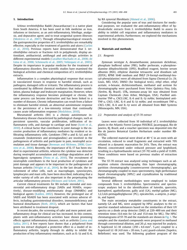
The main secondary metabolite constituents in the extract,namely GA and MG, were assigned by HPLC analyses to the re-spective standard substances based on the similarities of their UVspectra on a diode array detector (DAD) at 220–400 nm and theirretention times (6.6 min for GA and 15.0 min for MG). The HPLCchromatograms of ST-70 and the standards are shown in Fig. 1. Thesamples and the standard compounds were prepared in methanolthrough conventional dilution, and 20 mL was injected for analysis.A Supelcosil LC-18 column (250�4.6 mm2, 5 μm) coupled to aSupelcosil LC-18 (4.0 mm�20 mm, 5 mm) guard-column (Supelco,Bellefonte) was used. The following gradient program of the
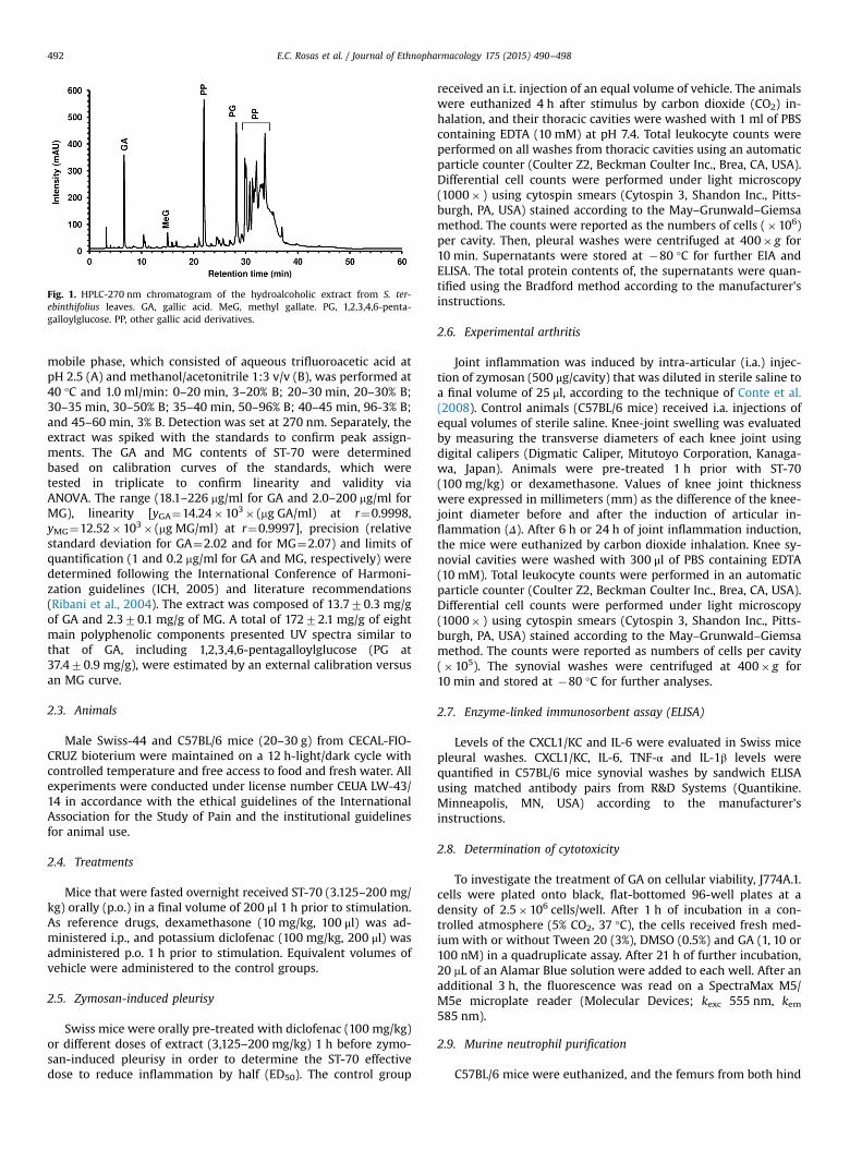
Fig. 1. HPLC-270 nm chromatogram of the hydroalcoholic extract from S. ter-ebinthifolius leaves. GA, gallic acid. MeG, methyl gallate. PG, 1,2,3,4,6-penta-galloylglucose. PP, other gallic acid derivatives.
E.C. Rosas et al. / Journal of Ethnopharmacology 175 (2015) 490–498492
mobile phase, which consisted of aqueous trifluoroacetic acid atpH 2.5 (A) and methanol/acetonitrile 1:3 v/v (B), was performed at40 °C and 1.0 ml/min: 0–20 min, 3–20% B; 20–30 min, 20–30% B;30–35 min, 30–50% B; 35–40 min, 50–96% B; 40–45 min, 96-3% B;and 45–60 min, 3% B. Detection was set at 270 nm. Separately, theextract was spiked with the standards to confirm peak assign-ments. The GA and MG contents of ST-70 were determinedbased on calibration curves of the standards, which weretested in triplicate to confirm linearity and validity viaANOVA. The range (18.1–226 mg/ml for GA and 2.0–200 mg/ml forMG), linearity [yGA¼14.24�103� (mg GA/ml) at r¼0.9998,yMG¼12.52�103� (mg MG/ml) at r¼0.9997], precision (relativestandard deviation for GA¼2.02 and for MG¼2.07) and limits ofquantification (1 and 0.2 mg/ml for GA and MG, respectively) weredetermined following the International Conference of Harmoni-zation guidelines (ICH, 2005) and literature recommendations(Ribani et al., 2004). The extract was composed of 13.770.3 mg/gof GA and 2.370.1 mg/g of MG. A total of 17272.1 mg/g of eightmain polyphenolic components presented UV spectra similar tothat of GA, including 1,2,3,4,6-pentagalloylglucose (PG at37.470.9 mg/g), were estimated by an external calibration versusan MG curve.
2.3. Animals
Male Swiss-44 and C57BL/6 mice (20–30 g) from CECAL-FIO-CRUZ bioterium were maintained on a 12 h-light/dark cycle withcontrolled temperature and free access to food and fresh water. Allexperiments were conducted under license number CEUA LW-43/14 in accordance with the ethical guidelines of the InternationalAssociation for the Study of Pain and the institutional guidelinesfor animal use.
2.4. Treatments
Mice that were fasted overnight received ST-70 (3.125–200 mg/kg) orally (p.o.) in a final volume of 200 μl 1 h prior to stimulation.As reference drugs, dexamethasone (10 mg/kg, 100 μl) was ad-ministered i.p., and potassium diclofenac (100 mg/kg, 200 μl) wasadministered p.o. 1 h prior to stimulation. Equivalent volumes ofvehicle were administered to the control groups.
2.5. Zymosan-induced pleurisy
Swiss mice were orally pre-treated with diclofenac (100 mg/kg)or different doses of extract (3,125–200 mg/kg) 1 h before zymo-san-induced pleurisy in order to determine the ST-70 effectivedose to reduce inflammation by half (ED50). The control group
received an i.t. injection of an equal volume of vehicle. The animalswere euthanized 4 h after stimulus by carbon dioxide (CO2) in-halation, and their thoracic cavities were washed with 1 ml of PBScontaining EDTA (10 mM) at pH 7.4. Total leukocyte counts wereperformed on all washes from thoracic cavities using an automaticparticle counter (Coulter Z2, Beckman Coulter Inc., Brea, CA, USA).Differential cell counts were performed under light microscopy(1000� ) using cytospin smears (Cytospin 3, Shandon Inc., Pitts-burgh, PA, USA) stained according to the May–Grunwald–Giemsamethod. The counts were reported as the numbers of cells (�106)per cavity. Then, pleural washes were centrifuged at 400� g for10 min. Supernatants were stored at �80 °C for further EIA andELISA. The total protein contents of, the supernatants were quan-tified using the Bradford method according to the manufacturer'sinstructions.
2.6. Experimental arthritis
Joint inflammation was induced by intra-articular (i.a.) injec-tion of zymosan (500 μg/cavity) that was diluted in sterile saline toa final volume of 25 μl, according to the technique of Conte et al.(2008). Control animals (C57BL/6 mice) received i.a. injections ofequal volumes of sterile saline. Knee-joint swelling was evaluatedby measuring the transverse diameters of each knee joint usingdigital calipers (Digmatic Caliper, Mitutoyo Corporation, Kanaga-wa, Japan). Animals were pre-treated 1 h prior with ST-70(100 mg/kg) or dexamethasone. Values of knee joint thicknesswere expressed in millimeters (mm) as the difference of the knee-joint diameter before and after the induction of articular in-flammation (Δ). After 6 h or 24 h of joint inflammation induction,the mice were euthanized by carbon dioxide inhalation. Knee sy-novial cavities were washed with 300 μl of PBS containing EDTA(10 mM). Total leukocyte counts were performed in an automaticparticle counter (Coulter Z2, Beckman Coulter Inc., Brea, CA, USA).Differential cell counts were performed under light microscopy(1000� ) using cytospin smears (Cytospin 3, Shandon Inc., Pitts-burgh, PA, USA) stained according to the May–Grunwald–Giemsamethod. The counts were reported as numbers of cells per cavity(�105). The synovial washes were centrifuged at 400� g for10 min and stored at �80 °C for further analyses.
2.7. Enzyme-linked immunosorbent assay (ELISA)
Levels of the CXCL1/KC and IL-6 were evaluated in Swiss micepleural washes. CXCL1/KC, IL-6, TNF-α and IL-1β levels werequantified in C57BL/6 mice synovial washes by sandwich ELISAusing matched antibody pairs from R&D Systems (Quantikine.Minneapolis, MN, USA) according to the manufacturer'sinstructions.
2.8. Determination of cytotoxicity
To investigate the treatment of GA on cellular viability, J774A.1.cells were plated onto black, flat-bottomed 96-well plates at adensity of 2.5�106 cells/well. After 1 h of incubation in a con-trolled atmosphere (5% CO2, 37 °C), the cells received fresh med-ium with or without Tween 20 (3%), DMSO (0.5%) and GA (1, 10 or100 nM) in a quadruplicate assay. After 21 h of further incubation,20 mL of an Alamar Blue solution were added to each well. After anadditional 3 h, the fluorescence was read on a SpectraMax M5/M5e microplate reader (Molecular Devices; kexc 555 nm, kem585 nm).
2.9. Murine neutrophil purification
C57BL/6 mice were euthanized, and the femurs from both hind
E.C. Rosas et al. / Journal of Ethnopharmacology 175 (2015) 490–498 493
legs were dissected, removed and freed of soft tissue attachments.The distal tip of the femur extremity was excised, and each femurwas washed with 2 ml of Ca2þ/Mg2þ-free HBSS–EDTA solution.Cell suspensions were then centrifuged at 400� g for 15 min at20 °C and resuspended in 2 ml Ca2þ/Mg2þ-free HBSS–EDTA. Thecells were purified via Percoll discontinuous gradient centrifuga-tion (65% and 72% diluted in Ca2þ/Mg2þ-free HBSS–EDTA). Briefly,the cells were centrifuged at 1200� g for 35 min at room tem-perature without braking, and polymorphonuclear cells were re-covered from the 65%/72% interface. Then, neutrophils werecounted in a Neubauer chamber, identified by cytospin cen-trifugation, and followed by Wright-Giemsa coloration accordingto the manufacturer's instructions.
2.10. Human neutrophil purification
Human neutrophils were isolated from citrate (3.8%)-treatedperipheral venous blood of healthy volunteers using a four-stepdiscontinuous Percoll gradient (Amersham Biosciences, San Fran-cisco, CA, USA). Erythrocytes were removed by hypotonic lysis.Isolated neutrophils (98% purity), estimated to be 496% viable byTrypan blue exclusion, were resuspended in RPMI 1640 medium.
2.11. Chemotaxis assay
Neutrophil chemotaxis was assayed in a 48-well Boydenchamber (Neuroprobe, Inc. Cabin John, MD, USA) using a 5-mmPVP-free polycarbonate filter (Nuclepore, Pleasanton, CA, USA).Briefly, purified murine neutrophils were incubated with ST-70(500 mg/ml) or dexamethasone (50 nM) for 1 h prior to the che-motaxis assay. Then, the cells were centrifuged at 400� g for10 min at room temperature and assayed in a chemotaxis cham-ber. The bottom wells of the chamber were filled with 29 ml of achemoattractant stimulant, CXCL-1/KC (250 nM). Pre-treatedneutrophils (1�105 cells/50 ml) were seeded in the upper com-partment. The chamber was then incubated at 37 °C in 5% CO2
atmosphere for 1 h, and the filter was fixed and stained by Wright-Giemsa coloration according to the manufacturer's instructions. Inanother set of assays, purified human neutrophils were incubatedwith one of the main polyphenols of ST-70, GA (100 nM), or dex-amethasone (50 nM) for 1 h prior to the chemotaxis assay. Then,the human cells were assayed in chemotaxis chambers, as de-scribed above, under fMLP (10 mM) stimulation. Neutrophils thathad migrated through the membrane were counted under a lightmicroscope (1000�magnification) in 15 random fields. The resultis expressed as the mean number of migrated neutrophils perfifteen fields.
2.12. Ulcerogenic activity and survival
Swiss mice that were fasted for 24 h were orally administeredeither different doses of ST-70 (100 or 400 mg/kg) or potassiumdiclofenac (100 mg/kg) diluted in filtered water to a final volumeof 200 ml. After 5 h, the mice were euthanized. The stomachs wereremoved, opened along the greater curvature and gently washedwith PBS. Gastric lesions were analyzed under a stereoscopic mi-croscope (Olympus, Japan) by a single-blind method. Additionally,Swiss mice received daily p.o. ST-70 (100 mg/kg), potassium di-clofenac (100 mg/kg) diluted in filtered water to a final volume of200 ml, or only filtered water (200 ml) for twenty days to assesssurvival rates.
2.13. Statistical analysis
All results are expressed as the means7standard errors of themeans (SEM) and were evaluated using one-way ANOVA followed
by the Student–Newman–Keuls test. A log-rank (Mantel–Cox) testwas used to compare the survival rates. The significance level wasset at Pr0.05.
3. Results
3.1. ST-70 inhibits leukocyte infiltration and inflammatory mediatorproduction in pleurisy
To determine the dose-response effect of ST70, it was appliedto zymosan-induced pleurisy. As shown in Fig. 2A and B, the oralpre-treatment with different doses of ST-70 (3.125–200 mg/kg)significantly inhibited (from 25 mg/kg) the migration into pleuralsites of total leukocytes, including neutrophils, 4 h after stimula-tion in a dose-dependent manner. According to the dose–responsecurve for leukocyte infiltration, the ED50 of ST-70 was 100 mg/kg(Fig. 2C). Potassium diclofenac, a nonsteroidal anti-inflammatorydrug (NSAID), was used as a reference inhibitor. As was observedwith 100 mg/kg ST-70, the oral pre-treatment with 100 mg/kgdiclofenac similarly inhibited total leukocyte and neutrophil ac-cumulation (Fig. S1).
The migration and activation of neutrophils during inflamma-tion results from several events, such as cytokine and chemokineaction. In zymosan pleural washes, increased CXCL-1/KC and IL-6levels were detected when compared with control groups. The oralpre-treatment with ST-70 (100 mg/kg) decreased the concentra-tions of CXCL-1/KC and IL-6 by 99.92% and 96.52%, respectively(Fig. 2D). A smaller decrease was observed after potassium diclo-fenac (100 mg/kg) pre-treatment (Fig. 2D).
3.2. ST-70 inhibits neutrophils migration on zymosan-inducedarthritis
We previously reported that the i.a. injection of zymosan(500 μg/cavity) induced an articular inflammatory response after6 h, which was characterized by a significant increase in edemaformation and massive neutrophil influx peaking at 24 h (Conteet al., 2008; Penido et al., 2006). In the present study, the analyseswere performed at 6 h and 24 h after stimulation because markedinflammatory reactions were already observed at these timepoints (Figs. 3 and 4). As shown in Fig. 3A and E, the oral pre-treatment with ST-70 (100 mg/kg) 1 h prior to zymosan injectionmarkedly reduced knee-joint thickness at 6 h and 24 h after sti-mulation. Notably, the oral treatment with ST-70 induced a similareffect as that achieved via the i.p. administration of dex-amethasone (10 mg/kg) (Fig. 3A and E). The treatments with ST-70or dexamethasone also inhibited total leukocyte, mononuclear celland neutrophil infiltration when compared with non-treated mice6 h after i.a. zymosan injection (Fig. 3B–D). However, 24 h afterstimulation, the ST-70 pre-treatment inhibited total leukocyte andneutrophil migration but failed to reduce mononuclear cell influxinto the synovial cavity (Fig. 3F–H). Interestingly, a single oraltreatment with ST-70 (100 mg/kg) was able to inhibit edema for-mation and leukocyte migration at all times analyzed after i.a.zymosan injection. It is noteworthy that ST-70 also inhibitedmurine neutrophil chemotaxis induced by CXCL-1/KC in vitro,demonstrating a direct effect of ST-70 on neutrophils (Fig. S2).
3.3. ST-70 reduces pro-inflammatory mediators in articular zymo-san-injected knee joints
To investigate whether ST-70 exerted its anti-inflammatoryeffect by modulating the production of inflammatory mediators,we analyzed its effect on the production of CXCL-1/KC, IL-6, TNF-αand IL-1β. As demonstrated in Fig. 4A–H, the articular cavity levels
Fig. 2. Study of the dose–response effect of the hydroalcoholic extract from S. terebinthifolius (ST-70) on cell migration and the production of inflammatory mediators. Effectof pre-treatment with ST-70 (3.125–200 mg/kg, p.o., 1 h before stimulation) on (A) total leukocyte and (B) neutrophil recruitment were analyzed 4 h after zymosan i.t.injection (100 mg/cavity; black columns). Control animals received saline i.t. injection (100 ml/cavity; white columns). (C) Dose–response log-curve of ST-70 treatment basedon neutrophil recruitment analysis; the ED50 was calculated as 100 mg/kg. (D) CXCL-1/KC and IL-6 production from thoracic cavity washes were measured by ELISA. Data arepresented as the means7SEM (n¼6). Significant differences (Po0.05) are indicated between stimulated and non-stimulated groups (*) and between treated and non-treated groups (þ).
E.C. Rosas et al. / Journal of Ethnopharmacology 175 (2015) 490–498494
of CXCL-1/KC, TNF-α, IL-1β and IL-6 increased at 6 h and at 24 hafter i.a. zymosan injection. ST-70 oral pre-treatment markedlyreduced zymosan-induced CXCL-1/KC, IL-6, TNF-α, and IL-1β pro-duction 6 h after i.a. injection (Fig. 4A–D). ST-70 (100 mg/kg) onlysignificantly inhibited CXCL-1/KC and IL-1β production 24 h after i.a. zymosan injection (Fig. 4E and H). Pre-treatment with dex-amethasone reduced the production of CXCL-1/KC, IL-6, TNF-α andIL-1β at both time points investigated.
3.4. Gallic acid from ST-70 exhibits anti-inflammatory activity
Phytochemical studies revealed that GA is one of the majorpolyphenol components of the ST-70 extract. To determine whe-ther this substance is responsible for the observed effect on neu-trophil migration, we evaluated the effect of GA on human neu-trophil chemotaxis. First, the effect of GA (1, 10 and 100 nM) was
assessed on cell viability using the resazurin assay; none of testedconcentrations were cytotoxic (100 nM Z95% viability). Pre-treatment with GA (100 nM) significantly reduced neutrophilchemotaxis (Fig. 5). This result suggests that GA is one of thebioactive component present in ST-70 extracts.
3.5. ST-70 does not induce gastric injury or death in mice
As shown in Fig. 6, animals that received a single oral dose ofsodium diclofenac (100 mg/kg) showed high indices of gastric le-sions (Fig. 6A) but did not exhibit decreased mucus secretions(Fig. 6B). Furthermore, 100% of the mice died up to the fifth dayafter daily sodium diclofenac oral administration (Fig. 6C). How-ever, ST-70 oral administration (100 or 400 mg/kg) induced fewgastric lesions when compared with the control group (non-trea-ted animals) and did not decrease the production of mucus
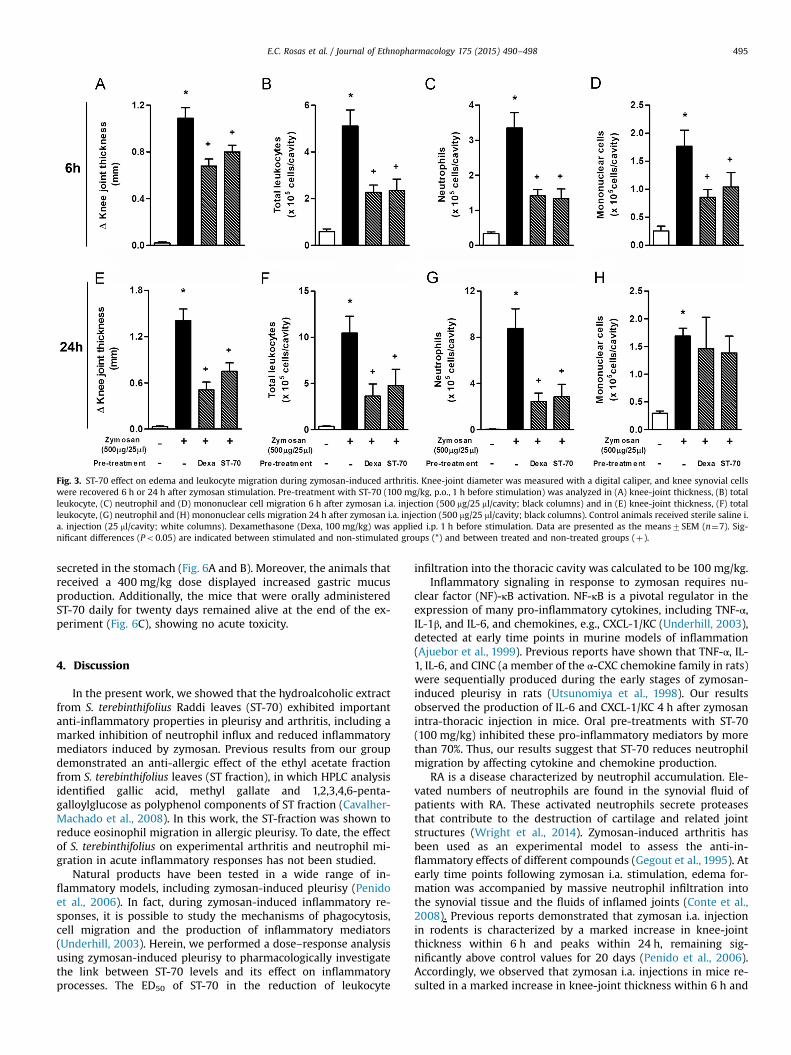
Fig. 3. ST-70 effect on edema and leukocyte migration during zymosan-induced arthritis. Knee-joint diameter was measured with a digital caliper, and knee synovial cellswere recovered 6 h or 24 h after zymosan stimulation. Pre-treatment with ST-70 (100 mg/kg, p.o., 1 h before stimulation) was analyzed in (A) knee-joint thickness, (B) totalleukocyte, (C) neutrophil and (D) mononuclear cell migration 6 h after zymosan i.a. injection (500 mg/25 ml/cavity; black columns) and in (E) knee-joint thickness, (F) totalleukocyte, (G) neutrophil and (H) mononuclear cells migration 24 h after zymosan i.a. injection (500 mg/25 ml/cavity; black columns). Control animals received sterile saline i.a. injection (25 ml/cavity; white columns). Dexamethasone (Dexa, 100 mg/kg) was applied i.p. 1 h before stimulation. Data are presented as the means7SEM (n¼7). Sig-nificant differences (Po0.05) are indicated between stimulated and non-stimulated groups (*) and between treated and non-treated groups (þ).
E.C. Rosas et al. / Journal of Ethnopharmacology 175 (2015) 490–498 495
secreted in the stomach (Fig. 6A and B). Moreover, the animals thatreceived a 400 mg/kg dose displayed increased gastric mucusproduction. Additionally, the mice that were orally administeredST-70 daily for twenty days remained alive at the end of the ex-periment (Fig. 6C), showing no acute toxicity.
4. Discussion
In the present work, we showed that the hydroalcoholic extractfrom S. terebinthifolius Raddi leaves (ST-70) exhibited importantanti-inflammatory properties in pleurisy and arthritis, including amarked inhibition of neutrophil influx and reduced inflammatorymediators induced by zymosan. Previous results from our groupdemonstrated an anti-allergic effect of the ethyl acetate fractionfrom S. terebinthifolius leaves (ST fraction), in which HPLC analysisidentified gallic acid, methyl gallate and 1,2,3,4,6-penta-galloylglucose as polyphenol components of ST fraction (Cavalher-Machado et al., 2008). In this work, the ST-fraction was shown toreduce eosinophil migration in allergic pleurisy. To date, the effectof S. terebinthifolius on experimental arthritis and neutrophil mi-gration in acute inflammatory responses has not been studied.
Natural products have been tested in a wide range of in-flammatory models, including zymosan-induced pleurisy (Penidoet al., 2006). In fact, during zymosan-induced inflammatory re-sponses, it is possible to study the mechanisms of phagocytosis,cell migration and the production of inflammatory mediators(Underhill, 2003). Herein, we performed a dose–response analysisusing zymosan-induced pleurisy to pharmacologically investigatethe link between ST-70 levels and its effect on inflammatoryprocesses. The ED50 of ST-70 in the reduction of leukocyte
infiltration into the thoracic cavity was calculated to be 100 mg/kg.Inflammatory signaling in response to zymosan requires nu-
clear factor (NF)-κB activation. NF-κB is a pivotal regulator in theexpression of many pro-inflammatory cytokines, including TNF-α,IL-1β, and IL-6, and chemokines, e.g., CXCL-1/KC (Underhill, 2003),detected at early time points in murine models of inflammation(Ajuebor et al., 1999). Previous reports have shown that TNF-α, IL-1, IL-6, and CINC (a member of the α-CXC chemokine family in rats)were sequentially produced during the early stages of zymosan-induced pleurisy in rats (Utsunomiya et al., 1998). Our resultsobserved the production of IL-6 and CXCL-1/KC 4 h after zymosanintra-thoracic injection in mice. Oral pre-treatments with ST-70(100 mg/kg) inhibited these pro-inflammatory mediators by morethan 70%. Thus, our results suggest that ST-70 reduces neutrophilmigration by affecting cytokine and chemokine production.
RA is a disease characterized by neutrophil accumulation. Ele-vated numbers of neutrophils are found in the synovial fluid ofpatients with RA. These activated neutrophils secrete proteasesthat contribute to the destruction of cartilage and related jointstructures (Wright et al., 2014). Zymosan-induced arthritis hasbeen used as an experimental model to assess the anti-in-flammatory effects of different compounds (Gegout et al., 1995). Atearly time points following zymosan i.a. stimulation, edema for-mation was accompanied by massive neutrophil infiltration intothe synovial tissue and the fluids of inflamed joints (Conte et al.,2008). Previous reports demonstrated that zymosan i.a. injectionin rodents is characterized by a marked increase in knee-jointthickness within 6 h and peaks within 24 h, remaining sig-nificantly above control values for 20 days (Penido et al., 2006).Accordingly, we observed that zymosan i.a. injections in mice re-sulted in a marked increase in knee-joint thickness within 6 h and
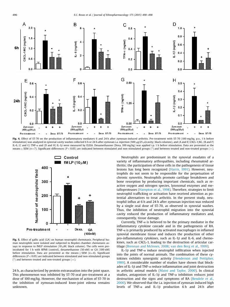
Fig. 4. Effect of ST-70 on the production of inflammatory mediators 6 and 24 h after zymosan-induced arthritis. Pre-treatment with ST-70 (100 mg/kg, p.o., 1 h beforestimulation) was analyzed in synovial cavity washes collected 6 h or 24 h after zymosan i.a. injection (500 mg/25 ml/cavity; black columns), and (A and E) CXCL-1/KC, (B and F)IL-6, (C and G) TNF-α and (D and H) IL-1β were measured by ELISA. Dexamethasone (Dexa, 100 mg/kg) was applied i.p. 1 h before stimulation. Data are presented as themeans7SEM (n¼7). Significant differences (Po0.05) are indicated between stimulated and non-stimulated groups (*) and between treated and non-treated groups (þ).
Fig. 5. Effect of gallic acid (GA) on human neutrophil chemotaxis. Peripheral hu-man neutrophils were isolated and subjected to Boyden chamber chemotaxis as-says in response to fMLP stimulation (10 mM; black column). The cells were pre-incubated for 1 h with RPMI (control), dexamethasone (50 nM) or GA (100 nM)before stimulation. Data are presented as the means7SEM (n¼4). Significantdifferences (Po0.05) are indicated between stimulated and non-stimulated groups(*) and between treated and non-treated groups (þ).
E.C. Rosas et al. / Journal of Ethnopharmacology 175 (2015) 490–498496
24 h, as characterized by protein extravasation into the joint space.This phenomenon was inhibited by ST-70 oral pre-treatment at adose of 100 mg/kg. However, the mechanism of action of ST-70 inthe inhibition of zymosan-induced knee-joint edema remainsunknown.
Neutrophils are predominant in the synovial exudates of avariety of inflammatory arthropathies, including rheumatoid ar-thritis; the participation of these cells in the pathogenesis of tissuelesions has long been recognized (Harris, 1991). However, neu-trophils do not seem to be responsible for the perpetuation ofchronic synovitis. Neutrophils promote cartilage breakdown andbone resorption by producing important chemicals, such as re-active oxygen and nitrogen species, lysosomal enzymes and me-talloproteases (Hampton et al., 1998). Therefore, strategies to limitneutrophil trafficking or activation have received attention as po-tential alternatives to treat arthritis. In the present study, neu-trophil influx at 6 h and 24 h after zymosan injection was reducedby a single oral dose of ST-70, as observed in synovial washes.Thus, the inhibition of neutrophil migration into the synovialcavity reduced the production of inflammatory mediators and,consequently, tissue damage.
Currently, TNF-α is believed to be the primary mediator in theinflammatory cytokine cascade and in the pathogenesis of RA.TNF-α is primarily produced by activated macrophages in inflamedsynovial membrane tissue and induces the production of otherpro-inflammatory cytokines, such as IL-1β and IL-6, and chemo-kines, such as CXCL-1, leading to the destruction of articular car-tilage (Brennan and McInnes, 2008; van den Berg et al., 1999).
IL-1β and TNF-α induce neutrophil infiltration when injectedinto the joints of normal animals. The combination of these cy-tokines exhibits synergistic activity (Henderson and Pettipher,1989). A considerable number of studies have shown that block-ades of IL-1β and TNF-α reduce inflammation and joint destructionin arthritic animal models (Maini and Taylor, 2000). In clinicalstudies, antagonism of IL-1β and TNF-α inhibition reduces jointdestruction and the signs and symptoms of RA (Bendele et al.,2000). We observed that the i.a. injection of zymosan induced highlevels of TNF-α and IL-1β production 6 h and 24 h after
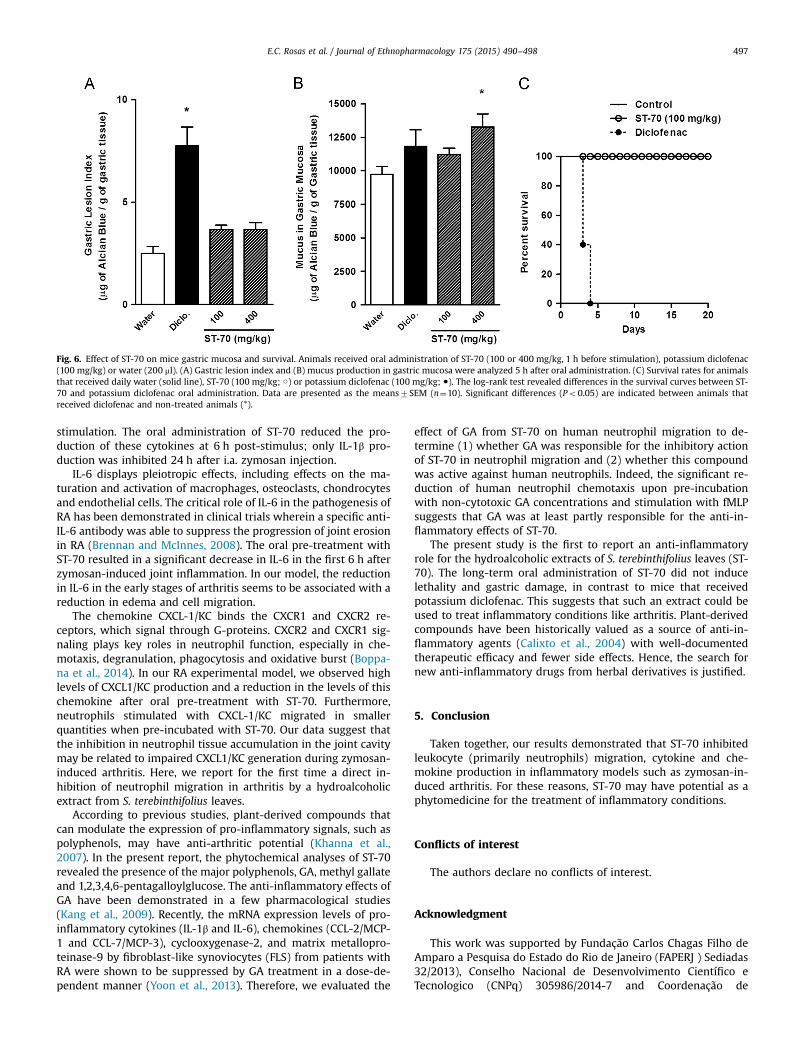
Fig. 6. Effect of ST-70 on mice gastric mucosa and survival. Animals received oral administration of ST-70 (100 or 400 mg/kg, 1 h before stimulation), potassium diclofenac(100 mg/kg) or water (200 ml). (A) Gastric lesion index and (B) mucus production in gastric mucosa were analyzed 5 h after oral administration. (C) Survival rates for animalsthat received daily water (solid line), ST-70 (100 mg/kg; ○) or potassium diclofenac (100 mg/kg; ●). The log-rank test revealed differences in the survival curves between ST-70 and potassium diclofenac oral administration. Data are presented as the means7SEM (n¼10). Significant differences (Po0.05) are indicated between animals thatreceived diclofenac and non-treated animals (*).
E.C. Rosas et al. / Journal of Ethnopharmacology 175 (2015) 490–498 497
stimulation. The oral administration of ST-70 reduced the pro-duction of these cytokines at 6 h post-stimulus; only IL-1β pro-duction was inhibited 24 h after i.a. zymosan injection.
IL-6 displays pleiotropic effects, including effects on the ma-turation and activation of macrophages, osteoclasts, chondrocytesand endothelial cells. The critical role of IL-6 in the pathogenesis ofRA has been demonstrated in clinical trials wherein a specific anti-IL-6 antibody was able to suppress the progression of joint erosionin RA (Brennan and McInnes, 2008). The oral pre-treatment withST-70 resulted in a significant decrease in IL-6 in the first 6 h afterzymosan-induced joint inflammation. In our model, the reductionin IL-6 in the early stages of arthritis seems to be associated with areduction in edema and cell migration.
The chemokine CXCL-1/KC binds the CXCR1 and CXCR2 re-ceptors, which signal through G-proteins. CXCR2 and CXCR1 sig-naling plays key roles in neutrophil function, especially in che-motaxis, degranulation, phagocytosis and oxidative burst (Boppa-na et al., 2014). In our RA experimental model, we observed highlevels of CXCL1/KC production and a reduction in the levels of thischemokine after oral pre-treatment with ST-70. Furthermore,neutrophils stimulated with CXCL-1/KC migrated in smallerquantities when pre-incubated with ST-70. Our data suggest thatthe inhibition in neutrophil tissue accumulation in the joint cavitymay be related to impaired CXCL1/KC generation during zymosan-induced arthritis. Here, we report for the first time a direct in-hibition of neutrophil migration in arthritis by a hydroalcoholicextract from S. terebinthifolius leaves.
According to previous studies, plant-derived compounds thatcan modulate the expression of pro-inflammatory signals, such aspolyphenols, may have anti-arthritic potential (Khanna et al.,2007). In the present report, the phytochemical analyses of ST-70revealed the presence of the major polyphenols, GA, methyl gallateand 1,2,3,4,6-pentagalloylglucose. The anti-inflammatory effects ofGA have been demonstrated in a few pharmacological studies(Kang et al., 2009). Recently, the mRNA expression levels of pro-inflammatory cytokines (IL-1β and IL-6), chemokines (CCL-2/MCP-1 and CCL-7/MCP-3), cyclooxygenase-2, and matrix metallopro-teinase-9 by fibroblast-like synoviocytes (FLS) from patients withRA were shown to be suppressed by GA treatment in a dose-de-pendent manner (Yoon et al., 2013). Therefore, we evaluated the
effect of GA from ST-70 on human neutrophil migration to de-termine (1) whether GA was responsible for the inhibitory actionof ST-70 in neutrophil migration and (2) whether this compoundwas active against human neutrophils. Indeed, the significant re-duction of human neutrophil chemotaxis upon pre-incubationwith non-cytotoxic GA concentrations and stimulation with fMLPsuggests that GA was at least partly responsible for the anti-in-flammatory effects of ST-70.
The present study is the first to report an anti-inflammatoryrole for the hydroalcoholic extracts of S. terebinthifolius leaves (ST-70). The long-term oral administration of ST-70 did not inducelethality and gastric damage, in contrast to mice that receivedpotassium diclofenac. This suggests that such an extract could beused to treat inflammatory conditions like arthritis. Plant-derivedcompounds have been historically valued as a source of anti-in-flammatory agents (Calixto et al., 2004) with well-documentedtherapeutic efficacy and fewer side effects. Hence, the search fornew anti-inflammatory drugs from herbal derivatives is justified.
5. Conclusion
Taken together, our results demonstrated that ST-70 inhibitedleukocyte (primarily neutrophils) migration, cytokine and che-mokine production in inflammatory models such as zymosan-in-duced arthritis. For these reasons, ST-70 may have potential as aphytomedicine for the treatment of inflammatory conditions.
Conflicts of interest
The authors declare no conflicts of interest.
Acknowledgment
This work was supported by Fundação Carlos Chagas Filho deAmparo a Pesquisa do Estado do Rio de Janeiro (FAPERJ ) Sediadas32/2013), Conselho Nacional de Desenvolvimento Científico eTecnologico (CNPq) 305986/2014-7 and Coordenação de
E.C. Rosas et al. / Journal of Ethnopharmacology 175 (2015) 490–498498
Aperfeiçoamento dePessoal de Nível Superior (CAPES) PROCAD149/2007. Luana B Correa and Tatiana A Padua are students fromthe Pós-Graduate Program in Cellular and Molecular Biology, Os-waldo Cruz Institute, Fiocruz, Rio de Janeiro, Brazil.
Appendix A. Supplementary Information
Supplementary data associated with this article can be found inthe online version at http://dx.doi.org/10.1016/j.jep.2015.10.014.
References
Ahmed, S., Pakozdi, A., Koch, A.E., 2006. Regulation of interleukin-1beta-inducedchemokine production and matrix metalloproteinase 2 activation by epigallo-catechin-3-gallate in rheumatoid arthritis synovial fibroblasts. Arthritis Rheum.54, 2393–2401.
Ajuebor, M.N., Das, A.M., Virág, L., Flower, R.J., Szabó, C., Perretti, M., 1999. Role ofresident peritoneal macrophages and mast cells in chemokine production andneutrophil migration in acute inflammation: evidence for an inhibitory loopinvolving endogenous IL-10. J. Immunol. 162, 1685–1691.
Bendele, A.M., Chlipala, E.S., Scherrer, J., Frazier, J., Sennello, G., Rich, W.J., Edwards,C.K., 2000. Combination benefit of treatment with the cytokine inhibitors in-terleukin-1 receptor antagonist and PEGylated soluble tumor necrosis factorreceptor type I in animal models of rheumatoid arthritis. Arthritis Rheum. 43,2648–2659.
Boppana, N.B., Devarajan, A., Gopal, K., Barathan, M., Bakar, S.A., Shankar, E.M.,Ebrahim, A.S., Farooq, S.M., 2014. Blockade of CXCR2 signalling: a potentialtherapeutic target for preventing neutrophil-mediated inflammatory diseases.Exp. Biol. Med. 239, 509–518.
Brennan, F.M., McInnes, I.B., 2008. Evidence that cytokines play a role in rheuma-toid arthritis. J. Clin. Investig. 118, 3537–3545.
Calixto, J.B., Campos, M.M., Otuki, M.F., Santos, A.R., 2004. Anti-inflammatorycompounds of plant origin. Part II. Modulation of pro-inflammatory cytokines,chemokines and adhesion molecules. Planta Med. 70, 93–103.
Carlini, E.A., Duarte-Almeida, J.M., Rodrigues, E., Tabach, R., 2010. Antiulcer effect ofthe pepper trees Schinus terebinthifolius Raddi (aroeira-da-praia) and Myr-acrodruon urundeuva Allemão, Anacardiaceae (aroeira-do-sertão). Braz. J.Pharmacogn., 140–146.
Cavalher-Machado, S.C., Rosas, E.C., Brito, Fe.A., Heringe, A.P., de Oliveira, R.R., Ka-plan, M.A., Figueiredo, M.R., Henriques, M., 2008. The anti-allergic activity ofthe acetate fraction of Schinus terebinthifolius leaves in IgE induced mice pawedema and pleurisy. Int. Immunopharmacol. 8, 1552–1560.
Conte, Fe.P., Barja-Fidalgo, C., Verri, W.A., Cunha, F.Q., Rae, G.A., Penido, C., Henri-ques, M., 2008. Endothelins modulate inflammatory reaction in zymosan-in-duced arthritis: participation of LTB4, TNF-alpha, and CXCL-1. J. Leukoc. Biol. 84,652–660.
de Lima, M.R., de Souza Luna, J., dos Santos, A.F., de Andrade, M.C., Sant'Ana, A.E.,Genet, J.P., Marquez, B., Neuville, L., Moreau, N., 2006. Anti-bacterial activity ofsome Brazilian medicinal plants. J. Ethnopharmacol. 105, 137–147.
Gegout, P., Gillet, P., Terlain, B., Netter, P., 1995. Zymosan-induced arthritis in rats. II.Effects of anti-inflammatory drugs. Life Sci. 56, PL389–PL394.
Guerrero, A.T., Verri, W.A., Cunha, T.M., Silva, T.A., Schivo, I.R., Dal-Secco, D., Canetti,C., Rocha, F.A., Parada, C.A., Cunha, F.Q., Ferreira, S.H., 2008. Involvement of LTB4in zymosan-induced joint nociception in mice: participation of neutrophils andPGE2. J. Leukoc. Biol. 83, 122–130.
Hampton, M.B., Kettle, A.J., Winterbourn, C.C., 1998. Inside the neutrophil
phagosome: oxidants, myeloperoxidase, and bacterial killing. Blood 92,3007–3017.
Harris, E.D., 1991. Pathogenesis of rheumatoid arthritis: its relevance to therapy inthe'‘90s. Trans. Am. Clin. Climatol. Assoc. 102, 260–268, discussion 268–270.
Henderson, B., Pettipher, E.R., 1989. Arthritogenic actions of recombinant IL-1 andtumour necrosis factor alpha in the rabbit: evidence for synergistic interactionsbetween cytokines in vivo. Clin. Exp. Immunol. 75, 306–310.
Kalden, J.R., 2002. Expanding role of biologic agents in rheumatoid arthritis. J.Rheumatol. Suppl. 66, 27–37.
Kang, M.S., Jang, H.S., Oh, J.S., Yang, K.H., Choi, N.K., Lim, H.S., Kim, S.M., 2009. Ef-fects of methyl gallate and gallic acid on the production of inflammatorymediators interleukin-6 and interleukin-8 by oral epithelial cells stimulatedwith Fusobacterium nucleatum. J Microbiol. 47, 760–767.
Khanna, D., Sethi, G., Ahn, K.S., Pandey, M.K., Kunnumakkara, A.B., Sung, B., Ag-garwal, A., Aggarwal, B.B., 2007. Natural products as a gold mine for arthritistreatment. Curr. Opin. Pharmacol. 7, 344–351.
Lama, A., Saikia, H., 2011. Targeted therapies for rheumatoid arthritis: a review. Int.J. Pharm. Sci. Res., 1116–1134.
Maini, R.N., Taylor, P.C., 2000. Anti-cytokine therapy for rheumatoid arthritis. Annu.Rev. Med. 51, 207–229.
McInnes, I.B., Schett, G., 2011. The pathogenesis of rheumatoid arthritis. N. Engl. J.Med. 365, 2205–2219.
Medeiros, K.C.P., Monteiro, J.C., Diniz, M.F.F.M., Medeiros, I.A., Silva, B.A., Piuvezam,M.R., 2007. Effect of the activity of the brazilian polyherbal formulation: Eu-calyptus globulus Labill, Peltodon radicans Pohl and Schinus terebinthifolius Raddiin inflammatory models. Braz. J. Pharmacogn., 23–28.
Medzhitov, R., 2010. Inflammation 2010: new adventures of an old flame. Cell 140,771–776.
Penido, C., Conte, F.P., Chagas, M.S., Rodrigues, C.A., Pereira, J.F., Henriques, M.G.,2006. Antiinflammatory effects of natural tetranortriterpenoids isolated fromCarapa guianensis Aublet on zymosan-induced arthritis in mice. Inflamm. Res.55, 457–464.
Pinto, L.G., Cunha, T.M., Vieira, S.M., Lemos, H.P., Verri, W.A., Cunha, F.Q., Ferreira, S.H., 2010. IL-17 mediates articular hypernociception in antigen-induced arthritisin mice. Pain 148, 247–256.
Ribani, M., Bottoli, C.B.G., Collins, C.H., Jardim ICSF, L.F.C.,M., 2004. Validação emmétodos cromatográficos e eletroforéticos. Quím. Nova 27, 771–780.
Roth, S.H., 2012. Coming to terms with nonsteroidal anti-inflammatory drug gas-tropathy. Drugs 72, 873–879.
Schmourlo, G., Mendonça-Filho, R.R., Alviano, C.S., Costa, S.S., 2005. Screening ofantifungal agents using ethanol precipitation and bioautography of medicinaland food plants. J. Ethnopharmacol. 96, 563–568.
Scott, D.L., Wolfe, F., Huizinga, T.W., 2010. Rheumatoid arthritis. Lancet 376,1094–1108.
Underhill, D.M., 2003. Macrophage recognition of zymosan particles. J. EndotoxinRes. 9, 176–180.
Utsunomiya, I., Ito, M., Oh-ishi, S., 1998. Generation of inflammatory cytokines inzymosan-induced pleurisy in rats: TNF induces IL-6 and cytokine-inducedneutrophil chemoattractant (CINC) in vivo. Cytokine 10, 956–963.
van den Berg, W.B., Joosten, L.A., Kollias, G., van De Loo, F.A., 1999. Role of tumournecrosis factor alpha in experimental arthritis: separate activity of interleukin1beta in chronicity and cartilage destruction. Ann. Rheum. Dis. 58 (Suppl. 1),SI40–SI48.
Velázquez, E., Tournier, H.A., Mordujovich de Buschiazzo, P., Saavedra, G., Schinella,G.R., 2003. Antioxidant activity of Paraguayan plant extracts. Fitoterapia 74,91–97.
Wright, H.L., Moots, R.J., Edwards, S.W., 2014. The multifactorial role of neutrophilsin rheumatoid arthritis. Nat. Rev. Rheumatol.
Yoon, C.H., Chung, S.J., Lee, S.W., Park, Y.B., Lee, S.K., Park, M.C., 2013. Gallic acid, anatural polyphenolic acid, induces apoptosis and inhibits proinflammatorygene expressions in rheumatoid arthritis fibroblast-like synoviocytes. Jt. BoneSpine 80, 274–279.


![010005545[1] Ethnopharmacology of Murcia](https://static.fdocuments.net/doc/165x107/5531de464a7959855b8b4643/0100055451-ethnopharmacology-of-murcia.jpg)