Control of circadian rhythms A2 AQA PSYA3 psychology biological rhythms
http://jbr.sagepub.com/Journal of Biological Rhythms
http://jbr.sagepub.com/content/17/6/526The online version of this article can be found at:
DOI: 10.1177/0748730402238235
2002 17: 526J Biol RhythmsManuel Miranda-Anaya, Paul A. Bartell and Michael Menaker
Circadian Rhythm of Iguana Electroretinogram: The Role of Dopamine and Melatonin
Published by:
http://www.sagepublications.com
On behalf of:
Society for Research on Biological Rhythms
can be found at:Journal of Biological RhythmsAdditional services and information for
http://jbr.sagepub.com/cgi/alertsEmail Alerts:
http://jbr.sagepub.com/subscriptionsSubscriptions:
http://www.sagepub.com/journalsReprints.navReprints:
http://www.sagepub.com/journalsPermissions.navPermissions:
http://jbr.sagepub.com/content/17/6/526.refs.htmlCitations:
What is This?
- Dec 1, 2002Version of Record >>
at INST DE FISIOLOGIA CELU LAR on May 7, 2013jbr.sagepub.comDownloaded from
JOURNAL OF BIOLOGICAL RHYTHMS / December 2002Miranda-Anaya et al. / CIRCADIAN RHYTHM OF ERG
Circadian Rhythm of Iguana Electroretinogram:The Role of Dopamine and Melatonin
Manuel Miranda-Anaya,2 Paul A. Bartell,3 and Michael Menaker1
Department of Biology and National Science Foundation Center for Biological Timing,University of Virginia, Charlottesville, VA 22904, USA
Abstract The amplitude of the b-wave of the electroretinogram (ERG) varies witha circadian rhythm in the green iguana; the amplitude is high during the day (orsubjective day) and low during the night (or subjective night). Dopamine andmelatonin contents in the eye are robustly rhythmic under constant conditions;dopamine levels are high during the subjective day, and melatonin levels arehigh during the subjective night. Dopamine and melatonin affect the amplitudeof the b-wave in an antagonistic and phase-dependent manner: dopamine D2-receptor agonists injected intraocularly during the subjective night producehigh-amplitude b-waves characteristic of the subjective day, whereas melatonininjected intraocularly during the subjective day reduces b-wave amplitude. Sec-tioning the optic nerve abolishes the circadian rhythms of b-wave amplitude andof dopamine content. The results of this study suggest that in iguana, a negativefeedback loop involving dopamine and melatonin regulates the circadianrhythm of the ERG b-wave amplitude that is at least in part generated in thebrain.
Key words ERG, circadian, dopamine, melatonin, optic nerve, iguana
Many behavioral and physiological changes in ani-mals are regulated by the circadian system, which isresponsive to daily changes in the levels of illumina-tion. The circadian system of vertebrates consists ofmultiple oscillators and photoreceptors located in dif-ferent tissues and their interactions, with the outputsfrom different oscillators directly regulating differentaspects of physiology and behavior (Menaker andTosini, 1995; Yamazaki et al., 2000).
The retinas of vertebrates contain circadian oscilla-tors that mediate physiological processes involved invisual sensitivity (Besharse et al., 1988; Herzog andBlock, 1999; Li and Dowling, 1998). Outputs of these
oscillators include melatonin and dopamine. Mela-tonin is synthesized rhythmically in cultured retinasof lamprey (Menaker and Tosini, 1995), fish (Cahill,1996), amphibians (Cahill and Besharse, 1995; Alonso-Gómez et al., 2000), reptiles (Tosini and Menaker,1998), and mammals (Tosini and Menaker, 1996a), anda circadian rhythm of ocular melatonin has beenreported in birds (Underwood and Siopes, 1984;Adachi et al., 1998). Melatonin is involved in the acti-vation of disk-shedding and cone movement inXenopus (Besharse et al., 1988) and decreases b-waveamplitude of the electroretinogram (ERG) in birds (Luet al., 1995; McGoogan and Cassone, 1999) and
526
1. To whom all correspondence should be addressed: Department of Biology, Gilmer Hall, University of Virginia, P.O. Box400328, Charlottesville, VA 22904-4328; e-mail: [email protected]. Current address: Departamento de Biología, Facultad de Ciencias, Universidad Nacional Autónoma de México, DF 04510.3. Current address: Forschungsstelle für Ornithologie der Max-Planck-Gesellschaft, D-82346 Erling-Andechs, Germany.
JOURNAL OF BIOLOGICAL RHYTHMS, Vol. 17 No. 6, December 2002 526-538DOI: 10.1177/0748730402238235© 2002 Sage Publications
at INST DE FISIOLOGIA CELU LAR on May 7, 2013jbr.sagepub.comDownloaded from
humans (Emser et al., 1993). It inhibits dopaminerelease from amacrine cells (Dubocovich, 1983), sug-gesting that it may influence dopamine-dependentmechanisms of light and dark adaptation.
Dopamine is an abundant neurotransmitter in theretina (Dowling, 1987; Nguyen-Legros, 1996), and cir-cadian rhythms in ocular content of dopamine havebeen described in fish (McCormack and Burnside,1993), birds (Adachi et al., 1998), and mammals (Doyleet al., 1999). Dopamine affects circadian rhythms ofmelatonin synthesis, disk shedding, rod elongation,and retinal pigment epithelium dispersion, as well ascone-rod inputs in the retina of Xenopus (Besharseet al., 1988; Cahill and Besharse, 1991; Krizaj et al.,1998). Dopamine also regulates the circadian rhythmof visual sensitivity in fish (Li and Dowling, 2000) andquail (Manglapus et al., 1999).
The ERG measures the global electrical response ofthe retina to a flash stimulus and is a useful tool toevaluate visual function (Dowling, 1987). ERG b-waveamplitude depends on postphotoreceptor electricalactivity and displays a circadian rhythm in constantconditions in fish (Li and Dowling, 2000), birds (Luet al., 1995; Manglapus et al., 1998; McGoogan andCassone, 1999; Wu et al., 2000), reptiles (Fowlkes et al.,1987; Shaw et al., 1993; Miranda-Anaya et al., 2000),and mammals (Brandenburg et al., 1983). In many ofthese studies, either melatonin or dopamine has beenproposed as a regulator of rhythms of ERG b-waveamplitude.
In the green iguana, an oscillator of unknown loca-tion regulates the circadian rhythm of ERG b-waveamplitude. Dopamine content in the eye peaks duringthe subjective day, whereas melatonin content peaksduring subjective night. Intraocular injections of thedopamine D2 receptor agonist quinpirole increase theamplitude of the b-wave during the subjective night,whereas melatonin injections decrease it during thesubjective day. Depleting the retinas of dopamineabolishes the circadian rhythm of ERG. Because sec-tioning the optic nerve abolishes the rhythm of retinaldopamine content, whereas the rhythm of melatoninsynthesis persists in cultured retinas, our data suggestthat the rhythm of dopamine content and the rhythmof melatonin synthesis are under the control of differ-ent circadian oscillators. We propose that the circadianrhythm of b-wave amplitude in the iguana is regu-lated by the interaction of the outputs (dopamine andmelatonin) of these two oscillators.
MATERIALS AND METHODS
Animal maintenance
Juvenile green iguanas (Iguana iguana) (30 to 50 g)obtained from Glades Herpetological Supply (FortMyers, FL, USA) were housed in plastic cages within awooden enclosure on a light:dark (LD) 12:12 cycle(lights-on 0600 h, lights-off 1800 h). White light wasprovided by a fluorescent lamp (20 µW) (Philips,Somerset, NJ, USA), and additional heat from a ther-mal pad maintained the cage temperature at 28 ± 2 °C.Animals were fed fresh green vegetables three timesper week, with water supplied ad libitum.
Melatonin Extractionand Radioimmunoassay
Eyes from halothane-anesthetized animals wereharvested in darkness using an infrared viewer (Find-R-Scope, FJW Optical Systems, Palatine, IL, USA),immediately frozen in individual Eppendorf tubes,and stored at –80 °C. Melatonin chloroform extractionand radioimmunoassay (RIA) were performed asdescribed by Rollag and Niswender (1976); this assayhas been validated for iguanas (Tosini and Menaker,1996b). Eyes were homogenized in 200 µL of phos-phate buffer with 0.9% NaCl and 1% gelatin (phos-phate-buffered saline gel) and sonicated using amicroultrasonic cell disrupter (Kontes, Vineland, NJ,USA). Melatonin was extracted in 1 mL of chloroformand washed with 200 µL of sodium-carbonate buffer(0.1 M, pH 10.25). Samples were spun and supernatantremoved. Aliquots were dried under nitrogen andsuspended in 0.5 mL of phosphate-buffered saline gel.RIAwas performed using melatonin antibody (R1055)and I125-melatonin analog (Covanzel, Vienna, VA,USA).
Dopamine Quantification byHigh-Pressure Liquid Chromatography
Levels of dopamine and its metabolites dihidroxy-phenylacetic acid (DOPAC) and homovanillic acid(HVA) were measured by high-performance liquidchromatography (HPLC) (Beckman System Gold®,San Ramon, CA, USA). Tissue was homogenized in200 µL of 80% 0.1 M perchloric acid/20% acetonitrile
Miranda-Anaya et al. / CIRCADIAN RHYTHM OF ERG 527
at INST DE FISIOLOGIA CELU LAR on May 7, 2013jbr.sagepub.comDownloaded from
solution and the supernatant spin-filtered through a0.2 µm nylon filter (PGC Scientifics, Gaithersburg,MD, USA). Twenty microliters of supernatant wereinjected into a Microsorb® Short-One C18 reversephase column (3 µm, 2.8 ! 10 cm) (Varian Associates,Walnut Creek, CA, USA). Mobile phase consisted of75 mM phosphate buffer with 25 mM EDTA, 1.7 mM 1-octanesulfonic acid, 0.01% triethylamine, and 7%acetonitrile (pH 3). Quantification was carried outwith an ESA Coulochem detector (ESA Inc., Bedford,MA, USA) fitted with a 5020 guard cell set at +400 mV,a 5011 analytical cell with a glassy carbon screeningelectrode set at 75 mV, and a measuring electrode at+250 mV. All separations were performed at a flowrate of 1.0 mL/min. Peaks and relative concentrationswere identified by comparison with known externalstandards.
ERGs
ERGs were obtained in constant darkness (DD) andin dim (5 µW/cm2) LD cycles (dLD 12:12) as describedpreviously (Miranda-Anaya et al., 2000). Light inten-sity was calibrated at the level of the head of the ani-mals using a radiometer (model 350, GrasebyOptronics, Orlando, FL, USA). A platinum-iridiumloop electrode was positioned on the cornea of a ure-thane-anesthetized animal (2.5 g/kg) and coveredwith clear silicone. The reference electrode was placedsubdermally in the front of the animal’s head. Animalswere held in a light-tight chamber with the tempera-ture maintained at 28 °C using copper tubing and a cir-culating bath system (Lauda MGW, Westbury, NY,USA). A 250 ms light pulse (50 µW/cm2) was givenusing a halogen lamp and delivered by a fiber opticlocated 3 mm from the cornea. Light irradiance waschosen to produce about 50% of maximal b-waveamplitude (Miranda-Anaya et al., 2000). ERGs wereevoked every 15 min during at least two consecutivecircadian cycles, amplified using a wide-band EEG-AC preamplifier (model 7P5 band-pass filter [3 to 1500Hz], Grass, Quincy, MA) coupled to a DC driver(Grass model 7DA), and recorded on a polygraph(Grass model 79D). B-wave amplitude was defined asthe voltage difference between the trough of the a-wave and the peak of the b-wave. In this study, the a-wave was not considered, since its amplitude wasbarely rhythmic and phase dependent effects couldnot be evaluated.
Protocol
Experiment 1: Melatonin andDopamine Content inControl Conditions
Twenty-one iguanas were transferred from LD12:12 to DD. Eyes from 3 animals were collected in thedark at projected circadian times (CTs) 2, 6, 10, 14, 17,20, and 23 on the second day in DD, and kept at –80 °Cuntil content determination. The period of the loco-motor activity rhythm of the iguana in DD is veryclose to 24 h; therefore, in this study we assume the cir-cadian time on the second day in DD as a projection ofthe LD conditions in which the animals were main-tained (e.g., circadian time 12 was considered the pro-jected transition from light to dark). Melatonin con-tent of one eye from each animal was determined byRIA, and dopamine content of the other eye was mea-sured using HPLC as described above.
Experiment 2: Effects of IntraocularInjections of Quinpirole, SKF38393,Eticlopride, and Melatonin onERG b-Wave Amplitude
Urethane-anesthetized iguanas were transferredfrom LD 12:12 to DD. During the second cycle in DD,and in dim red light, a total volume of 10 µL ofmelatonin or dopamine receptor agonist or antagonistwas injected through the sclera into the vitreous bodyof the eye using a Hamilton syringe fitted with a 26-gauge needle. Experimental animals received 5, 50, or500 µM doses of either the selective D2-receptor ago-nist quinpirole hydrochloride (in 1% 0.1 N HCl) ormelatonin (in 1% methyl alcohol); or a 100 µM dose ofeither the D2-receptor antagonist eticlopride or theD1-receptor agonist SKF38393 hydrobromide (in 1%0.1 N HCl) (Sigma, St. Louis, MO, USA). Controls wereinjected with 10 µL of vehicle (either 1% 0.1 N HCl, or1% methyl alcohol). Changes in ERG b-wave ampli-tude induced by these agents were measured fromanimals held in DD with only the test light required toevoke the ERG. Injections were given near the middleof the projected subjective night (MSN) (2300 to0100 h, EST) or near the middle of the projected subjec-tive day (MSD) (1100 to 1400 h), when the lowest andhighest amplitudes of ERG b-wave occur. For eachagent, five ERGs taken at 1-min intervals before injec-
528 JOURNAL OF BIOLOGICAL RHYTHMS / December 2002
at INST DE FISIOLOGIA CELU LAR on May 7, 2013jbr.sagepub.comDownloaded from
tion of the agent were used as a reference. After theinjection, ERGs were obtained every minute for 15min, and then every 15 min for at least 12 hours. Injec-tions of eticlopride were only given during the pro-jected early subjective day (0900 h) to observe its effecton the presence of the diurnal peak of the b-waveamplitude.
Experiment 3: ERG CircadianRhythm in Dopamine-Depleted Retinas
After 3 consecutive days of ERG recording in DDfrom intact animals (n = 6), intraperitoneal urethaneanesthesia was reinforced with a second dose of ure-thane (0.8 g/kg), enabling us to work with fully anes-thetized animals; injections of 6-hydroxy-dopamine(6-OHDA) (50 µmol in 20 µL) were then given in thevitreous body of both eyes. The anesthetized animalswere held in LD 12:12 (lights-on 0700-1900 h) for 3days after the injection, when a second dose of 6-OHDA was given. Four days after the second injec-tion, anesthesia was again reinforced and ERGs wereobtained for 3 more days in DD. During treatmentwith 6-OHDA, animals were administered a glucosesolution through a feeding tube inserted into theesophagus.
Experiment 4: ERG CircadianRhythm and Dopamine Contentin Optic Nerve–Sectioned Eyes
Animals were anesthetized with urethane; addi-tional anesthesia (10% procaine in saline) was injectedinto the palate. An incision was made in the palate,and the soft tissue was retracted with forceps until theoptic nerve could be seen. One optic nerve was cutusing a spring scissors, and the wound was packedwith Gelfoam. Sham operations were done in the sameway, except the optic nerve was not cut. ERG recordswere initiated immediately after surgery. In one groupof animals (n = 4), ERGs were recorded from both eyessimultaneously, the eye with intact optic nerve servingas a control. In a second group (n = 7), only one eye wasrecorded from before and after surgery, thus servingas its own control. At the end of each record, animalswere euthanized with an overdose of urethane and theoptic nerve was visually inspected to ensure it hadbeen completely severed. ERGs were obtained ineither DD or dim LD. A third group of iguanas wasused to evaluate daily changes in dopamine content ofoptic nerve–sectioned (ON-X) eyes in DD (n = 9),
using HPLC as described in experiment 1. For thesemeasurements, animals with one optic nerve sec-tioned were transferred from LD to DD, and both eyeswere collected in darkness during the MSD (n = 5) andin the MSN (n = 4) of the second cycle in DD. The intacteye of each animal was used as the control for theON-X eye.
Data Analysis
In each experiment, circadian rhythmicity of ERGb-wave amplitude was calculated at a 95% level ofconfidence using a fast Fourier transform–nonlinearleast squares estimation method developed by MartinStraume (National Science Foundation, Center forBiological Timing, University of Virginia; see Plautz,1997). Circadian variations in content of melatoninand dopamine (experiment 1) were evaluated usinganalysis of variance; the effect of intraocular injections(experiment 2) was evaluated using the Kruskall-Wallis nonparametric test and contrasted with theDunnett test by means of Sigma-Stat software (JandelScientific, San Rafael, CA, USA). The Mann-Whitneytest was used to compare dopamine content at twodifferent time points (experiment 4). A p value < 0.05was considered significant. Because the iguanas’ free-running periods are very close to 24 h, subjective timewas considered as the projection of the previous LDconditions in which the animals were kept before theexperiments. Thus, subjective day corresponds to theprevious interval between lights-on and lights-off.
RESULTS
Experiment 1: Melatonin andDopamine Content in DD Conditions
Both melatonin and dopamine content in the eyeshow robust circadian rhythms (p < 0.01). Figure 1Ashows that the average (± SEM) melatonin content ofeyes taken during the beginning of the subjectivenight (CTs 14 and 17, black bars) is more than fivetimes the level observed during the subjective day(gray bars). The highest levels of melatonin (407.08 ±SEM 9.13 pg/eye) occurred at CT 14, and the lowestlevels of melatonin (75.57 ± 6.55 pg/eye) at CT 2. Thepeak levels of dopamine and melatonin are nearly 180degrees out of phase with each other. The highest lev-els of dopamine occurred at CT 23 (2143.7 ± 61.6 pg/eye) (Fig. 1B) and the lowest levels (1450 ± 2.4 pg/eye)
Miranda-Anaya et al. / CIRCADIAN RHYTHM OF ERG 529
at INST DE FISIOLOGIA CELU LAR on May 7, 2013jbr.sagepub.comDownloaded from
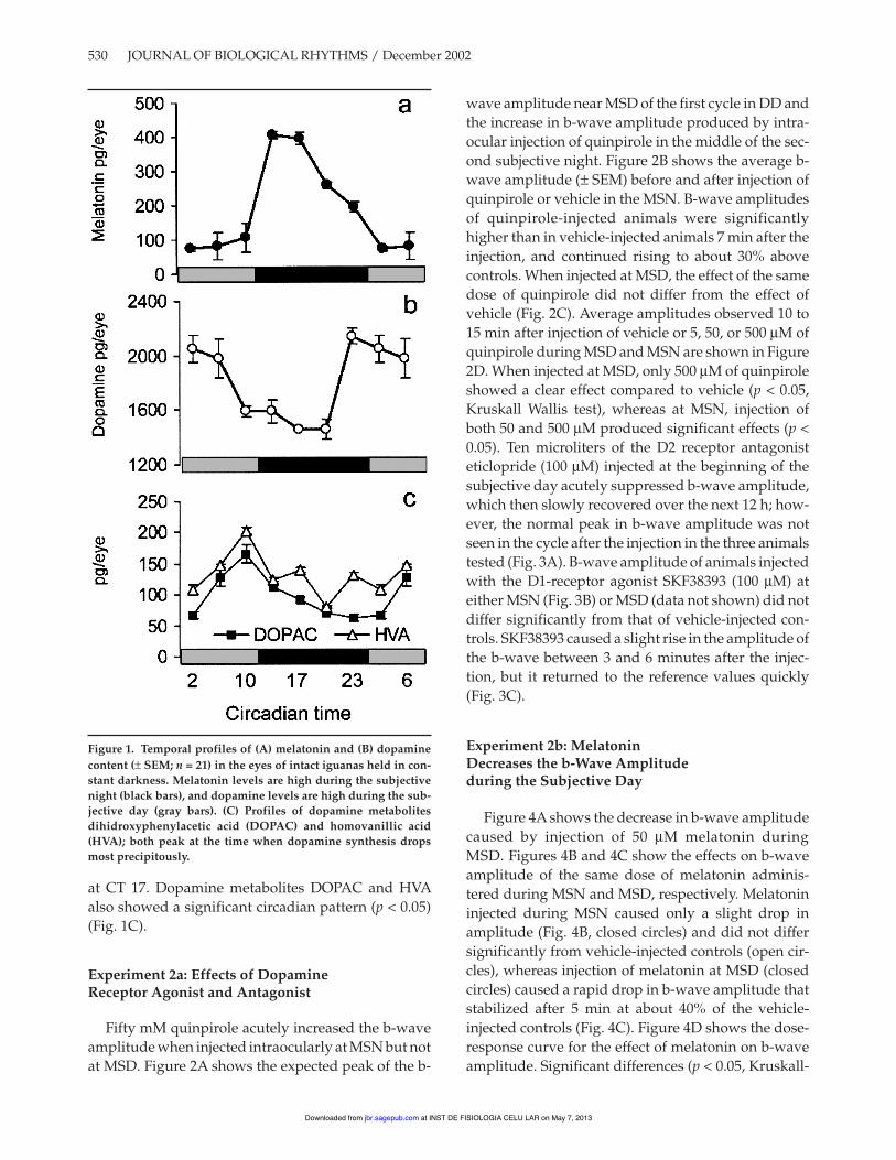
at CT 17. Dopamine metabolites DOPAC and HVAalso showed a significant circadian pattern (p < 0.05)(Fig. 1C).
Experiment 2a: Effects of DopamineReceptor Agonist and Antagonist
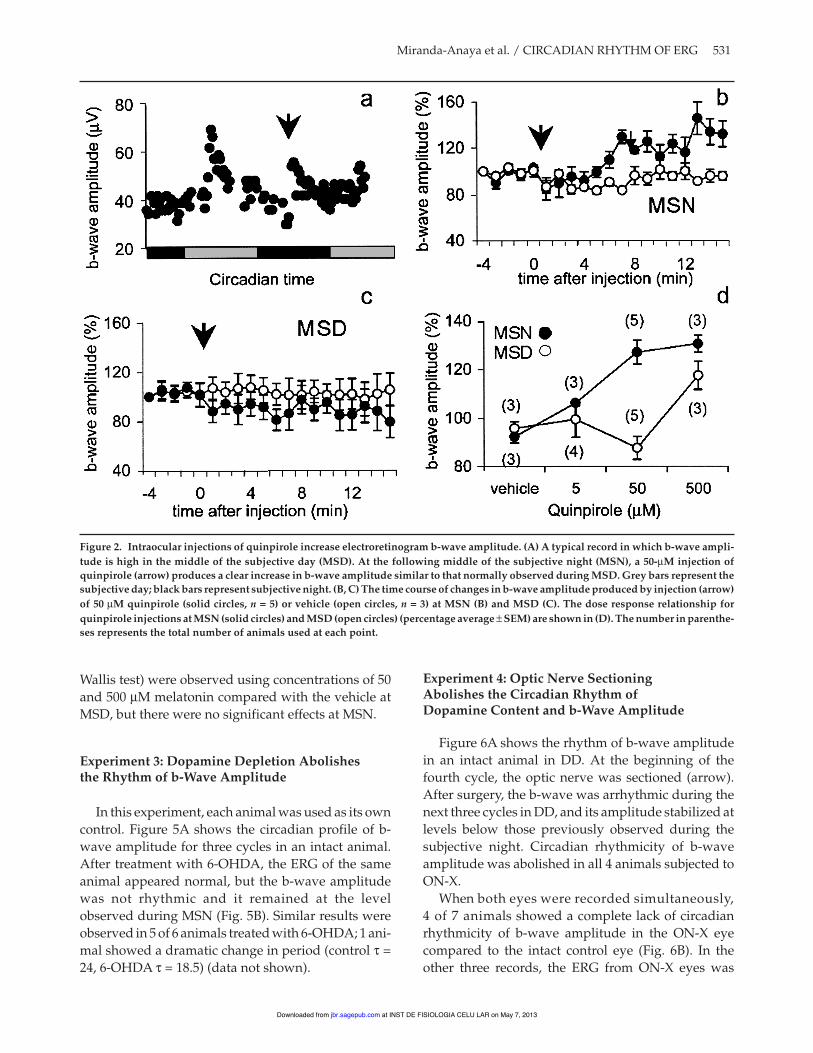
Fifty mM quinpirole acutely increased the b-waveamplitude when injected intraocularly at MSN but notat MSD. Figure 2A shows the expected peak of the b-
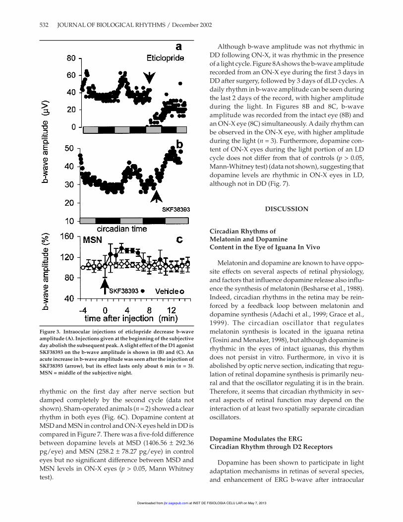
wave amplitude near MSD of the first cycle in DD andthe increase in b-wave amplitude produced by intra-ocular injection of quinpirole in the middle of the sec-ond subjective night. Figure 2B shows the average b-wave amplitude (± SEM) before and after injection ofquinpirole or vehicle in the MSN. B-wave amplitudesof quinpirole-injected animals were significantlyhigher than in vehicle-injected animals 7 min after theinjection, and continued rising to about 30% abovecontrols. When injected at MSD, the effect of the samedose of quinpirole did not differ from the effect ofvehicle (Fig. 2C). Average amplitudes observed 10 to15 min after injection of vehicle or 5, 50, or 500 µM ofquinpirole during MSD and MSN are shown in Figure2D. When injected at MSD, only 500 µM of quinpiroleshowed a clear effect compared to vehicle (p < 0.05,Kruskall Wallis test), whereas at MSN, injection ofboth 50 and 500 µM produced significant effects (p <0.05). Ten microliters of the D2 receptor antagonisteticlopride (100 µM) injected at the beginning of thesubjective day acutely suppressed b-wave amplitude,which then slowly recovered over the next 12 h; how-ever, the normal peak in b-wave amplitude was notseen in the cycle after the injection in the three animalstested (Fig. 3A). B-wave amplitude of animals injectedwith the D1-receptor agonist SKF38393 (100 µM) ateither MSN (Fig. 3B) or MSD (data not shown) did notdiffer significantly from that of vehicle-injected con-trols. SKF38393 caused a slight rise in the amplitude ofthe b-wave between 3 and 6 minutes after the injec-tion, but it returned to the reference values quickly(Fig. 3C).
Experiment 2b: MelatoninDecreases the b-Wave Amplitudeduring the Subjective Day
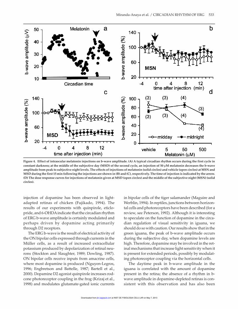
Figure 4A shows the decrease in b-wave amplitudecaused by injection of 50 µM melatonin duringMSD. Figures 4B and 4C show the effects on b-waveamplitude of the same dose of melatonin adminis-tered during MSN and MSD, respectively. Melatonininjected during MSN caused only a slight drop inamplitude (Fig. 4B, closed circles) and did not differsignificantly from vehicle-injected controls (open cir-cles), whereas injection of melatonin at MSD (closedcircles) caused a rapid drop in b-wave amplitude thatstabilized after 5 min at about 40% of the vehicle-injected controls (Fig. 4C). Figure 4D shows the dose-response curve for the effect of melatonin on b-waveamplitude. Significant differences (p < 0.05, Kruskall-
530 JOURNAL OF BIOLOGICAL RHYTHMS / December 2002
Figure 1. Temporal profiles of (A) melatonin and (B) dopaminecontent (± SEM; n = 21) in the eyes of intact iguanas held in con-stant darkness. Melatonin levels are high during the subjectivenight (black bars), and dopamine levels are high during the sub-jective day (gray bars). (C) Profiles of dopamine metabolitesdihidroxyphenylacetic acid (DOPAC) and homovanillic acid(HVA); both peak at the time when dopamine synthesis dropsmost precipitously.
at INST DE FISIOLOGIA CELU LAR on May 7, 2013jbr.sagepub.comDownloaded from
Wallis test) were observed using concentrations of 50and 500 µM melatonin compared with the vehicle atMSD, but there were no significant effects at MSN.
Experiment 3: Dopamine Depletion Abolishesthe Rhythm of b-Wave Amplitude
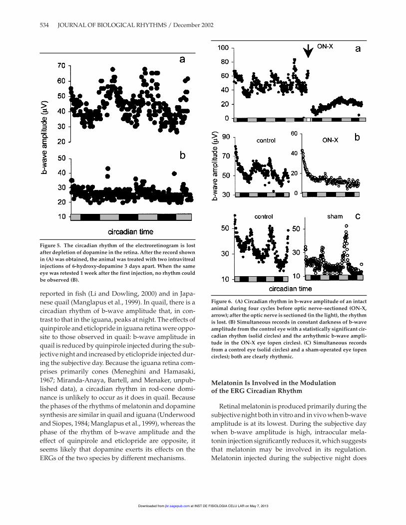
In this experiment, each animal was used as its owncontrol. Figure 5A shows the circadian profile of b-wave amplitude for three cycles in an intact animal.After treatment with 6-OHDA, the ERG of the sameanimal appeared normal, but the b-wave amplitudewas not rhythmic and it remained at the levelobserved during MSN (Fig. 5B). Similar results wereobserved in 5 of 6 animals treated with 6-OHDA; 1 ani-mal showed a dramatic change in period (control " =24, 6-OHDA " = 18.5) (data not shown).
Experiment 4: Optic Nerve SectioningAbolishes the Circadian Rhythm ofDopamine Content and b-Wave Amplitude
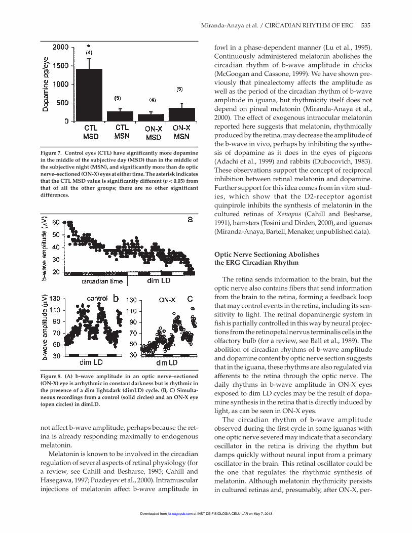
Figure 6A shows the rhythm of b-wave amplitudein an intact animal in DD. At the beginning of thefourth cycle, the optic nerve was sectioned (arrow).After surgery, the b-wave was arrhythmic during thenext three cycles in DD, and its amplitude stabilized atlevels below those previously observed during thesubjective night. Circadian rhythmicity of b-waveamplitude was abolished in all 4 animals subjected toON-X.
When both eyes were recorded simultaneously,4 of 7 animals showed a complete lack of circadianrhythmicity of b-wave amplitude in the ON-X eyecompared to the intact control eye (Fig. 6B). In theother three records, the ERG from ON-X eyes was
Miranda-Anaya et al. / CIRCADIAN RHYTHM OF ERG 531
Figure 2. Intraocular injections of quinpirole increase electroretinogram b-wave amplitude. (A) A typical record in which b-wave ampli-tude is high in the middle of the subjective day (MSD). At the following middle of the subjective night (MSN), a 50-µM injection ofquinpirole (arrow) produces a clear increase in b-wave amplitude similar to that normally observed during MSD. Grey bars represent thesubjective day; black bars represent subjective night. (B, C) The time course of changes in b-wave amplitude produced by injection (arrow)of 50 µM quinpirole (solid circles, n = 5) or vehicle (open circles, n = 3) at MSN (B) and MSD (C). The dose response relationship forquinpirole injections at MSN (solid circles) and MSD (open circles) (percentage average ± SEM) are shown in (D). The number in parenthe-ses represents the total number of animals used at each point.
at INST DE FISIOLOGIA CELU LAR on May 7, 2013jbr.sagepub.comDownloaded from
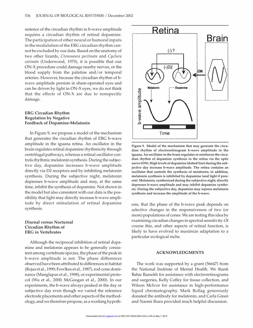
rhythmic on the first day after nerve section butdamped completely by the second cycle (data notshown). Sham-operated animals (n = 2) showed a clearrhythm in both eyes (Fig. 6C). Dopamine content atMSD and MSN in control and ON-X eyes held in DD iscompared in Figure 7. There was a five-fold differencebetween dopamine levels at MSD (1406.56 ± 292.36pg/eye) and MSN (258.2 ± 78.27 pg/eye) in controleyes but no significant difference between MSD andMSN levels in ON-X eyes (p > 0.05, Mann Whitneytest).
Although b-wave amplitude was not rhythmic inDD following ON-X, it was rhythmic in the presenceof a light cycle. Figure 8Ashows the b-wave amplituderecorded from an ON-X eye during the first 3 days inDD after surgery, followed by 3 days of dLD cycles. Adaily rhythm in b-wave amplitude can be seen duringthe last 2 days of the record, with higher amplitudeduring the light. In Figures 8B and 8C, b-waveamplitude was recorded from the intact eye (8B) andan ON-X eye (8C) simultaneously. A daily rhythm canbe observed in the ON-X eye, with higher amplitudeduring the light (n = 3). Furthermore, dopamine con-tent of ON-X eyes during the light portion of an LDcycle does not differ from that of controls (p > 0.05,Mann-Whitney test) (data not shown), suggesting thatdopamine levels are rhythmic in ON-X eyes in LD,although not in DD (Fig. 7).
DISCUSSION
Circadian Rhythms ofMelatonin and DopamineContent in the Eye of Iguana In Vivo
Melatonin and dopamine are known to have oppo-site effects on several aspects of retinal physiology,and factors that influence dopamine release also influ-ence the synthesis of melatonin (Besharse et al., 1988).Indeed, circadian rhythms in the retina may be rein-forced by a feedback loop between melatonin anddopamine synthesis (Adachi et al., 1999; Grace et al.,1999). The circadian oscillator that regulatesmelatonin synthesis is located in the iguana retina(Tosini and Menaker, 1998), but although dopamine isrhythmic in the eyes of intact iguanas, this rhythmdoes not persist in vitro. Furthermore, in vivo it isabolished by optic nerve section, indicating that regu-lation of retinal dopamine synthesis is primarily neu-ral and that the oscillator regulating it is in the brain.Therefore, it seems that circadian rhythmicity in sev-eral aspects of retinal function may depend on theinteraction of at least two spatially separate circadianoscillators.
Dopamine Modulates the ERGCircadian Rhythm through D2 Receptors
Dopamine has been shown to participate in lightadaptation mechanisms in retinas of several species,and enhancement of ERG b-wave after intraocular
532 JOURNAL OF BIOLOGICAL RHYTHMS / December 2002
Figure 3. Intraocular injections of eticlopride decrease b-waveamplitude (A). Injections given at the beginning of the subjectiveday abolish the subsequent peak. A slight effect of the D1 agonistSKF38393 on the b-wave amplitude is shown in (B) and (C). Anacute increase in b-wave amplitude was seen after the injection ofSKF38393 (arrow), but its effect lasts only about 6 min (n = 3).MSN = middle of the subjective night.
at INST DE FISIOLOGIA CELU LAR on May 7, 2013jbr.sagepub.comDownloaded from
injection of dopamine has been observed in light-adapted retinas of chicken (Fujikado, 1994). Theresults of our experiments with quinpirole, eticlo-pride, and 6-OHDAindicate that the circadian rhythmof ERG b-wave amplitude is certainly modulated andperhaps driven by dopamine acting primarilythrough D2 receptors.
The ERG b-wave is the result of electrical activity ofthe ON bipolar cells expressed through currents in theMüller cells, as a result of increased extracellularpotassium produced by depolarization of retinal neu-rons (Stockton and Slaughter, 1989; Dowling, 1987).ON bipolar cells receive inputs from amacrine cells,where most dopamine is produced (Nguyen-Legros,1996; Engbretson and Battelle, 1987; Bartell et al.,2000). Dopamine D2 agonist quinpirole increases rod-cone photoreceptor coupling in the frog (Krizaj et al.,1998) and modulates glutamate-gated ionic currents
in bipolar cells of the tiger salamander (Maguire andWerblin, 1994). In reptiles, junctions between horizon-tal cells and photoreceptors have been described (for areview, see Peterson, 1992). Although it is interestingto speculate on the function of dopamine in the circa-dian regulation of visual sensitivity in iguana, weshould do so with caution. Our results show that in thegreen iguana, the peak of b-wave amplitude occursduring the subjective day, when dopamine levels arehigh. Therefore, dopamine may be involved in the ret-inal mechanisms that increase light sensitivity when itis present for extended periods, possibly by modulat-ing photoreceptor coupling via the horizontal cells.
The daytime peak in b-wave amplitude in theiguana is correlated with the amount of dopaminepresent in the retina; the absence of a rhythm in b-wave amplitude in dopamine-depleted retinas is con-sistent with this observation and has also been
Miranda-Anaya et al. / CIRCADIAN RHYTHM OF ERG 533
Figure 4. Effect of intraocular melatonin injections on b-wave amplitude. (A) A typical circadian rhythm occurs during the first cycle inconstant darkness; at the middle of the subjective day (MSD) of the second cycle, an injection of 50 µM melatonin decreases the b-waveamplitude from peak to subjective night levels. The effects of injections of melatonin (solid circles) and vehicle (open circles) at MSN andMSD during the first 15 min following the injections are shown in (B) and (C), respectively. The time of injection is indicated by the arrow.(D) The dose response curves for injections of melatonin given at MSD (open circles) and the middle of the subjective night (MSN) (solidcircles).
at INST DE FISIOLOGIA CELU LAR on May 7, 2013jbr.sagepub.comDownloaded from
reported in fish (Li and Dowling, 2000) and in Japa-nese quail (Manglapus et al., 1999). In quail, there is acircadian rhythm of b-wave amplitude that, in con-trast to that in the iguana, peaks at night. The effects ofquinpirole and eticlopride in iguana retina were oppo-site to those observed in quail: b-wave amplitude inquail is reduced by quinpirole injected during the sub-jective night and increased by eticlopride injected dur-ing the subjective day. Because the iguana retina com-prises primarily cones (Meneghini and Hamasaki,1967; Miranda-Anaya, Bartell, and Menaker, unpub-lished data), a circadian rhythm in rod-cone domi-nance is unlikely to occur as it does in quail. Becausethe phases of the rhythms of melatonin and dopaminesynthesis are similar in quail and iguana (Underwoodand Siopes, 1984; Manglapus et al., 1999), whereas thephase of the rhythm of b-wave amplitude and theeffect of quinpirole and eticlopride are opposite, itseems likely that dopamine exerts its effects on theERGs of the two species by different mechanisms.
Melatonin Is Involved in the Modulationof the ERG Circadian Rhythm
Retinal melatonin is produced primarily during thesubjective night both in vitro and in vivo when b-waveamplitude is at its lowest. During the subjective daywhen b-wave amplitude is high, intraocular mela-tonin injection significantly reduces it, which suggeststhat melatonin may be involved in its regulation.Melatonin injected during the subjective night does
534 JOURNAL OF BIOLOGICAL RHYTHMS / December 2002
Figure 6. (A) Circadian rhythm in b-wave amplitude of an intactanimal during four cycles before optic nerve–sectioned (ON-X,arrow); after the optic nerve is sectioned (in the light), the rhythmis lost. (B) Simultaneous records in constant darkness of b-waveamplitude from the control eye with a statistically significant cir-cadian rhythm (solid circles) and the arrhythmic b-wave ampli-tude in the ON-X eye (open circles). (C) Simultaneous recordsfrom a control eye (solid circles) and a sham-operated eye (opencircles); both are clearly rhythmic.
Figure 5. The circadian rhythm of the electroretinogram is lostafter depletion of dopamine in the retina. After the record shownin (A) was obtained, the animal was treated with two intravitrealinjections of 6-hydroxy-dopamine 3 days apart. When the sameeye was retested 1 week after the first injection, no rhythm couldbe observed (B).
at INST DE FISIOLOGIA CELU LAR on May 7, 2013jbr.sagepub.comDownloaded from
not affect b-wave amplitude, perhaps because the ret-ina is already responding maximally to endogenousmelatonin.
Melatonin is known to be involved in the circadianregulation of several aspects of retinal physiology (fora review, see Cahill and Besharse, 1995; Cahill andHasegawa, 1997; Pozdeyev et al., 2000). Intramuscularinjections of melatonin affect b-wave amplitude in
fowl in a phase-dependent manner (Lu et al., 1995).Continuously administered melatonin abolishes thecircadian rhythm of b-wave amplitude in chicks(McGoogan and Cassone, 1999). We have shown pre-viously that pinealectomy affects the amplitude aswell as the period of the circadian rhythm of b-waveamplitude in iguana, but rhythmicity itself does notdepend on pineal melatonin (Miranda-Anaya et al.,2000). The effect of exogenous intraocular melatoninreported here suggests that melatonin, rhythmicallyproduced by the retina, may decrease the amplitude ofthe b-wave in vivo, perhaps by inhibiting the synthe-sis of dopamine as it does in the eyes of pigeons(Adachi et al., 1999) and rabbits (Dubocovich, 1983).These observations support the concept of reciprocalinhibition between retinal melatonin and dopamine.Further support for this idea comes from in vitro stud-ies, which show that the D2-receptor agonistquinpirole inhibits the synthesis of melatonin in thecultured retinas of Xenopus (Cahill and Besharse,1991), hamsters (Tosini and Dirden, 2000), and iguanas(Miranda-Anaya, Bartell, Menaker, unpublished data).
Optic Nerve Sectioning Abolishesthe ERG Circadian Rhythm
The retina sends information to the brain, but theoptic nerve also contains fibers that send informationfrom the brain to the retina, forming a feedback loopthat may control events in the retina, including its sen-sitivity to light. The retinal dopaminergic system infish is partially controlled in this way by neural projec-tions from the retinopetal nervus terminalis cells in theolfactory bulb (for a review, see Ball et al., 1989). Theabolition of circadian rhythms of b-wave amplitudeand dopamine content by optic nerve section suggeststhat in the iguana, these rhythms are also regulated viaafferents to the retina through the optic nerve. Thedaily rhythms in b-wave amplitude in ON-X eyesexposed to dim LD cycles may be the result of dopa-mine synthesis in the retina that is directly induced bylight, as can be seen in ON-X eyes.
The circadian rhythm of b-wave amplitudeobserved during the first cycle in some iguanas withone optic nerve severed may indicate that a secondaryoscillator in the retina is driving the rhythm butdamps quickly without neural input from a primaryoscillator in the brain. This retinal oscillator could bethe one that regulates the rhythmic synthesis ofmelatonin. Although melatonin rhythmicity persistsin cultured retinas and, presumably, after ON-X, per-
Miranda-Anaya et al. / CIRCADIAN RHYTHM OF ERG 535
Figure 8. (A) b-wave amplitude in an optic nerve–sectioned(ON-X) eye is arrhythmic in constant darkness but is rhythmic inthe presence of a dim light:dark (dimLD) cycle. (B, C) Simulta-neous recordings from a control (solid circles) and an ON-X eye(open circles) in dimLD.
Figure 7. Control eyes (CTL) have significantly more dopaminein the middle of the subjective day (MSD) than in the middle ofthe subjective night (MSN), and significantly more than do opticnerve–sectioned (ON-X) eyes at either time. The asterisk indicatesthat the CTL MSD value is significantly different (p < 0.05) fromthat of all the other groups; there are no other significantdifferences.
at INST DE FISIOLOGIA CELU LAR on May 7, 2013jbr.sagepub.comDownloaded from
sistence of the circadian rhythm in b-wave amplituderequires a circadian rhythm of retinal dopamine.The participation of other neural or humoral inputsin the modulation of the ERG circadian rhythm can-not be excluded by our data. Based on the anatomy oftwo other lizards, Ctenosaura pectinata and Cycluracarinata (Underwood, 1970), it is possible that ourON-X procedure could damage nearby nerves, or theblood supply from the palatine and/or temporalarteries. However, because the circadian rhythm of b-wave amplitude persists in sham-operated eyes andcan be driven by light in ON-X eyes, we do not thinkthat the effects of ON-X are due to nonspecificdamage.
ERG Circadian RhythmRegulation by NegativeFeedback of Dopamine-Melatonin
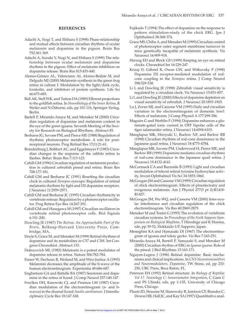
In Figure 9, we propose a model of the mechanismthat generates the circadian rhythm of ERG b-waveamplitude in the iguana retina. An oscillator in thebrain regulates retinal dopamine rhythmicity throughcentrifugal pathways, whereas a retinal oscillator con-trols rhythmic melatonin synthesis. During the subjec-tive day, dopamine increases b-wave amplitudedirectly via D2 receptors and by inhibiting melatoninsynthesis. During the subjective night, melatonindepresses b-wave amplitude and may, at the sametime, inhibit the synthesis of dopamine. Not shown inthe model but also consistent with our data is the pos-sibility that light may directly increase b-wave ampli-tude by direct stimulation of retinal dopaminesynthesis.
Diurnal versus NocturnalCircadian Rhythm ofERG in Vertebrates
Although the reciprocal inhibition of retinal dopa-mine and melatonin appears to be generally consis-tent among vertebrate species, the phase of the peak inb-wave amplitude is not. The phase differencesobserved have been attributed to differences in habitat(Rojas et al., 1999; Fowlkes et al., 1987), rod-cone domi-nance (Manglapus et al., 1998), or experimental proto-col (Wu et al., 2000; McGoogan et al., 2000). In ourexperiments, the b-wave always peaked in the day orsubjective day even though we varied the referenceelectrode placements and other aspects of the method-ology, and we therefore propose, as a working hypoth-
esis, that the phase of the b-wave peak depends onselective changes in the responsiveness of two (ormore) populations of cones. We are testing this idea byexamining circadian changes in spectral sensitivity. Ofcourse this, and other aspects of retinal function, islikely to have evolved to maximize adaptation to aparticular ecological niche.
ACKNOWLEDGMENTS
The work was supported by a grant (56647) fromthe National Institute of Mental Health. We thankBahie Rassekh for assistance with electroretinogramsand surgeries, Kelly Coffey for tissue collection, andWilson McIvor for assistance in high-performanceliquid chromatography. Mark Rollag generouslydonated the antibody for melatonin, and Carla Greenand Naomi Ihara provided much helpful discussion.
536 JOURNAL OF BIOLOGICAL RHYTHMS / December 2002
Figure 9. Model of the mechanism that may generate the circa-dian rhythm of electroretinogram b-wave amplitude in theiguana. An oscillator in the brain regulates or reinforces the circa-dian rhythm of dopamine synthesis in the retina via the opticnerve (ON). High levels of dopamine (dotted line) during the sub-jective day increase b-wave amplitude. The retina contains anoscillator that controls the synthesis of melatonin; in addition,melatonin synthesis is inhibited by dopamine (and light if pres-ent). Melatonin, synthesized during the subjective night, directlydepresses b-wave amplitude and may inhibit dopamine synthe-sis. During the subjective day, dopamine may repress melatoninsynthesis and increase the amplitude of the b-wave.
at INST DE FISIOLOGIA CELU LAR on May 7, 2013jbr.sagepub.comDownloaded from
REFERENCES
Adachi A, Nogi T, and Ebihara S (1998) Phase-relationshipand mutual effects between circadian rhythms of ocularmelatonin and dopamine in the pigeon. Brain Res792:361-369.
Adachi A, Suzuki Y, Nogi N, and Ebihara S (1999) The rela-tionship between ocular melatonin and dopaminerhythms in the pigeon: Effect of melatonin inhibition ondopamine release. Brain Res 815:435-440.
Alonso-Gómez AL, Valenciano AI, Alonso-Bedate M, andDelgado MJ (2000) Melatonin synthesis in the green frogretina in culture I: Modulation by the light/dark cycle,forskolin, and inhibitors of protein synthesis. Life Sci66:675-685.
Ball AK, Stell WK, and Tutton DA(1989) Efferent projectionsto the goldfish retina. In Neurobiology of the Inner Retina, RWeiler and N Osborne, eds, pp 101-116, Springer-Verlag,Berlin.
Bartell P, Miranda-Anaya M, and Menaker M (2000) Circa-dian regulation of dopamine and melatonin content inthe eye of the green iguana. Seventh Meeting of the Soci-ety for Research on Biological Rhythms. Abstract 85.
Besharse JG, Iuvone PM, and Pierce ME (1988) Regulation ofrhythmic photoreceptor metabolism: A role for post-receptoral neurons. Prog Retinal Res 17(1):21-61.
Brandenburg J, Bobbert AC, and Eggelmeyer F (1983) Circa-dian changes in the response of the rabbits retina toflashes. Behav Brain Res 7:113-123.
Cahill GM (1996) Circadian regulation of melatonin produc-tion in cultured zebrafish pineal and retina. Brain Res708:177-181.
Cahill GM and Besharse JC (1991) Resetting the circadianclock in cultured Xenopus eyecups: Regulation of retinalmelatonin rhythms by light and D2 dopamine receptors.J Neurosci 11:2959-2971.
Cahill GM and Besharse JC (1995) Circadian rhythmicity invertebrate retinae: Regulation by a photoreceptor oscilla-tor. Prog Retina Eye Res 14:267-291.
Cahill GM and Hasegawa M (1997) Circadian oscillators invertebrate retinal photoreceptor cells. Biol Signals6:191-200.
Dowling JE (1987) The Retina: An Approachable Part of theBrain, Belknap-Harvard University Press, Cam-bridge, MA.
Doyle S, Grace M, and Menaker M (1999) Retinal rhythms ofdopamine and its metabolites in C57 and C3H. Int Con-gress Chronobiol. Abstract 133.
Dubocovich ML (1983) Melatonin is a potent modulator ofdopamine release in retina. Nature 306:782-784.
Emser W, Dechoux R, Weiland M, and Wirz-Justice A (1993)Melatonin decreases the amplitude of the b-wave of thehuman electroretinogram. Experientia 49:686-687.
Engbretson GA and Battelle BA (1987) Serotonin and dopa-mine in the retina of lizard. J Comp Neurol 257:140-147.
Fowlkes DH, Karowski CJ, and Proenza LM (1987) Circa-dian modulation of the electroretinogram (a- and b-waves) in the diurnal lizard Anolis carolinensis. J Interdis-ciplinary Cycle Res 18:147-168.
Fujikado T (1994) The effect of dopamine on the response topattern stimulation-study of the chick ERG. Jpn JOphthalmol 38:368-374.
Grace MS, Chiba A, and Menaker M (1999) Circadian controlof photoreceptor outer segment membrane turnover inmice genetically incapable of melatonin synthesis. VisNeurosci 16:909-918.
Herzog ED and Block GD (1999) Keeping an eye on retinalclocks. Chronobiol Int 16:229-247.
Krizaj D, Gábriel R, Owen GW, and Witkovsky P (1998)Dopamine D2 receptor-mediated modulation of rod-cone coupling in the Xenopus retina. J Comp Neurol398:529-538.
Li L and Dowling JE (1998) Zebrafish visual sensitivity isregulated by a circadian clock. Vis Neurosci 15:851-857.
Li L and Dowling JE (2000) Effects of dopamine depletion onvisual sensitivity of zebrafish. J Neurosci 20:1893-1903.
Lu J, Zoran MJ, and Cassone VM (1995) Daily and circadianvariation in the electroretinogram of domestic fowl:Effects of melatonin. J Comp Physiol A 177:299-306.
Maguire G and Werblin F (1994) Dopamine enhances a glu-tamate-gated ionic current in OFF bipolar cells of thetiger salamander retina. J Neurosci 14:6094-6101.
Manglapus MK, Hiroyuki U, Buelow NF, and Barlow RB(1998) Circadian rhythms of rod-cone dominance in theJapanese quail retina. J Neurosci 18:4775-4784.
Manglapus MK, Iuvone PM, Underwood H, Pierce ME, andBarlow RB (1999) Dopamine mediates circadian rhythmsof rod-cone dominance in the Japanese quail retina. JNeurosci 19:4132-4141.
McCormack CA and Burnside B (1993) Light and circadianmodulation of teleost retinal tyrosine hydroxylase activ-ity. Invest Ophthalmol Vis Sci 34:1853-1860.
McGoogan JM and Cassone VM (1999) Circadian regulationof chick electroretinogram: Effects of pinealectomy andexogenous melatonin. Am J Physiol 277(5 pt 2):R1418-R1427.
McGoogan JM, Wu WQ, and Cassone VM (2000) Inter-ocu-lar interference and circadian regulation of the chickelectroretinogram. Vis Res 40:2869-2879.
Menaker M and Tosini G (1995) The evolution of vertebratecircadian systems. In Proceedings of the Sixth Sapporo Sym-posium on Biological Rhythms, T Hiroshige and K Honma,eds, pp 39-52, Hokkaido UP, Sapporo, Japan.
Meneghini KA and Hamasaki DI (1967) The electroretino-gram of iguana and tokay gecko. Vis Res 7:243-251.
Miranda-Anaya M, Bartell P, Yamazaki S, and Menaker M(2000) Circadian rhythm of ERG in Iguana iguana: Role ofthe pineal. J Biol Rhythms 15:163-171.
Nguyen-Legros J (1996) Retinal dopamine: Basic mecha-nisms and clinical implications. In CNS Neurotransmittersand Neuromodulators, Dopamine, TW Stone, ed, pp 233-250, CRC Press, Boca Raton, FL.
Peterson EH (1992) Retinal structure. In Biology of Reptilia:Vol 17. Neurology C: Sensoriomotor Integration, C Gans Cand PS Ulinski, eds, pp 1-135, University of ChicagoPress, Chicago.
Plautz JD, Straume M, Stanewsky R, Jamison CF, Brandes C,Dowse HB, Hall JC, and Kay SA(1997) Quantitative anal-
Miranda-Anaya et al. / CIRCADIAN RHYTHM OF ERG 537
at INST DE FISIOLOGIA CELU LAR on May 7, 2013jbr.sagepub.comDownloaded from
ysis of Drosophila period gene transcription in living ani-mals. J Biol Rhythms 12:204-217.
Pozdeyev ND, Doroshenko EM, Lavrikova EV, and EtingofRN (2000) The effects of melatonin and L-DOPA on thediurnal rhythms of free aminoacids content in rat retina. JBiol Rhythms 15:112-121.
Rojas LM, McNeil R, Cabana T, and Lachapelle P (1999)Behavioral, morphological and physiological correlatesof diurnal and nocturnal vision in selected wading birdspecies. Brain Behav Evol 53(5-6):227-242.
Rollag MR and Niswender GD (1976) Radioimmunoassayof serum concentrations of melatonin in sheep exposed todifferent lighting regimens. Endocrinology 98:482-489.
Shaw AP, Collazo R, Easterling K, Young CD, Cheryl D, andKarwoski C (1993) Circadian rhythm in the visual systemof the lizard Anolis carolinensis. J Biol Rhythms 8:107-124.
Stockton RA and Slaughter MM (1989) b-wave of theelectroretinogram: Areflection of ON bipolar cell activity.J Gen Physiol 93:101-122.
Tosini G and Dirden J (2000) Dopamine inhibits melatoninrelease in the mammalian retina: In vitro evidence.Neurosci Lett 286:119-122.
Tosini G and Menaker M (1996a) Circadian rhythms in cul-tured mammalian retina. Science 272:419-421.
Tosini G and Menaker M (1996b) The pineal complex andmelatonin affect the expression of the daily rhythm ofbehavioral thermoregulation in the green iguana. J CompPhysiol A 179:135-142.
Tosini G and Menaker M (1998) Multioscillatory circadianorganization in a vertebrate, Iguana iguana. J Neurosci18:1105-1114.
Underwood G (1970) The eye: The orbit and adnexae of adiurnal lizard. In Biology of Reptilia, Vol. 2, C Gans, ed, pp6-4, Academic Press, New York.
Underwood H and Siopes T (1984) Circadian organizationin Japanese quail. J Exp Zool 232:557-566.
Wu WQ, McGoogan JM, and Cassone VM (2000) Circadianregulation of visually evoked potentials in the domesticpigeon, Columbia livia. J Biol Rhythms 15:317-328.
Yamazaki S, Numano R, Abe M, Hida A, Takahashi R,Ueda M, Block GD, Sakaki Y, Menaker M, and Tei H(2000) Resetting central and peripheral circadian oscilla-tors in transgenic rats. Science 288:682-685.
538 JOURNAL OF BIOLOGICAL RHYTHMS / December 2002
at INST DE FISIOLOGIA CELU LAR on May 7, 2013jbr.sagepub.comDownloaded from